NOVEL BARRIERS TO THROMBOLYSIS:
THE ROLE OF MECHANICAL STRESS AND NEUTROPHIL EXTRACELLULAR TRAPS
PhD Thesis
Imre Varjú
Doctoral School of Molecular Medicine Semmelweis University
Supervisor: Krasimir Kolev, DSc
Official reviewers: Jolán Hársfalvi, DSc Béla Nagy Jr, PhD
Chairman of the final examination committee: Zoltán Benyó, DSc
Members of the final examination committee: Imre Bodó, PhD Nándor Müllner, PhD Budapest
2014
1
ABBREVIATIONS ... 4
1. INTRODUCTION ... 8
1.1. The fibrin net ... 9
1.1.1. Precursor and product– fibrinogen and fibrin... 9
1.1.2. Catalyst of formation- thrombin ... 11
1.1.3. Influence of blood components on structural parameters: fibre thickness and pore size ... 12
1.1.4. Influence of mechanical stress on structural parameters ... 14
1.2. The lysis of fibrin nets ... 16
1.2.1. Mechanism and morphology of fibrinolysis ... 17
1.2.1.1. Microscopic observations ... 17
1.2.1.2. Molecular model of fibrinolysis ... 18
1.2.2. Soluble components of the fibrinolytic system ... 20
1.2.2.1. Plasminogen and its activators... 20
1.2.2.2. Inhibitors of fibrinolysis ... 23
1.2.3. Cellular modulation of fibrinolysis ... 26
1.3. The role of neutrophils and neutrophil extracellular traps in haemostasis ………...29
1.3.1. Triggers of NET formation ... 29
1.3.2. Formation of NETs ... 31
1.3.2.1. NET formation as a form of cell death ... 31
1.3.2.2. Alternative ways of extracellular trap formation ... 31
1.3.3. Structure and composition of NETs ... 32
1.3.4. Intracellular events leading to NET formation ... 34
1.3.4.1. Signalling events ... 34
1.3.4.2. NADPH oxidase and ROS formation ... 35
1.3.4.3 Chromatin decondensation ... 37
1.3.4.4. Reorganization of membrane structures-the role of autophagy in NETosis ... 38
1.3.5. NETs and haemostasis ... 39
1.3.5.1. NETs and the vessel wall ... 40
1.3.5.2. NETs and platelets ... 40
1.3.5.3. NETs and red blood cells... 42
2
1.3.5.4. NETs and the coagulation system ... 42
1.3.5.5. NETs, thrombolysis, NET lysis ... 44
2. OBJECTIVES ... 46
2.1. Effects of mechanical stress ... 46
2.2. Effects of NET components ... 46
3. MATERIALS AND METHODS ... 47
3.1. Patients ... 47
3.2. Preparation of basic materials ... 47
3.2.1. Preparation of fibrin clots exposed to mechanical stress ... 47
3.2.2. Plasmin generation ... 48
3.2.3. Preparation of fibrin degradation products (FDP) ... 48
3.2.4. Preparation of neutrophil DNA ... 49
3.2.5. Expression and characteristics of fluorescent chimeric tPA variants ... 50
3.3. Structural studies ... 50
3.3.1. Scanning electron microscope (SEM) imaging of thrombi and clots ... 50
3.3.2. Morphometric analysis of fibrin structure in SEM images ... 51
3.3.3. Immunohistochemistry ... 52
3.3.4. Clot permeability assays ... 52
3.4. Mechanical studies-evaluation of fibrin rigidity ... 53
3.5. Intermolecular interactions-isothermal titration calorimetry (ITC) ... 54
3.6. Studies of fibrinolysis ... 54
3.6.1. Confocal microscopic imaging ... 54
3.6.2. Plasminogen activation assays... 55
3.6.3. Turbidimetry assays ... 56
3.6.4. Examination of clot lysis in microslide channels ... 57
3.6.5. Release of soluble fibrin degradation products (FDP) in the course of fibrinolysis ... 57
3.7. Enzyme inactivation assays ... 57
3.7.1. Defibrinogenated plasma-induced inactivation of plasmin ... 57
3.7.2. Inactivation of thrombin by antithrombin ... 58
3.8. Statistical procedures ... 58
4. RESULTS ... 59
3
4.1. Stressed fibrin lysis ... 59
4.1.1. Structural features of thrombi from patients... 59
4.1.2. Structural features of stretched fibrin clots ... 59
4.1.3. Lysis of stretched fibrin ... 62
4.2. Effect of neutrophil extracellular trap constituents on clot structure and lysis.………...66
4.2.1. Thrombi from patients ... 66
4.2.2. Structural studies ... 67
4.2.3. Inactivation kinetics of thrombin ... 70
4.2.4. Viscoelastic properties of fibrin ... 71
4.2.5. Studies on lysis of plasma clots... 72
4.2.6. Binding studies on fibrin degradation products and NET constituents .. 78
5. DISCUSSION ... 80
5.1. The effect of mechanical stress on structure and lytic susceptibility of fibrin ………...80
5.2. The effects of DNA, histones and neutrophil extracellular traps on structure, mechanical stability, and lytic properties of clots ... 82
5.2.1. DNA ... 83
5.2.2. Histones ... 85
5.2.3. DNA and histones, NETs ... 86
5.2.4. In vivo implications ... 87
6. CONCLUSIONS ... 89
7.1. SUMMARY ... 91
7.2. ÖSSZEFOGLALÁS ... 92
REFERENCES ... 93
LIST OF PUBLICATIONS ... 130
ACKNOWLEDGEMENTS ... 131
4 ABBREVIATIONS
2S, 3S: 2 and 3-times stretched fibrin α2-AP: α2-plasmin inhibitor
α2-MG: α2-macroglobulin
A340, A405: absorbance measured at 340 and 405 nm
ADAMTS-13: a disintegrin and metalloproteinase with a thrombospondin type 1 motif, member 13
Ag: antigen
Akt: protein kinase B apo(a): apoprotein (a) aPC: activated protein C Arg: arginine
AT: antithrombin C5a: complement 5a
CD: cluster of differentiation CXCL: CXC chemokine ligand DMSO: dimethyl-sulphoxide DNA: desoxy-ribonucleic-acid DVT: deep vein thrombosis
EDTA: ethylenediamine tetraacetic acid EGF: epidermal growth factor
EGTA: ethyleneglycol-bis-(β-aminoethylether)-N,N,N′,N′-tetraacetic acid ER: endoplasmic reticulum
ERK: extracellular signal-regulated kinase ET: extracellular trap
FV/FVIII/FIX: factor V/VIII/IX
FIXa/FXa/FXIIa/FXIIIa: activated factor IX/X/XII/XIII FcR: Fc receptor
FDP: fibrin degradation product Fg: fibrinogen
fMLP: formyl-Methionyl-Leucyl-Phenylalanine FpA, FpB: fibrinopeptide A and B
5 GAG: glucoseaminoglycan
GFP: green fluorescent protein Glu: glutamate
GM-CSF: granulocyte-monocyte colony stimulating factor GpIbα: glycoprotein Ibα
HEPES: 4-(2-hydroxiehyl)-1- piperazin-ethane-sulphonic acid HIF1α: Hypoxia inducible factor 1α
HIV-1: Human immunodeficiency virus 1 IFN: interferon
IL: interleukine Ile: isoleucine
K1-K5: kringle domains 1-5 Kd: dissociation constant
Km: Michaelis-Menten constant Ks: permeability constant Lp(a): lipoprotein a LPS: lipopolysaccharide
LRP1: LDL-receptor related protein 1 Lys: lysine
Mac-1: macrophage-1 antigen
MAPK: Mitogen activated protein kinase Mcl-1: myeloid cell leukemia-1
MEK: MAPK/ERK kinase MMP: matrix metalloproteinase MPO: myeloperoxidase
mTOR: mammalian target of rapamycin
NADPH: Nicotinamide Adenine Dinucleotide Phosphate Hydrogen NE: neutrophil elastase
NET: neutrophil extracellular trap NFκB: nuclear factor kappa B NS: non-stretched fibrin
PAD4: peptidyl-arginin-deiminase 4
6 PAI-1/-2: plasminogen-activator inhibitor-1/-2 PAR1: protease-activated receptor 1
PBS: Phosphate buffered saline PC: protein C
PF4: platelet factor 4 PHOX: phagocyte-oxidase
PI3K: Phosphatidyl-inositol-3-kinase PKB: protein-kinase B (also known as Akt) PKC: protein-kinase C
Plg: plasminogen (Glu, Lys)
PMA: phorbol 12-myristate 13-acetate PMN: polymorphonuclear cell
Pn: plasmin
proTh: prothrombin
Rac2: ras-related C3 botulinum toxin substrate 2 Raf: rat fibrosarcoma
Ras: rat sarcoma RBC: red blood cell RCL: reactive chain loop ROS: reactive oxygen species SAK: staphylokinase
SAK-Pn: staphylokinase-plasmin complex sctPA: single chain tPA
scuPA: single chain uPA SD: standard deviation
SDS-PAGE: sodium-dodecyl-sulphate polyacrylamide gel-electrophoresis SEM: scanning electron microscope
Ser: serine
Serpin: serine protease inhibitor SK: streptokinase
SK-Pn: streptokinase-plasmin complex SOD: superoxide-dismutase
7
SPPL: SPectrozyme-PL, Spectrozyme-plasmin: H-D-norleucyl-hexahydrotyrosyl- lysine-p-nitroanilide
Src: eukaryotic sarcoma tyrosine- kinase
TAFI: thrombin activatable fibrinolysis inhibitor TAFIa: active form of TAFI
TBS: TRIS buffered saline tctPA: two-chained tPA TF: tissue factor
TFPI: tissue factor pathway inhibitor Th: thrombin
TLR: toll like receptor TM: thrombomodulin
TNFα: tumor necrosis factor α
tPA: tissue type plasminogen activator tPA-GFP: GFP-tagged tPA
tPA-YFP:YFP-tagged tPA
TRIS: Tris(Hydroxymethyl)aminomethane uPA: urokinase type plasminogen activator uPAR: uPA-receptor
Val: valine
vWF: von Willebrand factor YFP: yellow fluorescent protein
8
1. INTRODUCTION
Cardio- and cerebrovascular diseases represent the major causes of death (35.8%) in the world according to recent data of World Health Organisation (1). The underlying cause in these cases is the formation of intravascular thrombi (composed of blood cells and plasma components embedded in a fibrin network), blocking the supply of oxygen and nutrients, therefore leading to the damage of the respective tissue. Statistics of mortality have shown improvement in developed countries within the last two decades, which can be partially accounted for the development of efficient tools regarding the prevention of thrombus formation (e.g. anticoagulants like warfarin and antiaggregants such as aspirin (2) and the therapeutic degradation of already formed clots by thrombolytic agents (e.g.
tPA). Despite this tendency, thrombolytic therapy often proves to be inefficient in the long term, and is accompanied by a serious risk for bleedings as a side effect (3,4).
Taken together, these facts point out the importance of the improvement of current thrombolytic therapeutic protocols, which requires determination of the factors influencing the efficiency of the respective enzymes in the dissolution of thrombi.
This thesis focuses on two of the numerous factors: mechanical stress to which fibrin formed in the circulation is exposed; and a recently recognized fundamental scaffold of venous and arterial thrombi: neutrophil extracellular traps (5) representing a web-like meshwork composed of DNA, histones and granular components released from granulocytes.
Since degradation of the fibrin scaffold itself is sufficient for the dissolution of thrombi, this introductory chapter gives a detailed description of fibrin structure and the factors influencing it. This is followed by the assessment of elements and regulation of fibrinolysis, the process of enzymatic degradation of the fibrin network. Finally, the chapter describes formation, structure and haemostatic effects of neutrophil extracellular traps.
9 1.1. The fibrin net
1.1.1. Precursor and product– fibrinogen and fibrin
Fibrinogen, the soluble, 340 kDa precursor of fibrin is a 45 nm long glycoprotein which consists of two peripheral ’D’ domains and a middle region (E domain) connected to each other by coiled-coil domains ((6), Fig. 1). The molecule is a heterohexamer containing 3 pairs of polypeptide chains (Aα, Bß, γ) linked together by disulphide- bridges (7-11).
Figure 1. Schematic structure of fibrinogen. The N-terminal regions of chains are found in the E region, while C-terminal sequences are localized in the peripheral regions, except for those of Aα chains. Black lines represent disulphide bridges, arrows point to sites of plasmin-mediated cleavage. For more detailed description, see text.
Modified from (12).
Proteolytic action of thrombin results in the cleavage of the N-terminals of Aα chains, releasing two fibrinopeptide A (FpA) molecules per fibrinogen, and leaving ‘desA fibrin’ behind (13-15). This leads to the exposure of two “A-knob” sequences, which are able to interact with C terminal “knobhole” regions found in γ chains of two other fibrin monomers. Aggregation of molecules in such a manner (head-to-head interactions stabilized by head-to-side linkages) results in a double-chained, half-staggered alignment of monomers with a longitudinal periodicity of 22.5 nm, and a lateral periodicity of ~5-10 nm (16), called a protofibril ((17-22), Fig. 2). Following this initial step, thrombin cleaves a further sequence (fibrinopeptide B (FpB)) from the N-terminals of Bß-chains, leading to the formation of ‘des AB fibrin’. The B-knobs exposed in these
10
molecules are partially responsible for the lateral aggregation of protofibrils and the branching of fibrin fibres (Fig. 2, (23)). Furthermore, following cleavage of FpB, αC-
Figure 2. Schematic assembly of the fibrin network. Fibrin(ogen) monomers are symbolized by rods with three (two peripheral and a central) nodules representing D and E domains. Further description in text. Modified from (12).
domains dissociate from E domains which makes them available for homophylic interactions, thereby promoting lateral fibril associations and assembly of an extensive fibre network (24,25). Fibre diameter values are typically in the 100-200 nm range, the structure of the fibres, however, is inhomogeneous. 70-80% of the fibre cross section is occupied by channels (26-28) that function like capillaries allowing the axial but not the radial diffusion of typically 50-90 kDa (diameter in the range of 10 nm in hydrated form (29)) proteins participating in fibrinolysis. The meshwork encloses pores with diameters in the range of 0.1 – 5 μm (30), which enable diffusion of bigger proteins up to 470 kDa (31).
11 1.1.2. Catalyst of formation- thrombin
Formation of thrombin from its zymogen (prothrombin) catalysed by FXa is a two-step process that takes place in the final stage of the coagulation cascade ((32-34), Fig. 3).
Hydrolysis of the first peptide bond mostly results in the formation of meizothrombin, which can be further converted to thrombin during the second cleavage causing the
Figure 3. Scheme of prothrombin activation. Human prothrombin consists of fragment 1 (F1), fragment 2 (F2), and the A and B chains of α-thrombin. Prothrombin is activated to α-thrombin by cleavage at Arg271 (R271) and Arg320 (R320).Regardless of the order of cleavages, α-thrombin and fragment 1.2 are generated. Modified from (35).
release of F1.2 zymogenic fragment (36). The formed two-chain serine protease, thrombin possesses an active site rich in negative charges allowing interaction with Arg-rich amino acid sequences (37), and two allosteric exosites (I and II). Exosite I is essential for binding to fibrinogen (38) and thrombomodulin (39), and takes part in the
12
direct (PAR1 (40), FV (41), FVIII (42,43)) and indirect (protein C (44), FXIII (45)) recognition of other substrates of thrombin. Exosite II is responsible for binding to heparin; and GpIbα found on the surface of platelets (46-48). Furthermore, in concert with exosite I, exosite II plays a role in the interaction with FV and FVIII (49,50). The primary endogenous inhibitors of thrombin (heparin cofactor II (51), protein C inhibitor (52), protease nexin 1 (53), and antithrombin (54)) belong to the serpin (serine protease inhibitor) family (55-57) (see also: 1.2.2.2.). The inhibition of thrombin exerted by serpins can be enhanced by GAGs like heparan sulphate and heparin, which are able to bind both serpins and exosite II (37).
1.1.3. Influence of blood components on structural parameters: fibre thickness and pore size
Concentrations of enzyme and substrate (thrombin and fibrinogen) are major determinants of fibre thickness: fibre diameter values show positive correlation with thrombin concentrations up to 10 nM, while above this value fibre thickness decreases (58) (Fig. 4.). In vivo fibrinogen concentrations vary in a narrower range (5-20
than thrombin concentrations, nevertheless this variation is also able to influence clot structure (29). However, in a plasma environment rich in macromolecules, the physicochemical behaviour of fibrinogen differs from the in vitro situation (59). As a consequence of the ‘space occupying’ effect by plasma proteins (e.g. albumin and immunoglobulins), participation of fibrinogen in chemical reactions (e.g. hydrolysis by thrombin) and binding interactions (e.g. with platelets) corresponds to that of its 10- times concentrated ideal solution. Another consequence is the self-association of molecules: according to sedimentation equilibrium studies, fibrinogen is dominantly present in a dimer form in the presence of 40 g/l albumin (60).
In addition, plasma components are also able to directly influence structural parameters of clots. FXIIIa, a calcium-dependent transglutaminase activated by thrombin, alters the molecular structure of fibrin by introducing covalent -glutamil-- amino-lysine isopeptide bonds between - (and, to a smaller extent, A-) chains of adjacent fibrin monomers, which also has severe consequences regarding mechanical and lytic resistance of the network (see 1.1.4.). Presence of immunoglobulins decreases the mass/length ratio of fibrin (thinner fibres are formed) (61,62), which can be partially
13
elucidated by direct inhibition of fibrin-polymerization (63,64). Activated protein C (65) and arginine (66) also contribute to the decrease of fibre diameters, while appearance of vessel wall components in the circulation causes thickening of the fibrin bundles (67).
The cellular components present in the bloodstream have further complex influence on clot structure. In vitro, red blood cells at cell counts near the physiological haematocrit values increase the average pore size approximately two-fold (68). Platelets in vivo form aggregates in the interior of clots. Fibrin strands originating from these zones (attached to glycoprotein IIb/IIIa receptors on the surface of platelets) are thinner and have a higher density (58). Contraction of platelets leading to retraction of thrombi (69) further modifies the structural and lytic parameters of clots (see 1.2.3.). Interaction of fibrin with phospholipids secreted upon thrombocyte activation (70) limits its availability for thrombin- (and plasmin-) mediated cleavage (71,72).
Figure 4. SEM images of pure fibrin clots. Clots contain 6 µM fibrinogen clotted with the indicated thrombin concentrations (in nM). Samples were prepared as described in 3.3.1.
14
Release of certain platelet-derived proteins also influences structural parameters, e.g.
actin contributes to the appearance of thinner fibres (73). Furthermore, besides providing a surface not only for the assembly of coagulation complexes but also FXIIIa, platelets contribute to the covalent modification of the meshwork by the secretion of their own transglutaminases.
1.1.4. Influence of mechanical stress on structural parameters
To maintain integrity of haemostasis, and to minimalize and localize the effects of clot formation, fibrin fulfils multiple criteria: it possesses not only firmness and plasticity at the same time (74), but also adequate permeability to allow the diffusion of fibrinolytic enzymes (75). FXIIIa-catalysed crosslinking profoundly alters viscoelastic properties of fibrin: both the elastic limit (the maximal extent of stretching, after the cessation of which the original structure can be regenerated) and the extensibility (extent of stretching that causes rupture of the polymer) of fibre strands show increase (76). In plasma clots, cross-linked structures bear 8.5-times higher elastic moduli compared to control. Following rupture, broken ends of fibres shrink nearly to their original size, which shows that stretching is largely accompanied by elastic alterations. The aforementioned effect of crosslinking is unique among biopolymers: as a comparison, introduction of crosslinks to collagen or keratin increases the stiffness and decreases the extensibility of these structures (77). The increased extensibility in the case of fibrin might be a consequence of axial alignment of crosslinks. Extensibility of whole fibrin nets is however 50-60% lower than that of individual fibres. This finding raises the assumption that disassembly of clots is not primarily due to rupture of single fibres, but more likely to dissociation at branching points of the fibrin network.
In vivo, stenosis of a blood vessel profoundly changes the rheological conditions around the obstruction. In addition to a several-fold increase of shear rate (78), the mechanical forces (radial, axial and circumferential) acting on the vessel wall show a heterogeneous pattern of relative strength at different locations of the stenotic region (stenosis throat, pre- and post-stenotic shoulder), but in all cases the axial force is two- to three-fold stronger than the radial force (79). Thus, if thrombi are formed at stenotic sites of blood vessels, the fibrin fibres on their surface will be exposed to enhanced
15
shear stress with well-defined directionality, which leads to the prediction of longitudinal alignment of these fibres.
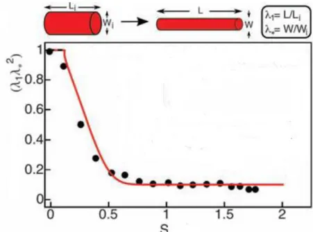
In vitro, stretching results in the decrease of clot volume (Fig. 5), which is a consequence of water expulsion (protein concentration of a 3-times stretched clot is 10 times higher compared to its non-stretched counterpart). SEM images show that fibrin
Figure 5. Effect of stretching on relative clot volume. S represents extent of stretching defined asS=(L/Li)-1, where Li is the initial and L is the stretched clot length.
Wi and W stand for initial and final diameter of cross section. Relative clot volume (λ1λ*2
) is defined as (L/Li)*(W/Wi)2. Modified from (80).
arrangement in non-stretched clots is essentially random, and stretching renders it ordered (80,81). The microscopic changes are accompanied by alterations on a molecular scale: upon stretching, tertiary structure of fibrin monomers changes, certain (possibly coiled-coiled (82)) domains unfold, which leads to exposure of hydrophobic amino acid residues. The latter form hydrophobic interactions which lead to tighter packing of protofibrils and the consequential expulsion of water (80,81).
16 1.2. The lysis of fibrin nets
Shortly after the formation of intravascular fibrin clots, fibrinolysis begins. This process can be divided into two steps (Fig. 6.): 1. activation of plasminogen (Plg) to plasmin 2.
proteolytic breakdown of the fibrin network (72). Plasmin, a serine protease formed by activators (e.g. tissue-type plasminogen activator (tPA), urokinase-type plasminogen activator (uPA)) from its zymogenic precursor plasminogen, plays a central role in the process. After its activation taking place on fibrin strands, cell surfaces, or in the circulation, plasmin digests fibrin releasing soluble fibrin degradation products (FDPs).
The most important end products are E and D fragments (central and peripheral domains of fibrin monomers, respectively, see before) (83), and D-dimers: two adjacent D domains ligated by FXIIIa activity, released from the covalently cross-linked fibrin.
Proteolysis of 25% of the total D-E connections is sufficient for complete lysis of the clot (84).
Figure 6. The two-step process of fibrinolysis. For description, see text.
The process of fibrinolysis is carried out by a multi-component system regulated by a set of interdependent biochemical reactions, the constituents of which will be described in detail within this section.
17 1.2.1. Mechanism and morphology of fibrinolysis 1.2.1.1. Microscopic observations
Confocal microscopic studies of fibrinolysis using labelled fibrinogen, Plg and Plg- activators revealed two phases of the process: a pre-lytic phase with accumulation of Plg on fibrin surface without any movement of the lysis front; and a final lytic phase with continuous thinning and eventual disappearance of fibres (85). The lytic zone is 5- 8 μm wide in the case of tPA (with uPA, it is thicker), but the pre-lytic zone penetrates deeper. Concentration of Plg in the latter zone can be up to 30-fold higher compared to its plasma concentrations. tPA shows similar accumulation, while uPA is only weakly bound to digested fibrin. These observations can be elucidated by binding data showing that plasminogen as well as tPA bind to fibrin with a dissociation constant of 10-8 - 10-6 M (86-88), whereas clots contain binding sites in the micromolar range (at least one per fibrin monomer). As a consequence, when fibrinolytic enzymes enter this adhesive environment, their diffusion slows down remarkably, which may lead to accumulation in a thin (pre)lytic layer. Activation of plasminogen leads to plasmin-induced exposure of additional C-terminal lysines resulting in the migration of lysine-binding fibrinolytic enzymes along concentration micro-gradients (89).
Further morphological information is gained by the help of scanning electron microscopy (SEM) using purified components: plasmin, Plg, tPA, fibrinogen and thrombin (90). SEM images of fibrin being digested show free, ‘cut’ fibre ends (appearing to have been transversely rather than longitudinally digested) and lack of fibrin strand continuity. More pronounced digestion results in lateral assembly of strands forming thick bundles and the increase of average pore size of fibrin clots (Fig.
7.). These studies were carried out according to an ‘external lysis’ model, where Plg was applied on clot surface. In vivo, however, clotting and lysis may occur at the same time, and lysis might proceed without the appearance of a distinct lytic front (‘internal lysis’). The latter model can be studied in a system where fibrin formation occurs in the presence of Plg and tPA (91). During this type of lysis, strands also appear to be preferentially digested transversely, and thinner individual fibres are digested faster than thicker ones. Frayed fibres form lateral interactions, and similarly to the findings of the former lysis model, average fibre diameter and pore size show transitional increase (91).
18
Figure 7. Effect of fibrinolysis on the macroscopic appearance of clot surfaces visualized by SEM. Clots were prepared from 6 µM fibrinogen and 0.2 µM plasminogen clotted with 3.5 nM thrombin. Lysis was initiated with 0.55µM tPA added to the surface. Numbers indicate minutes elapsed after the beginning of lysis. As lysis proceeds, cut-end fibres appear (10), and lateral aggregation of digested fibrin strands causes thickening of fibres (30). Preparation of samples is described in 3.3.1.
This is accompanied by increased turbidity, but not rigidity of the clots (92). In the late phase, decrease of absorbance and disassembly of the system into large fragments is seen.
1.2.1.2. Molecular model of fibrinolysis
Despite the fact that individual thin fibres lyse more quickly than thick fibres, in the case of whole fibrin clots, speed of lysis is mostly directly proportional to average fibre diameter (93). This phenomenon taken together with the aforementioned microscopic findings, supports the view that plasmin preferentially digests fibrin fibres in the transverse direction: under these circumstances plasmin might be more efficient in
19
digesting fibrin nets composed of thicker fibres, where the number of fibres in a given area is less than in a meshwork of fine fibres.
Plasmin binds near the end-to-end junction of two adjacent fibrin monomers, and, given its flexibility, is able to access cleavage sites on both of them. Hydrolysis of the susceptible peptide bonds generates C-terminal lysines which provide binding sites for additional plasmin (and also plasminogen, tPA) molecules. Since average distances between cleavage sites are shorter in the transverse than in the longitudinal direction (5- 10 nm and 22.5 nm, respectively) plasmin movement proceeds in the former direction, which eventually leads to the complete bisection of the fibre (16) (Fig. 8.).
Figure 8. ’Crawling’ model of plasmin. Rods with three (two peripheral and a central) nodules represent monomers of fibrin containing two D domains and an E domain, respectively. Plasmin is symbolized by a creature with a head (catalytic domain) and limbs (lysine binding Kringle domains). Conformational changes of plasmin allow the processive mechanism of action: A) Binding sites for plasmin are localized 22.5 nm away from each other longitudinally in fibrin fibres, but only 6 nm
20
away from each other in the fibre cross section. B) Plasmin rotates around a binding site. C) The induced conformational change allows binding of plasmin to another site.
D) Another conformational change restores initial state of plasmin, which enables cleavage of another monomer in the same cross section. Modified from (16).
Although not proved, this ‘crawling’ model of plasmin (16) is in good agreement with the multiple lysine binding sites and conformational changes of plasmin.
According to the model, while ‘crawling’, a plasmin(ogen) molecule is able to form a bridge between lysines located in two neighbouring protofibrils. This notion is supported by experimental data showing that Plg causes precipitation of FDP-s (94), and that Plg added to polymerizing fibrin results in increased fibre diameter (95,96).
1.2.2. Soluble components of the fibrinolytic system 1.2.2.1. Plasminogen and its activators
Human plasminogen is a 92 kDa, single-chain glycoprotein synthesized and secreted by the liver. Plasma concentration of the protein is approximately 2 M (97), and it can also be found in certain body fluids and tissues. The mature protein, Glu-plasminogen, named after its N-terminal amino acid, glutamate, consists of an N-terminal pre- activation peptide, 5 homologous Kringle-domains, and the catalytic serine protease domain (Fig. 9) (98). Cleavage (leading to formation of Lys-plasminogen) or non- proteolytic displacement of the pre-activation peptide has functional consequences:
susceptibility for certain activators and the affinity to fibrin increase. Kringle domains consisting of a polypeptide chain of around 80 amino acids stabilized by 3 disulphide bridges (99) are not unique to plasminogen, but can be found in other molecules influencing haemostasis (urokinase- and tissue type plasminogen activators (uPA and tPA), FXII, lipoprotein-a Lp(a), hepatocyte growth factors (100)). Kringle domains are responsible for binding plasminogen to small substances like Cl-, α,-diamino-acids, or
-aminocaproic acid, and also to lysine residues of fibrinogen, fibrin, and certain proteins of the extracellular matrix (101-103). Kringle 5 bears the highest affinity to- wards lysines located within the native peptide chain of fibrin (104,105), while Kringles1 and 4 preferentially bind to C-terminal lysines exposed in the course of fibrin
21
Figure 9. Plasminogen and its activators. Sites of cleavage for different proteases are shown. K1-5:Kringle 1-5; F: finger-domain; EGF: epidermal growth factor-like domain; SPD: serine protease-like catalytic domain. Long and short arrows at the top of the figure represent heavy and light chains, respectively. Modified from (97).
digestion (98). Interaction with lysine induces a profound conformational change in plasminogen: length of the molecule increases from 15 to 24 nm (106). Plasminogen in this ‘open’ conformation has similar characteristics to Lys-plasminogen formed by e.g.
plasmin-catalysed proteolytic cleavage of the pre-activation peptide (72).
The trypsin-like catalytic domain becomes active in the two-chain form of the molecule. This requires activation of plasminogen to plasmin by tPA or uPA mediated cleavage of the peptide bond Arg561-Val562 near Kringle 5 (107).
Tissue-type plasminogen activator (tPA) mainly synthesised by vascular endothelial cells, is a 70 kDa, single chain glycoprotein (108,109) that reaches a plasma concentration of 60-70 pM (110,111). However, only 20% of this quantity is found in free form, the rest is bound to its primary inhibitor, plasminogen activator inhibitor-1 (PAI-1). tPA consists of an N-terminal finger-domain, an epidermal growth factor
22
(EGF)-like domain, two Kringles, and a serine protease-type catalytic domain (Fig. 9).
Unlike most zymogens, this single-chain form of tPA (sctPA) possesses remarkable activity (about 10% of the two-chain from, tctPA formed following plasmin-mediated cleavage), however, in the absence of fibrin, its efficiency as a plasminogen-activator is low (112), which makes it a so-called fibrin-specific activator. tPA binds to low (lysine- independent (113)) and high (lysine-dependent (88)) affinity binding sites exposed in fibrin but not fibrinogen through its Kringle 2- and finger domains (114,115). Binding induces a conformational change similar to that of plasminogen, resulting in a 100-fold increase in the speed of plasminogen activation (116,117). Since binding sites for Plg and tPA in fibrin partially overlap, the two molecules come to close proximity, which increases the efficiency of plasmin generation. These mechanisms localized on fibrin surface ensure that formation of plasmin is conferred to fibrin deposits, this way sparing circulating fibrinogen from digestion (118).
Endothelial, epithelial, vascular smooth muscle cells, macrophages and granulocytes synthesize another type of plasminogen activators, uPA (urokinase-type, named after the fact that it appears in the urine (119)), the plasma concentration of which is approximately 2 ng/ml (120), but may vary under certain pathophysiological circumstances (121-123). It is secreted as a 55 kDa, single chain molecule (scuPA) consisting of an N-terminal EGF-like domain, a Kringle, and a catalytic domain homologous to that of tPA (Fig. 9). scuPA, possessing 1% of the final uPA activity, can be converted to its active two-chain form (tcuPA) by proteolytic action of kallikrein, FXIIa, trypsin, cathepsins (124,125), and plasmin. tcuPA is a fibrin-non-selective activator able to activate both fibrin-bound and free forms of Plg (126). The Kringle found in uPA domain is unable to bind lysine, but forms interactions with PAI-1 (127) and heparin (128). The EGF-like domain binds to receptors found on the surface of certain cells (uPAR), inducing cell migration and tissue remodelling (129), while the trypsin-like catalytic domain contributes to these processes by the cleavage of certain growth factors and metalloproteases (MMP-s) (97).
Streptokinase (SK) is a 47 kDa protein synthesized by the bacterium Streptococcus haemolyticus. Despite its name, SK possesses no enzymatic activity, however, it is able to form a 1:1 equimolar complex with Plg that functions as a Plg- activator (130,131). The formed plasmin cleaves SK, releasing an N-terminal peptide
23
that forms non-covalent interactions with the central fragment, thus inhibiting the binding of the SK-plasmin(ogen) complex to fibrin. This makes SK a fibrin non- selective activator, similarly to uPA (132).
Staphylokinase (SAK) is a 15.5 kDa protein synthesized by Staphylococcus aureus. Similarly to SK, SAK is not an enzyme, but is able to form a 1:1 complex with fibrin-bound plasmin. This interaction leads to a conformational change of the active centre which makes it similar to that of plasminogen activators, enabling the SAK- plasmin complex to convert Plg to plasmin (133). In the absence of fibrin, the complex formed between SAK and trace amounts of plasmin found in the plasma is quickly inhibited by alpha2-plasmin inhibitor (2-antiplasmin, 2-AP), therefore SAK is regarded as a fibrin-selective activator.
1.2.2.2. Inhibitors of fibrinolysis
TAFI (thrombin-activatable fibrinolysis inhibitor, other names: plasma procarboxypeptidase B, R, U (134,135)), a member of the metalloprotease family synthesized and secreted by the liver as a 60 kDa, extensively glycosylated (136) single- chain propeptide (134), reaches a plasma concentration of 220-270 nM (135,137). TAFI eliminates the C-terminal lysine residues exposed during plasmin-catalysed digestion of fibrin (138), which leads to reduction of the number of plasmin(ogen) binding sites.
Since plasmin bound to C-terminal lysines is known to be protected against α2-AP- mediated inhibition, TAFI also decreases the half-life of plasmin (139). Furthermore, TAFI slows down the conversion of Glu-Plg to Lys-Plg, which leads to hindered activation of plasminogen (140). Finally, higher concentrations of TAFI directly inhibit plasmin (141).
In order to gain its peptidase activity, TAFI needs to be proteolitically converted to its active form TAFIa (135,142). Thrombin is a weak activator, however, in the presence of thrombomodulin and calcium (138,143), the reaction speed increases more than 1000-fold (144). In comparison with thrombin alone, plasmin is 8-times more efficient, and the speed of activation increases in the presence of heparin, however, it is still far from that of the thrombin-thrombomodulin complex.
Heat-sensitivity of TAFI is remarkable: half-life of the molecule at 37°C is not more than a few minutes (145-148). Conformational change afterwards causes exposure
24
of peptide regions with high affinity towards α2-macroglobulin, which mediates the clearance of the molecule (147-149). FXIIIa plays an important role in the stabilization of TAFIa activity by covalently binding the molecule to fibrin (150).
PAI-1 (plasminogen activator inhibitor-1), the primary inhibitor of uPA and tPA, belongs to the family of serpins (serine protease inhibitors) (151). The molecule is a 50 kDa single chain glycoprotein synthesised by platelets (152), endothelial-, liver- and other, mainly perivascular cells (153,154). Basal plasma concentration of PAI-1 is generally low (0.4 nM), but reaches high local values in platelet-rich thrombi (155) and at sites of vessel injury (due to its high affinity towards vitronectin present in the extracellular matrix (156,157)). These local mechanisms presumably prevent premature lysis of thrombi.
PAI-1 forms a 1:1 complex with both uPA and tPA (158,159), however, fibrin- bound plasminogen activators are relatively protected from inhibition (151). The tertiary structure of PAI-1 contains a reactive centre loop (RCL) characteristic for serpins, which behaves as a ‘bait-substrate’ for the respective protease. Upon protease action, an Arg-Met peptide bond in the RCL is hydrolysed, and a consequential conformational change of the RCL N-terminal displaces the protease to the opposite side of the serpin (160). This leads to the disintegration of the serine protease active centre and the inhibition of dissociation of the complex (161-163). Upon cleavage of the RCL, the serpin forms a dead-end product, and the complex is eliminated from the circulation (164-166). In addition to the inhibition of plasminogen activators, PAI-1 exerts direct inhibitory effect on plasmin (167).
Similarly to TAFI, PAI-1 is fairly unstable (168), and binding to vitronectin (either in the plasma or in the extracellular matrix) prolongs its lifetime (169,170). This interaction induces a conformational change in the molecule that enables binding to integrins, making PAI-1 a modulator of cellular adhesion and motility (171-173).
Another member of the serpin family, PAI-2 is a 10-50-fold slower inhibitor of uPA and tctPA (in vitro) than PAI-1 (174-177) synthesised primarily by monocytes (178) and placental trophoblasts (179,180). The majority of PAI-2 molecules are found in the form of a 43 kDa non-glycosylated intracellular protein (179)), however, upon
25
stimulation by thrombin, it is secreted in the circulation as a 60-70 kDa glycoprotein (181-183). The polypeptide chain contains a glutamine-rich sequence which makes the molecule a good substrate for FXIIIa and other transglutaminases enabling covalent crosslinking of PAI-2 onto fibrin surface (184).
Besides its role in haemostasis, a growing amount of evidence supports the view that PAI-2 is also a regulator of intracellular proteolysis (185).
Figure 10. Regulation of fibrinolysis. A variety of negative and positive regulations is shown. For detailed description, see text.
α2-AP (α2-plasmin inhibitor/α2-antiplasmin), another serpin, is the primary plasmin inhibitor in humans. α2-AP is expressed as a 70 kDa, single chain glycoprotein in hepatocytes. The molecule reaches a concentration of 1 μM in plasma, where its half- life is approximately 3 days (186,187).
α2-AP exerts its anti-fibrinolytic activity through different mechanisms. 1) It forms a stable complex with plasmin (188). 2) Similarly to PAI-2 or TAFI, the molecule can be linked to Achains of fibrin by FXIIIa, which increases the lytic resistance of fibrin (189). 3) Lysine residues on the surface of α2-AP show high affinity towards Kringles found in plasmin(ogen) (188), and competitively inhibit the interaction between plasminogen and fibrin.
26
Lp(a) (lipoprotein a) is a plasma protein, which, similarly to LDL, contains an apolar lipid core and a surrounding phospholipid monolayer with embedded glycoproteins.
LDL contains apo B100, a 500 kDa glycoprotein, while in Lp(a), apolipoprotein(a) (apo(a)) is linked to apo B100 by disulphide bridges (190).
apo(a) bears structural homology with Plg: it has many isoforms containing Kringle 4-like (KIV, lysine-binding) (191) and Kringle 5-like (KV) structures, and an inactive serine protease-like region homologous to that of Plg (192). Instead of the Arg561-Val562 bond at the site of proteolytic cleavage in Plg, a Ser-Ile pair is found, which probably prevents recognition by proteases (193).
This structural homology between apo(a) and Plg results in competition regarding binding to lysine residues in fibrin (194-197), interactions with receptors on the surface of endothelial cells (198), platelets (199), and monocytes (200). apo(a) is also able to bind to the tertiary complex of Plg-tPA-fibrin, which prevents activation of Plg (201).Taken together, high levels of plasma Lp(a) are potentially anti-fibrinolytic, however, affinity of different Lp(a) isoforms towards fibrin depends on the number of Kringles: shorter isoforms show higher affinity, and therefore exert stronger inhibition on Plg activation (202).
α2-macroglobulin (α2-MG) is a 725 kDa homotetramer synthesised in the liver and found in the plasma in a concentration of approximately 3 μM. The molecule is able to bind to various enzymes, and also enzyme-substrate complexes (203). Plasmin and their activators are also able to bind to α2-MG, which results in a relatively slow inhibition of their activity (204) (Fig. 10). Cell surface- or fibrin bound plasmin molecules are protected from this type of inhibition (205). α2-MG possesses scavenger functions:
complexes containing α2-MG are internalized by LDL-receptor related protein 1 (LRP1), and are degraded in liver cells (206).
1.2.3. Cellular modulation of fibrinolysis
Intravascular thrombi are heterogeneous systems composed of fibrin scaffold and various soluble and cellular factors. To gain detailed information on fibrino- and thrombolysis in vivo, in addition to fibrin structure, the multidirectional interactions of
27
platelets, cells, extra- and intracellular proteins and lipids need to be taken into consideration.
In the course of formation of arterial thrombi, the platelet content of 10 ml of whole blood is compacted in a volume of 400 l, whereas the fibrinogen concentration of the same thrombi correspond to that in plasma (207), and Plg and 2-AP concentrations are substantially lower (110).
Presence of platelets leads to decreased velocity and hampered efficiency of fibrinolysis (208,209). This is partially due to release of PAI-1 (see 1.2.2.2.): high local concentrations of PAI-1 originate primarily from within platelets. During therapeutic fibrinolysis, however, their effect is ‘overcome’ by the applied concentrations of tPA (72).
Platelet-mediated retraction of thrombi (see 1.1.3.) leads to decreased mechanical (210) and lytic (208) susceptibility of clots, probably due to decrease of the fluid phase leading to limited diffusion of fibrinolytic enzymes. Furthermore, the fibres in platelet-rich areas of thrombi are more tightly packed and thinner than the ones in other clot regions, serving as a worse substrate for plasmin (58).
Histochemistry applied on arterial thrombi suggests that phospholipid concentrations (originating primarily from activated platelets (70)) exceed that of fibrin (207,211). Besides triggering the formation of coagulation complexes (e. g. tenase, prothrombinase (212)) phospholipids exert anti-fibrinolytic effects by limiting the diffusion of fibrinolytic enzymes (210) and directly inhibiting tPA- and plasmin- dependent fibrinolysis (211,72).
Platelet filopodia surrounding fibrin strands contain thick bundles of myosin (213), and high amounts of this protein are present in arterial thrombi (214). According to SEM studies, after 2 hours of initiation of thrombus formation, platelets start exhibiting morphological signs of necrosis (207). This leads to exposure of intracellular components, and allows interaction of fibrin and platelet-borne proteins (70). Myosin exerts multiple effects on fibrinolysis: hinders tPA-/uPA/plasmin-mediated lysis by serving as a source of ‘false’ binding sites for these proteins (214), while it weakens the interaction between the digested clot and the released FDPs, and (in higher concentrations) functions as a cofactor for tPA-induced plasminogen activation (215).
28
Red blood cells are no longer considered as passively trapped inert elements of thrombi, given their eptifibatide-sensitive interactions with fibrin(ogen) (216) resulting in altered fibrin structure (see 1.1.3.) and hindered fibrinolysis: red blood cells inhibit tPA induced plasmin generation on fibrin surface and tPA induced lysis of clots (217).
Neutrophil granulocytes (polymorphonuclear PMN cells) are present in thrombi in a smaller number than platelets, nevertheless, they play an important role in the formation and the elimination of thrombi. The role of neutrophil extracellular traps and other PMN-borne factors on fibrinolysis is discussed in the detail in the next section (1.3.).
29
1.3. The role of neutrophils and neutrophil extracellular traps in haemostasis As a response to inflammatory stimuli, polymorphonuclear (PMN, neutrophil) cells are able to expel a mixture of their nuclear and granular elements. These web-like composite structures are called neutrophil extracellular traps (NETs) that are able to entrap invading pathogens. NETs are composed of DNA, histones, granular enzymes and proteins (such as cathepsin G or elastase), and seem to be a universal tool of defence: humans, animals and even plants (218) are capable of extracellular trap formation, indicating that these webs provide an evolutionarily conserved protective mechanism.
Besides their protective function, a role for NETs is emerging in the pathogenesis of many diseases (219,220), and may be of interest regarding the pathogenesis of thrombosis. Stimulation of coagulation by NETs can result in unwanted thrombosis (221) and infection is a common event in the development of deep vein thrombosis (222,223). Targeting the release of nucleosomes, development of NETs and the availability of circulating histones could be a strategy for prevention or therapeutic intervention in venous thromboembolism, sepsis and other diseases involving cell death and lysis.
This chapter describes the formation and structure of NETs and discusses the possible connections and interrelations between this newly recognized form of innate immunity and components of the haemostatic system.
1.3.1. Triggers of NET formation
NETs can be formed in response to all major types of microbes (bacteria, fungi, protozoa, viruses) and their products, as well as inflammatory mediators, ROS, cell-cell interactions, and certain non-infectious or non-physiological stimuli. Table 1. shows a set of examples for various triggers.
30
Table 1. Triggers of NET formation. Several microbial and chemical stimuli have been identified. A summary based on (224-227).
Microbial stimuli Chemical stimuli
Bacteria
Enterococcus faecalis Escherichia coli
Haemophilus influenzae Helicobacter pylori Klebsiella pneumoniae Lactococcus lactis Listeria monocytogenes Mannheimia haemolytica
Mycobacterium tuberculosis/canettii Serratia marcescens
Shigella flexneri Staphylococcus aureus
Streptococcus dysgalactiae/pneumoniae Yersinia enterocolitica
Microbial toxins and components δ-Toxin from Staphylococcus epidermidis fMLP (+rapamycin)
Glucose oxidase
M1 protein-fibrinogen complex Lipophosphoglycan
Lipopolysaccharide (LPS) Panton-Valentine leukocidin
Inflammatory mediators and cytokines Antibodies
Calcium ions
GM-CSF + C5a/ LPS Hydrogen peroxide Interferon + eotaxin Interferon-α/γ + C5a Interleukin 1-β/8/23 Nitric oxide
Platelet activating factor Platelets through TLR-4 TNF-α
Fungi
Aspergillus fumigatus Candida albicans
Cryptococcus gattii/neoformans Protozoa
Leishmania
amazonensis/donovani/major/chagasi Other stimuli
Phorbol-12-myristate-13-acetate (PMA) PMA + ionomycin
Statins Virus
Feline Leukemia Virus HIV-1
Influenza A
31 1.3.2. Formation of NETs
1.3.2.1. NET formation as a form of cell death
NETs are the results of a unique cell death program that is different from apoptosis or necrosis (228). It is characterized by the loss of intracellular membranes before the plasma membrane integrity is compromised (NETosis). To release NETs, activated neutrophils undergo dramatic morphological changes (229). Minutes after activation by PMA, they flatten and firmly attach to the substratum, while showing a multitude of granules and a lobulated nucleus (230). During the next hour, the nucleus loses its lobules, the chromatin decondenses and swells, and the inner and outer nuclear membranes progressively detach from each other. Concomitantly, the granules disintegrate. After one hour, the nuclear envelope seems to disaggregate into vesicles and the contents of nucleoplasm, cytoplasm and granules are able to freely mix. After approximately 4 hours, the cells round up and seem to contract until the cell membrane ruptures and the internal components are ejected to the extracellular space (230,231). It is important to note, that depending on stimuli and donor, only a certain percentage of the activated neutrophils make NETs (230).
Apoptosis, another form of programmed cell death, is characterized by membrane blebbing, phosphatidylserine exposure on the cell surface, nuclear chromatin condensation and DNA fragmentation without membrane disintegration (225). Necrosis is characterized by PS exposure during the early steps, cellular swelling and bursting, and plasma membrane damage/rupture without nuclear membrane disintegration. The program of NETosis, on the other hand, shows disintegration of the nuclear envelope without DNA fragmentation; loss of internal membranes and organelles, and membrane rupture (and therefore PS exposure) after mixing of the nuclear and cytoplasmic elements.
1.3.2.2. Alternative ways of extracellular trap formation
Besides the above described, first observed form of NETosis (also called ‘suicidal’
NETosis), several other types have been reported (232).
32
In contrast with the PMA-induced 3-4 hour-long cell death program, a recently described form, ‘vital’ NETosis, leads to rapid NET formation without neutrophil cell death (233-235): Staphylococcus aureus appears to induce NETs in a rapid fashion (233), and LPS-activated platelets are also capable of inducing NETosis within minutes (236). ‘Vital’ NETosis does not only spare the neutrophil from ‘suicidal’ lysis, but transforms them into anuclear cytoplasts capable of chasing and imprisoning live bacteria (235). The third difference between ‘suicidal’ and ‘vital’ forms (besides timing and functional capacity of the involved neutrophils) is the mechanisms employed to create and cast out NETs: in contrast to the above described form, vital NETosis requires budding of the nuclear envelope, and vesicular trafficking of nuclear components to the plasma membrane, thereby delivering the NET out of the cell without requiring membrane perforation (233). Mitochondrial ETosis originally observed in eosinophils, and later in neutrophils could also be considered as a subtype of the ‘vital’ form (237,238).
1.3.3. Structure and composition of NETs
NETs released from neutrophils into the extracellular space consist of nuclear DNA and various histones decorated with granular proteins. NETs are fragile, complex structures (Fig. 11) composed of smooth ‘threads’, approximately 15-25 nm in diameter, which are likely to represent chains of nucleosomes from unfolded chromatin. High-resolution scanning electron microscopy (SEM) revealed that the NET threads are studded to variable extent with globuli of 30-50 nm (231) that contain the multiple cathelicidin
Figure 11. SEM images of NETs produced by PMA-activated neutrophils. Samples were prepared as described in 3.3.1. Images were taken at 10.000x magnification. Scale bars = 1 μm.
33
antimicrobial peptides which originate from the neutrophil granules (or lysosomes).
Several ‘threads’ can be wound into ‘cables’ that can be up to 100 nm in diameter (Fig.
11). These cables then form complex three-dimensional structures that, using SEM, can be hard to distinguish from fibrin networks (239). Analysis of cross sections of NETs by transmission electron microscopy (TEM) revealed that fibres are not surrounded by membranes (5). When produced in multiwell plates in vitro, NETs float within the medium, rather like a spider’s web does in moving air (240). The fact that they are
‘sticky’ as a result of their electrostatic charge and that they extend over areas of several microns makes them very effective at trapping (241), and possibly killing microorganisms (240).
DNA is a major structural component, because several intercalating dyes stain NETs strongly, and deoxyribonuclease (DNAse) treatment results in the disintegration of NETs, whereas protease treatment has no such effect (5). Accounting for approximately 70%, the most abundant component of NETs are histones (242). All core histones (H2A, H2B, H3, H4) as well as linker histones (H1) can be found in NETs, although in an enzymatically processed form (see later). The aforementioned globuli contain proteins and enzymes from the primary (azurophilic) granules (e.g. neutrophil elastase, cathepsin G, myeloperoxidase (MPO), bactericidal permeability increasing protein BPI), secondary (specific) granules (e.g. lactoferrin), and tertiary granules (e.g.
gelatinase or MMP-9, peptidoglycan recognition proteins PGRPs (243)) of neutrophils (244). Calprotectin, a heterodimer of cytosolic S100A8 and S100A9, represents one of the few examples for cytoplasmic components, which are rarely found in NETs (242).
These proteins exert various antimicrobial actions (245): MPO is responsible for microbicidal HOCl generation; serine proteases (neutrophil elastase NE, cathepsin G, proteinase 3, tryptase, neutrophil serine protease 4 NSP4 (246)) are able to inactivate bacteria by cleaving their virulence factors (5); cathelicidin LL37, BPI, defensins, and histones can disintegrate pathogen cell membranes challenging their viability (247,248);
calprotectin (242,249), calgranulin and lactoferrin chelate ions that are vital for microbial growth, altogether making NETs an effective tool virtually against all types of microbes.
NETs produced from mitochondrial DNA release have a slightly different structure (238). NE and MPO co-localize with mitochondrial DNA, but certain nuclear
34
(lamin B, nuclear matrix protein-45, poly-ADP-ribose polymerase, histones) and other (cytoplasmic caspase-3, beta-actin, mitochondrial cytochrome c, membrane markers CD15 and 16) elements are absent, which suggests a different type of host-NET interaction in the case of mitochondrion-derived NETs.
1.3.4. Intracellular events leading to NET formation
A unifying theory describing the subsequent steps of NET formation is still missing, but many mechanisms have been identified to contribute to NET expulsion.
1.3.4.1. Signalling events
The signalling mechanisms leading to the formation of NETs are poorly understood, and it is very likely that different triggers are able to induce NETosis through different pathways (Fig. 12,(250)).
The protein kinase C (PKC) enzyme family is comprised of conventional, novel and atypical isoforms (251). There are at least four conventional isoenzymes: PKCα, PKCβI, PKCβII and PKCγ. The novel isoenzyme group has four subtypes: PKCδ, PKCε, PKCη and PKCθ. The third group, atypical isoenzymes, consists of PKCζ and PKCι (251). PMA (phorbol-12-myristate-13-acetate), a widely used inducer of NETs, stimulates conventional (α, βI, βII, γ) and novel (δ, ε, η, θ) PKC by mimicking the activating ligand diacylglycerol (DAG) (251). PKC isoforms of all classes have been reported in neutrophils from healthy donors (252), and activation of PKC is critical in the generation of NETs (253). Nevertheless, an intricate antagonism is present between PKC isoforms in the regulation of a crucial element of NETosis, histone deimination:
PKCα has a dominant role in the repression of histone deimination, whereas PKCζ is essential in the activation of peptidyl arginine deiminase 4 (PAD4, see 1.3.4.3.) and the execution of NETosis. The precise balance between opposing PKC isoforms in the regulation of NETosis affirms the idea that NET release underlies specific and vitally important evolutionary selection pressures (254).
PKC activation (e.g. by PMA) is upstream of the Raf-MEK-ERK pathway (255) leading to phosphorylation of gp91phox (256) and p47phox (257) which initiates the assembly of the cellular or phagosomal membrane-bound and the cytosolic subunits of
35
another key player of NET formation, NADPH oxidase (see 1.3.4.2.). An alternative route for activation of ERK is also suggested through generation of reactive oxygen species (ROS) (258). The Raf-MEK-ERK pathway also upregulates the expression of antiapoptotic protein Mcl-1, which contributes to the inhibition of apoptosis and redirects the death program to NETosis (255).
The monomeric G-protein (rho small GTPase) Rac2 is also activated upstream of NADPH oxidase activation (259).
The role of PI3K-Akt-mTOR pathway is contradictory. Inhibition of mTOR leads to enhancement of fMLP-induced NETosis, because the pathway inhibits autophagy, a process that seems to enhance NET formation (e.g. by blocking apoptosis) (227). If a different trigger, lipopolysaccharide (LPS) is used, however, mTOR seems to support NETosis by exerting translational enhancement of HIF1α (260).
Certain triggers of NETosis act through a PKC/ROS-independent pathway, possibly mediated by Src kinase (261), which may be able to directly activate PAD4.
Cytoskeletal elements may also play a role in transmitting signals from the cell surface to the nucleus, e.g. inhibition of the cell surface receptor integrin Mac1- cytohesin1 (a guanine exchange factor)-actin cytoskeleton pathway results in inhibition of PAD4 activation and NET formation (262).
1.3.4.2. NADPH oxidase and ROS formation
Most signalling pathways activated by the triggers of NETosis converge to activate NADPH oxidase as a key enzyme of the process (263). Neutrophils isolated from patients with chronic granulomatous disease (CGD) caused by mutations in NADPH oxidase fail to produce NETs upon PMA-stimulation (230). Inhibition of the oxidase with diphenyleneiodonium (DPI) also prevents NETosis in response to several factors (264). Assembly of the NADPH oxidase responsible for the generation of ROS during the respiratory burst requires phosphorylation of the four cytosolic subunits (p47-phox, p40-phox, p67-phox and Rac) to enable their association with the membrane bound gp91phox-p22phox (cytochrome b558) complex. Once being in the active form, the enzyme generates ROS, out of which the most important seem to be the superoxide ions (O2-). O2- dismutates (either spontaneously or by superoxide dismutase (SOD) catalysis) to form H2O2. Further metabolization of H2O2 can lead to a variety of toxic oxygen de-
36
Figure 12. Intracellular steps leading to NET formation. Several signalling pathways can lead to NADPH oxidase activation and ROS formation, which triggers NE and PAD4 action on nuclear histones. Nuclear disintegration and decondensation leads to mixing of the granular and nuclear components, which are later expelled from the cell in the form of NETs. Dashed-end arrows represent inhibition, arrows pointing to the middle of another arrow represent activation of a step. Arrows with dotted lines stand for ambiguous relations. Gr: granule. For other abbreviations and explanation:
see text. Modified from (250).
rivatives, like the primary mediator of oxidative killing in the phagosome, HOCl, formed by myeloperoxidase (MPO) action. The importance of the latter enzyme is underlined by studies in patients suffering from MPO deficiency: the level of NETs they produced correlated negatively with the degree of the enzyme deficiency (265). How ROS generated during an oxidative burst contribute to NETosis is controversial. One
37
possibility is that they contribute directly to the observed morphological changes by causing direct membrane destruction (266). A proposed alternative is that ROS directly and indirectly (through activation of NF-κB) inactivate caspases (267-270), while exerting a possible autophagy-enhancing effect (250). Both mechanisms lead to an inhibition of apoptosis, ensuring that the already ongoing cell death program does not take an apoptotic route. ROS also play a crucial role in initializing the events that lead to chromatin decondensation, another key component of this type of cell death (Fig.
12.).
1.3.4.3 Chromatin decondensation
One option to weaken the interaction between DNA and highly positively charged histones is the enzymatic processing. At this moment, two enzymes seem to be of greatest importance: PAD4 (peptydilarginine deiminase 4) and NE (neutrophil elastase).
Peptydilarginine deiminases are enzymes catalysing citrullination (deimination), a posttranslational modification of arginine to citrulline. The process results in the loss of positive charge and hydrogen bond acceptors, therefore leading to weakened protein- protein, RNA-protein, and DNA-protein interactions. Out of the five PAD enzymes expressed in humans and mice (PAD1-4 and 6) (271), PAD2 and 4 are the most abundant in neutrophil granulocytes, and the latter seems to be critical in NET formation: PAD4-deficient mice are unable to decondense chromatin or form NETs (272), whereas overexpression of PAD4 is sufficient to drive chromatin decondensation to form NET-like structures in cells that normally do not form NETs (273).
PAD4, a 74 kDa protein that exists as a head-to-tail dimer (274,275) is the only member of the peptydilarginine deiminase family containing a nuclear localization signal that ensures its trafficking to the nucleus (274,276,277) (although not the only one to be found inside, e.g. PAD2 is also reported to be localized intranuclearly (278)).
The activation of PAD4 is calcium-dependent: binding of calcium to the C-terminal catalytic domain induces conformational changes that lead to the adequate positioning of critical active site residues (274). The calcium-dependency of the enzyme also serves as a possible connection between ROS generation (possibly leading to calcium release from the endoplasmic reticulum) and PAD4 activation. In addition, ROS are possible direct regulators of PAD4 (279). Cytoskeletal activity and autophagy may also be