Contents lists available atScienceDirect
Thrombosis Research
journal homepage:www.elsevier.com/locate/thromres
Review Article
Networks that stop the fl ow: A fresh look at fi brin and neutrophil extracellular traps
Imre Varjú
a,b, Krasimir Kolev
a,⁎aDepartment of Medical Biochemistry, Semmelweis University, Budapest, Hungary
bDepartment of Sociomedical Sciences, Mailman School of Public Health, Columbia University, New York, NY, USA
A R T I C L E I N F O
Keywords:
Neutrophil
Neutrophil extracellular trap Thrombosis
Fibrin Fibrinolysis
A B S T R A C T
Neutrophil extracellular traps (NETs) are DNA and histone-based networks enriched with granule-derived proteins cast out by neutrophils in response to various inflammatory stimuli. Another molecular network,fibrin is the primary protein scaffold that holds both physiological blood clots and pathological thrombi together.
There is mounting evidence that NETs andfibrin form a composite network within thrombi: in the past 10 years, a variety of molecular pathways have been revealed that help elucidate the nature of the NET-fibrin interaction.
Besides discussing the effects of various NET components on hemostasis, this review takes a closer look at the interaction of these individual effects, with novel perspectives on how the NET andfibrin networks stabilize each other. Similarities and molecular connections are also outlined between the processes responsible for the de- gradation (fibrinolysis and NET lysis) as well as elimination of these networks. In addition, the complex re- lationship of pathogens with the NET-fibrin network is discussed, with a particular focus on the role of peptidyl- arginyl deiminases (PADs) in NET formation as well as in pathogen intrusion, where PADs act as a virulence factor expressed by bacteria -an aspect that is currently left out from discussions in thefield.
1. Introduction
While Metchnikoff[1] and Ehrlich [2] examined neutrophils under the microscope as early as the 19th century, the discovery that these cells are capable of actively casting their nuclear constituents into the extracellular space was made only 15 years ago [3]. Since then, while neutrophil extracellular traps (NETs) have remained in the center of marked interdisciplinary attention, their exact nature has been criti- cally examined with some healthy skepticism (reviewed in [4,5]). It is becoming increasingly clearer that the process of NET formation (NE- Tosis [6]) is not a monolith, rather an umbrella term for multiple pathways that similarly result in active nuclear expulsion [7]. At minimum, the literature suggests suicidal, vital, and mitochondrial NETosis as distinct forms. However, as the concepts for the intracellular mechanism of NETosis are being continuously refined, ambiguities arise in terms of the exact requirements for these processes, implying that the main NETosis forms should be further divided (reviewed in [8]).
NADPH oxidase activity was originally deemed crucial for NET re- lease, supported by the observation that neutrophils from NADPH oxidase-deficient patients are incapable of NET formation [6,9], whereas the gene therapy for this enzyme deficiency restores their NET- forming capability [10]. However, later it was shown that certain
bacterial stimuli such as ionophores do not require oxidative burst to induce NETosis [11]. Furthermore, a NETosis-independent role for oxidative burst has been suggested as an alternative explanation for restored antimicrobial action at least in certain infections: NADPH oxidase activity enhances kynurenine formation from tryptophan [12].
This process is dependent on superoxide and is crucial in the host's ability tofight Aspergillus infections [13]. With regards to the role of myeloperoxidase (MPO), while its blockade results in inhibited NETosis in human neutrophils, in the same study, murine neutrophils have preserved their NETotic activity [14]. The role of neutrophil elastase (NE) has also been challenged as, in a thrombosis model, NE deficient murine neutrophils still produced NETs [15]. Autophagy was originally believed to be involved in NET formation [16], but later this has also been questioned [17]. The role of peptidyl-arginyl deiminases, PADs in NETosis is a much-debated topic which, given its wide implications for both immunity and hemostasis, will be discussed under a separate section. Besides ambiguities around the formation of NETs, there is mounting evidence that their structure and composition [18] is also far from uniform, which contributes to the difficulty offinding appropriate and comparable methods for the in/ex vivo detection of NETs [19].
While many of the basic steps and requirements of NETs have been questioned, the evidence that NETs contribute to pathological
https://doi.org/10.1016/j.thromres.2019.08.003
Received 17 May 2019; Received in revised form 18 July 2019; Accepted 5 August 2019
⁎Corresponding author at: Semmelweis University, Department of Medical Biochemistry, Tűzoltó utca 37-47, 1094 Budapest, Hungary.
E-mail address:Krasimir.Kolev@eok.sote.hu(K. Kolev).
Available online 07 August 2019
0049-3848/ © 2019 The Author(s). Published by Elsevier Ltd. This is an open access article under the CC BY-NC-ND license (http://creativecommons.org/licenses/BY-NC-ND/4.0/).
T
thrombosis [20] has been steadily accumulating in the past 10 years.
Accordingly, there is no shortage of excellent reviews on the topic [21–25]. Animal models of thrombosis have reported that submaximal ligation of inferior vena cava results in NET-rich thrombi [26], and during laser-induced arterial injury in mice, neutrophils are the first cells at the site of the injury [27]. NETs are abundant in human venous thrombus samples, particularly those in the organizing stages [28].
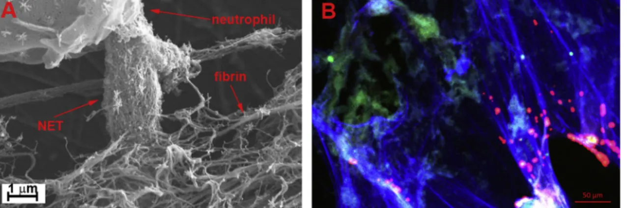
Similarly, arterial thrombi from patients who suffered heart attack [29,30], stroke [31], or peripheral arterial disease [32] contain NETs (Fig. 1). Remarkably, NETs alone have been shown to cause vascular occlusion in a non-canonical thrombosis model [33].
In light of thesefindings, NET/neutrophil inhibition has been sug- gested as a potential antithrombotic strategy, which has been ex- tensively discussed in a recent review in this journal [25]. To dissect the molecular basis of these clinical perspectives, the current review will specifically focus on the intersection of NETs with the two classic as- pects of hemostasis (coagulation andfibrinolysis). The mechanical and chemical stability of the fibrin-NET composite network will be ap- proached from two broad perspectives: from the aspect of thrombosis in the absence of infectious pathogens as well as from a pathogen per- spective.
2. NETs from a hemostatic perspective
The coupling of NETs and thrombus formation is reflected in the term immunothrombosis, which refers to the cooperation of hemostatic and immune systems in fighting pathogens [34]. In lower-level in- vertebrates, such as the horseshoe crab, hemocytes (nucleated im- munohemostatic cells) are responsible for both protection from patho- gens and sealing damage to prevent hemolymph loss (reviewed in [35]). In higher-level organisms, these two functionalities (immunity and hemostasis) appear to have begun an evolutionary separation process. Platelets, traditionally thought of as master regulators of he- mostasis, have lost their nuclei and accumulated coagulation factors andfibrinogen intracellularly (reviewed in [36]). These are released upon activation and contribute to the coagulation cascade, even though this seems to be redundant given thatfibrinogen is secreted by hepa- tocytes at micromolar concentrations in the circulation. At the end of the coagulation serine protease cascade, solublefibrinogen is cleaved, and an extracellular protein network is built up fromfibrin monomers (reviewed in [37]). At the same time, neutrophils, traditionally in- volved in immunity, have kept their nuclei and become responsible for the production of a complementary extracellular network that captures pathogens. Neutrophil nuclei are a pre-packaged source of NETs to be decondensed and cast out in the extracellular space upon microbial and inflammatory stimuli [7]. According to this view, both platelets and
neutrophils die while contributing to the higher cause, i.e. the forma- tion of a composite meshwork -resembling honey bees which can only sting humans once in their lives.
The two meshworks, NETs andfibrin are structurally intertwined (Fig. 1) and their co-localization has important functional con- sequences. In the following sections, the NET-fibrin link will be ex- amined first from a hemostatic perspective, focusing on the DNA backbone of NETs as well as on the effects of the two major proteins found in NETs: histones and elastase [38].
2.1. NETs stabilizing the clot I.: DNA
Throughout the literature, the DNA backbone of NETs has been overwhelmingly shown to exert prothrombotic and anti-fibrinolytic effects (reviewed in [39]). DNA induces coagulation through the in- trinsic pathway, which aligns well with the rule of thumb that highly negatively charged surfaces enhance the activation of factor XII (FXII), the initiator of this pathway (reviewed in [40]). While it is increasingly accepted that the intrinsic pathway is not required for physiological hemostasis, under pathological conditions, when DNA is released upon cellular damage (e.g. due to inflammation), this pathway can become crucial in initiatingfibrin formation (reviewed in [41]). Interestingly, another example of a negatively charged trigger of clotting is relevant in the course of NET formation, namely, polyphosphates released from histone-activated platelets [42,43]. Besides supporting the intrinsic pathway, DNA has been suggested to act as a cofactor surface in thrombin-dependent factor XI activation [44] and contribute to the propagation of the tissue factor-dependent coagulation pathway as well [45]. In addition, a variety of antifibrinolytic attributes of DNA have been described in the literature. Purified DNA has been shown to ac- celerate the formation of tissue-type plasminogen activator (tPA) – plasminogen activator inhibitor 1 (PAI-1) complexes [46], serve as a template for an inactive plasmin-fibrin-DNA complex [47], decelerate plasminogen-plasmin conversion by tPA on clot surfaces [48], bindfi- brin degradation peptides and delay their release fromfibrin clots [49], as well as intercalate intofibrinfibers and delay plasmin-mediated lysis of plasma clots [48,49]. Accordingly, thrombus samples from heart attack [30] and stroke [31] have shown faster ex vivo thrombolysis in the presence of DNAses in addition to tPA, just as in an in vitro NETosis- thrombosis model [48]. Nevertheless, these results mostly reflect the effect of pathologically high DNA concentrations within the micro- environment of NET-containing thrombi. Notably, lower concentrations of purified DNA might supportfibrinolysis through the facilitation of plasminogen activation by tPA in soluble phase [46].
Fig. 1.Intertwinedfibrin and NET scaffolds of thrombi. A: Scanning electron micrograph of NETs released by phorbol myristate acetate-activated isolated neu- trophils in afibrin clot. B: Confocal micrograph of a coronary thrombus removed with percutaneous coronary thromboaspiration from a 39-year old female patient with acute myocardial infarction and immunostained forfibrin (green) and citrullinated histone H3 (red) and stained for DNA with TOTO-3 (blue), (unpublished work of the authors). (For interpretation of the references to colour in thisfigure legend, the reader is referred to the web version of this article.)
2.2. NETs stabilizing the clot II.: histones
Another stream of publications examines the hemostatic effects of histones, highly positively charged DNA-binding molecules that re- present the majority of the proteins found in NETs [38]. Interest in the effect of polycationic polypeptides onfibrin formation and degradation goes back as far as the 1950s. Ginsburg and colleagues have shown that polylysine (which might mimic lysine-rich histones such as H1 [50]) inhibits streptokinase-inducedfibrinolysis [51] and the same substance facilitatedfibrin formation when added to staphylocoagulase and pro- thrombin [52]. With specific regards to histones, their interference with the hemostatic system has been observed in classic laboratory hemo- static tests mostly as prolonging clotting times [53], probably due to shielding the procoagulant phospholipid membranes [54]. Histone binding is thought to occur at phosphodiester bonds in phospholipids such as cardiolipin [54], which might mimic the phosphodiester sites in DNA, where nuclear histone binding normally occurs. In vivo, the majority of phospholipids are found in cellular membranes, where histone binding may exacerbate coagulation in various ways. Histones disrupt the anticoagulant endothelial barrier (reviewed in [55]) by in- ducing endothelial damage via pore formation or membrane destruc- tion and consequential ion influx [56,57]. During endothelial activation or even death [58] induced by histones, H2O2might be released, which is a further inducer of NETosis [6]. The contents of endothelial Weibel- Palade bodies are exocytosed alongside with von Willebrand factor (vWF) [59] that binds platelets and thus further supports thrombosis.
Histone binding to neutrophils also contributes to a vicious cycle by triggering further NET formation [60]. The interaction of histones with platelet membranes results in calcium influx either by pore formation [61] or opening of pre-existing channels [62], triggering activation of integrinα2bβ3[63] that facilitatesfibrin binding. As histones bind to fibrinogen [48], this chain of events could explain the unsaturable nature of histones binding to platelets [64]. In addition to less specific binding, histones also trigger platelet activation via toll-like receptors TLR2 and TLR4 [42] and potentiate thrombin-dependent platelet-acti- vation [65]. The in vivo result of these processes is a massive induction of thrombocytopenia and thrombosis [27,57,64]. Red blood cells (RBC), traditionally thought of as inert cells with regards to hemostasis have more recently been shown to mechanically [66] and chemically [67] strengthen clot structure, and to contribute to up to 40% of the thrombin-generating potential of the whole blood through phosphati- dylserine exposure in even a small fraction of the whole RBC population [68]. Histone binding enhances this membrane rearrangement [69], possibly further increasing the thrombogenicity of RBC during NETosis.
Besides their cellular effects, histones interfere with proteins of the coagulation cascade. They enhance prothrombin autoactivation [70]
and appear to impair antithrombin-induced thrombin inactivation [48].
Histones bindfibrinogen and fibrin, and get incorporated into poly- merizedfibrin in vitro, which results in mechanically and chemically more stable clots (49). Histones interfere with the thrombin-thrombo- modulin interaction [71], a prerequisite for the formation of the an- ticoagulant, histone-digesting activated protein C (APC) [57], which is also capable of inhibiting NETosis via PAR receptors on neutrophils [72]. Given the well-supported anticoagulant and anti-NET role of APC, it is not surprising that NETs offer histone-independent ways of pre- venting its activation: both neutrophil oxidases and neutrophil elastase (NE) are capable of inactivating it [73,74]. However, an aspect of the thrombin-thrombomodulin interaction often forgotten in reviews is the ability of the complex to activate not only protein C, but the thrombin- activatable fibrinolysis inhibitor (TAFI) as well [75]. The action of TAFI, on the other hand, is far from anticoagulant: it eliminates C- terminal lysines in fibrin that serve as binding sites for plasminogen [76]. Histone-mediated disruption, therefore, might also lead to sparing of these binding sites and accelerated lysis. However, the half-life of TAFI is remarkably low [77] and a myriad of opposing NET effects might override its influence onfibrinolysis in vivo. One additional way
in which histones might supportfibrinolysis is suggested by the ob- servation that histone 2B binds and recruits plasminogen on certain cell surfaces, which raises the possibility that it acts similarly in NETs [78].
2.3. NETs stabilizing the clot III.: elastase
Accounting for one-third of the cytosolic proteins found on NETs [38], NE has been shown to interfere with clotting and fibrinolysis alike. Earlier studies pointed to the role of elastase supporting fi- brinolysis. NE cleaves and inactivates the major plasma plasmin in- hibitor,α2-PI [79] and expresses directfibrinolytic activity [80]. NE is capable of clipping miniplasminogen out of plasminogen, which is more readily activatable than plasminogen [79], and once converted to miniplasmin, has a higher catalytic efficiency in digesting cross-linked fibrin than that of plasmin [81]. This might contribute to thefinding that, in the absence ofα1-antitrypsin, the major inhibitor of NE, in vitro fibrin clots containing neutrophils lyse spontaneously, unlike plasma clots [48], even in the absence of NETosis [82]. NE has been shown to cleave vWF and thereby release platelets from vessel walls under high shear stress [83]. Under certain circumstances, NE may also consume a portion of coagulation factors such as thrombin and factor X, releasing peptides with antimicrobial activity (e.g. GKY25) [84]. This antic- oagulant andfibrinolytic modality of elastase and neutrophils in gen- eral is in line with thefinding that plasminogen-knockout mice have increased number of neutrophils in their thrombi, and that neutrophils isolated from them show increased individual profibrinolytic activity [85].
Nevertheless, since the discovery of NETs, clot stabilizing effects of NE have been receiving more attention and are increasingly in- vestigated. Such effects include procoagulant properties, such as clea- vage of tissue factor pathway inhibitor [86], thrombomodulin as dis- cussed above [75], antithrombin [87] and certain proteins in the damaged vessel wall, making the subendothelium more thrombogenic [88]. Furthermore, NE has shown antifibrinolytic properties in certain experimental settings via cleavage of plasminogen activators (ur- okinase-type, uPA and tissue-type, tPA) [89].
2.4. Do DNA + histones + elastase equal NETs?
Studies on the isolated effects of NET components are invaluable even outside of the NETosis context as DNA and histones might be re- leased in a multitude of NETosis-independent ways (reviewed by Gould [39]). However, when trying to identify NET effects in the course of (immuno)thrombosis, the combined effects of individual NET compo- nents become a question of balance between the ratio of these mole- cules as well as the timing and the format of their release.
In vitro studies show that the effect of simultaneous addition of DNA and histones does not always lead to additive effects. For instance, DNA enhances mechanical stability offibrin clots, which is already increased in the presence of histones [49], while addition of DNA does not seem to have any additional effect on the tPA-induced degradation of plasma clots supplemented with histones [48]. In synthetic NET-like micro- webs, addition of DNA reduces the microbicidal activity of histones [90]. When added to coagulation assays in octamer forms, which is closer to their DNA-bound state, histones show reduced procoagulant activity [45]. Nuclear substance isolated from leukocytes suppresses in vitro coagulation less than pure histones isolated from the same ma- terial [53]. Isolated NETs have shown variable potential to induce NETs in platelet-independent assays [45,91], despite the fact that individual histones and DNA were both shown to possess in vitro procoagulant properties [91]. Since mixing activated neutrophils with whole blood does result in clot formation (20), it has been suggested that in vivo, NETs might contribute to clotting primarily through indirect means [45] (e.g. platelet activation). Taken together, these results imply an intricate fine-tuning mechanism during simultaneous release of DNA and histones, where these nuclear constituents might mutually silence
the hemostatic as well as the toxic effects of each other (on host cells and pathogens alike).
The presence of NE in NETs further increases the complexity of possible interactions. First of all, NE might continue to digest NET- bound histones in the extracellular space, thereby limiting their effects.
Secondly, the presence of DNA as well as NE-DNA interactions may be of crucial importance. Extrusion of a single neutrophil granule results in a remarkable local NE concentration of 5.33 mM, however, in an en- vironment with physiological antiprotease concentrations, the proteo- lytic effect of this release is limited to a 1.33μm radius for 12.4 ms [92,93]. On the other hand, in NETs, MPO co-localizing with DNA- bound NE is well-positioned for the rapid oxidation of Met358 ofα1- antitrypsin, resulting in the inactivation of the major NE inhibitor [94].
Access of inhibitors to NET-bound NE may also be impaired: DNA from bronchial secretions (which have been later shown to frequently con- tain NETs [95]) retards the inhibitory action ofα1-antitrypsin on NE [96]. Providing further means offine-tuning, DNA has been shown to be an inhibitor of NE activity [97], just like other negatively charged molecules that provide binding surface for the enzyme such as heparins [98]. This is in line with the effects of intragranular acidic proteogly- cans, which are thought to keep NE silent inside azurophil granules prior to its release triggered by the oxidative burst [99]. All of these interactions may be profoundly influenced by ionic strength or pH, factors that are very hard to predict in the local microcompartments of NETs in vivo. Nevertheless, these results suggest that DNA might act as a protective molecular leash for NE, which is confirmed by the ob- servation that NET-bound NE stays active in the circulation even after NET digestion by DNAses [100]. At the same time, soluble-phase NE (e.g. released during degranulation) might not be as protected from inhibitors, and may possess a different affinity towards various sub- strates than NET-bound NE. Such a modification of substrate specificity is not unprecedented in the regulation of serine proteases: thrombin for instance is known to go through a series of changes in substrate pre- ference when exiting the soluble phase and binding tofibrin, platelet surfaces, endothelium, or heparin (reviewed in [101]). This idea of different NE modalities during degranulation versus NETosis is sup- ported by the observation that certain stimuli predominantly induce NETosis (reviewed in [7]) while others lead to degranulation only (such as fMLP released from bacteria or damaged mitochondria [102] or tPA [103], a majorfibrinolysis activator). Intriguingly, recent work suggests that NE itself plays a key role in delineating degranulation from NE- Tosis, via its gasdermin-cleaving activity [104]. If NE is extruded from the cell via degranulation before it could process gasdermin in- tracellularly, gasdermin is unable to form vesicular pores that help channel NE into the nucleus, which has been shown to be an important step, at least in certain forms of NETosis [105].
2.5. Returning the favor: hemostasis-dependent and other ways of increasing NET stability
The previous sections illustrate the variety of pathways through which NETs might stabilize thrombi including direct interference (his- tones disrupting the thrombin-thrombomodulin interaction), cleavage of inhibitors (e.g. by NE), and covalent modification of inhibitors (ci- trullination and oxidation). On theflipside, however, little is known about whether the hemostatic system is able to return the favor and propagate NET formation or contribute to their stabilization.
Recently, it has been described that a serine protease of the intrinsic coagulation cascade, fXII (FXII), is able to promote NET formation in neutrophils via a uPA-receptor mediated mechanism, providing a link between inflammation-induced coagulation and NET formation [106].
Another protease cascade, the complement system represents a two- way link between the coagulation and NETosis (reviewed in [107]).
Thrombin is able to convert C5 and C3 to C5a and C3a, respectively, which attract leukocytes and C5a stimulates NETosis following inter- feron gamma-mediated priming of neutrophils [107]. The fact that
properdin, Factor B and C3 have been found bound to PMA-induced NETs and that MPO is known to activate properdin suggests that NETs might provide a surface for complement activation [107]. In turn this might support coagulation as previous studies have observed more bleeding propensity in complement-deficient mice [107]. Another consequence of complement binding is the increased chemical stability of NETs themselves: C1q binds to DNA and protects it from DNAse action either by shielding phosphodiester bonds or by directly in- hibiting DNAse 1 [107].
DNA oxidized by H2O2 or sodium-hypochlorite is resistant to di- gestion by the intracellular endonuclease TREX-1 [108]. Given that NETs are exposed to both MPO-derived hypochlorite and H2O2origi- nating from neutrophils or formed from superoxide released from ac- tivated platelets [109], DNA in NETs might possess increased resistance towards nucleases due to oxidation.
An intriguingfinding is that, similarly tofibrin, NETs are also sta- bilized by transglutaminase-dependent crosslinking [110], and that this crosslinking is critical to pathogen trapping. Lysine rich histones offer ample sites for transglutaminase action, and it has been shown earlier that histones are prone to tissue-type transglutaminase-mediated crosslinking, a process that might play a role in chromatin condensation during erythrocyte differentiation [111]. Extracellular crosslinking of histones in NETs showed no change when factor XIII (FXIII, the trans- glutaminase primarily responsible forfibrin crosslinking) was inhibited, which points to the involvement of neutrophil-derived transglutami- nases in the process [110]. Nevertheless, contribution of thrombin-ac- tivated platelets cannot be excluded as platelets trapped in thrombi eventually disintegrate and their own transglutaminase content might be released [112].
Another way to strengthen the ties between NETs and thefibrin network is offered by vWF which is secreted from endothelial cells in the presence of histones [59], or from the alpha granules of platelets activated during thrombosis [36]. vWF is known to bind to histones [113] as well as DNA [114] (possibly through its heparin binding sites).
This cross-binding of the two networks might further stabilize the thrombus mechanically, and even enzymatically, since vWF has been shown to inhibit plasmin-induced fibrinolysis [115] and binding to DNA might shield DNAse-susceptible sites. These observations are in line with the concept proposed by Martinod and Wagner [23] which suggests that vWF multimers themselves might form a third stabilizing scaffold in thrombi.Fig. 2summarizes some of the possible elements of thefibrin-vwF-NET interactions.
Besides superoxide and vWF, other factors released from platelets during activation might support NETosis, such as P selectin [116,117], interleukins (29) and High Mobility Group Box-1, HMGB-1 (reviewed in [118]).
Taking a bird's eye view, certain properties of the hemostatic end- productfibrin might also indirectly support NET formation by serving as an extracellular scaffold for the adherence and migration of leuko- cytes [119,120] during tissue injury. However, recent work suggests that in the circulation,fibrin formation might protect neutrophils from shear stress-induced NETosis [121]. Furthermore, thefibrin itself might capture and limit the availability of triggers of NET formation such as lipopolysaccharide or bacteria [122].
2.6. Clearance hand in hand? Degradation offibrin and NETs
On theflipside, connections between the degradation of bothfibrin and NETs are much less elaborated. Nevertheless, there is some evi- dence for the cooperation of systems that eliminatefibrin and extra- cellular DNA.
Classic fibrinolysis is mediated by the serine protease plasmin, which is synthesized and secreted by hepatocytes in its inactive pre- cursor form, plasminogen. Plasminogen is activated either by en- dogenous activators (such as tPA, mainly in the circulation, and uPA, mainly in tissues and on cell surfaces, reviewed in [123]), or by
exogenous activators from pathogens (see later). Regardless of its ac- tivation pattern, plasmin represents a plausible link between the de- gradation of thefibrin network and NETs as it has been shown to co- operate with DNAse I in the clearance of extracellular chromatin by cleaving histones [124]. This concerted action of plasmin and DNAses might be especially relevant in tissues where cellular death results in clot-like protein aggregates alongside with DNA released from dying cells [125]. Interestingly, this cooperation is not observed with the other major nuclease in the circulation, DNAse1-like 3, as this DNAse is sensitive to plasmin cleavage [124]. In addition to plasmin, APC has also been shown to cleave histones [57], however, several studies suggest that its efficiency is reduced when histones are bound to DNA [48,57]. Additionally, excess thrombin might lend a helping hand in cleaving histones [126], and also a host of other NET-bound proteins [127].
While many studies point to the primary importance of DNAses in terms of clearing NETs, a recent atomic force microscopy (AFM)-fo- cused structural study suggests that proteins are pivotal in determining the stability of NETs and that proteases could be effective in dis- mantling them [128]. While the authors used trypsin to demonstrate this phenomenon in their in vitro setting, they speculate that NET-borne proteases, such as NE or proteinase-3 might carry out a similar function in vivo, thereby contributing to a more dispersed NET structure or even the gradual clearance thereof. While this hypothesis needs further evidence, NETs would not be the only networks that induce their own decay:fibrin eventually facilitates its own proteolysis by serving as a cofactor in plasminogen activation by tPA [123].
Further similarities can be found in the cell-mediated clearance of fibrin and NETs, as it appears that leukocytes and endothelial cells cooperate in the elimination of both networks. Macrophages seem to play a major role in the clearance of NETs [129]. Macrophages and
neutrophils have been shown to phagocytosefibrin in the extracellular space, and it is likely that they do so intravascularly as well (reviewed in [130]). Underlining the role of endothelium in thrombus resolution, during the process of angiophagy, the endothelium engulfs clots up to a certain size, as demonstrated in an elegant study by Lam et al. [131].
Similarly, endothelial cells have been recently shown capable of pha- gocytosing NETs to a certain extent [132].
3. Requesting a seat at the table: the role of pathogens 3.1. The NET-fibrin meshwork from the pathogen's perspective
An intriguing aspect of the NET-fibrin merger is the modulatory role of pathogens during infection. The concept of immunothrombosis suggests that bothfibrin and NETs might be important in immobilizing, and perhaps eliminating bacteria [34]. Both networks form a fibrous structures that are tightly intertwined (Fig. 1).
Thefibrin network is generally formed of thicker fibers (ranging generally between 100 and 200 nm with pore diameters ranging from 0.1 to 5μm [133,134]. However, a multitude of factors influence the final structure (concentrations of thrombin,fibrinogen, ionic strength, pH, presence of plasma proteins, histones, DNA, cells, and even bacteria themselves [135], as reviewed in [37,123]. Furthermore, the network is largely heterogeneous (e.g. with higherfiber density and thinnerfibers in the proximity of platelets [136]). While the well-known structure of fibrin under SEM is typical for the inner parts of thrombi, a recent study has revealed that, on the outside, afibrin shield is formed that shows a dramatically altered structure and serves as a barrier to bacterial entry [137]. This barrier might also contribute to the long-known finding that, in the absence offlow, evenfibrinolytic enzymes have a difficult time diffusing into thefibrin clot: it takes about 10 days for uPA to Fig. 2.Synergy of NETs andfibrin thrombin thrombus stabilization. On the left: triggered neutrophils at the site of endothelial activation eject NETs (as represented by histone beads on DNA strings) [27]. In the middle: NETs themselves induce endothelial damage [56,57], which leads to vWF release (red line) [59]. Platelets bind to vWF and are activated by NETs [44]. On the right: activated platelets provide surface for coagulation complex assembly, secrete coagulation factors, and polyphosphates that activate factor XII, which in turn supports NET formation [106]. Coagulation cascade assembly leads to thrombin activation with multiple consequences. Thrombin catalyzes conversion offibrinogen intofibrin monomers (represented as dumbbells) that form double-strandedfibrin protofibrils. Thrombin also activates FXIII which introduces covalent crosslinks intofibrin and possibly between histones and other proteins in NETs [110,111]. Thrombin also catalyzes complement activation which supports NET formation [107]. (For interpretation of the references to colour in thisfigure legend, the reader is referred to the web version of this article.)
travel an axial distance of 1 cm [138]. According to SEM and AFM studies, NETs form thinner threads (15–25 nm) with globular domains of 30–50 nm diameter, containing NET-bound proteins and enzymes [3]. While this thinner network generally has smaller pores than that of fibrin, in some regions, aggregation of strands creates thicker threads (up to 100 nm [3]) and openings that couldfit bacterial cells [128].
Based on these data, and SEM images of composite NET-fibrin clots [48], it is tempting to speculate that fibrin and NETs complete each other: if bacteria slip through the pores of one network, they might still get caught up in another. Accordingly, bothfibrin [139] and NETs [3]
have been shown to capture bacteria, and NETs have been suggested to even kill pathogens in a variety of ways: oxidative action by MPO or H2O2, damage by proteases or histones, and limited access to divalent cations by the chelating effect of DNA [3].
3.2. Pathogen interference with NETs and hemostasis
Taken together, invading pathogens face a difficult task when trying to enter the bloodstream through the double-sieve formed by inter- twined NET andfibrin structures. No wonder that throughout evolu- tion, they have equipped themselves with a variety of tools to dismantle both networks, as summarized by some excellent reviews [140,141].
The need to degrade bothfibrin and DNA is especially well illu- strated by Streptococci which produce SK (streptokinase, a non-enzy- matic fibrin-independent activator of plasminogen) and SD (strepto- dornase, a DNAse) at the same time [142]. The intertwined nature of these two enzymes is also reflected in the fact that separating them poses a methodological challenge, and even many currently available SK preparations contain SD. In addition to secretion of a DNAse [143], Streptococcus pneumoniaeis also able to change the composition of its surface to decrease binding to NETs [144].
Another tool to dismantle the NET-fibrin conglomerate is the pro- motion of plasminogen activation resulting in fibrin and histone di- gestion. This route is exemplified by the enzyme Pla produced by Yersinia Pestis, which serves as a potent activator of plasminogen and uPA, inactivates plasminogen activator inhibitor-1 (PAI-1) andα2-an- tiplasmin, and cleaves TAFI, reducing its activatability by the thrombin- thrombomodulin complex (reviewed in [145]). While these effects are in line with the classic clinical picture of bubonic plague with massive hemorrhage, a study by Yun et al. reports that TFPI is also sensitive to Pla [146]. In agreement with the authors' discussion of thisfinding, one might speculate that the structural similarity between inhibitors of coagulation andfibrinolysis might have evolved in the host to serve as a protective mechanism to preserve hemostatic balance even in the face of detrimental effects of pathogen-triggered protease action. Further support for this theory is provided by the fact that NE released from neutrophils as a response to pathogens is also able to cleave both TFPI and α2-antiplasmin (see previous sections). Besides these broad ap- proaches to degrade the NET-fibrin sieve, some pathogens possess more specific tools of destruction, such as the histone-specific protease of Chlamydia trachomatis [147].
An important aspect of lysis of the NET-fibrin barrier by pathogens is the effect of the released digestion products. Lysis of host proteins such asfibrinogen may not only serve as a way to clear obstacles but potentially gives rise to peptides that bacteria can further metabolize to sustain themselves [148]. Furthermore, prior to its complete metabo- lism, the Bβ15–42 fragment liberated during the plasmin-mediated di- gestion of fibrin is capable of silencing leukocyte function [149]. Si- milarly, degradation of DNA in NETs might also contribute to dampening the action of leukocytes. In addition to a nuclease,Staphy- lococcus aureusalso secretes adenosine synthase which results in the formation of dATP [150], a long-known apoptosis trigger that NET- bound immune cells are susceptible to. In addition to turning the weapon of the host immunity against itself,S. aureusalso secretes sta- phylokinase [151] to generate plasmin and bind it on its own surface [152], thereby further aiding its escape from clots.
On theflipside, there are plenty of examples for pathogens hijacking fibrin rather thanfighting it. These include their own surfaces [153] as well as secreted molecules (such as staphylocoagulase [154]) that in- ducefibrin polymerization, and proteins that bindfibrin (e.g. protein M [155]). Interestingly, this versatility of approaches are not only seen with external parasites, but also with internal ones: tumor cells have been suggested to usefibrin formation to their advantage possibly by similarly shielding themselves from immune recognition [156], or, in certain cases, inducing fibrinolysis which is thought to help tumor dissemination and metastasis [157].
3.3. PADs-whose weapon are they really?
Citrullination, the conversion of peptidyl-arginyl residues to pep- tidyl-citrulline via deimination has been in the center of attention in the field of rheumatoid arthritis for a long time (reviewed in [158]), and PAD enzymes responsible for this irreversible post-translational mod- ification have been extensively investigated. Of particular relevance to the current review, several nuclear substrates of the subtype PAD4 have been identified: lamin C (during apoptosis [159]), PAD4 itself (either down-regulating its own action or not [160,161]), and various core [162] and linker histones [163]. Following the discovery of NETs, deimination of histones by PAD4 was soon identified as a hallmark of chromatin decondensation during NETosis [162]. Since then, the pic- ture has become more nuanced, and there is evidence that deimination might not be the only way of weakening histone-DNA interactions to enhance NET formation. NE travels to the nucleus after clipping gas- dermin [104] and possibly actin on its way [164], and processes his- tones [105]. Hypochlorite released by MPO is also suggested to reduce histone charge by chlorination [14]. Relatedly, pharmacological in- hibition of PAD4 leads to externalization of NET-like chromatin from human neutrophils, but with little histone citrullination [165]. Recent analysis has shown that PMA-induced NETs contain fewer citrullinated histones than ionophore-induced NETs [7], and certain authors explain this phenomenon by pointing out that PMA activates a PKC isoform that actually inhibits PAD4, rather than activating it [166]. Finally, plant cells are capable of forming NET-like structures, even though they do not express PAD enzymes [167].
Even if not a uniform requirement for all forms of NETosis, PAD4 expressed in neutrophils is still the only PAD with a nuclear localization signal [168]. This means that, when NETs are ejected, PAD4 reaches the extracellular space [169], which opens up a range of possible interac- tions, especially given that isolated PAD4 requires calcium concentra- tions that are more similar to the millimolar extracellular range than that of the intracellular milieu (reviewed in [170]). This characteristic of PADs continues to puzzle researchers, even if certain PAD4 binding proteins have been identified that might modulate its catalytic prop- erties in vivo [171]. PAD2, unlike PAD4, lacks a nuclear localization signal, however, it is still found inside the nucleus in certain cells [172]
and is also secreted during NETosis [169]. Hemostatic proteins that have been shown to be prone to either PAD4 or PAD2 action arefi- brinogen (the thrombin cleavage site of which contains a critical argi- nine that is lost during citrullination [173]), antithrombin (which is more prone to citrullination in the presence of heparin [174] and loses its inhibitory effect on thrombin activity [175]), and, according to a recent citrullinome analysis, inhibitors as well as activators of the plasminogen-plasmin axis [174]. Given this variety of hemostatic PAD substrates, the net effect of citrullination onfibrin stability is hard to predict, and it is possible that the presence of other NET components such as DNA and histones overrides the PAD effects in this complex environment.
Complements that provide a link between the coagulation cascade and NETosis, are also affected by citrullination. PAD 1, 2, and 4 have been shown to citrullinate C1q inhibitor (alongside with other serpins), which reduces its inhibitory activity [174]. LL37, an antimicrobial protein formed via proteinase-3-catalyzed cleavage from hCAP-18
[176], has a protective effect against DNAse action in certain assays [177]. Remarkably, LL37 is also prone to citrullination, which de- creases its antimicrobial efficiency [178]. Sorvillo et al. demonstrate in an elegant study that PAD4-catalyzed citrullination of ADAMTS 13 decreases its catalytic efficiency [179], which in vivo might result in increased half-life of vWF multimers that predispose to thrombosis.
What is often left out of the discussions about the role of PADs in immunothrombosis is that certain bacteria express PADs as a virulence factor, as reviewed by György et al. back in 2006 -at that time, un- related to NETosis [180]. The PAD secreted by Porphyromonas gingi- valis (PPAD) has been shown to contribute to the process of invasion, partially through creating a neutralizing NH4+-rich environment [181].
At the same time, PPAD is able to citrullinate chemokines and various proteins of the complement system, downsizing the immune reaction thereby [182]. PPAD seems to be more efficient in citrullinating NET- associated histone H3 than human PADs secreted during NETosis [183].
This phenomenon is well aligned with the dependence of (host- and pathogen-directed) histone toxicity on positively charged amino acid sidechains [183,184], and points in the direction of Konig and Anrade's argument about citrullination being a bacterial tactic rather than an antimicrobial one [166]. Nevertheless, controversy remains, as it has been shown that PAD4 is essential in the killing function of NETs [184].
It is possible that intracellular and extracellular histone citrullina- tion serve different functions. Intracellular citrullination might be useful in weakening the link between histone octamers and DNA, and also help unleash potent antibacterial histones from DNA once ex- ternalized. A small fraction of histones citrullinated might be sufficient for this purpose, and more toxic non-citrullinated histones may be also released. On the other hand, ‘extracellular’citrullination might be a defense mechanism to reduce toxicity of histones as demonstrated by neutrophils that otherwise would be prone to histone toxicity and which expresses PAD4 on their plasma membrane [185], as well as by bacteria that evolved to defend themselves from histone-mediated killing. To date, it remains unclear if citrullination of hemostatic pro- teins identified above can be induced by PPAD, and if so, if citrullina- tion occurs at the same or different sites.
4. Perspectives
The increasing number offindings around the interrelated effects of NETs and hemostasis call for further studies to decide which of the individually examined effects hold true in vivo. Furthermore, stronger emphasis should be placed on delineating the effects of different neu- trophil modalities on hemostasis, as it seems clear that degranulating neutrophils might have profoundly different effects on clotting than NET-forming ones. Relatedly, the question whether these modalities are merely dependent on different signals or represent different sub- populations of neutrophils is still open [186]. Further elaboration of NET effects should be attempted when examining how pathogens in- teract with the NET-fibrin meshwork: the case of PADs exemplifies the uncertainties in the current knowledge.
Furthermore, it is high time that citrullination gets thoroughly ex- amined in the context of thrombosis. To date, while citrullination has been extensively studied in the synovia overwhelmingly tied to rheu- matoid arthritis, no studies have been carried out on the hemostatic effects of the released PAD enzymes in blood. Given that citrullination has been shown to affect a series of human proteins in inflammatory diseases, bacterial PADs also deserve more attention. In the context of infections, it needs to be clearly outlined which relevant proteins might be citrullinated by which PADs (host or pathogen) and, if citrullination patterns are different, the implications of those differences should be investigated. Citrullination profile of thrombi should be evaluated in the absence and in the presence of bacteria and a series of in vivo and in vitro experiments are needed to decide which forms of deimination have a substantial effect in vivo, and whether citrullination of extra- cellular proteins is a bystander marker of NETosis or a pathogenetic
factor. Given that single amino acids (such as arginine [187]) or even the presence of cations (such as Zn2+[188]) can markedly alter clot structure, it is perhaps not far-fetched to suggest that an enzyme that is capable of covalently modifying multiple hemostatic players might profoundly alter the course of in vivofibrin formation and degradation.
Declaration of competing interest
The authors declare that the work was conducted in the absence of any commercial orfinancial relationships that could be construed as a potential conflict of interest.
Author contributions
IV wrote the initial draft of the manuscript, both authors con- tributed to manuscript revision, read and approved the submitted ver- sion.
Funding
This work was supported by the Hungarian National Research, Development and Innovation Office (NKFIH) (129528) and the Higher Education Institutional Excellence Programme of the Ministry of Human Capacities in Hungary for the Molecular Biology thematic programme of Semmelweis University. IV was further supported by a postdoctoral Fulbright Scholarship, a postdoctoral scholarship by the Tempus Public Foundation, and a scholarship by the Rosztoczy Foundation.
References
[1] E. Metchnikoff, Immunity in Infective Diseases, Cambridge University Press, Cambridge, 1905.
[2] P. Ehrlich, Methodologische Beiträge zur Physiologie und Pathologie der ver- schiedenen Formen der Leukocyten, Z.Klin. Med.1 (1880) 553–560.
[3] V. Brinkmann, U. Reichard, C. Goosmann, B. Fauler, Y. Uhlemann, D.S. Weiss, Y. Weinrauch, A. Zychlinsky, Neutrophil extracellular traps kill bacteria, Science 303 (5663) (2004) 1532–1535,https://doi.org/10.1126/science.1092385.
[4] V. Brinkmann, Neutrophil extracellular traps in the second decade, J Innate Immun. 10 (5–6) (2018) 414–421,https://doi.org/10.1159/000489829.
[5] W.M. Nauseef, P. Kubes, Pondering neutrophil extracellular traps with healthy skepticism, Cell. Microbiol. 18 (10) (2016) 1349–1357,https://doi.org/10.1111/
cmi.12652.
[6] T.A. Fuchs, U. Abed, C. Goosmann, R. Hurwitz, I. Schulze, V. Wahn, Y. Weinrauch, V. Brinkmann, A. Zychlinsky, Novel cell death program leads to neutrophil ex- tracellular traps, J. Cell Biol. 176 (2) (2007) 231–241,https://doi.org/10.1083/
jcb.200606027.
[7] E.F. Kenny, A. Herzig, R. Krüger, A. Muth, S. Mondal, P.R. Thompson, V. Brinkmann, H.V. Bernuth, A. Zychlinsky, Diverse stimuli engage different neutrophil extracellular trap pathways, Elife. 6 (2017) e24437, ,https://doi.org/
10.7554/eLife.24437.
[8] G. Sollberger, D.O. Tilley, A. Zychlinsky, Neutrophil extracellular traps: the biology of chromatin externalization, Dev. Cell 44 (5) (2018) 542–553,https://
doi.org/10.1016/j.devcel.2018.01.019.
[9] A. Hakkim, T.A. Fuchs, N.E. Martinez, S. Hess, H. Prinz, A. Zychlinsky, H. Waldmann, Activation of the Raf-MEK-ERK pathway is required for neutrophil extracellular trap formation, Nat. Chem. Biol. 7 (2) (2011) 75–77,https://doi.org/
10.1038/nchembio.496.
[10] M. Bianchi, A. Hakkim, V. Brinkmann, U. Siler, R.A. Seger, A. Zychlinsky, J. Reichenbach, Restoration of NET formation by gene therapy in CGD controls aspergillosis, Blood. 114 (13) (2009) 2619–2622,https://doi.org/10.1182/blood- 2009-05-221606.
[11] I. Neeli, M. Radic, Opposition between PKC isoforms regulates histone deimination and neutrophil extracellular chromatin release, Front. Immunol. 4 (2013) 38, https://doi.org/10.3389/fimmu.2013.00038.
[12] L. Romani, F. Fallarino, A. De Luca, C. Montagnoli, C. D'Angelo, T. Zelante, C. Vacca, F. Bistoni, M.C. Fioretti, U. Grohmann, B.H. Segal, P. Puccetti, Defective tryptophan catabolism underlies inflammation in mouse chronic granulomatous disease, Nature 451 (7175) (2008) 211–215,https://doi.org/10.1038/
nature06471.
[13] Q. Remijsen, P. Vandenabeele, J. Willems, T.W. Kuijpers, Reconstitution of pro- tection against Aspergillus infection in chronic granulomatous disease (CGD), Blood 114 (16) (2009) 3497,https://doi.org/10.1182/blood-2009-07-233312.
[14] K. Akong-Moore, O.A. Chow, M. von Köckritz-Blickwede, V. Nizet, Influences of chloride and hypochlorite on neutrophil extracellular trap formation, PLoS One 7 (8) (2012) e42984, ,https://doi.org/10.1371/journal.pone.0042984.
[15] K. Martinod, T. Witsch, K. Farley, M. Gallant, E. Remold-O'Donnell, D.D. Wagner, Neutrophil elastase-deficient mice form neutrophil extracellular traps in an ex- perimental model of deep vein thrombosis, J. Thromb. Haemost. 14 (3) (2016) 551–558,https://doi.org/10.1111/jth.13239.
[16] Q. Remijsen, T. Vanden Berghe, E. Wirawan, B. Asselbergh, E. Parthoens, R. De Rycke, S. Noppen, M. Delforge, J. Willems, P. Vandenabeele, Neutrophil extra- cellular trap cell death requires both autophagy and superoxide generation, Cell Res. 21 (2) (2011) 290–304,https://doi.org/10.1038/cr.2010.150.
[17] N. Germic, D. Stojkov, K. Oberson, S. Yousefi, H.U. Simon, Neither eosinophils nor neutrophils require ATG5-dependent autophagy for extracellular DNA trap for- mation, Immunology 152 (3) (2017) 517–525,https://doi.org/10.1111/imm.
12790.
[18] E.A. Chapman, M. Lyon, D. Simpson, D. Mason, R.J. Beynon, R.J. Moots, H.L. Wright, Caught in a trap? Proteomic analysis of neutrophil extracellular traps in rheumatoid arthritis and systemic lupus erythematosus, Front. Immunol. 10 (2019) 423,https://doi.org/10.3389/fimmu.2019.00423.
[19] E.A. Ryan, S. Boeltz, P. Amini, H.J. Anders, F. Andrade, R. Bilyy, S. Chatfield, I. Cichon, D.M. Clancy, J. Desai, T. Dumych, N. Dwivedi, R.A. Gordon, J. Hahn, A. Hidalgo, M.H. Hoffmann, M.J. Kaplan, J.S. Knight, E. Kolaczkowska, P. Kubes, M. Leppkes, A.A. Manfredi, S.J. Martin, C. Maueröder, N. Maugeri, I. Mitroulis, L.E. Munoz, D. Nakazawa, I. Neeli, V. Nizet, E. Pieterse, M.Z. Radic, C. Reinwald, K. Ritis, P. Rovere-Querini, M. Santocki, C. Schauer, G. Schett, M.J. Shlomchik, H.U. Simon, P. Skendros, D. Stojkov, P. Vandenabeele, T.V. Berghe, J. van der Vlag, L. Vitkov, M. von Köckritz-Blickwede, S. Yousefi, A. Zarbock, M. Herrmann, To NET or not to NET: current opinions and state of the science regarding the formation of neutrophil extracellular traps, Cell Death Differ. 26 (3) (2019) 395–408,https://doi.org/10.1038/s41418-018-0261-x.
[20] T.A. Fuchs, A. Brill, D. Duerschmied, D. Schatzberg, M. Monestier, D.D. Myers Jr., S.K. Wrobleski, T.W. Wakefield, J.H. Hartwig, D.D. Wagner, Extracellular DNA traps promote thrombosis, Proc. Natl. Acad. Sci. U. S. A. 107 (36) (2010) 15880–15885,https://doi.org/10.1073/pnas.1005743107.
[21] Y. Döring, O. Soehnlein, C. Weber, Neutrophil extracellular traps in atherosclerosis and atherothrombosis, Circ. Res. 120 (4) (2017 Feb 17) 736–743,https://doi.org/
10.1161/CIRCRESAHA.116.309692.
[22] L.L. Swystun, P.C. Liaw, The role of leukocytes in thrombosis, Blood 128 (6) (2016) 753–762,https://doi.org/10.1182/blood-2016-05-718114.
[23] K. Martinod, D.D. Wagner, Thrombosis: tangled up in NETs, Blood. 123 (18) (2014) 2768–2776,https://doi.org/10.1182/blood-2013-10-463646.
[24] Laridan E, Martinod K, De Meyer SF. Neutrophil extracellular traps in arterial and venous thrombosis. Semin. Thromb. Hemost. 2019; 45(1): 86–93. doi:https://doi.
org/10.1055/s-0038-1677040.
[25] S. Kapoor, A. Opneja, L. Nayak, The role of neutrophils in thrombosis, Thromb.
Res. 170 (2018) 87–96,https://doi.org/10.1016/j.thromres.2018.08.005.
[26] A. Brill, T.A. Fuchs, A.S. Savchenko, G.M. Thomas, K. Martinod, S.F. De Meyer, A.A. Bhandari, D.D. Wagner, Neutrophil extracellular traps promote deep vein thrombosis in mice, J. Thromb. Haemost. 10 (1) (2012) 136–144,https://doi.org/
10.1111/j.1538-7836.2011.04544.x.
[27] R. Darbousset, G.M. Thomas, S. Mezouar, C. Frère, R. Bonier, N. Mackman, T. Renné, F. Dignat-George, C. Dubois, L. Panicot-Dubois, Tissue factor-positive neutrophils bind to injured endothelial wall and initiate thrombus formation, Blood 120 (10) (2012) 2133–2143,https://doi.org/10.1182/blood-2012-06- 437772.
[28] A.S. Savchenko, K. Martinod, M.A. Seidman, S.L. Wong, J.I. Borissoff, G. Piazza, P. Libby, S.Z. Goldhaber, R.N. Mitchell, D.D. Wagner, Neutrophil extracellular traps form predominantly during the organizing stage of human venous throm- boembolism development, J. Thromb. Haemost. 12 (6) (2014) 860–870,https://
doi.org/10.1111/jth.12571.
[29] O.J. de Boer, X. Li, P. Teeling, C. Mackaay, H.J. Ploegmakers, C.M. van der Loos, M.J. Daemen, R.J. de Winter, A.C. van der Wal, Neutrophils, neutrophil extra- cellular traps and interleukin-17 associate with the organisation of thrombi in acute myocardial infarction, Thromb. Haemost. 109 (2) (2013) 290–297,https://
doi.org/10.1160/TH12-06-0425.
[30] A. Mangold, S. Alias, T. Scherz, T. Hofbauer, J. Jakowitsch, A. Panzenböck, D. Simon, D. Laimer, C. Bangert, A. Kammerlander, J. Mascherbauer, M.P. Winter, K. Distelmaier, C. Adlbrecht, K.T. Preissner, I.M. Lang, Coronary neutrophil ex- tracellular trap burden and deoxyribonuclease activity in ST-elevation acute cor- onary syndrome are predictors of ST-segment resolution and infarct size, Circ. Res.
116 (7) (2015) 1182–1192,https://doi.org/10.1161/CIRCRESAHA.116.304944.
[31] C. Ducroux, L. Di Meglio, S. Loyau, S. Delbosc, W. Boisseau, C. Deschildre, M. Ben Maacha, R. Blanc, H. Redjem, G. Ciccio, S. Smajda, R. Fahed, J.B. Michel, M. Piotin, L. Salomon, M. Mazighi, B. Ho Tin-Noe, J.P. Desilles, Thrombus neu- trophil extracellular traps content impair tPA-induced thrombolysis in acute is- chemic stroke, Stroke. 49 (3) (2018) 754–757,https://doi.org/10.1161/
STROKEAHA.117.019896.
[32] Á.Z. Farkas, V.J. Farkas, I. Gubucz, L. Szabó, K. Bálint, K. Tenekedjiev, A.I. Nagy, P. Sótonyi, L. Hidi, Z. Nagy, I. Szikora, B. Merkely, K. Kolev, Neutrophil extra- cellular traps in thrombi retrieved during interventional treatment of ischemic arterial diseases, Thromb. Res. 175 (2019) 46–52,https://doi.org/10.1016/j.
thromres.2019.01.006.
[33] M. Jiménez-Alcázar, C. Rangaswamy, R. Panda, J. Bitterling, Y.J. Simsek, A.T. Long, R. Bilyy, V. Krenn, C. Renné, T. Renné, S. Kluge, U. Panzer, R. Mizuta, H.G. Mannherz, D. Kitamura, M. Herrmann, M. Napirei, T.A. Fuchs, Host DNases prevent vascular occlusion by neutrophil extracellular traps, Science 358 (6367) (2017) 1202–1206,https://doi.org/10.1126/science.aam8897.
[34] B. Engelmann, S. Massberg, Thrombosis as an intravascular effector of innate immunity, Nat Rev Immunol. 13 (1) (2013) 34–45,https://doi.org/10.1038/
nri3345.
[35] C.N. Jenne, P. Kubes, Platelets in inflammation and infection, Platelets 26 (4) (2015) 286–292,https://doi.org/10.3109/09537104.2015.1010441.
[36] F. Rendu, B. Brohard-Bohn, The platelet release reaction: granules' constituents, secretion and functions, Platelets 12 (5) (2001) 261–273,https://doi.org/10.
1080/09537100120068170.
[37] J.W. Weisel, R.I. Litvinov, Fibrin formation, structure and properties, Subcell Biochem. 82 (2017) 405–456,https://doi.org/10.1007/978-3-319-49674-0_13.
[38] C.F. Urban, D. Ermert, M. Schmid, U. Abu-Abed, C. Goosmann, W. Nacken, V. Brinkmann, P.R. Jungblut, A. Zychlinsky, Neutrophil extracellular traps contain calprotectin, a cytosolic protein complex involved in host defense against Candida albicans, PLoS Pathog. 5 (10) (2009) e1000639, ,https://doi.org/10.1371/
journal.ppat.1000639.
[39] T.J. Gould, Z. Lysov, P.C. Liaw, Extracellular DNA and histones: double-edged swords in immunothrombosis, J. Thromb. Haemost. 13 (Suppl. 1) (2015) S82–S91, https://doi.org/10.1111/jth.12977.
[40] C. Naudin, E. Burillo, S. Blankenberg, L. Butler, T. Renné, Factor XII contact ac- tivation, Semin. Thromb. Hemost. 43 (8) (2017) 814–826,https://doi.org/10.
1055/s-0036-1598003.
[41] X. Delabranche, J. Helms, F. Meziani, Immunohaemostasis: a new view on hae- mostasis during sepsis, Ann. Intensive Care 7 (1) (2017) 117,https://doi.org/10.
1186/s13613-017-0339-5.
[42] F. Semeraro, C.T. Ammollo, J.H. Morrissey, G.L. Dale, P. Friese, N.L. Esmon, C.T. Esmon, Extracellular histones promote thrombin generation through platelet- dependent mechanisms: involvement of platelet TLR2 and TLR4, Blood 118 (7) (2011) 1952–1961,https://doi.org/10.1182/blood-2011-03-343061.
[43] F. Müller, N.J. Mutch, W.A. Schenk, S.A. Smith, L. Esterl, H.M. Spronk, S. Schmidbauer, W.A. Gahl, J.H. Morrissey, T. Renné, Platelet polyphosphates are proinflammatory and procoagulant mediators in vivo, Cell. 139 (6) (2009) 1143–1156,https://doi.org/10.1016/j.cell.2009.11.001.
[44] T.T. Vu, B.A. Leslie, A.R. Stafford, J. Zhou, J.C. Fredenburgh, J.I. Weitz, Histidine- rich glycoprotein binds DNA and RNA and attenuates their capacity to activate the intrinsic coagulation pathway, Thromb. Haemost. 115 (1) (2016) 89–98,https://
doi.org/10.1160/TH15-04-0336.
[45] D.F. Noubouossie, M.F. Whelihan, Y.B. Yu, E. Sparkenbaugh, R. Pawlinski, D.M. Monroe, N.S. Key, In vitro activation of coagulation by human neutrophil DNA and histone proteins but not neutrophil extracellular traps, Blood. 129 (8) (2017) 1021–1029,https://doi.org/10.1182/blood-2016-06-722298.
[46] A.A. Komissarov, G. Florova, S. Idell, Effects of extracellular DNA on plasminogen activation andfibrinolysis, J. Biol. Chem. 286 (49) (2011) 41949–41962,https://
doi.org/10.1074/jbc.M111.301218.
[47] T.J. Gould, T.T. Vu, A.R. Stafford, D.J. Dwivedi, P.Y. Kim, A.E. Fox-Robichaud, J.I. Weitz, P.C. Liaw, Cell-free DNA modulates clot structure and impairsfi- brinolysis in sepsis, Arterioscler. Thromb. Vasc. Biol. 35 (12) (2015) 2544–2553, https://doi.org/10.1161/ATVBAHA.115.306035.
[48] I. Varjú, C. Longstaff, L. Szabó, Á.Z. Farkas, V.J. Varga-Szabó, A. Tanka-Salamon, R. Machovich, K. Kolev, DNA, histones and neutrophil extracellular traps exert anti-fibrinolytic effects in a plasma environment, Thromb. Haemost. 113 (6) (2015) 1289–1298,https://doi.org/10.1160/TH14-08-0669.
[49] C. Longstaff, I. Varjú, P. Sótonyi, L. Szabó, M. Krumrey, A. Hoell, A. Bóta, Z. Varga, E. Komorowicz, K. Kolev, Mechanical stability andfibrinolytic resistance of clots containingfibrin, DNA, and histones, J. Biol. Chem. 288 (10) (2013) 6946–6956, https://doi.org/10.1074/jbc.M112.404301.
[50] M. Bustin, R.D. Cole, Regions of high and low cationic charge in a lysine-rich histone, J. Biol. Chem. 245 (6) (1970) 1458–1466 (PubMed PMID: 5462588).
[51] I. Ginsburg, A. De Vries, E. Katchalski, The action of some water-soluble poly- alpha-amino acids onfibrinolysis, Science 116 (3001) (1952) 15–16 (PubMed PMID: 14950165).
[52] N. Biezunski, E. Shafrir, A. De Vries, E. Katchalski, The action of poly-lysine on the conversion offibrinogen intofibrin by coagulase thrombin, Biochem J. 59 (1) (1955) 55–58 (PubMed PMID: 14351141; PubMed Central PMCID: PMC1216088).
[53] D.J. Giannitsis, St Pekker, Role of leukocyte nuclei in blood coagulation, Naturwissenschaften. 61 (12) (1974) 690.
[54] L.F. Pereira, F.M. Marco, R. Boimorto, A. Caturla, A. Bustos, E.G. De la Concha, J.L. Subiza, Histones interact with anionic phospholipids with high avidity; its relevance for the binding of histone-antihistone immune complexes, Clin. Exp.
Immunol. 97 (2) (1994) 175–180,https://doi.org/10.1111/j.1365-2249.1994.
tb06064.x.
[55] H. Qi, S. Yang, L. Zhang, Neutrophil extracellular traps and endothelial dysfunc- tion in atherosclerosis and thrombosis, Front. Immunol. 8 (2017) 928,https://doi.
org/10.3389/fimmu.2017.00928.
[56] T.J. Kleine, A. Gladfelter, P.N. Lewis, S.A. Lewis, Histone-induced damage of a mammalian epithelium: the conductive effect, Am. J. Phys. 268 (5 Pt 1) (1995) C1114-25, ,https://doi.org/10.1152/ajpcell.1995.268.5.C1114.
[57] J. Xu, X. Zhang, R. Pelayo, M. Monestier, C.T. Ammollo, F. Semeraro, F.B. Taylor, N.L. Esmon, F. Lupu, C.T. Esmon, Extracellular histones are major mediators of death in sepsis, Nat. Med. 15 (11) (2009) 1318–1321,https://doi.org/10.1038/
nm.2053.
[58] M. Saffarzadeh, C. Juenemann, M.A. Queisser, G. Lochnit, G. Barreto, S.P. Galuska, J. Lohmeyer, K.T. Preissner, Neutrophil extracellular traps directly induce epi- thelial and endothelial cell death: a predominant role of histones, PLoS One 7 (2) (2012) e32366, ,https://doi.org/10.1371/journal.pone.0032366.
[59] A. Michels, S. Albánez, J. Mewburn, K. Nesbitt, T.J. Gould, P.C. Liaw, P.D. James, L.L. Swystun, D. Lillicrap, Histones link inflammation and thrombosis through the induction of Weibel-Palade body exocytosis, J. Thromb. Haemost. 14 (11) (2016) 2274–2286,https://doi.org/10.1111/jth.13493.