Changing world of neutrophils
Csaba I. Timár, Ákos M. Lőrincz, Erzsébet Ligeti
Department of Physiology, Semmelweis University, Budapest, Hungary
Corresponding author: Erzsébet Ligeti M.D., Ph.D.
Department of Physiology, Semmelweis University H-1094 Budapest, Tűzoltó u. 37-47, Hungary phone: +361 266 7426, fax: +361 266 7480 email: ligeti.erzsebet@med.semmelweis-univ.hu
Abstract
Neutrophilic granulocytes are no longer regarded as cells involved only in the last phase of the immune response with one single – although vitally important – task: engulfing and killing of microorganisms marked by immunoglobulin or complement fragments. In recent years it was shown that neutrophils are actively involved in initiation and organization of the adaptive immune response by releasing various cytokines, interacting with all major types of immune cells, regulating their own lifespan, participating in the anaphylactic reaction and in several classically non-immune functions such as hemostasis, atherogenesis and even insulin resistance. The antibacterial effect is no longer restricted to killing and destruction of microorganisms sequestered in the phagosomal space. Bacteriostasis also occurs at certain locations of the extracellular space, by formation of neutrophil extracellular traps (NETs) that were shown in the last two years to have a significant role in prevention of dissemination of microorganisms. Extracellular vesicles represent a recently discovered form of intercellular communication carried out both by lipids, proteins and nucleic acids. In this review we also summarize the role of neutrophil-derived extracellular vesicles in modifying the function of other cell types as well as their direct antibacterial effect that differs significantly from mechanisms applied either by neutrophils or by the NETs.
Keywords:
Neutrophils; Neutrophil extracellular traps (NETs); Extracellular vesicles; Ectosomes;
Microvesicles; Antibacterial effect
Introduction
Neutrophilic granulocytes (also named polymorphonuclear granulocytes [PMN]) play a vital role in innate immune reactions, mainly in defense against pathogenic bacteria and fungi [20]. After their release from the bone marrow they circulate in blood and subsequently they emigrate to peripheral tissues where they find and combat the invaders. PMN are able to engulf particles of almost their own size [62]. Various pattern recognition receptors (PRR) allow only very slow rate of phagocytosis but opsonization of the particles by
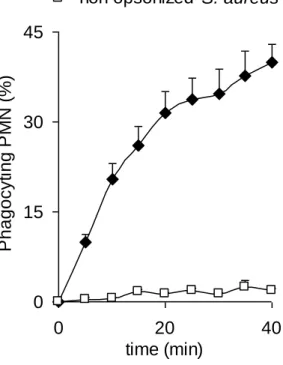
immunoglobulins or complement fragments augments the rate of phagocytosis by orders of magnitude (Fig. 1.). Isolated PMN are able to engulf up to 50 opsonized bacteria within 30 min (Fig 2.). Phagocytosed microorganisms are sequestered into phagosomes, that are sealed, membrane-surrounded compartments. Assembly and activation of the NADPH oxidase and opening of ion channels in the phagosomal membrane lead to abundant production of superoxide (O2.-), the first component of a cascade of reactive oxygen species (ROS).
Simultaneously, the membranes of different granule fractions fuse with the phagosomal membrane and the granule contents are released to the phagosome. Killing and degradation of the microorganism proceed in the small phagosomal space in a concentrated action of the involved ingredients, although the contribution of the individual factors may vary depending on the type of microorganism [89,91]. The molecular details of these processes have been worked out and summarized in recent excellent reviews [11,58,66,75,77,78,80,102,115].
Human pathology indicates the vital necessity of all the above steps as any disturbance of the migration, superoxide production, opsonization or granule production results in serious diseases with repeated, often life-threatening infections [30,46,52,100,112].
The established view of the neutrophils as effector cells coming into play only in the late phase of the immune response and having a single – although vital – task has been largely changed in the last one and a half decades and PMN are shown to play a role in divergent, unexpected functions. Also striking new properties of neutrophils have been discovered. This review highlights some of the new aspects and refers the reader to more specialized papers on these topics. Then we focus on the latest data revealing the contribution of PMN-derived extracellular vesicles both to intercellular communication and to antibacterial defense.
Recently discovered properties and functions of neutrophils
Protein synthesis
Neutrophils were regarded as terminally differentiated cells with very low level, if any, of protein synthesis. This view has been challenged in the 1990s, when it was discovered that stimulated PMN were able to synthetize and release various cytokines [19]. In early studies isolated neutrophils have been activated by lipopolysaccharide (LPS) and release of tumor necrosis factor (TNFα), interleukin 1β (IL-1β), IL-8, transforming growth factor β1 (TGF-β1) or IL-1 receptor antagonist (IL-1ra) was detected [19]. Later PMN were reported to produce a true arsenal of regulatory molecules, such as different chemokines, various pro- and anti-inflammatory cytokines, colony stimulating factors (CSF), and others [66]. The triggers consist of bacterial products as well as CSF, TNFα or interferons (IFN). Human and murine neutrophils differ, however, in the production of several cytokines [66].
Cytokine production expressed as amount per cell was significantly lower in PMN than in monocytes or lymphocytes, but in blood neutrophils largely outnumber any other type of leukocyte. In addition, neutrophils are the first cells that accumulate at an inflammatory site. Thus, cytokine production by neutrophils seemed to have physiological relevance.
Indeed, already the first in vivo experiments carried out in various animal models of inflammation demonstrated clearly the de novo synthesis of several cytokines both at the mRNA and at the protein level [99]. Determination of the gene expression profile of neutrophils from the bone marrow, circulating blood or a skin lesion of mice indicated that transmigration through the vessel wall initiates a new transcription program leading to the synthesis of regulatory and cell-fate determining proteins [11,105,106]. Phagocytosis of opsonized particles via Fc and/or complement receptors also altered the expression of numerous genes, many of them affecting proteins involved in apoptotic pathways [56,57].
Interaction with other immune cells
Neutrophils were regarded as effector cells of the humoral immune response, which eliminate certain pathogens marked by immunoglobulin or complement fragments, without any significant interaction with other immune cells. Discovery of the cytokine release from the neutrophils has profoundly changed this view [66,75].
Neutrophils produce IL-8, a peptide with strong chemotactic effect on neutrophils. A positive feedback via IL-8 may thus contribute to the fast migration of large numbers of neutrophils to inflammatory sites. Other chemokines secreted by PMN are chemoattractants to monocytes [102] or lymphocytes [66] triggering the second wave of cell migration, mostly of mononuclear cells. Furthermore, activated neutrophils produce lipid mediators such as
resolvins and protectins that inhibit neutrophil recruitment [25,58,66] and „find me” and „eat me” signals that initiate the elimination of aged PMN by macrophages [14].
In addition to chemotactic direction of migration, more substantial cross-talk has been demonstrated with all cell types of the immune reactions [66,75]. Some of these interactions are direct, like the effect of B-cell activating factor (BAFF) on survival, maturation and differentiation of B-cells [98], or activation of monocyte-derived dendritic cells (DC) via the integrin Mac1 and DC-SIGN (DC-specific ICAM3-grabbing non-integrin) [113]. In case of NK cells both a direct and an indirect activation by neutrophils has been described, the latter involving a PMN-DC-NK triangle including both contact and humoral effects [24].
Neutrophils are also able to interact with different subsets of T cells, and these interactions seem to be mostly reciprocal [66]. Cross-talk between neutrophils and other immune cells was not only observed in vitro, but has been supported in several animal models [66] and in cases of human pathology [75].
Neutrophils have even been proposed as antigen presenting cells. Earlier studies indicated that neutrophils were able to present exogenous antigen to CD8+ T-cells [7] and recent data have shown also antigen-presentation to Th1 and Th17 cells [2]. In confocal microscopic studies neutrophils migrating with fluorescently labeled pathogen were observed in draining lymph vessels [1] and lymph nodes [21]. Moreover, the chemokine receptor CCR7 has been identified as a key player in directing neutrophil migration from the interstitium to the draining lymph nodes [6].
Taken together, experimental evidence supports that neutrophils interact in a complex way with all the players of and exert a regulatory influence on the adaptive immune response.
Lifespan
Neutrophils are regarded as cells with a very short lifespan, spending 8 to 12 hours in the circulation [103]. If they are not involved in inflammatory processes, they undergo spontaneous apoptosis. However, various stimuli, such as cytokines (G-CSF, GM-CSF) and bacterial products were shown to prolong their survival [23]. Most recent in vivo
measurements estimate an extended half-life up to 5 days for circulating neutrophils [83], although there had been concerns about these data [64].
Interestingly, proteins known to regulate the cell cycle in dividing cells, such as survivin, cyclin-dependent kinases or proliferating cell nuclear antigen (PCNA) were
discovered to regulate the survival of neutrophils [119,120]. In contrast to proliferating cells,
in PMN these proteins are localized to the cytosol and act as inhibitors of the apoptotic pathway [120].
Additional functions of granule proteins
Neutrophils possess 4 different types of granules that develop in different stages of maturation in the bone marrow [11]. The contents of these granules are regarded as specific neutrophil proteins involved in killing and degradation of phagocytosed pathogens. However, some of the neutrophil granule proteins have been recently described in other functions or at other locations. As summarized in Table 1, neutrophil granule proteins also affect cell adhesion and migration, apoptosis, or clearance of apoptotic cells. Some of the hydrolases were shown to degrade extracellular (tissue factor pathway inhibitor, TFPI) or intracellular (insulin receptor substrate-1, IRS-1) proteins and influence thereby coagulation and insulin resistance, resp. Selected neutrophil granule proteins are also expressed upon stimulation of endothelial or epithelial cells [11].
Participation of PMN in vital function other than defense against bacteria and fungi Neutrophils were regarded as cells specialized in elimination of pathogens, mainly bacteria and fungi. In recent years their antimicrobial spectrum has been broadened by demonstration of their involvement in defense against HIV-1 [97], mycobacteria [68] and parasites such as malaria [85] and Leishmania [75,82].
However, the most surprising data support involvement of neutrophils in a vast array of different physiological and pathological processes. PMN were shown to participate in anaphylactic reactions initiated by IgG via murine FcγRIV or human FcγRIIA and mediated by release of platelet activating factor (PAF) from neutrophils [50].
Neutrophils are suggested to participate in atherogenesis as their presence has been detected in atherosclerotic plaques and PMN-depletion resulted in a reduction of the size of lesion [32,75]. Moreover, the neutrophil granule protein cathelicidin LL37 was reported to promote adhesion of monocytes to the vessel wall [31].
Insulin resistance is related to obesity, partly due to alteration of insulin signaling pathway by inflammatory mediators produced in the adipose tissue. Recently, significant accumulation of neutrophils has been demonstrated in adipose tissue and degradation of the signaling molecule IRS-1 was attributed to neutrophil-derived elastase [104].
Neutrophils were also shown to be involved in thrombus formation. They appear at the site of vessel injury instantaneously [27,116] and activated neutrophils express tissue factor [70]. PMN-depletion resulted in serious reduction of thrombus formation [27,116].
Last but not least, neutrophils have been implicated in modulation of tumor growth via several potential mechanisms [66].
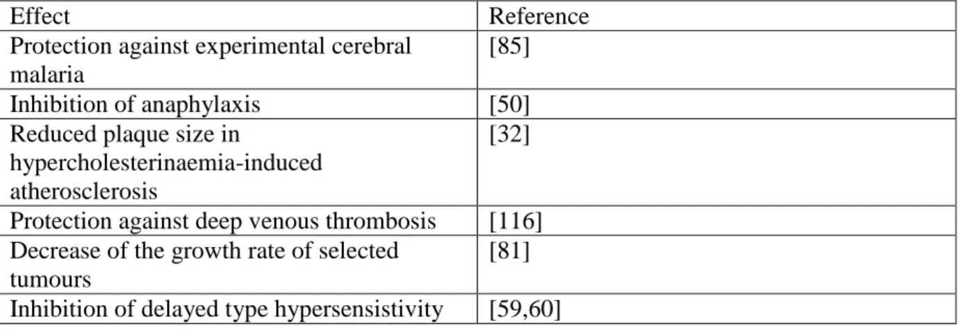
Many of the above results have been obtained or supported by detecting the changes following depletion of neutrophils in various animal models. Some remarkable examples are summarized in Table 2.
Extracellular killing by neutrophils
Discovery of the neutrophil extracellular traps (NETs) changed our view on bacterial killing and revealed that neutrophils were also capable to eliminate microorganisms in the extracellular space [15]. NETs represent filamentous structures composed of DNA, histones, granule proteins (mainly elastase and myeloperoxidase) and a few cytosolic proteins. They are formed by a unique mechanism of cell death, called NETosis, when intracellular membranes become disintegrated and granule proteins get access to the nucleus. Eventually, the plasma membrane ruptures and the protein-nucleic acid complex is released [16]. Formation of NETs requires superoxide and other ROS formed thereof. Neutrophils from patients with defective NADPH oxidase (chronic granulomatous disease, CGD) are unable to form NETs, but the process can be rescued by addition of H2O2 [37].
NETs were shown to kill various bacteria and Candida albicans [15,110] but most recently they were reported to be also involved in defense against human immunodeficiency virus [97]. The mechanism of microbe elimination is not fully understood but probably relies on multiple parallel pathways. Trapping of microorganisms prevents their dissemination and this phase may be based on surface charge interactions. Antibacterial proteins concentrated in NETs, such as histones, defensins and pentraxins, may have a direct toxic effect whereas granule enzymes, such as lysozyme or elastase, may destroy the microbial cell wall or virulence factors. The antimicrobial effect of NETs is critically dependent on the intact DNA structure, as it is inhibited by DNase treatment [15].
In vitro microbe killing effect of NETs was elegantly supported and extended by visualization of live neutrophils by confocal intravital microscopy. Upon bacterial challenge of the skin, extravascular NET formation was observed. In contrast to the in vitro data, in live tissues NET formation occurred within minutes and did not involve the death of neutrophils.
Instead, viable but anuclear granulocytes continued to crawl in a chemical gradient and to phagocytose [124]. In a septic model, rapid platelet-dependent NET formation was observed in liver sinusoids that increased trapping of bacteria by four-fold [71]. NETs were also shown to be formed and to be effective in certain viral infections [49]. In all these experiments, destruction of the DNA scaffold of NETs by iv. administered DNase resulted in an increase of bacteremia or number of virus-infected cells [49,71,124]. Thus, NET formation clearly
prevents dissemination of microorganisms.
In line with the previous observations, surface expression of nucleases is a critical component of pathogenicity for some bacteria [17] and dissemination of different strains correlated with the type of expressed DNase [117]. Furthermore, in a case of severe Aspergillosis in a CGD patient, negative correlation was found between the ability of neutrophils to form NETs and the severity of infection [10].
In addition to their role in killing of different microorganisms, in recent years NETs were reported to be involved in several other physiological and pathological processes. They were shown to activate dendritic cells via TLR9 receptors [38,61], linking thereby innate and adaptive immune processes. Activation of platelets via TLR2 and TLR4 receptors [101] and degradation of coagulation inhibitory proteins by proteases on the surface of NETs both promote blood clotting which also impairs bacterial dissemination [51,69]. On the other hand, formation of NETs was suggested to contribute to the viscosity of bronchial fluid in cystic fibrosis patients [67], to formation of autoantibodies in systemic lupus erythematosus [44] and to the pathogenesis of autoimmune vasculitis [54]. Most recently, the presence of NETs was demonstrated in atherosclerotic plaques in murine carotid arteries and in human tissues removed by endarterectomy [72] substantiating the involvement of neutrophils in
atherogenesis. Disruption of NETs in liver sinusoids by iv. DNase diminished liver damage in septic conditions [71].
Extracellular vesicles: new form of intercellular communication
The first mentioning about extracellular vesicles (EV) was in 1967 when „platelet dust” was described [121]. In the following years there were only sporadic publications, but the field has seen a real boom in the last decade. Extracellular vesicles are seen today as a common way of intercellular communication. Every investigated cell type is able to produce some form of extracellular vesicles, both spontaneously and upon stimulation. EVs are
present in all the body fluids, blood, urine, cerebrospinal fluid and milk having been the most intensively studies [18,107].
The size of EVs varies from approx. 30 nm to more than 1 µm. The smaller vesicles, having a diameter below 100 nm, are mostly referred to as exosomes, they arise from multivesicular bodies. The larger vesicles are named – depending on the authors - microvesicles, ectosomes or microparticles, and they are formed mostly by budding or shedding from the plasma membrane [22,43,107]. However, it seems that the size of EVs presents more a continuous spectrum than discrete populations.
Detection of EVs is based mostly on flow cytometric, light scattering or electric resistance measurements. All approaches present potential problems [43]. In flow cytometry the distinction of the smaller vesicles from the noise can be a hard task. Furthermore, immune complexes were shown to overlap with smaller vesicles [42], thus the vesicular structure should always be verified. Most convenient – and reliable – is the detection of vesicles above 300 nm stained with specific fluorescent ligand. Recently, the availability of convenient apparatus based on detection of light scattering or changes of the electric field resulted in the wide-spread usage of vesicle number as the unique parameter for characterization of a given preparation. However, the danger of vesicle fusion, presence of immune complexes or
aggregated proteins or contamination with viruses requires careful analysis of the preparation.
In our view, the safe approach is still the parallel usage of multiple techniques including electron microscopy for characterization and visualization of the EV preparations.
The composition of EVs is varied. They contain differently enriched collection of both membrane and cytosolic constituents. The discovery of their nucleic acid – mostly mRNA and miRNA – content raised wide perspectives for their application in diagnostics and therapy [111]. In our own experience, the same cells are able to produce EVs of different composition and different functional properties depending on the type of stimulus applied [109].
Differences between spontaneously formed and induced EVs has also been indicated in another study [47]
The physiological or pathological functions related to EVs present a long and colourful list. One of the earliest generally recognized functions was increase of blood coagulation initiated by tissue factor on EVs derived of endothelial cells and platelets.
Various tumor cells also produce large numbers of EVs with tissue factor and this could be part of the increase of coagulation generally observed in patients with tumor of different origin [22]. Secretion of leaderless proteins (e.g. IL-1β) occurs in EVs, too [4,88].
Transfer of plasma membrane receptors and proteins has also been demonstrated to occur via EVs. One of the earliest well-documented observations was that exosomes derived from dendritic cells were able to successfully present antigen to T-lymphocytes [90]. In other studies, macrophage-derived exosomes were reported to transfer pathogen-associated
molecular patterns of opportunistic intracellular pathogens to uninfected cells [9]. Perhaps the most striking result was the demonstration that oncogenic EGFR of glioblastoma cells was transferred to healthy cells, where it became functional and induced proliferation [3]. Transfer of chemokine receptors [65], FasL [55] and tissue factor [29] via EVs was also reported.
Transfer of miRNA has been demonstrated in many cell types [114].
Detailed knowledge is still missing on cell biological processes leading to formation and release of EVs as well as on their fate. However, their existence and role in
communication between different cell types can not be neglected.
Role of PMN-derived vesicles in communication
The first report on PMN-derived EVs (named in that paper microparticles) indicated their ability to increase secretion of the cytokine IL-6 from endothelial cells [73]. In a later work it has been demonstrated, that leukocyte-derived EVs were present in blood samples of healthy volunteers and the endogenous EVs were also able to upregulate IL-6 production and tissue factor expression in cultured endothelial cells [74]. In both studies an increase of EV number and effect was achieved by the chemotactic agent fMLP. Anti-neutrophil cytoplasmic antibodies (ANCA) also stimulate the release of microparticles from neutrophils, and these EVs activate endothelial cells to express intercellular adhesion molecule-1 and to secrete IL-6 and IL-8 [47].
Most of the data on PMN-derived EVs (called in these studies ectosomes) come from the laboratory of Dr. Schifferli [26,34-36,39,40,45,93-96]. They characterized the size and surface properties of EVs released upon fMLP-stimulation and came to the conclusion that both the in vitro and the in vivo generated EVs are right-side out [39,45]. Further, they demonstrated that PMN-derived EVs increased the secretion of the anti-inflammatory cytokine TGF-β1 from monocytes, whereas the stimulated release of IL-8, IL-10 and TNFα was decreased [40]. Secretion of TGF-β1 was induced also in monocyte-derived dendritic cells, where incubation with PMN-EVs interfered with the LPS-induced differentiation process indicated by a decrease of release of inflammatory cytokines, decrease of
phagocytosis and T-cell stimulation [34]. The inhibitory effects were initiated by membrane components of PMN-EVs which led to the activation of the Mer tyrosine kinase pathway in
monocytes, resulting eventually in inhibition of transcription factor NF-κB and down- regulation of several pro-inflammatory genes [35]. The rapid release of stored TGF-β1 was independent of the MerTK pathway whereas phosphatidylserine (PS) exposed on the surface of PMN-EVs was necessary but not sufficient for induction of TGF-β1 release [36].
Thus, EVs released from PMN upon chemotactic stimuli communicate a signal to monocytes and monocytic dendritic cells, reducing the inflammatory and favouring the anti- inflammatory response of these cells. However, the specificity of PMN-derived EVs in this process remains to be determined, as EVs produced by stored erythrocytes [94] and platelets [93] have also been reported to down-regulate macrophages and dendritic cells.
In line with the inhibitory effect of PMN-derived EVs upon macrophages, ectosomes released from Mycobacterium tuberculosis infected neutrophils decreased the activation of macrophages and prolonged the survival of M. tuberculosis [33]. In a recent study, PMN- derived microparticles were shown to stimulate efferocytosis and production of pro-resolving lipid mediators in macrophages [25].
Neutrophil-derived EVs were also shown to have prothrombotic effects. An increased number of platelet- and PMN-derived microparticles were found in the blood plasma of patients suffering from meningococcal sepsis. These microparticles expressed tissue factor and supported thrombin generation [79]. In another study, stimulation of neutrophils with bacterial endotoxin resulted in shedding of PAF containing microparticles which activated platelets [118]. More recently, the active form of the β2 integrin Mac-1was detected on the surface of EVs released from activated neutrophils, and the active integrin played a central role in binding to and activation of resting platelets [84].
PMN-derived EV with antibacterial effect
We observed that in isolated PMN different stimulating agents induced the formation of different number of EVs with different composition and different functional properties [109]. Particles (bacteria or zymosan) opsonized with pooled serum initiated the release of microvesicles in the highest quantity whereas chemotactic agents (fMLP, CXCL12),
cytokines (TNFα) or lipopolysaccharide (LPS) did not significantly increase the generation as compared to the spontaneous production. Other reports also demonstrated the release of EVs from neutrophils phagocytosing different pathogens [33,41].
Importantly, PMN-derived EVs initiated by opsonized particles were able to impair the growth of bacteria whereas EVs produced spontaneously or upon other stimuli did not interfere with bacterial growth (Fig.3). In induction of the formation of EVs with antibacterial
effect, opsonization of the particles with full serum was critical: heat-inactivation of complement factors resulted in generation of a large number of EV without antibacterial effect (Fig. 3).
The size of PMN-derived EVs ranged from 200 to 800 nm and did not depend on the type of initiating agent. In contrast, the composition of PMN-derived EVs with antibacterial effect differed significantly from that of ineffective EVs. Antibacterial EVs (aEV) were enriched in PMN granule proteins and β2 integrins. All types of PMN-derived EVs contained metabolic enzymes, many cytoskeletal proteins, some plasma membrane receptors and the membrane components of NADPH oxidase, however the cytosolic oxidase components were mostly missing. Apparently, formation of aEV is associated to specific sorting of cell
constitutents into the released EV.
The effect of phagocytosis-induced PMN-derived aEVs proved to be bacteriostatic rather than bacteriocidal and the mechanism of action differed in many respects from the effect of intact PMN (Table 3). PMN-derived aEVs did not engulf bacteria, although their size was comparable or even larger than that of bacteria. PMN-derived aEVs did not produce superoxide and their effect was not influenced by the inhibitor of NADPH oxidase, diphenyl- iodonium (DPI) (Fig. 4A). This observation is substantiated by the lack of the cytosolic components of the NADPH oxidase in aEVs. The most striking difference between the effect of PMN and antibacterial EVs was the total independence of opsonization in the latter case (Fig. 4B). Whether bacteria were opsonized in full serum or in complement-deficient serum or not opsonized at all, their growth was impaired to the same extent by antibacterial PMN- derived vesicles. As PMN have a very low killing activity for non-opsonized bacteria (Fig.
1.), under such conditions, PMN-derived antibacterial EVs proved to be significantly more effective than PMN themselves.
Finally, the spectrum of attacked bacteria is probably also different. Antibacterial EVs were effective against Staphylococcus aureus and Escherichia coli, but not against Proteus.
mirabilis, thus they showed some selectivity, which was however not paralleled with Gram staining.
The mechanism of action of PMN-derived antibacterial EVs is strongly associated with their ability to form large aggregates with bacteria (Fig. 5B.). In this process both the number and size of aggregates seem to be important. Summarizing all the experimental conditions investigated, a fairly good negative correlation was obtained between the
proportion of large (larger than 1.5 µm) aggregates and bacterial growth. Formation of large aggregates with bacteria seems to depend both on surface charge and density of β2 integrin on
PMN-derived EVs and requires continuous rearrangement of the actin cytoskeleton (Fig. 5C and D).
The impairment of bacterial growth by PMN-derived aEVs occurred in the
extracellular space and in this respect it is similar to the effect of NETs. However, there are numerous differences between NETs and antibacterial EVs (Table 3). Formation and hence effect of NETs depends on superoxide and ROS formed thereof whereas both production and effect of antibacterial EVs is independent of ROS. Generation of NETs requires firm
attachment of PMNs to a surface whereas antibacterial EVs were released equally well from suspended or adherent neutrophils. NETs also critically depend on intact DNA structure and can be disrupted by DNase treatment, whereas neither generation nor the antibacterial effect of PMN-derived EVs was influenced by DNase treatment. The kinetics of in vitro formation and the initiating agents are also widely different. Finally, the antibacterial effect of EVs depends on the intact vesicular structure, intact cytoskeletal organization (Fig. 5) and glucose supply whereas all these are not required for the antibacterial effect of NETs. However, involvement of granule proteins may be a common property: effect of NETs was shown to depend on granule proteases, and EVs are also enriched in these proteins, suggesting their functional role.
In vivo relevance of PMN-derived EVs
Blood serum contains EVs derived of many – if not all – cell types. The serum of healthy individuals contains also a detectable amount of PMN-derived EVs. Investigated under ex vivo conditions, these vesicles did not form aggregates with bacteria and did not interfere with their growth.
In bacteremic states, an increase in the number of PMN-derived vesicles has been reported. A six fold enrichment of PMN-derived EVs was reported in the blood serum of patients infected with S. aureus [109] and over 100-fold increase was detected in patients with meningococcal sepsis [79]. In 3 patients with acute peritonitis, the peritoneal fluid contained over tenfold higher number of PMN-derived microparticles than the control fluid from uninfected patients [87]. An increase of PMN-derived EVs was also reported in different inflammatory diseases (Table 4). Thus, bacterial stimulation results in an enhancement of EV- production also in circulating PMN, similarly to our observation made on isolated PMN.
Importantly, in our experiments, the PMN-derived EVs isolated from the serum of bacteremic patients infected with S. aureus, formed ex vivo large aggregates with added bacteria, similar to the observation made on antibacterial EVs initiated from isolated PMN.
Whether similar aggregates are formed between PMN-derived EV and bacteria in vivo, is presently not known.
The biological significance of the antibacterial effect of PMN-derived EVs detected under in vitro condition can not be assessed at present. The observation that antibacterial EVs are fully effective against non-opsonized bacteria, may represent an important factor in the early phase of innate immune reactions. However, on the basis of theoretical calculations, the effect of EVs against opsonized bacteria may also not be negligible. When presented as cell- equivalent, the antibacterial capacity of EVs against opsonized bacteria was smaller than that of PMN and was saturated by lower bacterial load. However, the protein content of EVs is rather low and if we relate the impairment of bacterial growth to protein content of PMN or EV, then we obtain a tenfold higher relative antibacterial capacity for EVs than for PMN. It is thus possible, that PMNs activated by opsonized bacteria, package their antibacterial arsenal into released vesicles that could capture the pathogen and impair its dissemination. Both NETs and blood clotting were shown earlier to prevent pathogen dissemination very effectively [69,71].
PMN-derived EVs with antibacterial capacity may thus represent another PMN-related extracellular mechanism to combat infectious agents.
Conclusion
After several decades, when the attention of immunologists was focused first on various subpopulations of lymphocytes as central players in adaptive immune processes, then later on exciting receptors of innate immunity, in the last period neutrophils have been
rediscovered. Several striking properties have been described and participation in a broad spectrum of unexpected functions has been revealed. Discovery of NETs that provide an extracellular mechanism in the fight against microorganisms was accepted first with certain skepticism. It took almost a decade till the in vivo significance of NET formation starts to be clarified. It will take certainly years before the true biological role of extracellular vesicles can be assessed. However, it is evident already now that the neutrophils are multifaceted cells that are involved in many more functions than phagocytosis and elimination of engulfed
microorganisms.
Acknowledgement
The authors are indebted to Professors W. Nauseef for providing green-fluorescent bacteria and Peter Enyedi for critical reading of the manuscript. Experimental work in the authors’ laboratory was supported by grants from the Hungarian Research Fund (OTKA K81277 and K75084).
References
1. Abadie V, Badell E, Douillard P, Ensergueix D, Leenen PJ, Tanguy M, Fiette L, Saeland S, Gicquel B, Winter N (2005) Neutrophils rapidly migrate via lymphatics after
Mycobacterium bovis BCG intradermal vaccination and shuttle live bacilli to the draining lymph nodes. Blood 106 (5):1843-1850. doi:blood-2005-03-1281 2. Abi Abdallah DS, Egan CE, Butcher BA, Denkers EY (2011) Mouse neutrophils are
professional antigen-presenting cells programmed to instruct Th1 and Th17 T-cell differentiation. Int Immunol 23 (5):317-326. doi:10.1093/intimm/dxr007
3. Al-Nedawi K, Meehan B, Micallef J, Lhotak V, May L, Guha A, Rak J (2008) Intercellular transfer of the oncogenic receptor EGFRvIII by microvesicles derived from tumour cells. Nat Cell Biol 10 (5):619-624. doi:10.1038/ncb1725
4. Andrei C, Dazzi C, Lotti L, Torrisi MR, Chimini G, Rubartelli A (1999) The secretory route of the leaderless protein interleukin 1beta involves exocytosis of endolysosome- related vesicles. Mol Biol Cell 10 (5):1463-1475
5. Barlow PG, Li Y, Wilkinson TS, Bowdish DM, Lau YE, Cosseau C, Haslett C, Simpson AJ, Hancock RE, Davidson DJ (2006) The human cationic host defense peptide LL-37 mediates contrasting effects on apoptotic pathways in different primary cells of the innate immune system. J Leukoc Biol 80 (3):509-520. doi:10.1189/jlb.1005560 6. Beauvillain C, Cunin P, Doni A, Scotet M, Jaillon S, Loiry ML, Magistrelli G, Masternak
K, Chevailler A, Delneste Y, Jeannin P (2011) CCR7 is involved in the migration of neutrophils to lymph nodes. Blood 117 (4):1196-1204. doi:10.1182/blood-2009-11- 254490
7. Beauvillain C, Delneste Y, Scotet M, Peres A, Gascan H, Guermonprez P, Barnaba V, Jeannin P (2007) Neutrophils efficiently cross-prime naive T cells in vivo. Blood 110 (8):2965-2973. doi:10.1182/blood-2006-12-063826
8. Berckmans RJ, Nieuwland R, Tak PP, Boing AN, Romijn FP, Kraan MC, Breedveld FC, Hack CE, Sturk A (2002) Cell-derived microparticles in synovial fluid from inflamed arthritic joints support coagulation exclusively via a factor VII-dependent mechanism.
Arthritis Rheum 46 (11):2857-2866. doi:10.1002/art.10587
9. Bhatnagar S, Shinagawa K, Castellino FJ, Schorey JS (2007) Exosomes released from macrophages infected with intracellular pathogens stimulate a proinflammatory response in vitro and in vivo. Blood 110 (9):3234-3244. doi:10.1182/blood-2007-03- 079152
10. Bianchi M, Hakkim A, Brinkmann V, Siler U, Seger RA, Zychlinsky A, Reichenbach J (2009) Restoration of NET formation by gene therapy in CGD controls aspergillosis.
Blood 114 (13):2619-2622. doi:10.1182/blood-2009-05-221606
11. Borregaard N, Sorensen OE, Theilgaard-Monch K (2007) Neutrophil granules: a library of innate immunity proteins. Trends Immunol 28 (8):340-345.
doi:10.1016/j.it.2007.06.002
12. Bournazou I, Mackenzie KJ, Duffin R, Rossi AG, Gregory CD (2010) Inhibition of eosinophil migration by lactoferrin. Immunol Cell Biol 88 (2):220-223.
doi:10.1038/icb.2009.86
13. Bournazou I, Pound JD, Duffin R, Bournazos S, Melville LA, Brown SB, Rossi AG, Gregory CD (2009) Apoptotic human cells inhibit migration of granulocytes via release of lactoferrin. J Clin Invest 119 (1):20-32. doi:10.1172/JCI36226
14. Bratton DL, Henson PM (2011) Neutrophil clearance: when the party is over, clean-up begins. Trends Immunol 32 (8):350-357. doi:10.1016/j.it.2011.04.009
15. Brinkmann V, Reichard U, Goosmann C, Fauler B, Uhlemann Y, Weiss DS, Weinrauch Y, Zychlinsky A (2004) Neutrophil extracellular traps kill bacteria. Science 303 (5663):1532-1535. doi:10.1126/science.1092385 303/5663/1532
16. Brinkmann V, Zychlinsky A (2012) Neutrophil extracellular traps: is immunity the second function of chromatin? J Cell Biol 198 (5):773-783. doi:10.1083/jcb.201203170 17. Buchanan JT, Simpson AJ, Aziz RK, Liu GY, Kristian SA, Kotb M, Feramisco J, Nizet V
(2006) DNase expression allows the pathogen group A Streptococcus to escape killing in neutrophil extracellular traps. Curr Biol 16 (4):396-400.
doi:10.1016/j.cub.2005.12.039
18. Camussi G, Deregibus MC, Bruno S, Cantaluppi V, Biancone L (2010)
Exosomes/microvesicles as a mechanism of cell-to-cell communication. Kidney Int 78 (9):838-848. doi:10.1038/ki.2010.278
19. Cassatella MA (1995) The production of cytokines by polymorphonuclear neutrophils.
Immunol Today 16 (1):21-26. doi:0167-5699(95)80066-2
20. Cassatella MA (2003) The Neutrophil. An Emerging Regulator of Inflammatory and Immune Response. Karger, Basel,
21. Chtanova T, Schaeffer M, Han SJ, van Dooren GG, Nollmann M, Herzmark P, Chan SW, Satija H, Camfield K, Aaron H, Striepen B, Robey EA (2008) Dynamics of neutrophil migration in lymph nodes during infection. Immunity 29 (3):487-496.
doi:10.1016/j.immuni.2008.07.012
22. Cocucci E, Racchetti G, Meldolesi J (2009) Shedding microvesicles: artefacts no more.
Trends Cell Biol 19 (2):43-51. doi:10.1016/j.tcb.2008.11.003
23. Colotta F, Re F, Polentarutti N, Sozzani S, Mantovani A (1992) Modulation of
granulocyte survival and programmed cell death by cytokines and bacterial products.
Blood 80 (8):2012-2020
24. Costantini C, Calzetti F, Perbellini O, Micheletti A, Scarponi C, Lonardi S, Pelletier M, Schakel K, Pizzolo G, Facchetti F, Vermi W, Albanesi C, Cassatella MA (2011) Human neutrophils interact with both 6-sulfo LacNAc+ DC and NK cells to amplify NK-derived IFN{gamma}: role of CD18, ICAM-1, and ICAM-3. Blood 117 (5):1677- 1686. doi:10.1182/blood-2010-06-287243
25. Dalli J, Serhan CN (2012) Specific lipid mediator signatures of human phagocytes:
microparticles stimulate macrophage efferocytosis and pro-resolving mediators. Blood 120 (15):e60-72. doi:10.1182/blood-2012-04-423525
26. Daniel L, Fakhouri F, Joly D, Mouthon L, Nusbaum P, Grunfeld JP, Schifferli J, Guillevin L, Lesavre P, Halbwachs-Mecarelli L (2006) Increase of circulating neutrophil and platelet microparticles during acute vasculitis and hemodialysis. Kidney Int 69 (8):1416-1423. doi:10.1038/sj.ki.5000306
27. Darbousset R, Thomas GM, Mezouar S, Frere C, Bonier R, Mackman N, Renne T, Dignat-George F, Dubois C, Panicot-Dubois L (2012) Tissue factor-positive
neutrophils bind to injured endothelial wall and initiate thrombus formation. Blood 120 (10):2133-2143. doi:10.1182/blood-2012-06-437772
28. Deban L, Russo RC, Sironi M, Moalli F, Scanziani M, Zambelli V, Cuccovillo I, Bastone A, Gobbi M, Valentino S, Doni A, Garlanda C, Danese S, Salvatori G, Sassano M, Evangelista V, Rossi B, Zenaro E, Constantin G, Laudanna C, Bottazzi B, Mantovani A (2010) Regulation of leukocyte recruitment by the long pentraxin PTX3. Nat Immunol 11 (4):328-334. doi:10.1038/ni.1854
29. Dewalt RI, Petkovich DA, Zahrt AN, Bruns HA, McDowell SA (2013) Host cell invasion by Staphylococcus aureus stimulates the shedding of microvesicles. Biochem Biophys Res Commun. doi:10.1016/j.bbrc.2013.01.122
30. Dinauer MC (2007) Disorders of neutrophil function: an overview. Methods Mol Biol 412:489-504. doi:10.1007/978-1-59745-467-4_30
31. Doring Y, Drechsler M, Wantha S, Kemmerich K, Lievens D, Vijayan S, Gallo RL, Weber C, Soehnlein O (2012) Lack of neutrophil-derived CRAMP reduces atherosclerosis in mice. Circ Res 110 (8):1052-1056.
doi:10.1161/CIRCRESAHA.112.265868
32. Drechsler M, Megens RT, van Zandvoort M, Weber C, Soehnlein O (2010)
Hyperlipidemia-triggered neutrophilia promotes early atherosclerosis. Circulation 122 (18):1837-1845. doi:10.1161/CIRCULATIONAHA.110.961714
33. Duarte TA, Noronha-Dutra AA, Nery JS, Ribeiro SB, Pitanga TN, Lapa ESJR, Arruda S, Boechat N (2012) Mycobacterium tuberculosis-induced neutrophil ectosomes decrease macrophage activation. Tuberculosis (Edinb) 92 (3):218-225.
doi:10.1016/j.tube.2012.02.007
34. Eken C, Gasser O, Zenhaeusern G, Oehri I, Hess C, Schifferli JA (2008)
Polymorphonuclear neutrophil-derived ectosomes interfere with the maturation of monocyte-derived dendritic cells. J Immunol 180 (2):817-824. doi:180/2/817 35. Eken C, Martin PJ, Sadallah S, Treves S, Schaller M, Schifferli JA (2010) Ectosomes
released by polymorphonuclear neutrophils induce a MerTK-dependent anti- inflammatory pathway in macrophages. J Biol Chem 285 (51):39914-39921.
doi:10.1074/jbc.M110.126748
36. Eken C, Sadallah S, Martin PJ, Treves S, Schifferli JA (2013) Ectosomes of
polymorphonuclear neutrophils activate multiple signaling pathways in macrophages.
Immunobiology 218 (3):382-392. doi:10.1016/j.imbio.2012.05.021
37. Fuchs TA, Abed U, Goosmann C, Hurwitz R, Schulze I, Wahn V, Weinrauch Y, Brinkmann V, Zychlinsky A (2007) Novel cell death program leads to neutrophil extracellular traps. J Cell Biol 176 (2):231-241. doi:10.1083/jcb.200606027
38. Garcia-Romo GS, Caielli S, Vega B, Connolly J, Allantaz F, Xu Z, Punaro M, Baisch J, Guiducci C, Coffman RL, Barrat FJ, Banchereau J, Pascual V (2011) Netting neutrophils are major inducers of type I IFN production in pediatric systemic lupus erythematosus. Sci Transl Med 3 (73):73ra20. doi:10.1126/scitranslmed.3001201 39. Gasser O, Hess C, Miot S, Deon C, Sanchez JC, Schifferli JA (2003) Characterisation and
properties of ectosomes released by human polymorphonuclear neutrophils. Exp Cell Res 285 (2):243-257. doi:S0014482703000557
40. Gasser O, Schifferli JA (2004) Activated polymorphonuclear neutrophils disseminate anti- inflammatory microparticles by ectocytosis. Blood 104 (8):2543-2548.
doi:10.1182/blood-2004-01-0361
41. Gonzalez-Cano P, Mondragon-Flores R, Sanchez-Torres LE, Gonzalez-Pozos S, Silva- Miranda M, Monroy-Ostria A, Estrada-Parra S, Estrada-Garcia I (2010)
Mycobacterium tuberculosis H37Rv induces ectosome release in human
polymorphonuclear neutrophils. Tuberculosis (Edinb) 90 (2):125-134.
doi:10.1016/j.tube.2010.01.002
42. Gyorgy B, Modos K, Pallinger E, Paloczi K, Pasztoi M, Misjak P, Deli MA, Sipos A, Szalai A, Voszka I, Polgar A, Toth K, Csete M, Nagy G, Gay S, Falus A, Kittel A, Buzas EI (2011) Detection and isolation of cell-derived microparticles are
compromised by protein complexes resulting from shared biophysical parameters.
Blood 117 (4):e39-48. doi:10.1182/blood-2010-09-307595
43. Gyorgy B, Szabo TG, Pasztoi M, Pal Z, Misjak P, Aradi B, Laszlo V, Pallinger E, Pap E, Kittel A, Nagy G, Falus A, Buzas EI (2011) Membrane vesicles, current state-of-the- art: emerging role of extracellular vesicles. Cell Mol Life Sci 68 (16):2667-2688.
doi:10.1007/s00018-011-0689-3
44. Hakkim A, Furnrohr BG, Amann K, Laube B, Abed UA, Brinkmann V, Herrmann M, Voll RE, Zychlinsky A (2010) Impairment of neutrophil extracellular trap degradation is associated with lupus nephritis. Proc Natl Acad Sci U S A 107 (21):9813-9818.
doi:10.1073/pnas.0909927107
45. Hess C, Sadallah S, Hefti A, Landmann R, Schifferli JA (1999) Ectosomes released by human neutrophils are specialized functional units. J Immunol 163 (8):4564-4573.
doi:ji_v163n8p4564 [pii]
46. Holland SM (2010) Chronic granulomatous disease. Clin Rev Allergy Immunol 38 (1):3- 10. doi:10.1007/s12016-009-8136-z
47. Hong Y, Eleftheriou D, Hussain AA, Price-Kuehne FE, Savage CO, Jayne D, Little MA, Salama AD, Klein NJ, Brogan PA (2012) Anti-neutrophil cytoplasmic antibodies stimulate release of neutrophil microparticles. J Am Soc Nephrol 23 (1):49-62.
doi:10.1681/ASN.2011030298
48. Houghton AM, Rzymkiewicz DM, Ji H, Gregory AD, Egea EE, Metz HE, Stolz DB, Land SR, Marconcini LA, Kliment CR, Jenkins KM, Beaulieu KA, Mouded M, Frank SJ, Wong KK, Shapiro SD (2010) Neutrophil elastase-mediated degradation of IRS-1 accelerates lung tumor growth. Nat Med 16 (2):219-223. doi:10.1038/nm.2084 49. Jenne CN, Wong CH, Zemp FJ, McDonald B, Rahman MM, Forsyth PA, McFadden G,
Kubes P (2013) Neutrophils recruited to sites of infection protect from virus challenge by releasing neutrophil extracellular traps. Cell Host Microbe 13 (2):169-180.
doi:10.1016/j.chom.2013.01.005
50. Jonsson F, Mancardi DA, Kita Y, Karasuyama H, Iannascoli B, Van Rooijen N, Shimizu T, Daeron M, Bruhns P (2011) Mouse and human neutrophils induce anaphylaxis. J Clin Invest 121 (4):1484-1496. doi:10.1172/JCI45232
51. Kambas K, Mitroulis I, Ritis K (2012) The emerging role of neutrophils in thrombosis-the journey of TF through NETs. Front Immunol 3:385. doi:10.3389/fimmu.2012.00385 52. Kang EM, Marciano BE, DeRavin S, Zarember KA, Holland SM, Malech HL (2011)
Chronic granulomatous disease: overview and hematopoietic stem cell transplantation.
J Allergy Clin Immunol 127 (6):1319-1326; quiz 1327-1318.
doi:10.1016/j.jaci.2011.03.028
53. Kantari C, Pederzoli-Ribeil M, Amir-Moazami O, Gausson-Dorey V, Moura IC, Lecomte MC, Benhamou M, Witko-Sarsat V (2007) Proteinase 3, the Wegener autoantigen, is externalized during neutrophil apoptosis: evidence for a functional association with phospholipid scramblase 1 and interference with macrophage phagocytosis. Blood 110 (12):4086-4095. doi:10.1182/blood-2007-03-080457
54. Kessenbrock K, Krumbholz M, Schonermarck U, Back W, Gross WL, Werb Z, Grone HJ, Brinkmann V, Jenne DE (2009) Netting neutrophils in autoimmune small-vessel vasculitis. Nat Med 15 (6):623-625. doi:10.1038/nm.1959
55. Kim JW, Wieckowski E, Taylor DD, Reichert TE, Watkins S, Whiteside TL (2005) Fas ligand-positive membranous vesicles isolated from sera of patients with oral cancer induce apoptosis of activated T lymphocytes. Clin Cancer Res 11 (3):1010-1020.
doi:11/3/1010
56. Kobayashi SD, Braughton KR, Whitney AR, Voyich JM, Schwan TG, Musser JM, DeLeo FR (2003) Bacterial pathogens modulate an apoptosis differentiation program in human neutrophils. Proc Natl Acad Sci U S A 100 (19):10948-10953.
doi:10.1073/pnas.1833375100
57. Kobayashi SD, Voyich JM, Buhl CL, Stahl RM, DeLeo FR (2002) Global changes in gene expression by human polymorphonuclear leukocytes during receptor-mediated phagocytosis: cell fate is regulated at the level of gene expression. Proc Natl Acad Sci U S A 99 (10):6901-6906. doi:10.1073/pnas.092148299
58. Kolaczkowska E, Kubes P (2013) Neutrophil recruitment and function in health and inflammation. Nat Rev Immunol 13 (3):159-175. doi:10.1038/nri3399
59. Kudo C, Yamashita T, Araki A, Terashita M, Watanabe T, Atsumi M, Tamura M, Sendo F (1993) Modulation of in vivo immune response by selective depletion of neutrophils using a monoclonal antibody, RP-3. I. Inhibition by RP-3 treatment of the priming and effector phases of delayed type hypersensitivity to sheep red blood cells in rats. J Immunol 150 (9):3728-3738
60. Kudo C, Yamashita T, Terashita M, Sendo F (1993) Modulation of in vivo immune response by selective depletion of neutrophils using a monoclonal antibody, RP-3. II.
Inhibition by RP-3 treatment of mononuclear leukocyte recruitment in delayed-type hypersensitivity to sheep red blood cells in rats. J Immunol 150 (9):3739-3746 61. Lande R, Ganguly D, Facchinetti V, Frasca L, Conrad C, Gregorio J, Meller S, Chamilos
G, Sebasigari R, Riccieri V, Bassett R, Amuro H, Fukuhara S, Ito T, Liu YJ, Gilliet M (2011) Neutrophils activate plasmacytoid dendritic cells by releasing self-DNA- peptide complexes in systemic lupus erythematosus. Sci Transl Med 3 (73):73ra19.
doi:10.1126/scitranslmed.3001180
62. Lee CY, Herant M, Heinrich V (2011) Target-specific mechanics of phagocytosis:
protrusive neutrophil response to zymosan differs from the uptake of antibody-tagged pathogens. J Cell Sci 124 (Pt 7):1106-1114. doi:10.1242/jcs.078592
63. Lee TD, Gonzalez ML, Kumar P, Grammas P, Pereira HA (2003) CAP37, a neutrophil- derived inflammatory mediator, augments leukocyte adhesion to endothelial
monolayers. Microvasc Res 66 (1):38-48. doi:S0026286203000104
64. Li KW, Turner SM, Emson CL, Hellerstein MK, Dale DC (2011) Deuterium and neutrophil kinetics. Blood 117 (22):6052-6053; author reply 6053-6054.
doi:10.1182/blood-2010-12-322271
65. Mack M, Kleinschmidt A, Bruhl H, Klier C, Nelson PJ, Cihak J, Plachy J, Stangassinger M, Erfle V, Schlondorff D (2000) Transfer of the chemokine receptor CCR5 between cells by membrane-derived microparticles: a mechanism for cellular human
immunodeficiency virus 1 infection. Nat Med 6 (7):769-775. doi:10.1038/77498 66. Mantovani A, Cassatella MA, Costantini C, Jaillon S (2011) Neutrophils in the activation
and regulation of innate and adaptive immunity. Nat Rev Immunol 11 (8):519-531.
doi:10.1038/nri3024
67. Manzenreiter R, Kienberger F, Marcos V, Schilcher K, Krautgartner WD, Obermayer A, Huml M, Stoiber W, Hector A, Griese M, Hannig M, Studnicka M, Vitkov L, Hartl D (2012) Ultrastructural characterization of cystic fibrosis sputum using atomic force and scanning electron microscopy. J Cyst Fibros 11 (2):84-92.
doi:10.1016/j.jcf.2011.09.008
68. Martineau AR, Newton SM, Wilkinson KA, Kampmann B, Hall BM, Nawroly N, Packe GE, Davidson RN, Griffiths CJ, Wilkinson RJ (2007) Neutrophil-mediated innate immune resistance to mycobacteria. J Clin Invest 117 (7):1988-1994.
doi:10.1172/JCI31097
69. Massberg S, Grahl L, von Bruehl ML, Manukyan D, Pfeiler S, Goosmann C, Brinkmann V, Lorenz M, Bidzhekov K, Khandagale AB, Konrad I, Kennerknecht E, Reges K, Holdenrieder S, Braun S, Reinhardt C, Spannagl M, Preissner KT, Engelmann B (2010) Reciprocal coupling of coagulation and innate immunity via neutrophil serine proteases. Nat Med 16 (8):887-896. doi:10.1038/nm.2184
70. Maugeri N, Brambilla M, Camera M, Carbone A, Tremoli E, Donati MB, de Gaetano G, Cerletti C (2006) Human polymorphonuclear leukocytes produce and express
functional tissue factor upon stimulation. J Thromb Haemost 4 (6):1323-1330.
doi:10.1111/j.1538-7836.2006.01968.x
71. McDonald B, Urrutia R, Yipp BG, Jenne CN, Kubes P (2012) Intravascular neutrophil extracellular traps capture bacteria from the bloodstream during sepsis. Cell Host Microbe 12 (3):324-333. doi:10.1016/j.chom.2012.06.011
72. Megens RT, Vijayan S, Lievens D, Doring Y, van Zandvoort MA, Grommes J, Weber C, Soehnlein O (2012) Presence of luminal neutrophil extracellular traps in
atherosclerosis. Thromb Haemost 107 (3):597-598. doi:10.1160/TH11-09-0650 73. Mesri M, Altieri DC (1998) Endothelial cell activation by leukocyte microparticles. J
Immunol 161 (8):4382-4387
74. Mesri M, Altieri DC (1999) Leukocyte microparticles stimulate endothelial cell cytokine release and tissue factor induction in a JNK1 signaling pathway. J Biol Chem 274 (33):23111-23118
75. Mocsai A (2013) Diverse novel functions of neutrophils in immunity, inflammation and beyond. J Exp Med in press
76. Nagaoka I, Tamura H, Hirata M (2006) An antimicrobial cathelicidin peptide, human CAP18/LL-37, suppresses neutrophil apoptosis via the activation of formyl-peptide receptor-like 1 and P2X7. J Immunol 176 (5):3044-3052. doi:176/5/3044
77. Nathan C (2006) Neutrophils and immunity: challenges and opportunities. Nat Rev Immunol 6 (3):9
78. Nauseef WM (2007) How human neutrophils kill and degrade microbes: an integrated view. Immunol Rev Oct. (219):14
79. Nieuwland R, Berckmans RJ, McGregor S, Boing AN, Romijn FP, Westendorp RG, Hack CE, Sturk A (2000) Cellular origin and procoagulant properties of microparticles in meningococcal sepsis. Blood 95 (3):930-935
80. Nourshargh S, Hordijk PL, Sixt M (2010) Breaching multiple barriers: leukocyte motility through venular walls and the interstitium. Nat Rev Mol Cell Biol 11 (5):12.
doi:10.1038/nrm2889
81. Pekarek LA, Starr BA, Toledano AY, Schreiber H (1995) Inhibition of tumor growth by elimination of granulocytes. J Exp Med 181 (1):435-440
82. Peters NC, Egen JG, Secundino N, Debrabant A, Kimblin N, Kamhawi S, Lawyer P, Fay MP, Germain RN, Sacks D (2008) In vivo imaging reveals an essential role for neutrophils in leishmaniasis transmitted by sand flies. Science 321 (5891):970-974.
doi:10.1126/science.1159194
83. Pillay J, den Braber I, Vrisekoop N, Kwast LM, de Boer RJ, Borghans JA, Tesselaar K, Koenderman L (2010) In vivo labeling with 2H2O reveals a human neutrophil lifespan of 5.4 days. Blood 116 (4):625-627. doi:10.1182/blood-2010-01-259028
84. Pluskota E, Woody NM, Szpak D, Ballantyne CM, Soloviev DA, Simon DI, Plow EF (2008) Expression, activation, and function of integrin alphaMbeta2 (Mac-1) on
neutrophil-derived microparticles. Blood 112 (6):2327-2335. doi:10.1182/blood-2007- 12-127183
85. Porcherie A, Mathieu C, Peronet R, Schneider E, Claver J, Commere PH, Kiefer-Biasizzo H, Karasuyama H, Milon G, Dy M, Kinet JP, Louis J, Blank U, Mecheri S (2011) Critical role of the neutrophil-associated high-affinity receptor for IgE in the pathogenesis of experimental cerebral malaria. J Exp Med 208 (11):2225-2236.
doi:10.1084/jem.20110845
86. Porro C, Lepore S, Trotta T, Castellani S, Ratclif L, Battaglino A, Di Gioia S, Martinez MC, Conese M, Maffione AB (2010) Isolation and characterization of microparticles in sputum from cystic fibrosis patients. Respir Res 11:94. doi:10.1186/1465-9921-11- 94
87. Prakash PS, Caldwell CC, Lentsch AB, Pritts TA, Robinson BR (2012) Human
microparticles generated during sepsis in patients with critical illness are neutrophil- derived and modulate the immune response. J Trauma Acute Care Surg 73 (2):401- 406; discussion 406-407. doi:10.1097/TA.0b013e31825a776d
88. Qu Y, Franchi L, Nunez G, Dubyak GR (2007) Nonclassical IL-1 beta secretion
stimulated by P2X7 receptors is dependent on inflammasome activation and correlated with exosome release in murine macrophages. J Immunol 179 (3):1913-1925.
doi:179/3/1913
89. Rada BK, Geiszt M, Kaldi K, Timar C, Ligeti E (2004) Dual role of phagocytic NADPH oxidase in bacterial killing. Blood 104 (9):2947-2953. doi:10.1182/blood-2004-03- 1005
90. Raposo G, Nijman HW, Stoorvogel W, Liejendekker R, Harding CV, Melief CJ, Geuze HJ (1996) B lymphocytes secrete antigen-presenting vesicles. J Exp Med 183 (3):1161-1172
91. Reeves EP, Lu H, Jacobs HL, Messina CG, Bolsover S, Gabella G, Potma EO, Warley A, Roes J, Segal AW (2002) Killing activity of neutrophils is mediated through activation of proteases by K+ flux. Nature 416 (6878):291-297. doi:10.1038/416291a
92. Rotondo R, Bertolotto M, Barisione G, Astigiano S, Mandruzzato S, Ottonello L, Dallegri F, Bronte V, Ferrini S, Barbieri O (2011) Exocytosis of azurophil and arginase 1- containing granules by activated polymorphonuclear neutrophils is required to inhibit T lymphocyte proliferation. J Leukoc Biol 89 (5):721-727. doi:10.1189/jlb.1109737 93. Sadallah S, Eken C, Martin PJ, Schifferli JA (2011) Microparticles (ectosomes) shed by stored human platelets downregulate macrophages and modify the development of dendritic cells. J Immunol 186 (11):6543-6552. doi:10.4049/jimmunol.1002788 94. Sadallah S, Eken C, Schifferli JA (2008) Erythrocyte-derived ectosomes have
immunosuppressive properties. J Leukoc Biol 84 (5):1316-1325.
doi:10.1189/jlb.0108013
95. Sadallah S, Eken C, Schifferli JA (2011) Ectosomes as immunomodulators. Semin Immunopathol 33 (5):487-495. doi:10.1007/s00281-010-0232-x
96. Sadallah S, Eken C, Schifferli JA (2011) Ectosomes as modulators of inflammation and immunity. Clin Exp Immunol 163 (1):26-32. doi:10.1111/j.1365-2249.2010.04271.x 97. Saitoh T, Komano J, Saitoh Y, Misawa T, Takahama M, Kozaki T, Uehata T, Iwasaki H,
Omori H, Yamaoka S, Yamamoto N, Akira S (2012) Neutrophil extracellular traps mediate a host defense response to human immunodeficiency virus-1. Cell Host Microbe 12 (1):109-116. doi:10.1016/j.chom.2012.05.015
98. Scapini P, Bazzoni F, Cassatella MA (2008) Regulation of B-cell-activating factor (BAFF)/B lymphocyte stimulator (BLyS) expression in human neutrophils. Immunol Lett 116 (1):1-6. doi:10.1016/j.imlet.2007.11.009
99. Scapini P, Lapinet-Vera JA, Gasperini S, Calzetti F, Bazzoni F, Cassatella MA (2000) The neutrophil as a cellular source of chemokines. Immunol Rev 177:195-203
100. Schmidt S, Moser M, Sperandio M (2012) The molecular basis of leukocyte recruitment and its deficiencies. Mol Immunol. doi:10.1016/j.molimm.2012.11.006
101. Semeraro F, Ammollo CT, Morrissey JH, Dale GL, Friese P, Esmon NL, Esmon CT (2011) Extracellular histones promote thrombin generation through platelet-dependent mechanisms: involvement of platelet TLR2 and TLR4. Blood 118 (7):1952-1961.
doi:10.1182/blood-2011-03-343061
102. Soehnlein O, Lindbom L (2010) Phagocyte partnership during the onset and resolution of inflammation. Nat Rev Immunol 10 (6):12. doi:10.1038/nri2779.
103. Summers C, Rankin SM, Condliffe AM, Singh N, Peters AM, Chilvers ER (2010) Neutrophil kinetics in health and disease. Trends Immunol 31 (8):318-324.
doi:10.1016/j.it.2010.05.006
104. Talukdar S, Oh da Y, Bandyopadhyay G, Li D, Xu J, McNelis J, Lu M, Li P, Yan Q, Zhu Y, Ofrecio J, Lin M, Brenner MB, Olefsky JM (2012) Neutrophils mediate insulin resistance in mice fed a high-fat diet through secreted elastase. Nat Med 18 (9):1407- 1412. doi:10.1038/nm.2885
105. Theilgaard-Monch K, Jacobsen LC, Borup R, Rasmussen T, Bjerregaard MD, Nielsen FC, Cowland JB, Borregaard N (2005) The transcriptional program of terminal granulocytic differentiation. Blood 105 (4):1785-1796. doi:10.1182/blood-2004-08- 3346
106. Theilgaard-Monch K, Knudsen S, Follin P, Borregaard N (2004) The transcriptional activation program of human neutrophils in skin lesions supports their important role in wound healing. J Immunol 172 (12):7684-7693. doi:172/12/7684
107. Thery C, Ostrowski M, Segura E (2009) Membrane vesicles as conveyors of immune responses. Nat Rev Immunol 9 (8):581-593. doi:10.1038/nri2567
108. Thewissen M, Damoiseaux J, van de Gaar J, Tervaert JW (2011) Neutrophils and T cells:
bidirectional effects and functional interferences. Mol Immunol 48 (15-16):2094- 2101. doi:10.1016/j.molimm.2011.07.006
109. Timar CI, Lorincz AM, Csepanyi-Komi R, Valyi-Nagy A, Nagy G, Buzas EI, Ivanyi Z, Kittel A, Powell DW, McLeish KR, Ligeti E (2013) Antibacterial effect of
microvesicles released from human neutrophilic granulocytes. Blood 121 (3):510-518.
doi:10.1182/blood-2012-05-431114
110. Urban CF, Reichard U, Brinkmann V, Zychlinsky A (2006) Neutrophil extracellular traps capture and kill Candida albicans yeast and hyphal forms. Cell Microbiol 8 (4):668-676. doi:10.1111/j.1462-5822.2005.00659.x
111. Valadi H, Ekstrom K, Bossios A, Sjostrand M, Lee JJ, Lotvall JO (2007) Exosome- mediated transfer of mRNAs and microRNAs is a novel mechanism of genetic exchange between cells. Nat Cell Biol 9 (6):654-659. doi:10.1038/ncb1596
112. van de Vijver E, Maddalena A, Sanal O, Holland SM, Uzel G, Madkaikar M, de Boer M, van Leeuwen K, Koker MY, Parvaneh N, Fischer A, Law SK, Klein N, Tezcan FI, Unal E, Patiroglu T, Belohradsky BH, Schwartz K, Somech R, Kuijpers TW, Roos D (2012) Hematologically important mutations: leukocyte adhesion deficiency (first update). Blood Cells Mol Dis 48 (1):53-61. doi:10.1016/j.bcmd.2011.10.004 113. van Gisbergen KP, Sanchez-Hernandez M, Geijtenbeek TB, van Kooyk Y (2005)
Neutrophils mediate immune modulation of dendritic cells through glycosylation- dependent interactions between Mac-1 and DC-SIGN. J Exp Med 201 (8):1281-1292.
doi:10.1084/jem.20041276
114. Vickers KC, Remaley AT (2012) Lipid-based carriers of microRNAs and intercellular communication. Curr Opin Lipidol 23 (2):91-97.
doi:10.1097/MOL.0b013e328350a425
115. Vignais PV (2002) The superoxide-generating NADPH oxidase: structural aspects and activation mechanism. Cell Mol Life Sci 59 (9):1428-1459
116. von Bruhl ML, Stark K, Steinhart A, Chandraratne S, Konrad I, Lorenz M, Khandoga A, Tirniceriu A, Coletti R, Kollnberger M, Byrne RA, Laitinen I, Walch A, Brill A, Pfeiler S, Manukyan D, Braun S, Lange P, Riegger J, Ware J, Eckart A, Haidari S, Rudelius M, Schulz C, Echtler K, Brinkmann V, Schwaiger M, Preissner KT, Wagner DD, Mackman N, Engelmann B, Massberg S (2012) Monocytes, neutrophils, and platelets cooperate to initiate and propagate venous thrombosis in mice in vivo. J Exp Med 209 (4):819-835. doi:10.1084/jem.20112322
117. Walker MJ, Hollands A, Sanderson-Smith ML, Cole JN, Kirk JK, Henningham A, McArthur JD, Dinkla K, Aziz RK, Kansal RG, Simpson AJ, Buchanan JT, Chhatwal GS, Kotb M, Nizet V (2007) DNase Sda1 provides selection pressure for a switch to invasive group A streptococcal infection. Nat Med 13 (8):981-985.
doi:10.1038/nm1612
118. Watanabe J, Marathe GK, Neilsen PO, Weyrich AS, Harrison KA, Murphy RC, Zimmerman GA, McIntyre TM (2003) Endotoxins stimulate neutrophil adhesion followed by synthesis and release of platelet-activating factor in microparticles. J Biol Chem 278 (35):33161-33168. doi:10.1074/jbc.M305321200
119. Witko-Sarsat V, Mocek J, Bouayad D, Tamassia N, Ribeil JA, Candalh C, Davezac N, Reuter N, Mouthon L, Hermine O, Pederzoli-Ribeil M, Cassatella MA (2010) Proliferating cell nuclear antigen acts as a cytoplasmic platform controlling human neutrophil survival. J Exp Med 207 (12):2631-2645. doi:10.1084/jem.20092241 120. Witko-Sarsat V, Pederzoli-Ribeil M, Hirsch E, Sozzani S, Cassatella MA (2011)
Regulating neutrophil apoptosis: new players enter the game. Trends Immunol 32 (3):117-124. doi:10.1016/j.it.2011.01.001
121. Wolf P (1967) The nature and significance of platelet products in human plasma. Br J Haematol 13 (3):269-288
122. Yang D, Chen Q, Schmidt AP, Anderson GM, Wang JM, Wooters J, Oppenheim JJ, Chertov O (2000) LL-37, the neutrophil granule- and epithelial cell-derived cathelicidin, utilizes formyl peptide receptor-like 1 (FPRL1) as a receptor to
chemoattract human peripheral blood neutrophils, monocytes, and T cells. J Exp Med 192 (7):1069-1074
123. Yang D, de la Rosa G, Tewary P, Oppenheim JJ (2009) Alarmins link neutrophils and dendritic cells. Trends Immunol 30 (11):531-537. doi:10.1016/j.it.2009.07.004 124. Yipp BG, Petri B, Salina D, Jenne CN, Scott BN, Zbytnuik LD, Pittman K,
Asaduzzaman M, Wu K, Meijndert HC, Malawista SE, de Boisfleury Chevance A, Zhang K, Conly J, Kubes P (2012) Infection-induced NETosis is a dynamic process involving neutrophil multitasking in vivo. Nat Med 18 (9):1386-1393.
doi:10.1038/nm.2847
Table 1. Non-classical functions of neutrophil granule proteins
Protein Location in the PMN
Other location/
effected cell
Function Reference Azurocidin Azurophilic
granules
Endothel cells Monocyte adhesion
[63]
Cathelicidin LL37
Specific granules
Epithel cells, skin, lung
Apoptosis [5]
Cathelicidin LL37
Specific granules
Neutrophils Activation [76]
Cathelicidin LL37
Specific granules
Monocyte Chemoattractant [122]
Cathepsin G Azurocidin Defensin
Azurophil granules
Monocyte Chemoattractant [123]
Elastase Azurophil granules
Lung cells IRS-1 hydrolysis
[48]
Elastase Azurophil granules
Hepatocytes IRS-1 hydrolysis, insulin resistance
[104]
Elastase Cathepsin G
Azurophil granules
Thrombus formation
Hydrolysis of tissue factor pathway inhibitor
[69]
Pentraxin 3 Specific granules
Endothel cells Binding to P- selectin inhibition of neutrophil transmigration
[28]
Lactoferrin Specific granules
Endothel cells Inhibition of migration of neutrophils and eosinophils
[12,13]
Proteinase 3 (PR3)
Azurophil granules
Macrophages Inhibition of uptake of aged neutrophils into macrophages
[53]
Arginase 1 Gelatinase granules
T-cell Inhibition of T- cell activation
[58,92,108]
Table 2. Involvement of PMN in non-classical functions as revealed by PMN-depletion in animal models
Effect Reference
Protection against experimental cerebral malaria
[85]
Inhibition of anaphylaxis [50]
Reduced plaque size in
hypercholesterinaemia-induced atherosclerosis
[32]
Protection against deep venous thrombosis [116]
Decrease of the growth rate of selected tumours
[81]
Inhibition of delayed type hypersensistivity [59,60]
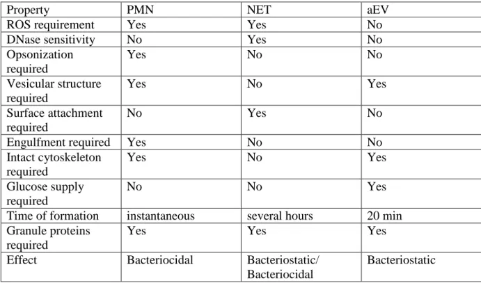
Table 3. Comparison of the in vitro antibacterial properties of intact PMN, neutrophil extracellular traps (NETs) and PMN-derived antibacterial EVs (aEVs)
Property PMN NET aEV
ROS requirement Yes Yes No
DNase sensitivity No Yes No
Opsonization required
Yes No No
Vesicular structure required
Yes No Yes
Surface attachment required
No Yes No
Engulfment required Yes No No
Intact cytoskeleton required
Yes No Yes
Glucose supply required
No No Yes
Time of formation instantaneous several hours 20 min Granule proteins
required
Yes Yes Yes
Effect Bacteriocidal Bacteriostatic/
Bacteriocidal
Bacteriostatic
Table 4. Increased production of PMN-derived EVs in pathological conditions
State/disease Investigated fluid Extent of elevation Ref
Cystic fibrosis Sputum Approx. 2-fold [86]
Chronic vasculitis Blood serum Approx. 2.fold [26]
Acute vasculitis Blood serum Approx. 5-fold [26]
ANCA-associated vasculitis
Blood serum 50-100 fold [47]
Rheumatoid arthritis Synovial fluid ? [8]
Figure Legends
Fig. 1. Difference in the phagocytosis of opsonized and non-opsonized bacteria by PMN.
Uptake of green fluorescent S. aureus was followed in time in a flow cytometer.
Fig. 2. Phagocytosis of opsonized bacteria by PMN. Phagocytosis of green fluorescent S.
aureus by PMN labeled with red-fluorescent antibody against the surface marker CD11b.
Phagocytosis was followed in a laser scanning confocal microscope. Pictures were taken of the same visual field at the indicated time-points.
Fig. 3. Differences in number and functional properties of PMN-derived EV induced by different agents. EV formation was induced by S. aureus opsonized in full serum or complement-depleted serum, or tumor necrosis factor (TNFα) or occurred spontaneously (marked “none”). The number of produced EV was determined by flow cytometry (empty columns); bacterial growth was followed by a semi-automated technique (shaded columns).
Fig. 4. Comparison of the effect of (A) inhibition of NADPH oxidase and (B) opsonization on the antibacterial effect of PMN or PMN-derived antibacterial EV. NADPH oxidase was inhibited by 5 µM diphenylene-iodonium (DPI). S. aureus was opsonized with full serum.
Fig. 5. Antibacterial EVs form large aggregates with bacteria. The relation of green
fluorescent S. aureus with different PMN-derived EVs (stained red) was followed for 30 min in a confocal laser scanning microscope. A: spontaneously formed EVs; B. EVs induced by opsonized particles; C. EVs induced by opsonized particles treated with 10 µM cytochalasin B (CB); D. EVs induced by opsonized particles treated with 10 µM latrunculin A. Note that inhibition of continuous rearrangement of the actin cytoskeleton by CB or latrunculin prevents the formation of large aggregates between bacteria and antibacterial EVs.
Figure 1
0 15 30 45
0 20 40
time (min)
Phagocyting PMN (%)
opsonized non-opsonized
S. aureus S. aureus
Figure 2
0’ 10’
20’ 30’
Figure 3
inductor of PMN
Count of produced EV Antibacterial effect of EV
Figure 4
0 50 100 150
growth rate (%)
ops. of
S.aureus
― + ― +
PMN aEV
n=4, SEM
B
0 50 100 150 200
growth rate (%)
5 μM DPI
― + ― +
PMN aEV
n=4, SEM
A
Figure 5
4 μm
B
4 μm
C
4 μm
A
4 μm