https://doi.org/10.1007/s10555-021-10014-2
The role of the metabolite cargo of extracellular vesicles in tumor progression
Mária Harmati1 · Mátyás Bukva1,2,3 · Tímea Böröczky1,2,3 · Krisztina Buzás1,2 · Edina Gyukity‑Sebestyén1
Received: 22 October 2021 / Accepted: 15 December 2021
© The Author(s) 2021
Abstract
Metabolomic reprogramming in tumor and stroma cells is a hallmark of cancer but understanding its effects on the metabolite composition and function of tumor-derived extracellular vesicles (EVs) is still in its infancy. EVs are membrane-bound sacs with a complex molecular composition secreted by all living cells. They are key mediators of intercellular communication both in normal and pathological conditions and play a crucial role in tumor development. Although lipids are major com- ponents of EVs, most of the EV cargo studies have targeted proteins and nucleic acids. The potential of the EV metabolome as a source for biomarker discovery has gained recognition recently, but knowledge on the biological activity of tumor EV metabolites still remains limited. Therefore, we aimed (i) to compile the list of metabolites identified in tumor EVs isolated from either clinical specimens or in vitro samples and (ii) describe their role in tumor progression through literature search and pathway analysis.
Keywords Extracellular vesicles; Metabolites · Cancer · Metastasis Abbreviations
AA Amino acid Aa Arachidonic acid ATP Adenosine triphosphate C1P Ceramide-1-phosphate CAF Cancer-associated fibroblast
CD81sEV CD81-expressing small extracellular vesicle CDE CAF exosome
CEA Carcinoembryonic antigen
Cer Ceramide
CTCL Cutaneous T-cell lymphoma D-2-HG D-2-hydroxyglutarate DAG Diacylglycerol EC Endothelial cell ELV Exosome-like vesicle
EV Extracellular vesicle FA Fatty acid
FFA Free fatty acid
HIF-1α Hypoxia-inducible factor-α HNC Head and neck cancer
HNSCC HNC squamous cell carcinoma ISEV International Society for Extracellular
Vesicles
lEV Large extracellular vesicle LPE Lysophosphatidylethanolamine
MISEV Minimal Information for Studies of Extracel- lular Vesicles
MPE Malignancy pleural effusion MVB Multivesicular body PC Phosphatidylcholine PCa Prostate cancer
PE Phosphatidylethanolamine PG Phosphoglycerol
PI Phosphatidylinositol PS Phosphatidylserine sEV Small extracellular vesicle SM Sphingomyelin
TCA Tricarboxylic acid cycle TG Triacylglycerol
TME Tumor microenvironment TPE Tuberculosis pleural effusion
* Edina Gyukity-Sebestyén e.gyukity.sebestyen@gmail.com
1 Laboratory of Microscopic Image Analysis and Machine Learning, Institute of Biochemistry, Biological Research Centre – Eötvös Loránd Research Network, 6726 Szeged, Hungary
2 Department of Immunology, University of Szeged, 6720 Szeged, Hungary
3 Doctoral School of Interdisciplinary Medicine, University of Szeged, 6720 Szeged, Hungary
1 Introduction
Under normal and pathological conditions, most cells secrete a range of membrane-bound extracellular vesicles (EVs). Although their physical characteristics overlap, EVs are highly heterogeneous, and several subpopulations have been described. Microvesicles and exosomes are pri- mary subtypes of EVs differentiated by their biogenesis, release pathway, size, content, and function [1, 2].
Initially, EV secretion was thought to be a cellular waste disposal mechanism. Since then, it has been clearly demonstrated that EVs play a key role in intercellular com- munication by mediating horizontal transfer of diverse molecular content between adjacent and distal cells [3–5].
These delivery vehicles are excellently equipped to pro- tect their cargo inside the lipid bilayer from extracellular enzymes, and they are able to cross different biological barriers, such as the blood–brain barrier [6, 7]. There is also accumulating evidence that they fulfill the two main criteria of the EV-mediated communication: (i) selective packaging of signaling content into the newly formed vesi- cles, and (ii) selective delivery of EVs to target cells [2].
Recently, it has been recognized that metabolite con- tent of EVs may have a prominent role in EV-mediated communication in tumor diseases. Despite the technical challenges of EV metabolite analysis, investigation of the
EV metabolite cargo, its role in tumor progression, and potential in clinical diagnosis deserve further attention.
In this review, we provide insight into EV biology and the technical aspects of EV studies. We also describe the cur- rent knowledge on the functional role of the EV-transferred metabolites in tumor progression.
2 Tumor EV biology and research
2.1 Biogenesis of EVsBased on their biogenesis pathways, EVs are divided into two major classes—ectosomes (or microvesicles) and exosomes [5, 8, 9] (Fig. 1). The membrane budding step, a common feature in this pathway, is similar in both classes. In addition, both EV types bud away from the cytoplasm result- ing in the same membrane orientation, which is identical to the orientation of the plasma membrane [12]. In the case of ectosomes, this budding step occurs outward at the plasma membrane and results in a direct release of EVs ranging from ~ 50 nm to 1 μm in diameter. In contrast, exosomes are formed as intraluminal vesicles (ILVs) through inward budding of endosomes, which develop into multivesicular bodies (MVBs). These MVBs may fuse with lysosomes for degradation or fuse with the plasma membrane resulting in the extracellular release of ILVs as exosomes (40–160 nm in diameter) [8]. The precise molecular mechanisms of EV
Fig. 1 Biogenesis and isolation methods of EVs. The left side of the figure shows a schematic overview of the main EV biogenesis path- ways. The bottom left of the figure shows how EVs are classified by biogenesis (exosomes and microvesicles) and by size (small and medium/large EVs), indicating the overlap in the size range of the dif- ferent EV types. The right side of the figure shows the main isolation
methods and the comparison of their most important indicators such as yield, and co-isolated contaminants [10, 11]. Abbreviations: ILV, intraluminal vesicle; MVB, multivesicular body; EVs, extracellular vesicles; SN, supernatant; UC, ultracentrifugation; DG-UC, density gradient-ultracentrifugation; SEC, size-exclusion chromatography.
This figure was created at BioRender.com
biogenesis have only recently started to be understood. The main driver of exosomal biogenesis is the endosomal sorting complex required for transport (ESCRT), but the existence of ESCRT-independent routes has also been proven [4, 13].
Despite their different biogenesis routes, intracellular mech- anisms and sorting machineries of ectosomes and exosomes partially overlap. Shared features of different EVs make it difficult to distinguish between vesicle subpopulations [14].
2.2 Composition of EVs
EVs convey numerous proteins (e.g., tetraspanins, chaper- ones, biogenesis factors, signaling molecules), nucleic acids (e.g., miRNA and other non-coding RNAs, mRNA, DNA), small metabolites (e.g., sugars, amino acids, vitamins), and lipids (e.g., phosphatidylserine, cholesterol, ceramide), which are all selectively packed into vesicles in a cell type- dependent manner [14]. As EVs are distinct entities of the complex intercellular communication, their molecular fin- gerprint depends on the quality and state of the donor cell, and it is often influenced by microenvironmental stimuli [5, 15, 16].
2.3 EV signaling and uptake mechanisms
The mechanisms EVs used to interact with the cell sur- face and transfer their cargo into the target (recipient) cells are not fully understood. Literature data suggest that these mechanisms depend on the origin and type of EV as well as the target cell [14, 17].
EVs may induce a phenotypic response in the recipient cell without internalization; receptor–ligand interactions may be sufficient to elicit signal transduction. Alterna- tively, EVs may transfer their cargo by direct fusion with the plasma membrane, and they may also be internalized via an active endocytic process, i.e., clathrin-, caveolin-, and lipid raft-mediated endocytosis, macropinocytosis, or phagocytosis [18]. Once in the cell, intraluminal EVs may fuse with the endosomal limiting membrane to release their content into the cytoplasm and elicit phenotypic responses in the recipient cell [14, 18, 19].
2.4 EV terminology
Since the origin and the physical and functional character- istics of EVs are diverse, several terms have been used for EVs in the literature. The prefixes micro- and nano- refer to their size (microvesicles, microparticles, nanovesicles, nanoparticles); ecto- and exo- refer to their presence out- side the cells (ectosomes, exosomes, exovesicles). Other terms, such as oncosomes and tolerosomes, indicate their origin or function, respectively [13]. Although the nomen- clature is continuously evolving, the International Society
for Extracellular Vesicles (ISEV) recommends the use of
“extracellular vesicle” as the “generic term for particles naturally released from the cell that is delimited by a lipid bilayer and cannot replicate.” They also suggest the use of operational terms for EV subtypes that refer to their (i) physical characteristics (small or medium/large EVs), (ii) biochemical composition (CD81+ EVs), or (iii) conditions of release (hypoxic EVs) [1, 20]. Here, we use the terms found in the referenced articles.
2.5 Role of EVs in cancer
Exosomes and other classes of EVs are important media- tors of cell–cell communication and play an essential role in cancer biology. It has long been well known that cancer cells secrete higher amounts of EVs than healthy cells. EVs in higher numbers have been detected in the plasma of cancer patients as well as in tumor cell cultures [21, 22].
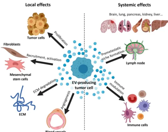
Exosomes contribute substantially to tumor progression, invasion, and metastasis by horizontally transmitting a vari- ety of surface and signaling molecules, oncogenic proteins, and nucleic acids to target cells, thereby altering their behav- ior [23, 24]. For instance, locally, in the tumor microenvi- ronment (TME), tumor-derived EVs may convey resistance to neighboring tumor cells. These EVs can also re-educate fibroblasts and mesenchymal stem cells or activate endothe- lial cells, thereby inducing angiogenesis. Systemically, EVs have a crucial role in immune modulation and pre-metastatic niche formation [7, 25–28] (Fig. 2). However, communica- tion is not unidirectional in tumors. On the contrary, a com- plex, systemic communication network develops in parallel with the tumor evolution [29].
As mentioned above, the characteristic molecular finger- print of small EVs (sEV), i.e., exosomes, is not independent of the parent/donor cell status. The metabolic status of can- cer cells influences exosome secretion and content. Hypoxia, starvation, and acidosis are among the typical metabolic conditions that cancer cells undergo in the TME. Notably, all of these conditions have been shown to influence not only the rate of exosome secretion, but also the molecular composition of exosomes [30].
2.6 EV isolation and characterization methods Before launching any investigation, one must consider the complexities of working with EVs. For instance, prepa- rations obtained using isolation procedures that target exosomes may contain other EVs as contaminants due to the overlapping physical and biomolecular features of exosomes and microvesicles. Research guidelines for EVs are provided in the Minimal Information for Studies of Extracellular Vesi- cles (MISEV) to support the transparency and reproducibil- ity of EV studies [1].
A standardized method for the isolation of EVs from cell culture supernatants or body fluids (blood, urine, saliva, etc.) has not yet been established. There are several alternative approaches to isolate and purify EVs. Important factors to consider when choosing a method include the type and vol- ume of the EV source, the target EV subtype, the target EV yield and purity of isolates, and the downstream metabolite analysis technique. Depending on the isolation method and the EV source, abundant serum proteins (albumin, globu- lins) and various lipoproteins (chylomicrons, HDL, LDL, and VLDL) as well as nucleic acids on the surface of EVs may contaminate the EV isolates due to their similar physi- cal properties. These co-isolated contaminants may interfere with the particle number and size distribution measurements and mislead biomarker analyses or functional assays [10].
Previous research has shown that EV subgroups have a unique biochemistry and function [31, 32]. This is consistent with the results of Luo et al. who found that different types of vesicles from pleural effusions showed unique metabolic enrichments [33]. Isolation methods themselves may also modify EV composition. In prostate cancer cell line models, the metabolic signature varies according to the conditions of cell culture [34]. Due to the broad range of physical and chemical characteristics of distinct metabolites, it is hardly possible to quantify all metabolites using a single approach.
Therefore, it is necessary to select an appropriate method for metabolite analysis. Gas chromatography-mass spectrom- etry (GC–MS), liquid chromatography-mass spectrometry (LC–MS), and capillary electrophoresis-mass spectrometry
(CE-MS) are the most common mass spectrometric meth- ods used for metabolomic analyses. Additionally, in recent years, improved analytical techniques have emerged, such as ion chromatography-mass spectrometry (IC-MS) to ana- lyze highly hydrophilic compounds [35, 36] or the capillary IC-MS as a selective and specific method to analyze ani- onic metabolites [37], e.g., nucleotides, sugar phosphates, and organic acids. Williams and colleagues have collected and highlighted several practical pitfalls in the field of EV metabolomics research [38].
2.7 EV metabolomics
Although lipids are dominant components of EVs, the vast majority of the EV cargo studies have investigated the pro- tein and nucleic acid content of EVs; only a few researches have analyzed the lipid or small metabolite composition [38]. In line with this observation, EV databases, such as Vesiclepedia, ExoCarta, EVpedia, or miREV, mainly con- tain protein, mRNA, and miRNA entries with less lipid and metabolite data [39–42].
The composition of EVs is comparable to that of the source donor cells, but they are also enriched in certain lipids such as cholesterol, phosphatidylserine (PS), phos- phatidylcholine (PC), and phosphatidylinositol (PI), sug- gesting that EV may operate as cell-to-cell lipid mediators [43]. From a practical and clinical standpoint, studying the metabolomics of EVs isolated from human biofluids is the most suited approach, since the metabolome of these
Fig. 2 Tumor-derived EVs have both local and systemic effects.
These EVs can alter the TME, modulate immune responses and prepare distant tissue sites for metastasis. This figure shows some examples of the tumor EV effects. Abbreviation:
ECM, extracellular matrix; the figure was created based on [28]
at BioRender.com
vesicles contains a goldmine of disease biomarkers. The EV metabolome’s potential as a source for biomarkers was first demonstrated by comparative metabolomics of plasma- derived EVs from endometrial cancer patients and healthy controls, which revealed valuable differences in these two groups [44].
3 Functional role of EV‑transferred metabolites in cancer
Numerous studies have shown that the tumor- and tumor stroma-derived EVs alter the metabolism of the recipient cells. Several studies highlight the differences in the metabo- lite profile of EVs between diseased and normal states as well as between various stages of tumors and/or suggest
biomarkers for diagnosis, prognosis, or treatment schedule choice [45, 46]. At the same time, the knowledge on the biological activity of tumor EV metabolites remains limited.
The primary aim of this review is to collect this knowledge focusing on the role of EV metabolites in tumor progression.
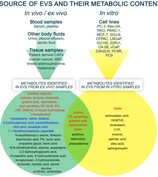
We collected a list of metabolites identified in tumor EVs isolated from either clinical specimens or in vitro samples (Fig. 3) and describe their functional effects according to main metabolite types.
3.1 Amino acids, amines, and their derivatives 3.1.1 Amino acids
Amino acids (AA) present in the metabolome of EVs have been investigated in both in vitro and ex vivo experiments.
Fig. 3 Summary Venn diagram of EV metabolites. The figure summarizes the metabolites identified in the literature according to their source and expression. The top of the figure shows the sources of the EVs investigated in the differ- ent studies, and the identified metabolites are shown at the bottom. The different colors and font styles indicate the expres- sion state of metabolites. The figure was created using GIMP
In these studies, both cell culture supernatants and patient- derived biofluids such as urine, serum, or plasma have been used as sources of EVs.
Recent findings indicate that the AA content of EVs secreted by the cells may be a source of nutrients for the recipient cells by entering into different metabolic pathways or by acting on cell motility and proliferation through other pathways. The results of Onozato et al. revealed that certain AAs—histidine, arginine, glutamine, cysteine, lysine, and tyrosine—are significantly enriched in the exosome-eluted fraction from healthy human serum, but no functional analy- ses were performed [47].
Numerous studies have reported the increased expression of AAs or their derivatives in tumors, but so far, no clear consensus on a shared set of AAs across various malignan- cies has been achieved. Palviainen and colleagues observed that proline was upregulated in all EVs derived from pros- tate cancer (PCa), cutaneous T-cell lymphoma (CTCL), and colon cancer (CC) cell lines (PC3, Mac-2A, RKO) when compared to their respective controls [48]. Proline is a unique AA that plays a key function not only in protein biosynthesis but also in cancer metabolism as a regulatory AA. Altered proline biosynthesis in tumor tissue leads to increased proliferation and biomass production [49, 50].
During the degradation of proline, the p53 gene-induced proline dehydrogenase/proline oxidase pathway produces adenosine triphosphate (ATP) for autophagy and reac- tive oxygen species (ROS) for apoptosis [51]. Surazynski et al. have shown that proline can inhibit the degradation of hypoxia-inducible factor-α (HIF-1α) via the von Hippel- Lindau protein-dependent proteasomal pathway [52]. HIF- mediated pathways have a significant impact on metabolic response, erythropoiesis, angiogenesis and vascular tone, cell proliferation and differentiation, survival, and apoptosis;
thus, they are crucial factors in cancer [53].
Luo and colleagues compared the metabolic profile of large EVs (lEVs) and sEVs in malignancy pleural effusion (MPE) and tuberculosis pleural effusion (TPE) samples [33].
In the lEV samples, more AAs were decreased in MPE, such as phenylalanine, tryptophan, leucine, valine, ornithine, and betaine; in contrast, threonate and glutaric acid were elevated in the MPE lEV samples. Luo and colleagues have identified a relationship between these metabolite variations in lEVs and biological and clinical parameters. The levels of carcinoembryonic antigen (CEA) and pleural adenosine deaminase show significant correlations with different AA levels in lEVs, but this correlation was moderate in sEVs [33]. Aspartate, a metabolite that plays an important role in protein synthesis and is a precursor of cell signaling mol- ecules, has been found in MPE EVs [33].
Altadill et al. have identified significant amounts of AAs and AA derivatives in sEVs isolated from the supernatants of the PANC1 human pancreatic carcinoma cell line [44].
Although they did not perform functional assays, the mol- ecules identified have previously been shown to be involved in tumor development and metabolic pathways. Aminoadipic acid is a well-known intermediate in the synthesis of acetyl- CoA; therefore, it is closely linked to the tricarboxylic acid (TCA) cycle and cellular energy balance [54]. Aminoadipic acid plays a role in the synthesis of lysine, various modi- fications of which may contribute to tumor development through several metabolic pathways [55]. Aminoadipic acid is also known to have direct effects on various cells, such as enhancing glial cell migration and glioblastoma aggressive- ness [56, 57].
Other studies have also pointed to the involvement of the TCA cycle. Palviainen et al. investigated the effect of the biochemical composition of lEVs and sEVs isolated from supernatants of two prostate cancer cell lines (PCa, VCaP) in silico and found that AAs present in vesicles mainly affect the TCA cycle, thereby providing energy to fuel the inten- sive metabolism of the rapidly dividing recipient tumor cells for [34]. Zhao et al. have shown that cancer-associated fibro- blasts (CAFs) secrete exosomes to regulate the metabolism of recipient cancer cells [58]. They detected particularly high levels of glutamine, arginine, glutamate, proline, alanine, threonine, serine, asparagine, valine, and leucine in prostate CAF-derived exosomes (CDEs). Additionally, in pancreatic CDEs, they found high levels of glutamine, threonine, phe- nylalanine, valine, isoleucine, glycine, arginine, and serine [58]. Zhao et al. provided a compelling proof-of-concept that AAs in CDEs can supply TCA cycle metabolites to cancer cells under both complete and nutrient-deprived conditions.
Using isotope tracing, they demonstrated that these metabo- lites are used as precursor metabolites by the recipient can- cer cells for proliferation and also to restore the levels of the TCA cycle metabolites [58].
Puhka et al. isolated lEVs and sEVs from serum and urine samples of healthy volunteers and PCa patients and detected a high concentration of ornithine in PCa urine and plasma EVs in contrast to healthy EVs [59]. Their results emphasize the importance of the non-proteinogenic AA ornithine in addition to the proteinogenic AAs discussed above. Ornith- ine has previously been described as an important precursor of polyamines, which show elevated levels during carcino- genesis [59]. Gökmen et al. found that ornithine levels can be useful to distinguish patients with malignant skin tumors from healthy subjects [60].
Vallabhaneni et al. have directly investigated the effect of sEVs secreted by patient-derived mesenchymal stem cells on MCF-7 breast tumor mouse xenograft models [61].
Their findings showed that sEV treatment accelerated tumor growth compared to the control group. They hypothesized that—among other factors—the high concentrations of glu- tamic acid determined in sEVs may enhance cell prolifera- tion, as glutamine can not only contribute to the TCA cycle
but can also serve as a carbon and nitrogen source for all major macromolecules [61].
3.1.2 Amines
In addition to ornithine, Puhka et al. found elevated levels of an aliphatic polyamine called spermidine in PCa EVs. The high amount of spermidine may be caused by the high activ- ity of the enzyme ornithine decarboxylase, the rate-limiting enzyme in the polyamine synthase pathway [59]. Various studies have shown that polyamine biosynthesis is upregu- lated in actively growing cells, including cancer cells. The elevated level of polyamines in the TME has a role in cancer cell transmigration into the circulation leading to metastasis formation and helps cancer cells escape recognition by the immune system [62]. N,N-Dimethylaniline, a member of the amines group, has been detected in serum-derived exosomes from head and neck cancer (HNC) patients [45].
3.1.3 Derivatives
Clos-Garcia et al. found increased levels of acylcarnitines (the acetylated forms of L-carnitine) in the urinary EVs from PCa patients [63]. Puhka and coworkers previously sug- gested that variable carnitine levels in PCa EVs correlated with a metabolic shift towards β-oxidation of fatty acids (FA) [59]. Altadill et al. have shown the presence of N-ara- chidonyl L-serine in plasma exosome-like vesicles (ELVs) obtained from patients with endometrioid adenocarcinoma [44]. N-arachidonyl L-serine has been found to promote cell migration, proliferation, and angiogenesis [64].
3.2 Lipids
Lipids are a main class of biological compounds with a wide variety of structural and signaling roles. Apart from sterols, most lipids have hydrophobic side chains and polar head groups, and the rich lipid diversity is the result of different combinations of side chains and head groups. Despite their comparable molecular complexity, there is a better under- standing of the function of proteins than that of lipids, which are called as the “Cinderellas” of cell biology by Muro et al.
[65].
Exosomes predominantly contain lipids, including diglyc- erides, sphingolipids, phospholipids, and phosphoglycerolip- ids, and they are enriched in specific lipids, such as choles- terol, PS, PC, and PI, which may function as cell-to-cell lipid mediators [43]. Exosomes may also carry specific bioactive lipids, including leukotrienes and prostaglandins [66].
There is accumulating evidence that the lipid content of EVs and parental cells differ; for instance, elevated levels of diacylglycerols (DAG), ceramides (Cer), sphingomyelin (SM), PC, phosphatidylethanolamines (PE) and FA were
detected in EVs [31, 67, 68]. In line with this, Luo et al.
identified phosphoglycerolipids, sphingolipids, and glyc- erolipids as major differential lipid species in lEVs and sEVs when comparing TPE with MPE enrichment of specific lipid metabolites in EVs may affect the cellular function of target cells and reflect the metabolic state of parent cells [33].
Lipids have a key role in the production and biological functions of EVs [33]. Sphingolipids, such as Cer, are criti- cal not only in the formation and release of EVs [69], but in the regulation of cell survival and inflammation as well [70].
SM, PS, PC, PI, and cholesterol may occur in four times higher amounts in EVs than in parental cells, which contrib- utes to the increased membrane rigidity of exosomes, and their role in the recognition and internalization of exosomes [43].
3.2.1 Fatty acids
Paolino et al. showed that FA and protein compositions of plasma-derived sEVs from stage 0–I, II, and III–IV mela- noma patients could reflect disease stages. FA analysis of CD81-expressing sEVs (CD81sEVs) revealed that several FA species are more abundant in EVs obtained from can- cer patients than those from healthy donors. They also dis- cussed the role of these FAs in disease progression [46].
For instance, higher levels of lauric (C12:0) and myristic (C14:0) acids in stage II and III–IV CD81sEVs may result from the accelerated metabolism of advanced cancer [71];
also, higher oleic acid (C18:1) levels in stage II and III–IV may increase membrane fluidity supporting the adhesion and migration, since the correlation between C18:1 and the metastatic potential has already been established [72].
Elevated levels of saturated FAs (FA 18:0) were observed in MPE EVs compared to EVs in TPE [33], and FAs can also be used to provide energy through β-oxidation and accelerate lung tumorigenesis [73].
Wojakowska and colleagues detected heptanoic acid in serum exosomes, but not in whole serum from HNC cancer patients. However, there were no significant differences in heptanoic acid levels of the vesicles between the healthy controls and pre-treated and post-treated cancer samples [45].
According to Schlaepfer et al., EVs may support growth following reoxygenation in a survival response of prostate cancer cells to hypoxic stress. Palmitic and oleic FAs trans- ferred by hypoxic EVs may serve a dual purpose; they can be used for membrane synthesis and ATP generation as they are built into phospholipids or utilized as fuel in the mito- chondria in the oxygenated recipient cells in the periphery of the tumors. This way, hypoxic PCa EVs may contribute to the overall aggressiveness of the tumor [74].
Other FAs in EVs, e.g., arachidonic acid (Aa), can also be delivered to intracellular membrane-localized enzymes,
which enable bioactive lipid generation and stimulate growth and motility of the target cells [74]. Indeed, Aa is the pre- cursor of important proliferative and inflammatory modu- lators, e.g., eicosanoids and prostaglandins [63]. However, Clos-Garcia et al. found reduced levels of Aa in PCa urine EVs [75]. They hypothesized that the increased metabolism resulted decreased vesicular Aa levels, as elevated concen- trations of its metabolic products (prostaglandin E2, PGE2; 12-hydroxyeicosatetraenoic acid, 12-HETE) were detected in malignant prostatic tissue [76, 77]. These studies high- light the significant role of EVs in Aa metabolism and PCa development.
In general, the potential of FAs, such as Aa to support cancer progression, has been reported in several previous papers [78–82]. For instance, Liu et al. have found increased serum levels of free fatty acids (FFA), Aa, linoleic acid (LA), and 15-HETE in lung adenocarcinoma patients. The group concluded that there is considerable basic evidence supporting the contribution of FFAs in tumor development and progression in lung cancer [83]. In PANC1 human pancreatic carcinoma cell-derived ELVs, the presence of 15-HETrE was shown and Pham et al. highlighted that this polyunsaturated FA participates in tumorigenesis and modu- lates Aa metabolism [84].
3.2.2 Sphingolipids, glycerophospholipids, and triacylglycerols
Several studies have investigated the lipid composition of cancer EVs. Altadill et al. have shown that ELVs iso- lated from the plasma of healthy controls or patients with endometrioid adenocarcinoma have significant amounts of glycerophospholipids (probably due to the exosomal membrane) and sphingolipids (29% of the total metabolite cargo of ELVs). They listed PI (16:0/22:4), PE (22:2/16:1), galactosylceramide (GalCer) (d18:2/16:0), glycerophos- phocholine (GPCho) (18:0/14:0), or triacylglycerol (TG) (12:0/12:0/20:5) as the abundant lipids. In addition, the pres- ence of phosphoglycerol (PG) (16:0/16:0), a precursor of cardiolipin, was validated and highlighted in plasma ELVs [44]. Cardiolipin is located in the inner membrane of mito- chondria, and its concentration and distribution changes in mitochondria were observed in several diseases, including cancer [85].
In the study of Altadill et al., the metabolome of PANC1 pancreatic cell line-derived ELVs was also dominated by glycerophospholipids and sphingolipids with a proportion of 56% [44].
Lipids are sensitive biomarkers of pathophysiological changes. Significantly increased levels of several lysophos- phatidylethanolamines (LPE), Cer, and PC were observed in MPE EVs compared to TPE vesicles [33]. Previous studies revealed that these lipids play a critical role in the immune
response, cellular signaling, and proliferation. For instance, Kachler et al. found that Cers are related to metastasis and immune evasion in lung cancer [86]. Similarly, Luo et al.
revealed an association between the levels of most PCs, PIs, and SMs and the clinical parameters of CEA and others [33].
CEA is an important tumor marker for colorectal and other carcinomas and plays a role in cell adhesion, signal trans- duction, and innate immunity [87]. The close relationships described by Luo et al. also suggest that the metabolites investigated are suitable for phenotypic characterization of MPE and TPE [33].
In the same study, more TGs were found in MPE EVs compared to EVs of TPE samples [33]. TGs are considered to be the main energy storage molecules, and elevated levels of TGs have also been observed in lung cancer tissues [88].
Additionally, 12 metabolites including PEs, DAGs, hexa- Cer, malic acid, and palmitic acid were elevated in MPE- lEVs. In general, more sphingolipids and glycerophospholip- ids were enriched in lEVs, while more FAs and glycolipids were enriched in sEVs. In addition, unique metabolic enrich- ment signatures were found both in TPE and MPE EVs pro- viding the opportunity to track the unique biogenesis and function of the two EV subgroups in TPE and MPE [33].
DAGs are important messenger molecules in intercellular communication [89]. Nishida-Aoki et al. have shown that unsaturated DAGs are enriched in EVs from highly meta- static breast cancer. They also proved that the biological activity of the EVs to induce protein kinase D (PKD)/PKCµ phosphorylation in endothelial cells leads to neoangiogen- esis. As DAG-mediated PKC activation occurs in many other cancer-related functions, such as cell proliferation and immune reactions, they concluded that DAG in cancer EVs may contribute to the EV-mediated education of the recipi- ent cells to support tumor progression [90].
Clos-Garcia et al. found a selective decrease of Cers in urine EVs that correlates with PCa aggressiveness suggest- ing that Cers may have both cell-autonomous and non-cell- autonomous functions to limit cancer progression [63]. Kuc et al. showed that ceramide-1-phosphate (C1P) is a modu- lator of pancreatic cancer stem cell (PCSC) migration and fibronectin-specific based adhesion. They also identified pancreatic ductal adenocarcinoma (PDAC) cells as a source of C1P and concluded that C1P-containing EVs might recruit PCSCs to sustain tumor growth and C1P release could be a mechanism that facilitates tumor progression [91].
Kelleher et al. reported that PS-expressing EVs derived from ascites fluids and solid tumors of ovarian cancer patients induce a rapid and reversible arrest of the T cell receptor signaling in the CD4 + and CD8 + T cells through a DAG kinase-mediated inactivation of DAG. This finding offers therapeutic strategies, such as targeting PS-expressing EVs or the application of anti-PS antibodies or DAG kinase
inhibitors (DGKi), which may enhance the patients’ T-cell responses to their tumor [92].
The lipid content of EVs has a crucial role in the adap- tive response of tumors as well. Jung et al. found that phos- pholipid signatures of tumor EVs are related to gefitinib- resistance in non-small-cell lung cancer cells [93]. As a survival response to hypoxic stress, human PCa cells and EVs accumulate triglycerides, which support growth fol- lowing reoxygenation [74].
3.2.3 Cholesterol and steroids
Cholesterol levels in EVs have been extensively studied using a wide range of experimental methods, and findings indicate that cholesterol is essential for the biogenesis, secre- tion, membrane stability and uptake of the vesicles as well [94]. As cholesterol is involved in the entire journey of EVs, it has a fundamental role in the EV-mediated signaling as well.
The human SOJ-6 pancreatic tumor cell-derived exosomes were shown to induce (glyco)protein ligand- independent apoptosis and inhibit the Notch-1 pathway in differentiated carcinoma cells, which indirectly favors the growth of undifferentiated tumor cells [95]. Beloribi et al.
hypothesized that SOJ-6 exosomes interacted with tumor cells through cholesterol-rich membrane microdomains and exosomal lipids were the key elements to induce apoptosis.
Through designing Synthetic Exosome-Like Nanoparticles (SELN) based on the lipid composition of SOJ-6 exosomes enriched in cholesterol and SM and depleted in phospho- lipids, they proved the role of lipids (i) in the interaction of SELNs and tumor cells and (ii) in induced cell death with inhibition of the Notch-1 pathway [96].
Clos-Garcia et al. detected an elevated level of dehydroe- piandrosterone sulfate (DHEAS), an intermediary metabolite of androgen synthesis, in PCa urinary EVs, which suggests a potential role for EVs in androgen signaling in neighboring or distal cells [63].
3.3 Carbohydrates, carbonic acids 3.3.1 Carbohydrates
Tumor cells possess an extraordinary capacity to regulate their energy metabolism as part of their tumor survival strat- egies [97]. One of the primary metabolic features of tumor cells is the Warburg effect, also known as aerobic glycoly- sis, which is characterized by an elevated rate of glycolysis even in the presence of oxygen. A large amount of glycolytic intermediates might be used to satisfy the metabolic require- ments of proliferating cells [98].
Puhka et al. studied the metabolic profile of platelet- and urinary-derived EVs from PCa patients, and in both EV
samples they observed a high concentration of D-ribose 5-phosphate, which is a major product of the cytosolic pentose-phosphate pathway and a key precursor for NAD+ and nucleotide biosynthesis [59]. Additionally, not only the D-ribose 5-phosphate concentration was increased in EVs, but also enzymes related to the pentose-phosphate pathway, such as glucose-6-phosphate dehydrogenase, transketolase, and transaldolase [99]. Numerous studies have demonstrated that the pentose-phosphate pathway serves an essential role for a cancer cell growth regulation and that the enzymes and metabolites delivered by EVs may contribute to the intense proliferation and cancer progression [100].
Furthermore, Wojakowska and colleagues studied the metabolic profiles of serum and serum-derived exosomes in HNC patients. Forty-six metabolites were identified in serum-derived exosome samples, including levoglucosan and 2,3-diphosphoglyceric acid. Metabolites that were detected in cancer but not in control samples were associ- ated with energy metabolism [45].
3.3.2 Carbonic acids
Vallabhaneni et al. [61] found lactic acid in EVs secreted by mesenchymal stem/stromal cells from patients. The pres- ence of lactic acid in the TME was shown to be linked to the improved capacity of tumor cells to withdraw hypoxic and nutrient-deprived core environments. Moreover, low pH caused by lactic acid is a known strategy of cancer cells to evade immune surveillance [101]. It is also worth mention- ing that low pH, which is one of the hallmarks of cancer, enhanced exosome release and uptake in a melanoma cell line model [102].
Oncometabolites are common cellular metabolites that show abnormal accumulation in malignancies in comparison to non-proliferating cells and possess pro-oncogenic proper- ties. These compounds are the products of cancer cell gene mutations or hypoxia-driven enzyme promiscuity. Accu- mulation of these oncometabolites in cancer cells results in metabolic and epigenetic changes, post-translational modi- fications, and other tumor-promoting effects [103]. Succi- nate, D-2-hydroxyglutarate (D-2-HG), L-2-hydroxyglutarate (L-2-HG), and fumarate are the four oncometabolites iden- tified so far. All four oncometabolites are produced in the mitochondria (during TCA cycle) and can induce compara- ble changes in cancer cells, such as hypermethylation and pseudohypoxia, which results in metabolic and epigenetic changes, post-translational modifications and other tumori- genic characteristics [48].
Succinate, together with the three other oncometabolites, is a small molecule that accumulates in cancer cells as a result of gain-of-function or loss-of-function mutations in genes encoding energy metabolism enzymes. Elevated levels of succinate were measured in EVs from prostate, CTCL,
and CC cell lines (PC-3, Mac-2A, RKO) compared to their respective control EVs (PNT2, PBMC, CCD841) [48]. In addition, succinate promotes tumorigenesis through a num- ber of ways, such as generating epigenetic modifications and increasing cancer cell angiogenesis, invasion, and migration [104, 105]. Elevated levels of succinate levels have been found in cancer tissues, and biofluids of patients with vari- ous malignancies, including prostate and colorectal cancer [106], and hepatocarcinoma.
TCA cycle intermediates such as succinate, fumarate, and L-2-HG can alter the response of both the innate and adaptive immune systems. Through inhibition of histone and DNA demethylases, 2-HG and fumarate can also alter the epigenetic landscape of cells [107]. Endogenous fuma- rate was reported to suppress GAPDH via succination in macrophages [108]. Succinate, fumarate, and L-2-HG can inhibit prolyl-hydroxylases (PHDs) in normoxic environ- ments leading to a pseudohypoxic state [109]. Inhibition of PHD enzymes results in stabilization of HIFs [110]. The HIF system plays a critical role in the regulation of a broad range of cellular and systemic responses to hypoxia. Thus, HIF- mediated pathways affect metabolic adaptation by increasing glucose uptake, lactate production, while decreasing respi- ration. HIF1α is a key regulator of EV production under hypoxia [111, 112].
Zhao et al. reported elevated lactate and acetate levels in both prostate and pancreatic CDEs [58]. Moreover, inves- tigation of intra-exosomal metabolites revealed high citrate and pyruvate concentrations, as well as the significant pres- ence of α-ketoglutarate, fumarate, and malate. These metab- olites together with others – such as AAs – can replenish TCA cycle metabolites, and act as a source for lipid biosyn- thesis. Pyruvate is converted to acetyl-CoA by mitochondrial pyruvate dehydrogenase (PDH), while acetate is transferred into cells and transformed to acetyl-CoA through acetyl- CoA synthase [113–116]. Acetyl-CoA is the first step in lipid biosynthesis, which helps proliferating cells meet their biosynthetic needs. Recent findings implicate that exosomes of the TME can participate in the induced metabolic rewir- ing in cancer cells [117–119].
3.4 Adenosine and other purine metabolites
Extracellular adenosine can be produced by cells, or it can be generated from extracellular ATP. Adenosine has an extremely short half-life (10 s) in the extracellular envi- ronment due to its quick uptake by cells and irreversible conversion to inosine [120, 121]. Considerable research has been conducted in the last few years on the diverse roles and associated mechanisms of extracellular adenosine sign- aling. Extracellular adenosine has a wide variety of effects on cell cycle control, immunoregulation, and cytokine regulation via both direct and indirect processes, eventually
contributing to the development of malignant diseases [122].
Adenosine has been detected in urinary EVs from prostate cancer patients [59]. Sayner et al. have demonstrated that EVs encapsulate cAMP to offer a second messenger com- partment [123]. Ludwig et al. have recently reported that exosomes from HNC squamous cell carcinoma (HNSCC) cell lines contain cAMP and adenosine as well as adeno- sine metabolites, i.e. inosine, hypoxanthine and xanthine.
This exosomal repository of adenosine and inosine pro- vides a unique and key pathway for the distal transport of these purines. By shielding them from the quick uptake and metabolism, exosomes can shuttle adenosine and inosine across cells, tissues, and organ systems. Ludwig and col- leagues demonstrated that exosomes from HNSCC culture supernatant contain a variety of purine metabolites, the most abundant of which are adenosine and inosine. Purine metabolites, including adenosine, were much more abundant in exosomes isolated from the plasma of HNSCC patients than in exosomes obtained from normal donors. Exosomes from patients with early-stage illness and no lymph node metastases had considerably higher levels of adenosine and 5'-GMP. At the same time, exosomal levels of purine metabolites were reduced in patients with advanced cancer and nodal involvement. Decreased purine concentration in circulating exosomes may be the result of purine metabolites being primarily used for cellular maintenance and prolifera- tion in metastatic tumor cells instead of being packaged into exosomes and exported outside of the cell. This suggests that the molecular composition of tumor-derived exosomes and circulating exosomes is quantitatively and, possibly, qualitatively different in advanced stages compared to early malignancies [124].
Clayton and colleagues demonstrated that exosomes pro- duced by several cancer cell types have a high capacity for ATP and 5’-AMP phosphohydrolysis, which is partly attrib- uted to the exosomal expression of CD39 and CD73 [125].
Exosomes can carry out both hydrolytic steps sequentially to convert extracellular ATP to adenosine. Exosome-produced adenosine can induce a cAMP response in adenosine A2A receptor-positive but not A2A receptor-negative cells.
A recently discovered pathway, the adenosine A2B recep- tor-mediated signaling for exosome-induced angiogenesis contributes to the reprogramming of endothelial cells (ECs) to an angiogenic phenotype by direct interaction, and also to the reprogramming of other cell types found in the TME, such as macrophages [126]. As previously described, A2B receptor stimulates the growth of ECs, induces angiogen- esis, leads to VEGF production and upregulation of eNOS in ECs, and stimulates macrophages to release pro-angiogenic factors [127].
Tadokoro et al. showed another mechanism leading to an increase in extracellular adenosine levels, in which perforin secreted by CD8 + cytotoxic T cells disrupts the membrane
of breast adenocarcinoma-derived EVs, and adenosine pas- sively diffuses out. Adenosine from EVs acts as an immuno- suppressive metabolite by binding to the adenosine receptor and inhibits perforin secretion by cytotoxic T lymphocytes [128].
Hypoxanthine, a purine derivative, is a potential interme- diate in the metabolism of adenosine and also in the synthe- sis of nucleic acids. Glyceraldehyde 3-phosphate (G-3-P) is an intermediate in glycolysis. Luo and colleagues detected elevated levels of hypoxanthine and G-3-P in the lEVs in the MPE in comparison to lEVs of TPE [33], which may indicate accelerated glycolysis and nucleic acid formation.
However, Ronquist et al. reported that human semi- nal prostasomes contain glycolytic enzymes [129]. They detected the full set of glycolytic enzymes in PCa cell- derived exosomes and observed that both types of vesi- cles were capable of producing ATP when substrates were available [130]. Moreover, they reported a marked distinc- tion between the high ATPase activity of prostasomes and the low ATPase activity of a malignant cell (PC3)-derived exosomes, which leads to a larger net gain of ATP in these latter exosomes. In contrast to prostasomes, the net ATP gain of metastatic PCa cell line exosomes was considerable due to their downregulated ATPase activity. This group also found that normal and prostate cancer cells uptake EVs (prostas- omes and PC3 exosomes) in an energy-dependent manner.
This uptake mechanism required a continuous glycolytic flux and extracellular ATP production by EVs and/or intracellu- larly by recipient cells in conjunction with the presence of a functioning vacuolar-type H( +)-ATPase (V-ATPase) [130].
3.5 Other metabolites
Few additional compounds in HNC serum-derived EVs were found to be markedly downregulated compared to healthy controls. These include citric acid, 4-hydroxybenzoic acid, and propylene glycol, while 1-hexadecanol was markedly upregulated. A few other metabolites, 1,1-dimethoxyhep- tane, oxoadipic acid, paramethadione could only be detected in EVs, but not in whole serum samples [45].
Folate (B9 vitamin) as a cancer-associated metabolite was identified in greater amounts in CTCL and in PCa cell line-derived EVs compared to control EVs [48]. Along with folate, overexpression of pantothenic acid (B5), niacin (B3), thiamine (B1), and pyridoxine (B6) may boost one-carbon metabolism directly or indirectly through their roles as coen- zymes, hence promoting cancer development [131]. One- carbon units are required for nucleotide synthesis, methyla- tion, and reductive metabolism, all of which contribute to the rapid proliferation of cancer cells. Also in cell line-derived EVs from PCa and CTLC, Palviainen et al. have detected elevated levels of creatinine [48].
3.6 Pathway analysis of EV metabolites
In order to explore the relationship between metabolites found in the literature and the pathways potentially involved, we performed pathway analyses (Fig. 4). This involved a total of 62 metabolites obtained from the processed litera- ture, and 28 of these metabolites were strongly associated with 15 different pathways. These pathways are mainly those related to AAs, but significant associations were also found for glutathione, glyoxylate, dicarboxylate metabolism, TCA cycle, pantothenate and CoA biosynthesis.
In many cases, metabolites in the same column are subse- quent steps in a metabolic pathway based on MetaboAnalyst.
For example, in arginine biosynthesis glutamate is converted into ornithine in four steps, which in turn is converted into arginine in two steps (with the addition of aspartate). Dur- ing the reaction, fumarate is released as a side product. The metabolism of glutathione serves as another example, in which glutathione is synthesized using cysteine, glutamate and glycine in subsequent steps, and then reacts with orni- thine and spermidine to form trypanothione. With regard to carbohydrate metabolism, succinate, fumarate and citrate are associated with the TCA cycle, in which these molecules act as both substrates and products.
These results show a predominance of pathways associ- ated with AA metabolism, but this analysis has serious limi- tations. For example, the metabolites from different sources were examined using different methods. The outcome of the pathway analysis is strongly influenced by the fact that most functional metabolomics studies have explicitly focused on AAs, resulting in a high number of AAs and their associ- ated pathways. Nevertheless, the results suggest that non- AA metabolites transported by EVs may also play a role in amino metabolism. In addition, the results reveal that certain metabolites, such as glutamine/glutamate, may exert a wide range of effects on the metabolism of recipient cells entering a number of pathways.
4 Conclusion and future directions
The stroma is a dynamic environment that is constantly evolving. Tumor-stroma interactions alter the microenviron- ment, making it more permissive towards cancer cells [132, 133]. Throughout the course of carcinogenesis, tumor cell hierarchies and various cellular components in the microen- vironment co-evolve [134].
Understanding how cancer cells interact with the TME is critical for designing medicines that can halt tumor devel- opment and spread. Several studies have demonstrated that sEVs can facilitate communication between cancer cells and stromal cells inside the TME [135, 136]. EVs have emerged as a crucial mode of communication between different cell
types in the TME. EVs transfer information across cells and reprogram the recipients [23, 24, 137]. In other words, cur- rent research indicates that EVs have the capacity to influ- ence recipient cell proliferation, survival, and immune effec- tor status [25–28].
It is also important to note that cancer changes cell metabolism. Typical metabolic conditions in the TME include hypoxia, starvation, and acidosis [133, 138]. The
metabolic rewiring alters the secretion rate and metabo- lite content of cancer-derived EVs as well, but this cargo remains poorly characterized. Most of the current studies focus on the analysis of RNA [139, 140], or protein pro- files [31] of EVs and related effects, and little is known about their metabolite cargo and function in recipient cells, which makes this field a fresh area of vesicle research.
Fig. 4 A matrix representa- tion of metabolites and their associated pathways. Shades of red indicate the P values, which refers to the significance level of the association with different pathways. The significance increases from left to right of the graph. From top to bot- tom, the number of pathways associated with a metabolite decreases. The figure was cre- ated using RAWGraphs and GIMP
Metabolites of EVs from human body fluids represent a goldmine of tumor biomarkers. However, the lack of a con- sensual approach for the separation of EVs is one a major obstacle to the advancement of EV research. In addition, the metabolite profile of EVs are potentially influenced by pre-analytical factors including external (storage, handling, analysis method) and internal (enzyme activity, sample contamination) factors. This may also account for the het- erogeneity of experimental outcomes in EV research, since single or multiple EV subtypes with varying compositions and purities reveal method-dependent EV content and func- tion. Given the wide range of constantly evolving EV isola- tion techniques, analysis methods, and applications avail- able, MISEV2018 was unable to provide standard protocol recommendations yet. In our conclusion, there is not a gold standard method, which is optimal for all sample types and volumes, EV subtypes, and budgets. The use of EV-TRACK [141] may help choose the most advantageous method for EVs isolation and characterization.
There are several gaps in the knowledge on the effects of metabolites from cancer EVs. This review aimed to col- lect the current results on the biological activity of tumor EV metabolites (Fig. 3, Fig. 4). Innovative metabolomics technology, methods and their applications in clinical phar- macology have made significant progress during the last few years. The study of EV metabolites has shown remarkable potential and provides a new perspective in understanding cancer progression. Once methodologies are standardized, this knowledge will serve as a novel tool to identify new diagnostic biomarkers of cancer, to explain pathological mechanisms, to find possible therapeutic targets, to predict the biochemical and physiological effects of therapies, and to aid following up treatments of cancer.
It is important to note that investigating one or a few mol- ecules in a subgroup of EVs allows only a partial insight into the functional role of the studied EV population. In contrast, analysis of the whole EV molecular pattern of the whole EV set would provide more relevant results. This approach has recently been applied in clinical studies for tumor diagnostic purposes [142–144].
5 Methodology
This review is based on 95 articles related to the metabo- lomic research of EVs. The publications on the metabo- lomics of tumor EVs were selected based on the follow- ing criteria: (a) any type of EVs were investigated, (b) EVs were isolated by any method, (c) source of EVs were either ex vivo or in vitro samples, (d) ex vivo samples were tumor tisssues, CAFs, stromal cells or different body fluids of can- cer patients (e) in vitro samples were tumor cell lines, (f) methods and equipment used in metabolomics studies were
not necessarily identical across the studies. In some studies, metabolomic analyses were not accompanied by functional assays. In these cases, information on the potential tumor/
metabolome-related effects of the molecule in question was gathered from other, non-EV-related research.
Based on the literature, pathway analyses were performed using MetaboAnalyst 5.0 software with the Kyoto Encyclo- pedia of Genes and Genomes database. Pathway enrichment and pathway topology were determined by hypergeometric test and relative-betweenness centrality method. Figures were created using RawGraphs 2.0 and CytoScape 3.9.
Acknowledgements The authors wish to thank Lilla Pintér for her contribution.
Funding Open access funding provided by ELKH Biological Research Center. This study was supported by the following research grants:
GINOP-2.2.1–15-2017–00052, ÚNKP-20–3-New National Excel- lence Program of the Ministry for Innovation and Technology from the National Research, Development and Innovation Fund; University of Szeged, Faculty of Medicine, Szent-Györgyi Albert Research Fund (2021).
Declarations
Conflict of interest The authors declare no competing interests.
Open Access This article is licensed under a Creative Commons Attri- bution 4.0 International License, which permits use, sharing, adapta- tion, distribution and reproduction in any medium or format, as long as you give appropriate credit to the original author(s) and the source, provide a link to the Creative Commons licence, and indicate if changes were made. The images or other third party material in this article are included in the article's Creative Commons licence, unless indicated otherwise in a credit line to the material. If material is not included in the article's Creative Commons licence and your intended use is not permitted by statutory regulation or exceeds the permitted use, you will need to obtain permission directly from the copyright holder. To view a copy of this licence, visit http:// creat iveco mmons. org/ licen ses/ by/4. 0/.
References
1. Théry, C., Witwer, K. W., Aikawa, E., Alcaraz, M. J., Ander- son, J. D., Andriantsitohaina, R., Antoniou, A., Arab, T., Archer, F., Atkin-Smith, G. K., Ayre, D. C., Bach, J. M., Bachurski, D., Baharvand, H., Balaj, L., Baldacchino, S., Bauer, N. N., Baxter, A. A., Bebawy, M., … Zuba-Surma, E.
K. (2018). Minimal information for studies of extracellular vesicles 2018 (MISEV2018): A position statement of the Inter- national Society for Extracellular Vesicles and update of the MISEV2014 guidelines. Journal of extracellular vesicles, 7(1), 1535750. https:// doi. org/ 10. 1080/ 20013 078. 2018. 15357 50 2. Raposo, G., & Stahl, P. D. (2019). Extracellular vesicles:
A new communication paradigm? Nature reviews. Molecu- lar cell biology, 20(9), 509–510. https:// doi. org/ 10. 1038/
s41580- 019- 0158-7
3. Hessvik, N. P., & Llorente, A. (2018). Current knowledge on exosome biogenesis and release. Cellular and molecular life
sciences : CMLS, 75(2), 193–208. https:// doi. org/ 10. 1007/
s00018- 017- 2595-9
4. Bebelman, M. P., Smit, M. J., Pegtel, D. M., & Baglio, S. R.
(2018). Biogenesis and function of extracellular vesicles in can- cer. Pharmacology & Therapeutics, 188, 1–11. https:// doi. org/
10. 1016/j. pharm thera. 2018. 02. 013
5. van Niel, G. & Théry C. (2020). Extracellular vesicles: eat glutamine and spit acidic bubbles. The EMBO Journal, 39(16):e105119. https:// doi. org/ 10. 15252/ embj. 20201 05119 6. Simeone, P., Bologna, G., Lanuti, P., Pierdomenico, L., Guag-
nano, M. T., Pieragostino, D., Del Boccio, P., Vergara, D., Mar- chisio, M., Miscia, S., & Mariani-Costantini, R. (2020). Extracel- lular Vesicles as Signaling Mediators and Disease Biomarkers across Biological Barriers. International journal of molecular sciences, 21(7), 2514. https:// doi. org/ 10. 3390/ ijms2 10725 14 7. Tao, S. C., & Guo, S. C. (2020). Role of extracellular vesicles in
tumour microenvironment. Cell communication and signaling : CCS, 18(1), 163. https:// doi. org/ 10. 1186/ s12964- 020- 00643-5 8. Kalluri, R., & LeBleu, V. S. (2020). The biology, function, and
biomedical applications of exosomes. Science (New York, N.Y.), 367(6478), eaau6977. https:// doi. org/ 10. 1126/ scien ce. aau69 77 9. Möller, A., & Lobb, R. J. (2020). The evolving translational
potential of small extracellular vesicles in cancer. Nature reviews. Cancer, 20(12), 697–709. https:// doi. org/ 10. 1038/
s41568- 020- 00299-w
10. Brennan, K., Martin, K., FitzGerald, S. P., O’Sullivan, J., Wu, Y., Blanco, A., Richardson, C., & Mc Gee, M. M. (2020). A comparison of methods for the isolation and separation of extracellular vesicles from protein and lipid particles in human serum. Scientific Reports, 10(1), 1039. https:// doi. org/ 10. 1038/
s41598- 020- 57497-7
11. Sun, L., & Meckes, D. G., Jr. (2018). Methodological approaches to study extracellular vesicle miRNAs in Epstein−Barr virus- associated cancers. International Journal of Molecular Sciences, 19(9). https:// doi. org/ 10. 3390/ ijms1 90928 10
12. Mathieu, M., Martin-Jaular, L., Lavieu, G., & Théry, C. (2019).
Specificities of secretion and uptake of exosomes and other extra- cellular vesicles for cell-to-cell communication. Nature cell biol- ogy, 21(1), 9–17. https:// doi. org/ 10. 1038/ s41556- 018- 0250-9 13. Colombo, M., Raposo, G., & Théry, C. (2014). Biogenesis,
Secretion, and Intercellular Interactions of Exosomes and Other Extracellular Vesicles. Annual Review of Cell and Developmental Biology, 30(1), 255–289. https:// doi. org/ 10. 1146/ annur ev- cellb io- 101512- 122326
14. van Niel, G., D’Angelo, G., & Raposo, G. (2018). Shedding light on the cell biology of extracellular vesicles. Nature reviews.
Molecular cell biology, 19(4), 213–228. https:// doi. org/ 10. 1038/
nrm. 2017. 125
15. Harmati, M., Gyukity-Sebestyen, E., Dobra, G., Janovak, L., Dekany, I., Saydam, O., Hunyadi-Gulyas, E., Nagy, I., Farkas, A., Pankotai, T., Ujfaludi, Z., Horvath, P., Piccinini, F., Kovacs, M., Biro, T., & Buzas, K. (2019). Small extracellular vesicles convey the stress-induced adaptive responses of melanoma cells. Scientific reports, 9(1), 15329. https:// doi. org/ 10. 1038/
s41598- 019- 51778-6
16. Harmati, M., Tarnai, Z., Decsi, G., Kormondi, S., Szegletes, Z., Janovak, L., Dekany, I., Saydam, O., Gyukity-Sebestyen, E., Dobra, G., Nagy, I., Nagy, K., & Buzas, K. (2017). Stress- ors alter intercellular communication and exosome profile of nasopharyngeal carcinoma cells. Journal of oral pathology &
medicine : Official publication of the International Association of Oral Pathologists and the American Academy of Oral Pathology, 46(4), 259–266. https:// doi. org/ 10. 1111/ jop. 12486
17. Herrmann, I. K., Wood, M., & Fuhrmann, G. (2021). Extra- cellular vesicles as a next-generation drug delivery platform.
Nature nanotechnology, 16(7), 748–759. https:// doi. org/ 10. 1038/
s41565- 021- 00931-2
18. Mulcahy, L. A., Pink, R. C., & Carter, D. R. (2014). Routes and mechanisms of extracellular vesicle uptake. Journal of extracel- lular vesicles 3. https:// doi. org/ 10. 3402/ jev. v3. 24641
19. French, K. C., Antonyak, M. A., & Cerione, R. A. (2017). Extra- cellular vesicle docking at the cellular port: Extracellular vesicle binding and uptake. Seminars in cell & developmental biology, 67, 48–55. https:// doi. org/ 10. 1016/j. ceb. 2015. 04. 013
20. Witwer, K. W., & Théry, C. (2019). Extracellular vesicles or exosomes? On primacy, precision, and popularity influencing a choice of nomenclature. Journal of extracellular vesicles, 8(1), 1648167. https:// doi. org/ 10. 1080/ 20013 078. 2019. 16481 67 21. Johnsen, K. B., Gudbergsson, J. M., Andresen, T. L., & Simon-
sen, J. B. (2019). What is the blood concentration of extracel- lular vesicles? Implications for the use of extracellular vesicles as blood-borne biomarkers of cancer. Biochimica et biophysica acta. Reviews on cancer, 1871(1), 109–116. https:// doi. org/ 10.
1016/j. bbcan. 2018. 11. 006
22. Cappello, F., Logozzi, M., Campanella, C., Bavisotto, C. C., Marcilla, A., Properzi, F., & Fais, S. (2017). Exosome levels in human body fluids: A tumor marker by themselves? European journal of pharmaceutical sciences : Official journal of the Euro- pean Federation for Pharmaceutical Sciences, 96, 93–98. https://
doi. org/ 10. 1016/j. ejps. 2016. 09. 010
23. Yang, E., Wang, X., Gong, Z., Yu, M., Wu, H., & Zhang, D.
(2020). Exosome-mediated metabolic reprogramming: The emerging role in tumor microenvironment remodeling and its influence on cancer progression. Signal transduction and targeted therapy, 5(1), 242. https:// doi. org/ 10. 1038/ s41392- 020- 00359-5 24. Parayath, N. N., Padmakumar, S., & Amiji, M. M. (2020). Extra- cellular vesicle-mediated nucleic acid transfer and reprogram- ming in the tumor microenvironment. Cancer letters, 482, 33–43.
https:// doi. org/ 10. 1016/j. canlet. 2020. 04. 009
25. Gulei, D., Petrut, B., Tigu, A. B., Onaciu, A., Fischer-Fodor, E., Atanasov, A. G., Ionescu, C., & Berindan-Neagoe, I. (2018).
Exosomes at a glance - common nominators for cancer hallmarks and novel diagnosis tools. Critical reviews in biochemistry and molecular biology, 53(5), 564–577. https:// doi. org/ 10. 1080/
10409 238. 2018. 15082 76
26. Nogués, L., Benito-Martin, A., Hergueta-Redondo, M., & Pei- nado, H. (2018). The influence of tumour-derived extracellular vesicles on local and distal metastatic dissemination. Molecular aspects of medicine, 60, 15–26. https:// doi. org/ 10. 1016/j. mam.
2017. 11. 012
27. Marar, C., Starich, B., & Wirtz, D. (2021). Extracellu- lar vesicles in immunomodulation and tumor progression.
Nature immunology, 22(5), 560–570. https:// doi. org/ 10. 1038/
s41590- 021- 00899-0
28. Kahlert, C., & Kalluri, R. (2013). Exosomes in tumor microenvi- ronment influence cancer progression and metastasis. Journal of molecular medicine (Berlin, Germany), 91(4), 431–437. https://
doi. org/ 10. 1007/ s00109- 013- 1020-6
29. Maia, J., Caja, S., Strano Moraes, M. C., Couto, N., & Costa- Silva, B. (2018). Exosome-Based Cell-Cell Communication in the Tumor Microenvironment. Frontiers in cell and developmen- tal biology, 6, 18. https:// doi. org/ 10. 3389/ fcell. 2018. 00018 30. Kucharzewska, P., Christianson, H. C., Welch, J. E., Svensson, K.
J., Fredlund, E., Ringnér, M., Mörgelin, M., Bourseau-Guilmain, E., Bengzon, J., & Belting, M. (2013). Exosomes reflect the hypoxic status of glioma cells and mediate hypoxia-dependent activation of vascular cells during tumor development. Proceed- ings of the National Academy of Sciences of the United States of America, 110(18), 7312–7317. https:// doi. org/ 10. 1073/ pnas.
12209 98110