NOVEL BARRIERS TO THROMBOLYIS:
THE ROLE OF MECHANICAL STRESS AND NEUTROPHIL EXTRACELLULAR
TRAPS
PhD thesis booklet
Imre Varjú
Semmelweis University
Doctoral School of Molecular Medicine
Supervisor: Krasimir Kolev, DSc Official reviewers: Jolán Hársfalvi, DSc
Béla Nagy Jr, PhD
Chairman of the final examination committee:
Zoltán Benyó, DSc
Members of the final examination committee:
Imre Bodó, PhD Nándor Müllner, PhD
Budapest, 2014
1
INTRODUCTION
Cardio- and cerebrovascular diseases represent the major causes of death (35.8%) in the world according to recent data of World Health Organisation. The underlying cause in these cases is the formation of intravascular thrombi (composed of blood cells and plasma components embedded in a fibrin network), blocking the supply of oxygen and nutrients, therefore leading to the damage of the respective tissue. The fact that degradation of the fibrin scaffold (Fig. 1.) itself is sufficient for the dissolution of thrombi is exploited by
Figure 1. The two-step process of fibrinolysis. I.: activation of the inactive precursor plasminogen. The physiologically and therapeutically most relevant plasminogen activator is tPA (tissue-type plasminogen activator). II.: Fibrin degradation catalysed by the active enzyme, plasmin.
2
therapeutic thrombolysis, representing a modality aiming the reconstitution of blood flow.
However, thrombolytic therapy often proves to be inefficient in the long term, and is accompanied by a serious risk for bleedings as a side effect. Improvement of current thrombolytic therapeutic protocols requires determination of the factors influencing the efficiency of the respective enzymes in the dissolution of thrombi.
This thesis focuses on two of the numerous factors:
mechanical stress to which fibrin formed in the circulation is exposed; and a recently recognized scaffold of venous and arterial thrombi: neutrophil extracellular traps (NETs) representing a web-like meshwork (Fig. 2.) composed of DNA, histones and granular components released from granulocytes as a response to various (inflammatory) stimuli.
Figure 2. SEM (scanning electron microscopic) images of neutrophil extracellular traps released by activated neutrophils. Scale bar: 1 µm.
3
OBJECTIVES
Aims of the studies on the effects of mechanical stress:
-To examine thrombi from patients to seek for possible effects of intravascular mechanical forces
-To build a model system in which fibrin structure approximates that of the external region of certain thrombi exposed to shear stress exerted by blood flow
-To assess structural and lytic properties of stretched fibrin clots
Aims of the studies on the effects of NET components:
-To detect DNA and histone content in arterial thrombi from patients
-To investigate the effect of NET components on fibrin structure in pure fibrin clots and more complex plasma systems -To assess mechanical properties of clots containing DNA, histones, or their combinations
-To study the process of fibrinolysis in plasma clots containing DNA ± histones or NETs derived from activated neutrophils
4
MATERIALS AND METHODS
Patients
Thirteen patients with no inherited or acquired thrombophilic state were enrolled in the study. At the time of thrombectomy all patients received heparin treatment. Written informed consent was obtained from all patients and the study protocol was approved by the institutional and regional ethical board.
Scanning electron microscopic imaging
Pieces of thrombi from patients or fibrin- and plasma clots were fixed in glutaraldehyde, dehydrated, critical point dried with CO2, and sputter coated with gold. Images taken with scanning electron microscope EVO40 were analysed to determine the diameter of the fibrin fibres and the area of the fibrin network pores using self-designed scripts running under the Image Processing TOOLBOX v. 7.0 of Matlab R2010a.
Immunohistochemistry
Cryosections of thrombi frozen immediately after surgery were attached to lysine-coated slides. Sections were fixed in acetone and air-dried, followed by incubation with bovine serum albumin to eliminate nonspecific binding of antibodies.
5
Subsequently, slides were washed and DNA was stained with TOTO-3. For double immunostaining the sections were incubated with mouse monoclonal anti-human fibrin antibody, rabbit anti-human histone H1 antibody, fluorescent goat anti- mouse immunoglobulin antibody and fluorescent goat anti- rabbit immunoglobulin antibody. Confocal fluorescent images were taken using a Zeiss LSM710 confocal laser scanning microscope equipped with a 20x1.4 objective.
Clot permeability assays
Fibrinogen or human citrated plasma supplemented with CaCl2
± additives (DNA ± histone) were clotted with thrombin in plastic pipette tips. Thereafter, 10 mM 4-(2-hydroxiethyl)-1- piperazin-ethane-sulphonic acid (HEPES) buffer pH 7.4 containing 150 mM NaCl was permeated through the clots with a constant head pressure. KS (permeability coefficient or Darcy-constant) was calculated from the equation . .
S . . K Q L
t A P
where Q = permeated volume of buffer (cm3); = viscosity of buffer (N.s.cm-2); L = clot length (cm); A = average cross- sectional area of the clot (cm2); t = time (s); ΔP = pressure drop (N.cm-2).
6 Evaluation of fibrin rigidity
Fibrinogen pre-mixed with DNA ± histones was clotted with thrombin. The clotting mixture was transferred to the plate of HAAKE RheoStress 1 oscillation rheometer thermostatted at 37 °C. Measurements of storage modulus (G’) and loss modulus (G’’) were taken at 1 Hz in the course of 15 min with HAAKE RheoWin data manager software v. 3.50.0012.
Following this 15-min clotting phase, determination of the flow limit of the fibrin gels was performed in the same samples.
Isothermal titration calorimetry (ITC)
The enthalpy changes accompanying the interaction of DNA/histone and fibrin degradation products (FDP), fibrinogen or plasminogen were measured using isothermal titration method on VP-ITC microcalorimeter. The proteins were injected in a series of aliquots into the cell of the calorimeter containing DNA or histones and the heat increment of each addition was recorded by the instrument. The heat data for the interactions were evaluated according to the single-site algorithm with ITC Data Analysis 7.0 software.
7 Confocal microscopic imaging
Clots were prepared from fibrinogen or human plasma supplemented with Alexa Fluor® 546-conjugated fibrinogen plasminogen and clotted with thrombin in uncoated IBIDI VI 0.4 μ-slides. In certain cases, human granulocyte DNA and/or histone were also added to the mixture. Thereafter tPA-GFP or tPA-YFP supplemented with plasminogen was added to the edge of the clot and the fluorescence was monitored with LSM710 taking sequential images of the fluid-fibrin interface with identical exposures and laser intensities using a Plan- NeofluarX20/0.5 objective.
Plasminogen activation assays
In 96-well microtiter plates, plasma supplemented with CaCl2
± DNA and/or histone was clotted with thrombin. tPA and chromogenic plasmin substrate Spectrozyme-Plasmin (SPPL) were pipetted on the surface of the clot. The forming plasmin generated p-nitroaniline, the absorbance of which was continuously recorded at 405 nm (A405) with Zenith 200rt spectrophotometer.
To detect plasminogen activation on a stretched substrate, plasminogen was added to fibrinogen before clotting
8
in elastic silicon tubes. After stretching the tubes, the shell filled with buffer around the retracted fibrin was replaced with tPA. After incubation at 37 °C for various times, the total fluid phase was removed and its volume was measured. The concentration of plasmin in the fluid phase was determined from the enzyme activity measured on SPPL.
Turbidimetry assays Plasma supplemented with plasminogen, CaCl2 and DNA ±
histone or (activated) granulocytes were clotted with thrombin in 96-well microtiter plates. Lysis was initiated by addition of tPA. Clot dissolution was followed by measuring the light absorbance at 340 nm at 37 °C with a Zenyth 200rt microplate spectrophotometer. The time needed to reduce the turbidity of the clot to a given fraction of the maximal value (T50 to reach 0.5Amax, T10 to reach 0.1Amax) was used as a quantitative parameter of fibrinolytic activity.
Examination of clot lysis in microslide channels
Mixtures of plasma supplemented with CaCl2, thrombin and DNA ± histone were pipetted into channnels of IBIDI slides.
Following clotting, plasmin was added, and lysis was followed
9
by time lapse photoscanning of the transparent fluid/opaque clot boundary.
Enzyme inactivation assays
A mixture of plasmin, CaCl2, defibrinogenated plasma ± DNA were incubated at room temperature for 10 seconds, and then diluted in SPPL. The change of absorbance at 405 nm was measured with a Beckman DU 7500 spectrophotometer.
Mixtures of antithrombin, thrombin, histone/DNA ± heparin were incubated for various times at room temperature.
Residual thrombin-induced clotting time was measured with a coagulometer KC-1A at 37 °C after addition of fibrinogen.
Statistical procedures
Theoretical distributions of the data on fibre diameter and pore area measured in SEM images were fitted to the empirical data sets and compared using Kuiper’s test and Monte Carlo simulation procedures. The statistical evaluation of other experimental measurements in this work was performed with the Kolmogorov–Smirnov test (Statistical TOOLBOX 7.3 of Matlab); values of p<0.05 were considered statistically significant.
10 RESULTS
Stressed fibrin lysis
Structural features of thrombi from patients and stretched clots While in all cases the core of the ex vivo thrombi contained a random fibrin network, in 4 specimens the gel pores on the surface were elongated in one direction resulting in longitudinal alignment of fibres accompanied by their tighter packing in the transverse direction. Morphometric analysis of SEM images showed that both fibre diameter and pore area were lower in these areas compared to the interior regions.
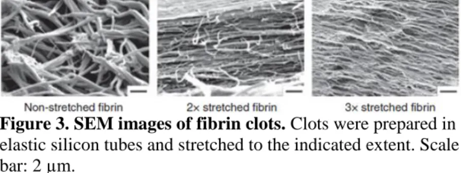
Since stretching of fibrin clots changed the arrangement of the fibres to a similar pattern (both fibre diameter and pore area were decreased, see Fig. 3.), stretched fibrin was used as a model system to evaluate the impact of mechanical stress on the structure and lytic susceptibility of fibrin.
Figure 3. SEM images of fibrin clots. Clots were prepared in elastic silicon tubes and stretched to the indicated extent. Scale bar: 2 µm.
11 Lysis of stretched fibrin
The amount of plasmin generated by tPA on the surface of fibrin (containing plasminogen) and released in the fluid phase decreased two- to three-fold, if stretched fibrin was used as a template instead of its non-stretched counterpart. In the presence of SPPL, which is able to penetrate into the clot, the detected plasmin activity was similarly lower on stretched fibrin, showing that the effect of the modified fibrin structure on the apparent plasmin generation is based on changes in plasminogen activation rather than in plasmin retention in the clot. The release of soluble FDP from stretched fibrin clots was also slower.
In order to evaluate separately the direct fibrin solubilisation by plasmin, plasminogen-free fibrin clots were treated with plasmin and the course of their dissolution was monitored. The SEM images of non-stretched fibrin digested for 45 min with plasmin showed many truncated fibres in the remnant fibrin, whereas only few fibres presented signs of digestion in the stretched fibrin.
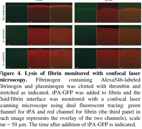
The mechanism of fibrinolytic resistance induced by stretching of fibrin was approached with the help of fluorescent confocal microscopy (Fig. 4.). When tPA-GFP was applied to the surface of non-stretched fibrin, a distinct zone of tPA
12
accumulation was formed at the fluid/fibrin interface within several minutes, which moved a distance of about 75 µm in 50 min as plasmin was formed and it dissolved the fibrin. This interfacial tPA-enriched zone on the surface of stretched fibrin did not move at all in the first hour of observation. Thus, the modified ultrastructure of fibrin in clots exposed to mechanical stress impedes tPA binding/penetration into fibrin and delays the lytic process in this experimental setup.
Figure 4. Lysis of fibrin monitored with confocal laser microscopy. Fibrinogen containing Alexa546-labeled fibrinogen and plasminogen was clotted with thrombin and stretched as indicated. tPA-GFP was added to fibrin and the fluid/fibrin interface was monitored with a confocal laser scanning microscope using dual fluorescent tracing: green channel for tPA and red channel for fibrin (the third panel in each image represents the overlay of the two channels), scale bar = 50 µm. The time after addition of tPA-GFP is indicated.
13
Effects of NET components on clot structure and lysis Thrombi from patients
Surgically removed thrombi analysed using immunohistochemistry and SEM (Fig. 5.) showed variable but widespread staining for DNA, and histones were also present though not so widely dispersed and in some cases were coincident with fibrin aggregates. Based on these findings, model thrombi containing DNA ± histones were used to study the effect of NET components on clot structure and fibrinolysis, as described in the next sections.
Figure 5. Fibrin, histone and DNA content of arterial thrombi. The thrombi rich in red blood cells (TO) or in fibrin (GI) according to the SEM images showed limited DNA- and histone-positive regions in contrast to the leukocyte-rich (TJ) thrombus. Scale bars: 50 µm in confocal panels, 2 µm in SEM panels. H1: histone H1.
14
Structural studies and thrombin inactivation kinetics
At low concentrations of thrombin, the presence of histones enhanced the otherwise small effects of DNA on fibre diameter values resulting in the appearance of thicker fibres, while at higher thrombin concentrations DNA and histones alone had opposing effects in a plasma environment: DNA caused thickening of fibres, while histones caused a decrease of diameter values.
In fibrin clots, the presence of histones increased the permeability constant approximately 4-fold, even in the presence of additional DNA. In the more complex plasma environment, however, the opposite effect was seen: the presence of histones reduced KS values by almost 50%. The effect of DNA alone on clot permeability was consistent in both systems: a significant negative effect was observed.
Since thrombin concentrations alone are also able to influence structural parameters of clots, the effects of NET constituents on the inactivation of thrombin by antithrombin were investigated. Histones were effective in protecting thrombin from inactivation even in the presence of heparin, and increasing concentrations of DNA were able to partially attenuate this effect in the presence of physiological antithrombin concentration.
15
Viscoelastic properties of clots containing NET components Striking differences were seen in the shear stress necessary to disassemble the fibrin as presented in Fig. 6. In the presence of
DNA alone the curves can be interpreted as increased sensitivity of fibrin to mechanical shear so that the shear stress needed to disassemble fibrin (where viscosity approaches zero) is reduced in comparison to the situation without DNA.
However, when histones are added to fibrin, and to a greater extent when histones are added to fibrin+DNA, the clots become more stable and resistant to shear forces.
B Figure 6. Rheometer studies showing the effect of DNA and
histones on the critical shear stress needed to disassemble fibrin. Red: pure fibrin; green, magenta, blue, black: fibrin containing 50 and 100 µg/ml DNA, 300 µg/ml histone, histone+100 µg/ml DNA, respectively. τ: shear stress, η:
viscosity.
16 Studies on lysis of plasma clots
According to confocal microscopy, DNA and histones alone had a negligible effect on the tPA-front penetration in plasma clots, however, when both components were added simultaneously, the relative run distance of the lysis front after 30 minutes was reduced by approximately 25%.
Plasminogen activation assays on plasma clot surface using chromogenic substrate SPPL showed that apparent velocity of p-nitroaniline formation detected at A405 was reduced by DNA alone by 20%, while histones ± DNA increased this value.
Turbidimetry assays revealed that DNA and histones prolonged the average time elapsed until 90% clot lysis when added separately or together, while the time needed for 50%
lysis remained mostly unchanged, and ITC studies showed that FDPs (>150 kDa) bind to both DNA and histones with a higher affinity than fibrinogen and plasminogen, possibly contributing to the retardation of lysis in the presence of these NET constituents.
Presence of NETs generated by phorbol-myristate- acetate (PMA)-activated granulocytes incorporated in the clots reproduced the effect of isolated components (Fig. 7.): T10
17
increased approximately 2-fold, while T50 was unaffected.
NETosis inhibitor Cl-Amidine moderated the NET effects on T10 values (Fig. 7, inset).
While incorporation of histones ± DNA did not influence plasmin-induced microscopic lytic front movement velocity in IBIDI slides, DNA alone reduced front velocity by 40%, in line with the DNA-mediated enhancement of the defibrinogenated plasma-induced inactivation of plasmin.
Figure 7. tPA-induced lysis of clots containing activated neutrophils. Clots were prepared as described in Materials and Methods. A340 turbidity values were normalized and converted to clot integrity. Mean curves of 8 measurements from a representative experiment are shown. Inset: Effect of Cl- Amidine and its vehiculum dimethylsulphoxide (DMSO) on T10 in clots containing PMA-activated neutrophils. T10 values are normalized for control to give relative units. Mean and standard deviation of means of 3 experiments with 6-8 parallels is shown.
18
CONCLUSIONS
(1) Stretching of fibrin results in structural changes: a thinner fibres and diminished pores are formed. The distribution of these parameters is more homogeneous in stretched clots compared to that of non-stretched clots.
(2) Structural changes are accompanied by decreased lytic susceptibility of stretched fibrin clots: tPA- as well as plasmin- mediated lysis is hindered on the stretched substrate.
(3) Major NET components (DNA and histones) are present in arterial thrombi.
(4) Presence of DNA and histones in clots formed with low concentrations of thrombin results in the formation of thicker fibres. DNA alone decreases permeability, while histones have opposing effects in the purified and plasma systems (positive and negative effect, respectively).
(5) Histones slow down the antithrombin-mediated inactivation of thrombin even in the presence of heparin, while the addition of DNA partially reverses this effect.
(6) DNA alone decreases mechanical stability of fibrin clots, histones ± DNA increase clot resistance against shear forces.
19
(7) Plasma clots containing either NETs from activated neutrophils or purified NET components (DNA ± histones) are resistant to tPA-mediated lysis, while DNA alone hinders plasmin-induced lysis.
(8) Retardation of fibrinolysis by NET constituents is partially elucidated by their affinity towards fibrin degradation products.
Binding of DNA and histone to these clot fragments may contribute to hindered disassembly of thrombi.
Implications:
(1) Our findings indicate that intravascular thrombi exposed to increased circulatory shear forces (e.g. in bifurcations) might be more difficult to dissolve. Our results also add to the earlier observations that thrombi going through the process of retraction (platelet contraction leading to stretching of the fibrin network) are less susceptible to fibrinolysis.
(2) Disruption of the DNA-histone matrix of thrombi (e.g. by DNAses or proteases capable of histone degradation, such as activated protein C) may enhance the effectiveness of current thrombolytic therapies aiming the dissolution of the fibrin meshwork.
20 LIST OF PUBLICATIONS
[1] Varjú I, Sótonyi P, Machovich R, Szabó L, Tenekedjiev K, Silva MM, Longstaff C, Kolev K. (2011) Hindered dissolution of fibrin formed under mechanical stress.
J Thromb Haemost, 9(5):979-86. doi: 10.1111/j.1538- 7836.2011.04203.x.
Impact factor: 5.731
[2] Longstaff C, Varjú I, Sótonyi P, Szabó L, Krumrey M, Hoell A, Bóta A, Varga Z, Komorowicz E, Kolev K. (2013) Mechanical stability and fibrinolytic resistance of clots containing fibrin, DNA, and histones. J Biol Chem, 288(10):6946-56. doi: 10.1074/jbc.M112.404301.
Impact factor: 4.600
[3] Varjú I, Kolev K. Fibrinolysis at the Interface of Thrombosis and Inflammation — The Role of Neutrophil Extracellular Traps. In: Kolev K (Ed.), Fibrinolysis and Thrombolysis. InTech, Rijeka, 2014: pp. 31-59. doi:
10.5772/57259.
Impact factor: -