DOES SUBINHIBITORY CONCENTRATIONS OF CLINICALLY IMPORTANT ANTIBIOTIC INDUCE
BIOFILM PRODUCTION OF ENTEROCOCCUS FAECIUM STRAINS?
FATMA NESLIHAN YUKSEL, NESLIHAN TASKALE KARATUG and MUSTAFAAKCELIK*
Department of Biology, Faculty of Science, Ankara University, Ankara, Turkey
(Received: 11 April 2017; accepted: 7 August 2017)
Biofilm structures are the most resistant form of active microorganisms against sanitation, disinfection, and sterilization processes. One of the specific properties of biofilm is the development of antibiotic resistance that can be up to 1,000-fold greater than planktonic cells. Enterococcus faecium is a human pathogen that causes nosocomial bacteremia and at the present time, it is well known that most of the chronic infections are biofilm-based. Recent evidence suggested that subinhibitory concentrations (sub-MICs) of antibiotics have an important role in the evolution of antibiotic resistance and induction on biofilm formation. Based on this information, we aimed to determine the effect of subinhibitory antibiotic concentrations on biofilm formation and the role of the antibiotic concentrations on the enterococcal surface protein gene (esp). To determine the impact of clinically important antibiotics on biofilm production, crystal violet assay was used. Then, the effect of sub-MICs of antibiotics on the expression of theespgene was investigated by quantitative real-time PCR. Biofilm production assays show that MIC/2 of erythromycin (ERT; 512μg/ml), MIC/32 of vancomycin (VAN; 16μg/ml), MIC/64 of streptomycin (STR; 32μg/ml), and MIC/128 of kanamycin (KAN; 4 μg/ml) values induce maximum biofilm production compared with the control. According to q-PCR results, sub-MIC values of ERT, VAN, and STR antibiotics were found to enhanceespgene expression. In addition, despite the increasing biofilm production after KAN treatment, the antibiotic was not effective on theespexpression.
Keywords: Enterococcus faecium, sub-MIC, biofilm formation, esp gene, quantitative real-time PCR
*Corresponding author; E-mail:akcelik@science.ankara.edu.tr
Introduction
Antibiotics target essential bacterial structures, such as cell wall and cellular pathways including DNA, RNA, and protein synthesis mechanism. They have been used to treat several infectious diseases. The long-term use of antibiotics in recent years has resulted in appearance of multidrug-resistant (MDR) bacterial pathogens, such as Enterococci [1, 2].
Enterococci, known as opportunistic pathogens, are naturally found in intestinal microflora and oral cavity of humans and animals. Two most common Enterococcus species (Enterococcus faecalis and Enterococcus faecium) are capable of producing biofilms, which are bacterial communities attached to a biotic or an abiotic substrate encased in a matrix.E. faeciumis an important global cause of biofilm-related infections. Biofilms are dependent on multiple genetic factors, such as esp, gelE, and fsr locus [3, 4]. Cell wall-associated protein implicated in biofilm formation is an enterococcal surface protein (Esp) coded by espgene. It wasfirst identified inE. faecalisas a large surface-anchored protein from infection-derived isolates [5]. An esp homologue has been identified in E. faecium and this gene is localized on pathogenicity islands in both species [6–8]. Studies suggested that there was a strong correlation betweenespand the forming of biofilms. Toledo-Arana et al. [9] reported that 93.5% ofesp-positive isolates could form biofilms on polystyrene, whereas none of the esp-negative isolates could produce biofilms. The investigators suggested that the N-terminal domain of Esp is sufficient for biofilm production, mutation on the N-terminal domain region of Esp inE. faecalisstrain causes less biofilm production [10]. The esp-positive strains were also identified as strong biofilm producers compared with esp-negative isolates [11, 12]. In addition to that, researchers presented that presence of a higher glucose concentration in the growth medium-regulated biofilm production [12–14]. In spite of that, other studies suggested that there was no association between the presence ofespand biofilm-forming ability and theespgene was not necessary for the production of biofilm in E. faecalisand E. faecium [15–18]. While some studies showed that espis a certain factor for biofilm formation, others presented that biofilm production needs other necessary factors with esp.
Recent studies suggested that sub-MICs of antibiotics acted as signaling molecules mediating variety of cell processes, such as gene transcription and expression, quorum sensing, inter- or intra-species communication, and biofilm formation [19–23]. In addition, low concentrations of antibiotics may stimulate different stress responses that might enable horizontal transfer of antibiotic resis- tance genes among bacterial communities, which are found on biofilm [23–26].
Studies showed that some using antibiotic concentrations below the MIC can significantly induce biofilm formation in a variety of Gram-positive and Gram- negative bacterial species. Subinhibitory concentration of an aminoglycoside antibiotic tobramycin induced biofilm formation in Pseudomonas aeruginosa andEscherichia coli [27]. Similarly, gentamicin (GEN) (>64 mg/ml, 2×MIC) and tetracycline (>128 mg/ml, 4×MIC) were determined as the most effective antibiotics againstSalmonellaInfantis biofilm formation; however, biofilm struc- ture was induced with sub-MICs of nalidixic acid, spectinomycin, tetracycline, and neomycin antibiotics treatment [28]. Balaji et al. [29] proved that 1/16 MIC value offluoroquinolones increased biofilm formation, whereas 1/2 MIC value of them occurred inhibition effect on clinical isolates of Streptococcus pyogenes biofilm. The proof presented that inhibition and biofilm formation were dose- dependent [29]. Previous studies showed that sub-MIC amoxicillin antibiotic levels induce methicillin-resistantStaphylococcus aureusbiofilm and this biofilm was thicker, contained more pillar and channel structures compared with the control [30]. Kafil et al. [31] investigated the effects of ampicillin (AMP), vancomycin (VAN), GEN, and ceftizoxime antibiotics on biofilm formation and gene expression of colonization factors, such asE. faecalisantigen A gene (efaA), aggregation substance gene (asa1), endocarditis and biofilm-associated pilli gene (ebpA),esp, and collagen adhesin gene (ace), inE. faecalis. They found that AMP, VAN, and ceftizoxime did not have any significant effect on biofilm formation while GEN induced biofilm formation. And also for 12 strains, GEN, VAN, and AMP increased expression ofesp in the ratio of 50.9%, 89.1%, 131%, respec- tively, by contrast, ceftizoxime reduced expression ofesp(35%) [31]. One of the most important goals in clinical microbiology is to prevent biofilm-associated infections. However, strategies for the treatment of biofilm-related infections should not be according to antibiotic concentrations that are effective only against to planktonic cells. Currently, very little is known about the mechanism of antibiotic induced biofilm formation in genusEnterococcus.This study aims that quantifying the use of low concentrations of antibiotics induces enterococcal biofilm formation and biofilm-related gene expression.
Material and Methods Bacterial isolates and culture conditions
Two biofilm producerE. faecium strains, isolated from rectal sample, were selected for this research.E. faecalisOG1RF was used as biofilm producer control
strain. All strains were obtained from Prokaryote Genetic Laboratory Culture Collection of Ankara University (Ankara, Turkey). Glycerol stock cultures were activated in Tryptic Soy Broth (TSB, Merck, Germany) for overnight (18 h) at 37 °C.
Determination of minimum inhibitory concentration (MIC)
The MIC values of antibiotics were determined by the Clinical and Laboratory Standards Institute broth microdilution method using Mueller–Hinton Broth (Oxoid, UK). The strains were treated with clinically important antibiotics, which are commonly used for the treatment ofEnterococcusinfections (TableI).
E. faecalisATCC 29212 was used as the control strain and each experiment was performed in duplicate.
Effect of subinhibitory concentrations (sub-MICs) of antibiotics on enterococcal biofilm formation
To determine induction of antibiotics on biofilm production level of E. faeciumisolate, chloramphenicol (CHL), kanamycin (KAN), erythromycin (ERT), AMP, VAN, streptomycin (STR), and GEN antibiotics were selected. A method described by Extremina et al. [32] and Baldassarri et al. [33] was used to test the microorganisms for biofilm formation with minor modifications. The serial twofold dilution of the antibiotics (from MIC/2 to MIC/128), 107 CFU/ml (OD595=0.07) overnight inE. faeciumculture, and TSB supplemented with 1% glucose were added to 96 well plates and incubated at 37 °C for 48 h. Negative control was only test broth, and positive control was only bacteria. After 48 h growth at 37 °C, the plates were gently washed thrice with phosphate buffered saline. The plates were allowed to dry
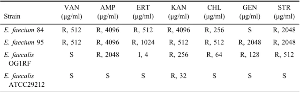
Table I.Antibiotic resistance phenotypes and MIC values of the strains
Strain
VAN (μg/ml)
AMP (μg/ml)
ERT (μg/ml)
KAN (μg/ml)
CHL (μg/ml)
GEN (μg/ml)
STR (μg/ml)
E. faecium84 R, 512 R, 4096 R, 512 R, 4096 R, 256 S R, 2048
E. faecium95 R, 512 R, 4096 R, 1024 R, 512 R, 512 R, 2048 R, 2048 E. faecalis
OG1RF
S R, 2048 I, 4 R, 256 R, 64 R, 128 R, 512
E. faecalis ATCC29212
S S S R, 32 S S S
Note:AMP: ampicillin; GEN: gentamicin; KAN: kanamycin; STR: streptomycin; CHL: chloramphenicol;
ERT: erythromycin; VAN: vancomycin; R: resistance; I: intermediate-level resistance; S: susceptible;
MIC: minimum inhibitory concentration.
for 1 h at 60 °C and thenfixed using methanol (95%). For biofilm quantification, 200μl of 1% crystal violet (CV) solution was added to each well and the plates were allowed to stand for 30 min. The wells were subsequently washed thrice with steriled H2O to wash off the excess CV. CV bounded to the biofilm was extracted with 200μl of ethanol–acetone (80/20% v/v) and the absorbance of the extracted CV was measured at 595 nm on ELISA Reader (ThermoScientific, Multiskan Go, USA).
Determination of esp gene expression by quantitative real-time PCR assay
Statistically significant sub-MIC antibiotic values leading to biofilm formation in E. faecium 95 were selected for esp gene expression experiments. Antibiotic- induced total RNA of clinicalE. faecium95 strain was extracted using Promega RNA Isolation kit (Promega, USA). RNA concentrations were measured by NanoDrop 2000 (ThermoScientific, USA). cDNA synthesis was carried out using Transcriptor First Strand cDNA Synthesis Kit (Roche, Switzerland). Quantitative real-time PCR was done byespprimers;esp-F 5′TGGTGATGGAAACCCTGACGA-3′, andesp-R 5′-TTGCGCTTTGTGACCTGTTCC-3′ [34]. The q-PCR assay was performed in Roche Light Cycler®480 II (Roche, Switzerland). The q-PCR amplifications were performed in 10μl reactions containing 1×Hot FirePol®EvaGreen® qPCR master mix (Solis BioDyne, Esthonia), which includes Hot FirePol® DNA Polymerase, EvaGreen® qPCR Buffer, 2.5 mM MgCl2, ultrapure dNTPs, EvaGreen® dye and RNase-free H2O, 0.5 pmol each primer, and 1μl of the respective template cDNA dilution. The q-PCR assay was optimized to the initial activation step of 95 °C for 15 min, followed by 40 cycles of denaturation at 95 °C for 15 s, annealing at 62 °C for 20 s for all the studied genes, extension at 72 °C for 20 s. All experiments were performed in triplicate. The threshold cycle (Ct) of each well and data acquisition were carried out using a software program from Real-Time Analysis Software Programme (Light Cycler®480 SW 1.5.0 SP4). The delta Ct (ΔCt) method was used for PCR single gene data analysis. The normalized (ΔCt) for esp gene was calculated by subtracting the mean Ct of the 16S rDNA housekeeping gene from the Ct of esp.
Statistical analysis
All biofilm assays were performed in triplicate. In CV quantitative analysis, the results were calculated by subtracting the median OD595of the triplicates of the control (test broth and E. faecium isolate, without antibiotic) from the median OD595of the triplicates of the sample. Statistical analysis was carried out by SPSS (version 18, USA). One-way ANOVA test was preferred for microtiter plate assay data. Ap value less than 0.05 was considered statistically significant.
Results and Discussion
In this study, two E. faecium strains isolated from clinical samples were subjected. Isolates were investigated for resistance against seven antibiotics that are important to clinical treatment of Enterococcus infections (Table I). Both strains were resistant to AMP, KAN, STR, CHL, ERT, and VAN at high levels. In addition, E. faecium 95 strain showed resistance to GEN (MIC; 2,048 μg/ml), whereasE. faecium84 strain was sensitive. According to this result, both strains were detected to exhibit multidrug resistance to clinically important antibiotics.
Biofilm producingE. faecalisOG1RF as a control strain was found sensitive to VAN only, whereas non-biofilm producer E. faecalis ATCC29212 was sensitive to all treated antibiotics except KAN (TableI). Considering this assay results, it can be indicated that the strains capable of producing biofilms are more resistant to antibiotics. MDR strains may have acquired antibiotic-resistant genes between bacteria that are present inside the biofilm structure causing to be in- hospital adapted clones. In addition, the uncontrolled use of the most common antibiotics may have influenced the rise in prevalence of enterococcal infections in humans. Recent reports showed that E. faecium strains isolated from clinical samples had high degree of resistance to antibiotics [35–38]. In comparing the biofilm-forming strains (BIO+) with the non-biofilm-forming strains (BIO−), BIO+ strains were high frequency resistant than BIO− strains in clinical E. faeciumisolates [39]. This observation was also declared in some other studies conducted by Sindhanai et al. [40] and Bhardwaj et al. [41].These results support that the biofilm forming promotes the virulence profile to microorganisms [41].
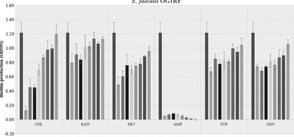
The adherence ability of biofilm was determined by estimation of obtained OD values ofE. faeciumclinical isolates according to growth conditions including planktonic culture and different sub-MIC antibiotic dilutions-broth media supple- mented with 1% glucose. After 48 h exposure to sub-MIC antibiotics, the biofilm exhibited for these antibiotics on the plates the highest adherence ratio, which was statistically significant difference (p<0.05). The activities of antimicrobial agents were tested at sub-MICs against E. faecium 95, 84, and E. faecalis OG1RF biofilms are shown in Figures1–3, respectively. According to biofilm induction with sub-MIC of antibiotics results; CHL, AMP, and GEN antibiotic values did not induce biofilm formation ofE. faecium95 strain. On the other hand, sub-MIC antibiotic values of inducing maximum biofilm formation in E. faecium 95 were MIC/128 of KAN (4μg/ml), MIC/2 of ERT (512μg/ml), MIC/32 of VAN (16μg/ml), and MIC/64 of STR (32 μg/ml) compared with control. The results proved that the different concentrations of each antibiotic promoted the maximum biofilm production. In addition, concentration of antibiotic decreased except ERT,
and the biofilm formation was also induced. Only the ERT was the cause of the increased biofilm formation onE. faecium84 strain whose value was found to be MIC/4 (256μg/ml) whereas tested sub-MIC antibiotic values had any significant difference on biofilm formation ofE. faecalisOG1RF. This study presented that
Figure 1.Biofilm production levels ofE. faecium95 strain following incubation with sub-MIC antibiotic levels. PC: positive control; CHL: chloramphenicol; KAN: kanamycin; ERT:
erythromycin; AMP: ampicillin; VAN: vancomycin; STR: streptomycin; GEN: gentamicin
Figure 2.Biofilm production levels ofE. faecium84 strain following incubation with sub-MIC antibiotic levels. PC: positive control; CHL: chloramphenicol; KAN: kanamycin; ERT:
erythromycin; AMP: ampicillin; VAN: vancomycin; STR: streptomycin
strains formed different amounts of biofilm productivity. E. faecium 84 and E. faecalis OG1RF were more powerful biofilm producers compared with E. faecium 95. Strains producing high levels of biofilm in the absence of antibiotics may not form biofilm induction in the presence of sub-MIC antibiotic values [42], for instance, sub-MIC levels of azithromycin showed inhibition effect onP. aeruginosabiofilm formation [43]. The demonstration of differences in the formation of antibiotic-induced biofilms according to strains shows that a single model system cannot be established for treatment. In this case, it is necessary to conduct strain-based control.
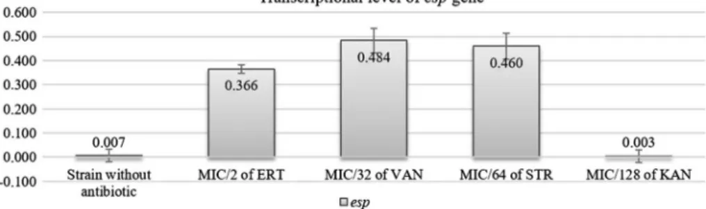
Quantitive real-time PCR assay was used to evaluate the effect of sub-MIC of KAN, ERT, VAN, and STR onespgene as one of the responsible genes on colonization of enterococci in selected strainE. faecium95. Transcriptions ofesp gene were strongly increased by MIC/2 of ERT (512 μg/ml), MIC/32 of VAN (16 μg/ml), and MIC/64 of STR (32 μg/ml). Although the MIC/128 of KAN (4μg/ml) induced biofilm formation, this concentration has no effect on the esp expression (Figure 4). As the similar results, Kafil et al. [31] showed, although expression ofespgene increased with GEN and VAN, it was reduced with ceftizoxime and AMP antibiotics. There are several virulence factors relating to biofilm, such as ace, esp, efaA, ebpA, and asa1 in Enterococci [32, 44]. As based to this study, KAN-induced biofilm formation may not only depend on the induction of esp but also other virulence genes. Other components, such as extracellular DNA, may also be effective on the biofilm.
Figure 3.Biofilm production levels ofE. faecalisOG1RF strain following incubation with sub-MIC antibiotic levels. PC: positive control; CHL: chloramphenicol; KAN: kanamycin; ERT:
erythromycin; AMP: ampicillin; STR: streptomycin; GEN: gentamicin
We showed that virulence gene expression patterns can be changed by exposure to antibiotics below MIC and there is no single mechanism of antibiotic-induced biofilm formation. There are limited studies about the role of antibiotics on enterococcal biofilm formation. More studies are needed to determine whether there is a relationship between biofilm inducibility and response to therapy. Genes responsible for biofilm formation should be investigated by knockout studies.
Conclusions
Antibiotic dosages in clinical treatment are generally chosen according to MICs of antibiotics against planktonic cells. Although MIC has been used as a gold standard to determine antimicrobial sensitivities of bacteria, the MIC value is not predictive of a particular antibiotic choosing in clinical efficacy because of the biofilm forming ability in bacteria. In addition, cells buried in deep within a biofilm matrix may be exposed to sub-MIC concentrations of antibiotics because of diffusion gradients. The data from our experiments showed that certain concentrations of chosen antibiotics stimulate biofilm production of clinically isolatedE. faeciumstrain. Commonly using antibiotics as VAN in treatment of clinical enterococcal infections provokes the biofilm formation at low concentrations. We concluded that antibiotic concentrations for struggle with pathogens need to be designated carefully. In addition, induction ofespexpression via sub-MICs of antibiotics may cause difficulties at accomplished by antibiotic therapy for eradicating persistent enterococcal infections associated with biofilms. Future studies as generation of mutant libraries will probably elucidate the mechanisms of biofilm induction by sub- MIC antibiotic levels.
Figure 4.espgene expression rate after antibiotic exposure inE. faecium95
Conflict of Interest The authors declare no conflict of interest.
References
1. Aka, S. T., Haji, S. H.: Sub-MIC of antibiotics induced biofilm formation ofPseudomonas aeruginosa in the presence of chlorhexidine. Braz J Microbiol46, 149–154 (2015).
2. Weiner, L. M., Webb, A. K., Walters, M. S., Dudeck, M. A., Kallen, A. J.: Policies for controlling multidrug-resistant organisms in US healthcare facilities reporting to the National Healthcare Safety Network, 2014. Infect Control Hosp Epidemiol,37, 1105–1108 (2016).
3. Mohamed, J. A., Huang, D. B.: Biofilm formation by enterococci. J Med Microbiol56, 1581–1588 (2007).
4. Paganelli, F. L., Willems, R. J. L., Jansen, P., Hendrickx, A., Zhang, X., Bonten, M. J. M., Leavis, H. L.:Enterococcus faeciumbiofilm formation: Identification of major autolysin AtlAEfm, associated Acm surface localization, and AtlAEfm-independent extracellular DNA release. mBio4, e00154-13 (2013).
5. Shankar, V., Baghdayan, A. S., Huycke, M. M., Lindahl, G., Gilmore, M. S.: Infection- derivedEnterococcus faecalisstrains are enriched in esp, a gene encoding a novel surface protein. Infect Immun67, 193–200 (1999).
6. Eaton, T. J., Gasson, M. J.: A variant enterococcal surface protein Esp (fm) inEnterococcus faecium; distribution among food, commensal, medical, and environmental isolates. FEMS Microbiol Lett 216, 269–275 (2002).
7. Shankar, N., Baghdayan, A. S., Gilmore, M. S.: Modulation of virulence within a pathogenicity island in vancomycin-resistant Enterococcus faecalis. Nature 417, 746–750 (2002).
8. Leavis, H., Top, J., Shankar, N., Borgen, K., Bonten, M., van Embden, J., Willems, R. J. : A novel putative enterococcal pathogenicity island linked to the esp virulence gene of Enterococcus faeciumand associated with epidemicity. J Bacteriol186, 672–682 (2004).
9. Toledo-Arana, A., Valle, J., Solano, C., Arrizubieta, M. J., Cucarella, C., Lamata, M., Amorena, B., Leiva, J., Penadés, J. R., Lasa, I.: The enterococcal surface protein, Esp, is involved in Enterococcus faecalis biofilm formation. Appl Environ Microbiol 67, 4538–4545 (2001).
10. Tendolkar, P. M., Baghdayan, A. S., Shankar, N.: The Nterminal domain of enterococcal surface protein, Esp, is sufficient for Esp-mediated biofilm enhancement inEnterococcus faecalis. J Bacteriol187, 6213–6222 (2005).
11. Tendolkar, P. M., Baghdayan, A. S., Shankar, N.: Putative surface proteins encoded within a novel transferable locus confer a high-biofilm phenotype to Enterococcus faecalis.
J Bacteriol188, 2063–2072 (2006).
12. Diani, M., Esiyok, O. G., Ariafar, M. N., Yuksel, F. N., Altuntas, E. G., Akcelik, N.: The interactions between esp, fsr, gelE genes and biofilm formation and pfge analysis of clinical Enterococcus faeciumstrains. Afr J Microbiol Res8, 129–137 (2014).
13. Pillai, S. K., Sakoulas, G., Eliopoulos, G. M., Moellering, R. C., Jr., Murray, B. E., Inouye, R. T.: Effects of glucose on fsr-mediated biofilm formation in Enterococcus faecalis.
J Infect Dis190, 967–970 (2004).
14. Tendolkar, P. M., Baghdayan, A. S., Gilmore, M. S., Shankar, N.: Enterococcal surface protein, Esp, enhances biofilm formation by Enterococcus faecalis. Infect Immun 72, 6032–6039 (2004).
15. Sandoe, J. A., Witherden, I. R., Cove, J. H., Heritage, J., Wilcox, M. H.: Correlation between enterococcal biofilm formation in vitro and medical-device-related infection potential in vivo. J Med Microbiol52, 547–550 (2003).
16. Dworniczek, E., Wojciech, L., Sobieszczanska, B., Seniuk, A.: Virulence of Enterococcus isolates collected in Lower Silesia (Poland). Scand J Infect Dis37, 630–636 (2005).
17. Raad, I. I., Hanna, H. A., Boktour, M., Chaiban, G., Hachem, R. Y., Dvorak, T., Lewis, R., Murray, B. E.: Vancomycin-resistant Enterococcus faecium: Catheter colonization, esp gene, and decreased susceptibility to antibiotics in biofilm. Antimicrob Agents Chemother 49, 5046–5050 (2005).
18. Ramadhan, A. A., Hegedus, E.: Biofilm formation and esp gene carriage in enterococci.
J Clin Pathol58, 685–686 (2005).
19. Davies, J.: Are antibiotics naturally antibiotics? J Ind Microbiol Biotechnol33, 496–499 (2006).
20. Romero, D., Traxler, M. F., L´opez, D., Kolter, R.: Antibiotics as signal molecules. Chem Rev111, 5492–5505 (2011).
21. Sengupta, S., Chattopadhyay, M. K., Grossart, H. P.: The multifaceted roles of antibiotics and antibiotic resistance in nature. Front Microbiol4, 47 (2013).
22. Andersson, D. I., Hughes, D.: Microbiological effects of sub lethal levels of antibiotics. Nat Rev Microbiol12, 465–478 (2014).
23. Balcázar, J. L., Subirats, J., Borrego, C. M.: The role of biofilms as environmental reservoirs of antibiotic resistance. Front Microbiol,6, 1216 (2015).
24. Beaber, J. W., Hochhut, B., Waldor, M. K.: SOS response promotes horizontal dissemina- tion of antibiotic resistance genes. Nature427, 72–74 (2004).
25. Miller, C., Thomsen, L. E., Gaggero, C., Mosseri, R., Ingmer, H., Cohen, S. N.: SOS response induction by beta-lactams and bacterial defense against antibiotic lethality.
Science305, 1629–1631 (2004).
26. Maiques, E., Úbeda, C., Campoy, S., Lasa, I., Novick, R. P., Barbé, J., Penadés, J. R.:
β-lactam antibiotics induce the SOS response and horizontal transfer of virulence factors in Staphylococcus aureus. Appl Environ Microbiol188, 2726–2729 (2006).
27. Hoffman, L. R., D’Argenio, D. A., MacCoss, M. J., Zhang, Z., Jones, R. A., Miller, S. I.:
Aminoglycoside antibiotics induce bacterial biofilm formation. Nature436, 1171–1175 (2005).
28. Tezel, B. U., Akçelik, N., Yüksel, F. N., Karatug, N. T., Akçelik, M.: Effects of sub-MIC˘ antibiotic concentrations on biofilm production of Salmonella Infantis. Biotechnol Bio- technol Equip30, 1184–1191 (2016).
29. Balaji, K., Thenmozhi, R., Pandian, S. K.: Effect of subinhibitory concentrations of fluoroquinolones on biofilm production by clinical isolates of Streptococcus pyogenes.
Indian J Med Res137, 963–971 (2013).
30. Mlynek, K. D., Callahan, M. T., Shimkevitch, A. V., Farmer, J. T., Endres, J. L., Marchand, M., Bayles, K. W., Horswill, A. R., Kaplana, J. B.: Effects of low-dose amoxicillin on Staphylococcus aureusUSA300 biofilms. Antimicrob Agents Chemother60, 2639–2651 (2016).
31. Kafil, H. S., Mobarez, A. M., Moghadam, M. F., Hashemi, Z. S., Yousefi, M.: Gentamicin induces efaA expression and biofilm formation inEnterococcus faecalis. Microb Pathog92, 30–35 (2016).
32. Extremina, C. I., Costa, L., Aguiar, A. I., Peixe, L., Fonseca, A. P.: Optimization of processing conditions for the quantification of enterococci biofilms using microtitreplates.
J Microbiol Methods84, 167–173 (2011).
33. Baldassarri, L., Cecchini, R., Bertuccini, L., Ammendolia, M. G., Losi, F., Arciola, C. R., Montanaro, L., Di Rosa, R., Gherardi, G., Dicuonzo, G., Orefici, G., Creti, R.:
Enterococcus spp. produces slime and survives in rat peritoneal macrophages. Med Microbiol Immunol190, 113–120 (2001).
34. Diani (2016): Determination biofilm producing characteristics of Enterococcus strains isolated from Turkey originated cheese samples, PhD thesis, Ankara University Biotechnology Institute, Turkey.
35. Mengeloglu, F. Z., Çak˘ ır, D., Terzi, H. A.: Comparison of resistance in isolates of Enterococcus faecalisandEnterococcus faecium. J Microbiol Infect Dis1, 10–13 (2011).
36. Banerjee, T., Anupurba, S.: Prevalence of virulence factors and drug resistance in clinical isolates of Enterococci: A study from North India. J Pathogens 2015, 692612 (2015).
37. Kolli, H. R., Swarnalatha, K., Reddy, B. S., Singh, M., Kabra, V.: Isolation and identification of vancomycin resistance Enterococci from clinical specimens. Med Sci 6, 9–14 (2016).
38. Sundaram, M., Kavita, Y., Mohiddin, S. K.: Antibiogram of Enterococcal species isolated from clinical specimens in a tertiary care teaching hospital. J Evolution Med Dent Sci5, 2955–2958 (2016).
39. Sie´nko, A., Wieczorek, P., Majewski, P., Ojdana, D., Wieczorek, A., Olsza´nska, D., Tryniszewska, E.: Comparison of antibiotic resistance and virulence between biofilm- producing and non-producing clinical isolates ofEnterococcus faecium. Acta Biochimica Pol62, 859–866 (2015).
40. Sindhanai, V., Avanthiga, S. S., Chander, V. C. S.: Antibiotic susceptibility pattern of biofilm forming and biofilm non formingEnterococcispecies. IOSR J Dental Med Sci15, 33–37 (2016).
41. Bhardwaj, S. B., Mehta, M., Sood, S., Sharma, J.: Biofilm formation by drug resistant Enterococci isolates obtained from chronic periodontitis patients. J Clin Diagn Res 11, DC01–DC03 (2017).
42. Kaplan, J. B.: Antibiotic-induced biofilm formation. Int J Artif Organs34, 737–751 (2011).
43. Favre-Bonte, S., Kohler, T., Van Delden, C.: Biofilm formation by Pseudomonas aeruginosa: Role of the C4-HSL cell-to-cell signal and inhibition by azithromycin.
J Antimicrob Chemother52, 598–604 (2003).
44. Kafil, H. S., Mobarez, A. M., Moghadam, M. F.: Adhesion and virulence factor properties ofEnterococciisolated from clinical samples in Iran. Indian J Pathol Microbiol56, 238–42 (2013).