DISTRIBUTION OF GENES ENCODING
RESISTANCE TO MACROLIDES, LINCOSAMIDES, AND STREPTOGRAMINS AMONG METHICILLIN-
RESISTANT STAPHYLOCOCCUS AUREUS STRAINS ISOLATED FROM BURN PATIENTS
SAEED KHOSHNOOD1,2, FATEMEH SHAHI1,3*, NABI JOMEHZADEH4, EFFATABBASIMONTAZERI1*, MORTEZASAKI1,3, SEYEDMOJTABAMORTAZAVI2
and LEILA MAGHSOUMI-NOROUZABAD3,5
1Department of Microbiology, Faculty of Medicine, Ahvaz Jundishapur University of Medical Sciences, Ahvaz, Iran
2Student Research Committee, School of Medicine, Bam University of Medical Sciences, Bam, Iran
3Student Research Committee, Ahvaz Jundishapur University of Medical Sciences, Ahvaz, Iran
4Abadan School of Medical Sciences, Abadan, Iran
5Nutrition and Metabolic Diseases Research Center, Ahvaz Jundishapur University of Medical Sciences, Ahvaz, Iran
(Received: 7 February 2019; accepted: 11 March 2019)
The increasing resistance to macrolide, lincosamide, and streptogramin B agents among methicillin-resistantStaphylococcus aureus(MRSA) is a worldwide problem for the health community. This study aimed to investigate the prevalence ofermA, ermB, ermC, andmsrAin MRSA strains isolated from burn patients in Ahvaz, southwest of Iran. A total of 76 isolates ofS. aureuswere collected from January to May 2017 from Taleghani Burn Hospital in Ahvaz. Among 76S. aureusstrains collected, 60 (78.9%) isolates were MRSA. The antimicrobial susceptibility testing for MRSA showed extreme high resistance rate to clarithromycin (100%) and azithromycin (100%), followed by erythromycin (98.3%). The PCR assay revealed that the frequency rates ofmsrA, ermA, andermCgenes were 23 (38.3%), 28 (46.7%), and 22 (36.7%), respectively. In addition, none of the MRSA isolates had theermB gene. Because of the high prevalence of macrolide and lincosamide resistance found in MRSA isolates from infections of burn patients in Ahvaz, southwest of Iran, it is recommended that local periodic survey be performed for controlling the dissemination of antimicrobial resistance.
Keywords: Staphylococcus aureus,ermgenes, MRSA, clindamycin, Iran
*Corresponding authors; E-mails:Shahiftm66@gmail.com;eam1043@gmail.com
First published online May 17, 2019
Introduction
Infection is the most serious complication among burned patients, which is difficult to control and remains to be the leading cause of morbidity and mortality in these patients. In addition, invasive infections caused by antibiotic-resistant bacteria, which are responsible for 28%–65% of burn deaths globally, should be considered as a potential risk and their resistance pattern must be identified as soon as possible [1,2].
Staphylococcus aureusis known to be one of the most common burn wound pathogens worldwide. Colonization ofS. aureuson the surface of burn wounds could be associated with delayed wound healing, increased treatment costs through the need for expensive antibiotics, prolonged duration of stay at burn centers, and increased need for surgical interventions [3, 4].
Since the discovery of thefirst effective antimicrobials in medical science,S.
aureushas demonstrated rapid development of antibiotic resistance, as well as developed resistance to the most variety of antibiotics [5]. Although β-lactam antibiotics are the main compounds utilized to treat staphylococci-related infec- tions, the emergence of methicillin-resistantS. aureus(MRSA) and alterations in antimicrobial resistance pattern has caused renewed interest in the use of anti- biotics, such as macrolide, lincosamide, and streptogramin B (MLSB), for the treatment of these infections [6].
This group of antibiotics, in spite of their different chemical structure, has a similar mode of action and has been classified in the same group. They inhibit protein synthesis by binding to the subunit 23S rRNA of the bacterial 50s ribosomal subunits [7]. Among MLSB, clindamycin, due to its pharmacokinetic properties such as good oral absorption, excellent penetration in the skin, and tolerability, is a frequent choice for some staphylococcal infections, particularly skin and soft-tissue infections. However, extensive use of these antibiotics has led to the emergence of resistance to them [8].
Resistance to MLSB antibiotics among staphylococci more often involves the following two mechanisms, such as the active efflux of the antimicrobial agent by an ATP-dependent pump encoded bymsrAgene and the ribosomal binding site modification by 23S rRNA methylases mediated by one or moreermgenes (ermA, ermB, ermC, andermF) among whichermAandermCare predominant genes [9].
Mechanism of ribosomal target site modification can be either constitutive or inducible. S. aureus isolates with constitutive resistance show resistance to erythromycin and clindamycin onin vitrotesting, whereas isolates with inducible resistance show resistance to erythromycin but appear sensitive to clindamycin on disk diffusion testing [10, 11]. The aim of this study was to investigate the molecular detection of MLSB resistance genes (ermA, ermB, ermC, mecA, and
msrA) and antibiotic resistance profiles in MRSA strains isolated from burn patients using polymerase chain reaction (PCR) technique in southwest Iran.
Materials and Methods Bacterial isolates
In this cross-sectional study, clinical samples were collected from burn patients, admitted to the Taleghani Burn Hospital, Ahvaz, Iran, from January to May 2017. The research was approved by the ethical committee of Ahvaz Jundishapur University of Medical Sciences, Khuzestan, Iran. As a part of the Ahvaz Jundishapur University of Medical Sciences policy, written informed consent was obtained from all patients. The study was conducted in accordance with the Declaration of Helsinki. The specimens included were urine, blood, abscess, deep wound, and endotracheal secretion. The samples were cultured on 10% sheep blood agar and Mannitol salt agar (Merck, Darmstadt, Germany).
Presumptive staphylococcal colonies (growth on mannitol salt agar, Gram- positive, and catalase-positive cocci) were tested for production of DNase and coagulase. Isolates with positive reactions (DNase-positive and coagulase- positive) were considered as S. aureus[12].
Cefoxitin and oxacillin disk diffusion method
Susceptibility tests for S. aureus isolates were performed by the Kirby– Bauer disk diffusion method as recommended by Clinical and Laboratory Standards Institute (CLSI) using oxacillin (1 μg) and cefoxitin (30 μg) disks.
The inhibition zones for the oxacillin disk with diameter≤10 mm for S. aureus were considered to be resistant and the inhibition zone for cefoxitin with diameters of≥20 and≤19 mm were considered susceptible and resistant, respectively [13].
Epsilometer test
The Epsilometer test (E-test) was conducted for quantitative antimicrobial susceptibility testing using E-test strips (Liofilchem, Italy). A standard bacterial suspension equal to 0.5 McFarland inoculated on Mueller–Hinton agar (MHA) plates; then, E-test strips of tigecycline, linezolid, teicoplanin, vancomycin, and quinupristin/dalfopristin were placed on the medium surface and incubated at 35 °C for 24 h for detection of minimum inhibitory concentration (MIC). The MIC was
read at the lowest concentration at which the border of the elliptical inhibition zone intercepted the scale on the strip.
Oxacillin–salt agar screening
The presence of MRSA was confirmed by oxacillin–salt agar screening test.
This test was performed according to CLSI recommendations [13]. For each isolate, 1 ml of standard 0.5 McFarland suspension was cultured on an MHA medium containing oxacillin (at a concentration of 6 μg/ml of media) and 4% NaCl. The plates were incubated in ambient air at 35 °C for 24 h. Any growth on the plate was indicated as oxacillin resistance.
MRSA antibiotic susceptibility pattern
Susceptibility testing of MRSA isolates against erythromycin (15 μg), clarithromycin (15μg), azithromycin (15μg), vancomycin (30μg), clindamycin (2 μg), linezolid (30 mg), teicoplanin (30 μg), trimethoprim/sulfamethoxazole (1.25/23.75μg), quinupristin/dalfopristin (15μg), tigecycline (15μg), gentamicin (10 μg), and rifampin (5 μg) disks (Mast, Merseyside, United Kingdom) was determined by the Kirby–Bauer disk diffusion method on MHA, according to the procedures described by the CLSI guidelines. On following this, inducible clindamycin resistance was determined using the D-zone test according to these guidelines [13]. S. aureusATCC 25923 was used as the reference strain.
Amplification of 16S rRNA gene specific forS. aureusandermA, ermB, ermC, mecA, andmsrAgenes
DNA was extracted from bacterial colonies by the simple boiling method as previously described [4]. In brief, a few bacterial colonies were suspended in 400 ml of tris ethylene diaminetetraacetic acid buffer (pH 8.0), and the solution was heated at 100 °C for 10 min and then centrifuged at 15,000 rpm for 15 min.
The supernatant was used as template DNA in PCR.
The PCR assay was performed in 25μl contained a DNA template (50 ng), 100μM concentrations (each) of the four dNTPs, 1 U of Taq DNA polymerase (Cinnagen, Iran), 5μl of Taq buffer (5×), 25 pM of each of forward and reverse primersermA, ermB, ermC, andmsrA(TableI). The PCR mixtures were subjected to thermal cycling (4 min at 94 °C, followed by 30 cycles of 30 s at 94 °C for denaturation, 30 s for annealing extension, and extension at 72 °C for 30 s). Afinal elongation at 72 °C for 5 min was achieved in a DNA thermal cycler [14].
PCR products were analyzed by 1% agarose gel electrophoresis in 1×tris-borate- EDTA buffer at pH 8.3. The amplification products were photographed and their size was determined using a 100-bp molecular size marker [14].
Statistical analysis
Descriptive data were analyzed using SPSS v.22.0 statistics software (IBM Corporation, Armonk, NY, USA).χ2andt-tests were used to analyze intergroup significance. In addition, p<0.05 was considered statistically significant.
Results Bacterial isolates
From the total screened samples during 6 months, 76S. aureuswere isolated by biochemical tests and 16S rRNA gene PCR. Using cefoxitin and oxacillin disk diffusion and oxacillin–salt agar screening and PCR for themecAgene, 60 (79%) ofS.
aureus were identified as being methicillin-resistant. Out of 60 MRSA isolates studied, 34 (56.7%) and 26 (43.3%) strains were collected from male and female patients, respectively. The sample sources according to the hospital wards were as follows: the internal women, internal men, pediatric, intensive care unit, and surgery, repair, and outpatient department (OPD), with proportions of 6 (10%), 12 (20%), 9 (15%), 26 (43.3%), 2 (3.3%), 4 (6.7%), and 1 (1.7%), respectively. About 88.3%
(53/60) of isolates were obtained from wound culture specimens, 1 (1.7%) from urine culture, 3 (5%) from blood culture, and 3 (5%) from endotracheal secretion culture.
Table I.Primers and their target genes used in this study
Primer Sequence (5′–3′) Product size (bp) Reference
16S rRNA F: GAA AGC GTG GGG ATC AAA CA 340 [15]
R: TTG CGG GAC TTA ACC CAA CA
ermA F: GAT TTC GTT CCT CGA CC 139 [15]
R: TAT CTT ATC GTT GAG AAG GGA TT
ermB F: CTA TCT GAT TGT TGA AGA AGG ATT 142 [15]
R: TTT ACT CTT GGT TTA GGA TGA AA
ermC F: CTT GTT GAT CAC GAT AAT TTC C 190 [15]
R: ATC TTT TAG CAA ACC CGT ATT C
msrA F: TCC AAT CAT TGC ACA AAA TC 163 [16]
R: AAT TCC CTC TAT TTG GTG GTC
mecA F: ACGGTAACATTGATCG-CAACG 176 [15]
R: GGCCAATTCCACATTGTTTCG
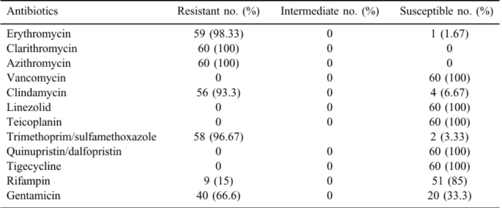
Antibiotic resistance pattern
According to disk diffusion results, all MRSA strains were resistant to clarithromycin and azithromycin. In addition, the majority of the strains was resistant to erythromycin 59 (98.33%), clindamycin 56 (93.3%), and trimethoprim/
sulfamethoxazole 58 (96.67%), whereas, using the E-test, all MRSA isolates were susceptible to teicoplanin with a maximum range 0.25 (MIC≤4 mg/ml), linezolid= 0.19 (MIC≤4 mg/ml), vancomycin=0.5 (MIC≤2 mg/ml), and tigecycline=0.25 (MIC≤4 mg/ml) and showed resistance to oxacillin (MIC≥4 mg/ml). Besides, the D-test results showed that 30 (50%) of the MRSA isolates have the inducible clindamycin resistance phenotype. The resistance profile for all isolates to macrolides and other tested antibiotics is listed in Table II. Fifty-nine (98.3%) isolates were simultaneously resistant to erythromycin, azithromycin, and clarithromycin (cross- resistance); whereas only 1 (1.7%) isolate had various macrolide susceptibility pattern.
This isolate was susceptible to erythromycin and was resistant to azithromycin and clarithromycin. The highest antimicrobial resistance was related to wound specimens with 100% resistance to erythromycin, clarithromycin, and azithromycin and 93.3%
resistance to clindamycin.
MDR profiles
The results of the susceptibility testing showed that all 60 MRSA isolates were resistant to at least two antibiotics, and the majority of isolates (N=58, 96.6%) was multidrug-resistant (MDR) with five diverse patterns (Table III).
Table II.Prevalence of resistance to the tested antibiotics among MRSA isolates using the disk diffusion andt-test methods
Antibiotics Resistant no. (%) Intermediate no. (%) Susceptible no. (%)
Erythromycin 59 (98.33) 0 1 (1.67)
Clarithromycin 60 (100) 0 0
Azithromycin 60 (100) 0 0
Vancomycin 0 0 60 (100)
Clindamycin 56 (93.3) 0 4 (6.67)
Linezolid 0 0 60 (100)
Teicoplanin 0 0 60 (100)
Trimethoprim/sulfamethoxazole 58 (96.67) 2 (3.33)
Quinupristin/dalfopristin 0 0 60 (100)
Tigecycline 0 0 60 (100)
Rifampin 9 (15) 0 51 (85)
Gentamicin 40 (66.6) 0 20 (33.3)
Note:MRSA: methicillin-resistantStaphylococcus aureus.
Most isolates (50%) had an antibiotic resistance profile of number III (erythromycin– clarithromycin–azithromycin–clindamycin–trimethoprim–sulfamethoxazole– gentamicin).
PCR
MRSA isolates were screened for the presence of ermB, ermA, ermC, and msrA genes as the main causative agents of resistance to macrolides. The frequency rates of msrA, ermA, and ermC genes in MRSA isolates were 23 (38.3%), 28 (46.7%), and 22 (36.7%), respectively. In addition, 32 (53.3%) MRSA isolates harbored at least one of the four investigated genes. Seventeen (28.3%) macrolide-resistant MRSA harbored ermA, msrA, and ermC genes simultaneously.
In contrast, the ermB gene was absent in all MRSA isolates. All (100%) msrA-, ermA-, and ermC-positive isolates were resistant to clarithromycin, azithromycin, and erythromycin and 98.33% of clindamycin-resistant isolates harbored genes of ermA andmsrA. Statistical analyses showed that among the MRSA isolates, difference in prevalence of ermA, ermC, and msrA genes was significant in clarithromycin- and clindamycin-resistant MRSA isolates.
Discussion
S. aureusremains a major cause of wound infection in patients with burn injuries [17]. Infection by MRSA has been observed to be higher than 50% in burn units. The increase in antibiotic resistance of this pathogen involved in wound infections is a great therapeutic problem and worsens the prognosis of burn patients [18]. The high frequency of infections caused by MRSA and its diverse antimicrobial resistance patterns had led to the use of MLSB antibiotics in the
Table III.Multidrug-resistant profiles of methicillin-resistantStaphylococcus aureusisolates Multidrug-resistant profile Phenotypic resistance Number of isolates (%)
I ERY–CLR–AZM–SXT 2 (3.3)
II ERY–CLR–AZM–CLI–SXT 16 (26.6)
III ERY–CLR–AZM–CLI–SXT–GEN 30 (50.0)
IV ERY–CLR–AZM–CLI–SXT–RIF–GEN 9 (15.0)
V CLR–AZM–CLI–SXT–GEN 1 (1.6)
Note: ERY: erythromycin; CLR: clarithromycin; AZM: azithromycin; CLI: clindamycin; SXT:
trimethoprim–sulfamethoxazole; RIF: rifampicin; GEN: gentamicin.
treatment of these infections [19]. At present, the widespread use of these antibiotics in treatment of infections caused byS. aureushas led to the emergence of MLSB-resistant strains [19,20].
In this study, 79% of the isolatedS. aureusstrains was identified as MRSA by the application of the cefoxitin disk and oxacillin–salt agar screening, which is comparable to the 77.9% prevalence of MRSA in Iranian burn patients [21]. In a study from capital of Iran, Abbasian et al. [22] reported prevalence rate of 64.2%
for MRSA in a burn hospital. The prevalence rate of MRSA in Iranian burn centers is different in various regions. However, several studies revealed the increasing prevalence of MRSA in our country [21]. These inconsistencies in the prevalence of MRSA among various regions might be due to the different antibiotic use patterns and dissimilar infection control strategies [22].
In this study, according to results of disk diffusion testing, most of the MRSA isolates showed high resistance rate to macrolide antibiotics including 100% resistance against clarithromycin and azithromycin and 98.3% against erythromycin, respectively. In a previous study performed by Seifi et al. [23], lower resistance rate (88.6%) was reported for erythromycin in clinical isolates of MRSA. In this study, similar to the previous report by Goudarzi et al. [14], the majority of the erythromycin-resistant isolates had cross-resistance to other macrolides. This study revealed a high level of resistance to clindamycin (93.3%) that was similar to the study from a regional burn center in Southeastern China [24]. Furthermore, this study indicated that more than 90% of MRSA isolates were MDR (resistance to three or more unique antimicrobial drug classes), which were in accordance with the results of another investigation from Iran carried out by Goudarzi et al. [25].
Moreover, ourfinding revealed that the vancomycin, linezolid, teicoplanin, and tigecycline were the most effective antibiotics against MRSA that was parallel with the findings reported by Amissah et al. [1] and Ohadian Moghadam et al.
[26]. Therefore, the mentioned antibiotics can still be used for treatment of the infections caused by MRSA in burn patients in our region. In contrast to another report from Iran [25], our results showed low frequency of resistance to quinupristin/dalfopristin (10%) and rifampicin (15%) in MRSA isolates that is probably due to the low prescribing of these antibiotics in our region.
In this study, the molecular assay identified the ermA gene as the most frequent (46.7%) resistance gene in the MRSA strains isolated from burn patients.
In addition, none of the MRSA isolates had theermBgene that was in line with the report by Lina et al.[27]. Thesefindings were in disagreement with the study by Fasihi et al.[28] performed in Kerman, Iran, in which an incidence of 11% and 3.5% was reported forermAandermBgenes, respectively. It has been reported
that the prevalence of theermBin staphylococci isolated from animal sources is higher than those isolated from human specimens [29].
Furthermore, the erm andmsr genes have been reported in Denmark, the United Kingdom, and Tunisia. In Tunisia and Denmark,ermBandermA genes were the most common clindamycin- and erythromycin-resistant genes, respec- tively, but in this study,ermCwas the most common [28]. In this study, according to the results of PCR, the prevalence ofermCgene was lower than that of theermA andmsrAgenes, whereas most studies reportermCas the most frequent genetic determinant [30,31]. RegardingmsrA, we found the incidence rate of 38.3% in MRSA isolates. In a study from Serbia, themsrAwas the most common resistance gene [32].
The dissimilarities in the prevalence rate of MLSB resistance genes in different studies may be explained by the heterogeneous nature of erythromycin resistance, or it may be due to the loss of small plasmids that carryermandmsr genes [28]. We identified the ermA+msrA+ermC gene combinations in 28.3% of the MRSA isolates. Similarly, the simultaneous presence of two or more MLSB resistance genes has been reported in previous studies from different countries [33, 34].
Conclusions
This study has investigated the frequency of MLSB resistance genes in MRSA strains isolated from burn patients using PCR method. This was thefirst study to investigate the frequency of these genes in MRSA isolated from burn patients in our region, which demonstrated theermAgene as the most common MLSB resistance gene among erythromycin-resistant isolates.
Because of the high prevalence of macrolide and lincosamide resistance found in MRSA isolates from infections of burn patients in Ahvaz, Iran, a knowledge about susceptibility patterns may provide crucial information for controlling the dissemination of antimicrobial resistance and it is recommended that local periodic survey be performed.
Acknowledgements
The authors would like to thank the Department of Microbiology, Faculty of Medicine, Ahvaz Jundishapur University of Medical Sciences for their coopera- tion. They would also like to appreciate the Vice Chancellor for Research affairs, Ahvaz Jundishapur University of Medical Sciences, Ahvaz, Iran, and Tropical and
Infectious Diseases Research Center of the University for theirfinancial (grant no.
91126) and executive support. FS and EAM contributed equally to this work.
Conflict of Interest The authors declare no competing interests.
References
1. Amissah, N. A., van Dam, L., Ablordey, A., Ampomah, O. W., Prah, I., Tetteh, C. S., van der Werf, T. S., Friedrich, A. W., Rossen, J. W., van Dijl, J. M., Stienstra, Y.: Epidemiology ofStaphylococcus aureusin a burn unit of a tertiary care center in Ghana. PLoS One12, e0181072 (2017).
2. Emaneini, M., Bigverdi, R., Kalantar, D., Soroush, S., Jabalameli, F., Khoshgnab, B. N., Asadollahi, P., Taherikalani, M.: Distribution of genes encoding tetracycline resistance and aminoglycoside modifying enzymes inStaphylococcus aureusstrains isolated from a burn center. Ann Burns Fire Disasters26, 76–80 (2013).
3. Kooistra-Smid, M., Nieuwenhuis, M., Van Belkum, A., Verbrugh, H.: The role of nasal carriage in Staphylococcus aureus burn wound colonization. FEMS Immunol Med Microbiol57, 1–3 (2009).
4. Alebachew, T., Yismaw, G., Derabe, A., Sisay, Z.:Staphylococcus aureusburn wound infection among patients attending Yekatit 12 hospital burn unit, Addis Ababa, Ethiopia.
Ethiop J Health Sci22, 209–213 (2012)
5. Boucher, H. W., Corey, G. R.: Epidemiology of methicillin-resistant Staphylococcus aureus. Clin Infect Dis46, 344–349 (2008).
6. Coutinho, V. D., Paiva, R. M., Reiter, K. C., de-Paris, F., Barth, A. L., Machado, A. B.:
Distribution ofermgenes and low prevalence of inducible resistance to clindamycin among staphylococci isolates. Braz J Infect Dis14, 564–568 (2010).
7. Zarifian, A., Setayesh, Y., Askari, E., Amini, A., Rahbar, M., Naderinasab, M.: Inducible clindamycin resistant Staphylococcus aureus in Iran: A systematic review and meta- analysis. J Med Microbiol4, 43–52 (2015)
8. Moosavian, M., Shoja, S., Rostami, S., Torabipour, M., Farshadzadeh, Z.: Inducible clindamycin resistance in clinical isolates of Staphylococcus aureusdue to ermgenes, Iran. Iran J Microbiol6, 421–427 (2014).
9. Gherardi, G., De Florio, L., Lorino, G., Fico, L., Dicuonzo, G.: Macrolide resistance genotypes and phenotypes among erythromycin-resistant clinical isolates ofStaphylococ- cus aureusand coagulase-negative staphylococci, Italy. FEMS Immunol Med Microbiol 55, 62–67 (2009).
10. Adhikari, R. P., Shrestha, S., Barakoti, A., Amatya, R.: Inducible clindamycin and methicillin resistantStaphylococcus aureusin a tertiary care hospital, Kathmandu, Nepal.
BMC Infect Dis17, 483–487 (2017).
11. Lall, M., Sahni, A. K.: Prevalence of inducible clindamycin resistance inStaphylococcus aureusisolated from clinical samples. Med J Armed Forces India70, 43–47 (2014).
12. Burch, M. L., Ballinger, M. L., Yang, S. N., Getachew, R., Itman, C., Loveland, K., Osman, N., Little, P. J.: Thrombin stimulation of proteoglycan synthesis in vascular smooth muscle is mediated by protease-activated receptor-1 transactivation of the transforming growth factorβ-type I receptor. J Biol Chem285, 26798–26805 (2010).
13. Clinical and Laboratory Standards Institute (CLSI): Methods for Dilution Antimicrobial Susceptibility Tests for Bacteria That Grow Aerobically. CLSI document M7-A4. CLSI, Wayne, PA, 2018.
14. Goudarzi, G., Tahmasbi, F., Anbari, K., Ghafarzadeh, M.: Distribution of genes encoding resistance to macrolides among staphylococci isolated from the nasal cavity of hospital employees in Khorramabad, Iran. Iran Red Crescent Med J 18, e25701 (2016).
15. Aktas, Z., Aridogan, A., Kayacan, C. B., Aydin, D.: Resistance to macrolide, lincosamide and streptogramin antibiotics in staphylococci isolated in Istanbul, Turkey. J Microbiol45, 286–290 (2007).
16. Martineau, F., Picard, F. J., Grenier, L., Roy, P. H., Ouellette, M., Bergeron, M. G.:
Multiplex PCR assays for the detection of clinically relevant antibiotic resistance genes in staphylococci isolated from patients infected after cardiac surgery. The ESPRIT Trial. J Antimicrob Chemoth46, 527–534 (2000).
17. Norbury, W., Herndon, D. N., Tanksley, J., Jeschke, M. G., Finnerty, C. C.: Infection in burns. Surg Infect17, 250–255 (2016).
18. Mehmood Khan, T., Leng Kok, Y., Bukhsh, A., Lee, L. H., Chan, K. G., Goh, B. H.:
Incidence of methicillin resistantStaphylococcus aureus(MRSA) in burn intensive care unit: A systematic review. Germs8, 113–125 (2018).
19. Shantala, G. B., Adithi, S. S., Rahul, R. K., Nagarathnamma, T.: Detection of inducible clindamycin resistance in clinical isolates ofStaphylococcus aureusby the disc diffusion induction test. J Clin Diagn Res5, 35–37 (2011).
20. Foster, T. J.: Antibiotic resistance in Staphylococcus aureus. Current status and future prospects. FEMS Microbiol Rev41, 430–449 (2017).
21. Emaneini, M., Beigverdi, R., van Leeuwen, W. B., Rahdar, H., Karami-Zarandi, M., Hosseinkhani, F., Jabalameli, F.: Prevalence of methicillin-resistantStaphylococcus aureus isolated from burn patients in Iran: A systematic review and meta-analysis. J Glob Antimicrob Resist12, 202–206 (2018).
22. Abbasian, S., Farahani, N. N., Mir, Z., Alinejad, F., Haeili, M., Dahmardehei, M., Mirzaii, M., Khoramrooz, S. S., Nasiri, M. J., Darban-Sarokhalil, D.: Genotypic characterization of Staphylococcus aureusisolated from a burn centre by usingagr, spaandSCCmectyping methods. New Microbes New Infect26, 15–19 (2018).
23. Seifi, N., Kahani, N., Askari, E., Mahdipour, S., Naderi, N. M.: Inducible clindamycin resistance inStaphylococcus aureusisolates recovered from Mashhad, Iran. Iran J Micro- biol4, 82–86 (2012).
24. Chen, K., Lin, S., Li, P., Song, Q., Luo, D., Liu, T., Zeng, L., Zhang, W.: Characterization of Staphylococcus aureus isolated from patients with burns in a regional burn center, Southeastern China. BMC Infect Dis18, 51 (2018).
25. Goudarzi, H., Seyedjavadi, S. S., Udo, E. E., Beiranvand, E., Fazeli, M., Goudarzi, M.:
Molecular characterization and distribution of class 1 integron-bearing methicillin resistant Staphylococcus aureusstrains in burn patients, Tehran, Iran, Jundishapur. J Microbiol10, e40592 (2017).
26. Ohadian Moghadam, S., Pourmand, M. R., Aminharati, F.: Biofilm formation and antimicrobial resistance in methicillin-resistantStaphylococcus aureusisolated from burn patients, Iran. J Infect Dev Ctries8, 1511–1517 (2014).
27. Lina, G., Quaglia, A., Reverdy, M. E., Leclercq, R., Vandenesch, F., Etienne, J.:
Distribution of genes encoding resistance to macrolides, lincosamides, and streptogramins among staphylococci. Antimicrob Agents Chemother43, 1062–1066 (1999).
28. Fasihi, Y., Saffari, F., Kandehkar Ghahraman, M. R., Kalantar-Neyestanaki, D.: Molecular detection of macrolide and lincosamide-resistance genes in clinical methicillin-resistant Staphylococcus aureus isolates from Kerman, Iran. Arch Pediatr Infect Dis 5, e37761 (2017).
29. Argudín, M. A., Tenhagen, B. A., Fetsch, A., Sachsenröder, J., Käsbohrer, A., Schroeter, A., Hammerl, J. A., Hertwig, S., Helmuth, R., Bräunig, J., Mendoza, M. C.: Virulence and resistance determinants in GermanStaphylococcus aureusST398 isolates from non-human origin. Appl Environ Microbiol77, 3052–3060 (2011).
30. Piatkowska, E., Piatkowska, J., Przondo-Mordarska, A.: The strongest resistance of Staphylococcus aureusto erythromycin is caused by decreasing uptake of the antibiotic into the cells. Cell Mol Biol Lett17, 633–645 (2012).
31. Ogbolu, D. O., Alli, O. A., Oluremi, A. S., Onifade, C. O.: Erythromycin resistance determinants in clinical Gram positive cocci isolated from Nigerian patients. J Clin Diagn Res12, 5–10 (2018).
32. Miši´c, M.,Čuki´c, J., Vidanovi´c, D.,Šekler, M., Mati´c, S., Vukašinovi´c, M., Baski´c, D.:
Prevalence of genotypes that determine resistance of staphylococci to macrolides and lincosamides in Serbia. Front Public Health28, 200 (2017).
33. Faccone, D., Togneri, A. M., Podesta, L., Perez, M., Gagetti, P., Sanchez, S., Romero, G., Corso, A.: MRSA pediatric clone expressingermCpluslnuAgenes causing nosocomial transmission and healthcare workers colonization in a neonatal intensive care unit. Infect Genet Evol25, 78–80 (2014).
34. Cetin, E. S., Gunes, H., Kaya, S., Aridogan, B. C., Demirci, M.: Distribution of genes encoding resistance to macrolides, lincosamides and streptogramins among clinical staph- ylococcal isolates in a Turkish university hospital. J Microbiol Immunol Infect43, 524–529 (2010).