AUTHOR PROOF
COPY
Not f or
publica tion
O R I G I N A L R E S E A R C H
Correlation Between Bio fi lm-Formation and the Antibiotic Resistant Phenotype in Staphylococcus aureus Isolates: A Laboratory-Based Study in
Hungary and a Review of the Literature
5 AQ1 AQ2This article was published in the following Dove Press journal:
Infection and Drug Resistance
Seyyed Askhan Senobar Tahaei1 Anette Stájer 2
Ibrahim Barrak 2 Eszter Ostorházi3 Dóra Szabó3 Márió Gajdács 1,3 AQ3
1Department of Pharmacodynamics and Biopharmacy, Faculty of Pharmacy, University of Szeged, Szeged, 6720, Hungary
AQ4 ;2Department of
Periodontology, Faculty of Dentistry, University of Szeged, Szeged, 6720, Hungary;3Institute of Medical Microbiology, Faculty of Medicine, Semmelweis University, Budapest, 1089, Hungary
Introduction: Staphylococcus aureus (S. aureus) is an important causative pathogen in human infections. The production of biofilms by bacteria is an important factor, leading to treatment failures. There has been significant interest in assessing the possible relationship
10 between the multidrug-resistant (MDR) status and the biofilm-producer phenotype in bac- teria. The aim of our present study was to assess the biofilm-production rates in clinical methicillin-susceptible S. aureus[MSSA] and methicillin-resistant S. aureus[MRSA] iso- lates from Hungarian hospitals and the correlation between resistance characteristics and their biofilm-forming capacity.
15 Methods: A total of three hundred (n=300)S. aureusisolates (corresponding to MSSA and MRSA isolates in equal measure) were included in this study. Identification of the isolates©was carried out using the VITEK 2 ID/AST system and matrix-assisted laser desorption/ionization time-of-flight mass spectrometry (MALDI-TOF MS). Antimicrobial susceptibility testing was performed using the Kirby–Bauer disk diffusion method and E-tests, confirmation of MRSA
20 status was carried out using PBP2a agglutination assay. Biofilm-production was assessed using the crystal violet (CV) tube-adherence method and the Congo red agar (CRA) plate method.
Results: There were significant differences among MSSA and MRSA isolates regarding susceptibility-levels to
©commonly usedantibiotics (in case of erythromycin, clindamycin and ciprofloxacin: p<0.001, gentamicin: p=0.023, sulfamethoxazole/trimethoprim: p=0.027, rifam-
25 pin: p=0.037). In the CV tube adherence-assay, 37% (n=56) of MSSA and 39% (n=58) of MRSA isolates were positive for biofilm-production, while during the use of CRA plates, 41%
(n=61) of MSSA and 44% (n=66) of MRSA were positive; no associations were found between methicillin-resistance and biofilm-production. On the other hand, erythromycin, clindamycin and rifampin resistance was associated with biofilm-positivity (p=0.004, p<0.001 and p<0.001,
30 respectively). Biofilm-positive isolates were most common from catheter-associated infections.
Discussion:Our study emphasizes the need for additional experiments to assess the role biofilms have in the pathogenesis of implant-associated and chronicS. aureusinfections.
Keywords: Staphylococcus aureus, MSSA, MRSA, biofilm, antibiotic resistance, crystal violet, Congo red agar, phenotypic assay
Introduction
Staphylococcus aureus (S. aureus) is a Gram-positive, catalase-positive and bacitra- 35 cin-resistant coccus, which is a common colonizer of the human body. These bacteria are frequently found on mucosal surfaces (eg, the nares, the throat and the rectum)
Correspondence: Márió Gajdács Tel +36 62-341-330
Email gajdacs.mario@szte.hu AQ5
–
open access to scientific and medical research Open Access Full Text Article
and moist regions of the skin (eg, axilla, groin and perineum).1,2According to recent data, 60% of the popula- 40 tion is transiently colonized, while in 30%, this colonization is persistent.3S. aureus (both MSSA [methicillin-sensitive S. aureus] and MRSA [methicillin-resistantS. aureus]) is an exceptionally successful and adaptable pathogen, relevant in both community-associated and nosocomial infections.4 45 They are an important cause of skin and soft tissue infec- tions (SSTIs), osteoarticular infections, medical device- related infections, pneumonia, infective endocarditis and bacteremia (in addition, through hematogenous spread, this microorganism may cause a wide range of secondary 50 pathologies).5,6MRSA wasfirst identified in 1961 and has emerged as thefirst multidrug-resistant (MDR) bacterium in human medicine.7 MRSA is resistant to all β-lactam anti- biotics (with the exception of©fifth-generationcephalospor- ins), severely narrowing safe and effective treatment 55 options; additionally, these strains often possess a battery of resistance-determinants against other antibiotic groups (eg, fluoroquinolones, macrolides, tetracyclines, aminogly- cosides), thus earning the name “superbug” for the first time.8 Methicillin-resistance is mediated by modifications 60 in penicillin-bindings proteins (namely PBP2a/2c/2ʹ), owing to the presence of mecA or mecC genes. Initially, MRSA was mainly associated with nosocomial infections (hospital- associated MRSA; HA-MRSA), however, some 20–30 years after its initial description, community-associated 65 MRSA (CA-MRSA) infections have also emerged.4,9 Since the 2000s, extensive research regarding livestock- associated MRSA (LA-MRSA) has been published, both due to its impact in veterinary medicine (for animal hus- bandry and for companion animals) and due to the possible 70 relevance of animals as vectors for MRSA transmission.10 The prevalence of MRSA infections shows large geogra- phical differences: it is around 1–10% in Northern Europe, 15–30% in the United States, 40–50% in Southern and Eastern Europe, while it may exceed 80% in some parts 75 of Asia.11Risk factors associated with acquiring an MRSA infection©includeadvanced age (≥60 years), prolonged hos- pital stay, prior antimicrobial treatment and the use of nasogastric tubes or endovascular catheters.12MRSA infec- tions are associated with decreased quality of life (QoL), 80 excess mortality and substantial economic costs, compared
to MSSA infections.4,12
The emergence of MDR isolates in human infections considerably limits clinicians in administering adequate antimicrobial therapy.13,14 A variety of resistance- 85 determinants have been described in the literature (both
intrinsic resistance and genes acquired on mobile genetic elements), allowing bacteria to withstand otherwise lethal doses of antibiotics.15 In addition to these resistance- determinants, the production of biofilms by bacteria is
90 another important factor leading to treatment failures.16 The first record on the existence of bacterial biofilms was published by Henrici (1933), while a recent publica- tion by the National Institute of Health (NIH) suggested
that in in vivoconditions, 60% of all infections are caused AQ6 95 by bacteria embedded in biofilms.17,18Biofilms are aggre- gates of mono-species or multispecies bacterial commu- nities, enveloped in a protective extracellular matrix.19,20 This matrix is typically made up of secreted exopolysac- charides (EPS), environmental DNA (eDNA), proteins,
100 surfactants, lipids and water.21 Biofilms allow bacterial communities to attach to and persist on inanimate surfaces and inside the body. The initial step of biofilm-production is the attachment of bacteria to relevant surfaces (most commonly coarse or hydrophobic surfaces, such as cathe-
105 ters, implanted medical devices and other biomaterials), with the aid of EPS, surface proteins, fimbriae and pili.22 After the development of the mature biofilm, bacteria residing inside this protective structure will be in different metabolic states: bacteria in the surface layer of the bio- film will be aerobic and©metabolically active; while in the 110 deeper layers, due to nutrient deficiency and lower oxygen concentrations, bacteria are fermentative and dormant.23In essence, biofilms provide double protection against anti- biotics: as most antibiotics are only effective against
115 actively-replicating (ie, planktonic) cells, the eradication of these persisters is an important challenge; additionally, the thick biofilm also©acts as a pharmacokinetic barrier, limiting the diffusion of antimicrobials and other noxious agents in the vicinity of the pathogens.24,25This may result
120 in minimal inhibitory concentrations (MICs) 10–10,000- times higher against bacteria embedded in biofilms.26 Owing to this resistance against antibiotics and the protec- tive effects of biofilms against harsh environmental stres- sors (eg, sheer forces, drying) and the immune system (eg,
125 phagocytosis), it is unsurprising that biofilms are an important virulence factor in the development of skin and soft tissue infections, catheter- (intravascular or urin- ary) and medical device-associated infections, oral infec- tions, dental caries and chronic infections.27–30
130 At present, the group of“ESKAPE”bacteria–which is a list©consisting ofMDR pathogens, including MRSA–is considered as the most concerning in respect to their resistance rates, clinical impact and mortality.31 As most
of the ESKAPE-members are biofilm-producers, there has 135 been significant interest in assessing the possible relation- ship between their MDR status and the biofilm-producer phenotype.32 Although several studies have provided experimental data on the subject, corresponding to both MDR and wild-type Gram-negative (eg, Escherichia 140 coli,33 Klebsiella spp.,34 Pseudomonas aeruginosa,35 Acinetobacter spp.36) and Gram-positive bacteria (eg, MSSA/MRSA,37 Enterococcus spp.38), the findings of these studies are often controversial. With this in mind, the aim of our present study was to assess the rates of 145 biofilm-production in various clinical MSSA and MRSA isolates from Hungarian©hospitals with phenotypic meth- ods, and the potential correlation between the resistance characteristics and their biofilm-forming capacity.
Materials and Methods
150
Collection of Isolates
A total of three hundred (n=300) S. aureusisolates (corre- sponding to MSSA and MRSA isolates in equal measure;
n=150 isolates, respectively) were included in this study, which were kindly provided by a tertiary-care teaching 155 hospital and two smaller regional hospitals in Hungary.
The study uses a cross-sectional study design; the microor- ganisms were isolated between 2019.01.01©and 2020.01.01., including n=100 isolates from catheter-associated infections (CAI-SA), skin and soft tissue infections (SSTI-SA) and 160 urinary tract infections (UTI-SA). During our experiments, S. aureus ATCC 29213 (MSSA; positive for biofilm- production, icaAB gene negative), S. aureusATCC 43300 (MRSA; positive for biofilm-production,icaAB gene posi- tive), S. aureus ATCC 12600 (MSSA; non-biofilm produ- 165 cing, icaAB gene negative), S. epidermidis ATCC 35984 (positive for biofilm-production,icaAB gene positive) and S. epidermidisATCC 12224 (non-biofilm producing,icaAB gene negative) were used as control strains, obtained from the American Type Culture Collection (ATCC; Manassas, 170 VI, USA).39 Stock cultures were stored at −80 °C in a cryopreservation medium (700 µL trypticase soy broth + 300 µL 50% glycerol).
Bacterial Identi fi cation
Identification of S. aureus isolates was carried out using 175 the VITEK 2 ID/AST automated system (bioMérieux, Marcy-l’Étoile, France) and matrix-assisted laser deso- rption/ionization time-of-flight mass spectrometry (MALDI-TOF MS; Bruker Daltonics, Bremen,
Germany). During the MALDI-TOF assay, bacterial cells from fresh overnight cultures were transferred to 180 a stainless-steel target. An on-target extraction was per- formed by adding 1 µL of 70% formic acid prior to the matrix. After drying at room temperature, the cells were covered with 1 µL matrix (α-cyano-4-hydroxy cinnamic
185 acid in 50% acetonitrile/2.5% trifluoro-acetic acid; Bruker Daltonics, Bremen, Germany). Mass spectrometry ana- lyses were performed by the Microflex MALDI Biotyper (Bruker Daltonics, Bremen, Germany) in positive linear mode across the m/z range of 2 to 20 kDa; for each
190 spectrum, 240 laser shots at 60 Hz in groups of 40 shots per sampling area were collected. The MALDI Biotyper RTC 3.1 software (Bruker Daltonics, Bremen, Germany) and the MALDI Biotyper Library 3.1 were used for spec- trum analysis. As a result of the MALDI-TOF spectrum
195 analysis, a log(score) value was provided, indicating the reliability of MALDI-TOF MS identification. The log- (score) values were evaluated as follows: a log(score)
<1.69 showed unreliable identification, 1.70–1.99 cor- responded to probable genus-level identification, 2.00–-
200 2.29 corresponded to reliable genus-level identification, while a score ≥2.30 corresponded to reliable species- level identification.40 All isolates included in the study were re-identified asS. aureusbefore further experiments.
Antimicrobial Susceptibility Testing, Resistance Detection
205Antimicrobial susceptibility testing (AST) was performed either using the Kirby–Bauer disk diffusion method or E-tests (Liofilchem, Teramo, Italy) on Mueller–Hinton agar (MHA) plates. During testing, the susceptibilities to
210 erythromycin (ERY; 15 µg), clindamycin (CLI; 2 µg), ciprofloxacin (CIP; 5 µg), gentamicin (GEN; 10 µg), sul- famethoxazole/trimethoprim (SXT; 23.75/1.25 µg), vanco- mycin (VAN; E-test), tigecycline (TIG; 15 µg), linezolid (LZD; 10 µg), fusidic acid (FUS; 10 µg),©quinupristin/
215 dalfopristin (QDP; 15 µg), rifampicin (RIF; 5 µg) and ceftaroline (CFT; 5 µg) were determined. Interpretation of testing results and classification of isolates as MDR (being non-susceptible to at least one antimicrobial agent in three or more antimicrobial classes) was based on
220 EUCAST standards and breakpoints v. 9.0. (http://www.
eucast.org). VITEK 2 ID/AST (bioMérieux, Marcy- l’Étoile, France) was used for the verification of discrepant results. During data analysis, intermediate results were grouped with and reported as resistant. Inducible CLI
225 resistance was detected using the ERY-CLI D test; these strains were also reported as resistant.41
Methicillin-resistance was verified using mannitol salt agar (MSA) plates using cefoxitin (FOX) disks (zone diameters under 22 mm were considered positive for 230 methicillin-resistance) and PBP2ʹ Latex Agglutination Test (Thermo Fisher Scientific Hungary GmbH, Budapest, Hungary). A MRSA strain was automatically considered to be resistant to all β-lactam antibiotics other than CFT.41 MSSA S. aureus ATCC 29213 and MRSA 235 S. aureus ATCC 43300 were used as quality control
strains.
Detection of Bio fi lm-Production by the Tube-Adherence Method
Assessment of biofilm-formation was carried out in the 240 tube-adherence method described previously.42 In short, glass tubes containing 1 mL of sterile trypticase soy broth (bioMérieux, Marcy-l’Étoile, France) were inocu- lated with 1 µL of the overnight culture of a respective bacterial strains. Respective tubes were then incubated 245 statically for 24 h at 37 °C. Verification of planktonic growth was observed visually. After the incubation period, the supernatant was then discarded, the adhered cells were rinsed three times with phosphate buffer saline (PBS;
Sigma-Aldrich; Budapest, Hungary) and the tubes were 250 patted dry on a paper towel. The contents of the tubes were treated with a 1 mL solution of 0.1% crystal violet (CV; Sigma-Aldrich; Budapest, Hungary) to stain the adhered biomass; the tubes were incubated for 3 h at room temperature with the staining solution. The CV 255 solution was then discarded and the tubes were again rinsed three times with PBS and the tubes were patted dry on a paper towel. Biofilm-formation was observed visually; in case of the appearance of visible biofilm lining at the bottom and on wall of the glass tubes, the strain was 260 considered a biofilm-producer in this assay.33,42All experi- ments were performed in triplicate and were evaluated by two independent researchers.
Detection of Bio fi lm-Production by the Congo Red Agar Method
265 Biofilm-formation of the isolates©was also evaluated on Congo Red Agar (CRA) plates, based on the previously described protocol.43 Briefly, CRA plates were prepared using trypticase soy agar supplemented with 5% sucrose and 40 μg/mL Congo red dye (Sigma-Aldrich; Budapest,
270 Hungary). Congo red is a secondary diazo dye, which can be used as a pH indicator (with a detectable color change at pH 3.0–5.2). Strains were cultured on trypticase soy agar plates at 37 °C for 16–©18 h; cells were resuspended in trypticase soy broth at a density of OD600=2;©10 µl of
275 the suspension was spotted on CRA plates. The inoculated CRA plates were incubated at 37 °C in aerobic conditions for 24 h, followed by incubation at room temperature before the reading of the plates for an additional 24 h. The isolates were assessed for their colony-
280 morphologies: black colonies with a dry consistency and rough surface edges were considered as biofilm-producers in this assay, while red colonies with smooth, round and shiny surface were read as negative for biofilm- production.43All experiments were performed in triplicate
285 and were evaluated by two independent researchers.
Statistical Analysis
Descriptive statistical analysis (including means and per- centages to characterize data) was performed using Microsoft Excel 2013 (Microsoft Corp.; Redmond, WA,
290 USA). Additional statistical analyses were performed with IBM SPSS Statistics for Windows 24.0 (IBM Corp., Armonk, NY, USA), using the χ2-test.44 p values©<0.05 were considered statistically significant. Additionally, con- sistency-assessment was also performed between the
295 results of the two different biofilm-production studies (CV assay vs CRA agar).30
Ethical Considerations
Clinical, personal and epidemiological data pertaining to the affected patients were not collected or provided during
300 the study, bacterial isolates were only identifiable based on their serial number; therefore, our present study was not subject to ethics review.
Results
Antibiotic Susceptibility of MSSA and MRSA Isolates Included in the Study
305 Out of the testedS. aureusisolates, the following suscept- ibilities were detected overall: complete susceptibility (100%; n=300) was seen for VAN, CFT, QDP, FUS, LZD and TIG 100%; on the other hand, varying levels of310 resistance were observed for other antibiotics, such as GEN 90% (n=271), SXT 89% (n=268), RIF 86%
(n=257), ERY 54% (n=162), CIP 51% (n=153) and CLI 48% (n=144). The detailed susceptibilities for every group
of isolates (namely CAI-SA, SSTI-SA and UTI-SA) are 315 presented in Table 1. There were significant differences among MSSA and MRSA isolates regarding susceptibil- ity-levels to©commonly usedantibiotics (ERY: p < 0.001, χ2= 156.52, degrees of freedom [DOF]: 1; CLI: p < 0.001, χ2= 155.63, DOF: 1; CIP: p < 0.001,χ2= 192.03, DOF: 1;
320 GEN: p < 0.023,χ2= 12.03, DOF: 1; SXT: p = 0.0027,χ2
= 8.95, DOF: 1; RIF: p = 0.0037, χ2 = 8.95, DOF: 1).
Similarly, the subset of MRSA isolates could be classified as MDR more commonly (85% [n=128] vs 8% [n=12]), compared to isolates from the MSSA group.
325
Association of MSSA/MRSA Status and Resistance to Other Antibiotics with Bio fi lm-Production
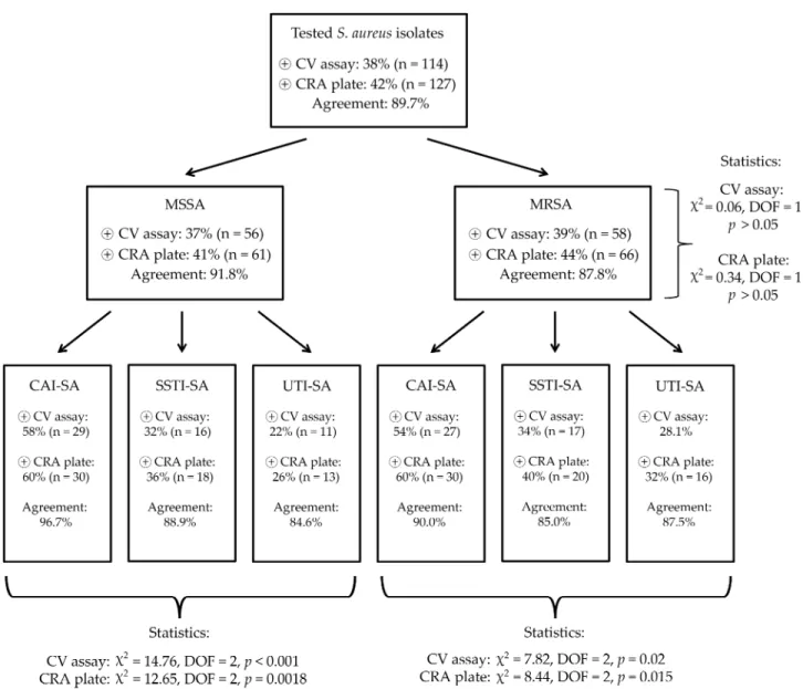
In the CV tube adherence-assay, 37% (n=56) of MSSA and 39% (n=58) of MRSA isolates were positive for biofilm- 330 production, while during the use of CRA plates, 41% (n=61) of MSSA and 44% (n=66) of MRSA were positive; no asso- ciations were found between methicillin-resistance and bio- film-production (p>0.05 in both cases). The agreement between the results of the two phenotypic testing methods 335 was 0.897 (89.7%) overall (91.8% in case of MSSA and 87.8% in case of MRSA isolates). Interestingly, biofilm- production was more commonly detected from both MSSA and MRSA CAI-SA isolates (p<0.0001 and p=0.0018 for MSSA, p=0.02 and p=0.015 for MRSA, respectively), com- 340 pared to the isolates from other origins. The detailed distribu- tion of biofilm-positive S. aureus among the isolates of different origin, the results of the statistical analyses and the agreements among the CV adherence assay and CRA plates are presented inFigure 1. Among the control strains,S. aureus 345 ATCC 29213,S. aureusATCC 43300,S. epidermidisATCC 35984 were positive for biofilm-production in both phenoty- pic assays, whileS. aureusATCC 12600 andS. epidermidis ATCC 12224 were both negative.
The relationship between biofilm-production and resis- 350 tance to other antibiotics was also assessed; during these analyses, only the results from the CRA plates were consid- ered. It was found that resistance to ERY (p = 0.004,χ2= 8.12, DOF: 1), CLI (p < 0.001,χ2= 44.57, DOF: 1) and RIF (p <
0.001, χ2 = 96.95, DOF: 1) was associated with biofilm- 355 positivity; in fact, 37 out of 43 (86%) of RIF-resistant isolates were biofilm-producers. On the other hand, this association was not shown for other antibiotics (ie, CIP, GEN, SXT;
p>0.05).
Discussion and Review of the Literature
360S. aureusinfections are associated with considerable mor- bidity, mortality and economic costs for the healthcare institutions worldwide.45 Owing to the adaptability, the plethora of virulence factors and the increasing levels of antimicrobial resistance in S. aureus, treatment of these 365 infections is a considerable challenge for clinicians.46 Biofilm-formation has been classified as an important defense mechanism and pathogenic hallmark for both MSSA and MRSA isolates, both as a means to persist in
370 the environment (eg, on a hospital ward) and in the host during infections.47It has been described that the staphy- lococcal colonization of the skin is dependent of the bio- film-formation of these bacteria; in addition, S. aureus strains adhere to damaged skin and mucosal surfaces more easily, leading to the development of SSTIs.48 At 375 the same time, all inserted and implanted medical devices (contact lenses, cardiac pacemakers, prosthetic valves, cerebrospinal fluid shunts, implanted catheters and syn- thetic joints) are at risk to be associated with S. aureus
380 infections (eg, endocarditis, osteomyelitis, bacteremia).49 In the present study, we have investigated n=150 MSSA and n=150 MRSA isolates – originating from clinical materials – regarding their antibiotic susceptibilities and their biofilm-forming capacities using two phenotypic tests, namely the CV tube-adherence assay and the plate- 385 based CRA medium. There are a plethora of methods available for the characterization of the biofilm-forming capacity of bacteria – self-developed and chromogenic media (both in liquid and in plate form), staining methods,
390 assessment via measurements with a spectrophotometer or electron microscopy and most recently,flow chamber sys- tems–however, these are pronounced differences among these methods in the price, reproducibility, high- throughput nature and the in vivo adaptability of the results.50–52Our two utilized methods have been described 395 for a number of years (the CV tube-based assay was described by Christensen et al in 1982,53 while the CRA method was developed by Freeman et al in 1989);54 although subsequent studies have demonstrated that these
400 methods needed to be modified to improve accuracy and sensitivity, these methods are cheap, easy to perform, the criteria for their evaluation©are straightforward and their results are comparable to other, more expensive assays.39 In our study, no significant association was noted between
405 MSSA/MRSA-status and biofilm-production in either
Table1AntibioticSusceptibilityRatesAmongS.aureusIsolatesIncludedinThisStudy CAI-SASSTI-SAUTI-SAOverall MSSA (n=50)
MRSA (n=50)
Overall (n=100)
MSSA (n=50)
MRSA (n=50)
Overall (n=100)
MSSA (n=50)
MRSA (n=50)
Overall (n=100) MSSA (n=150)
MRSA (n=150) Erythromycin90%(n=45)20%(n=10)55%(n=55)86%(n=43)16%(n=8)51%(n=51)94%(n=47)18%(n=9)56%(n=56)90%(n=135)18%(n=27) Clindamycin84%(n=42)14%(n==7)49%(n=49)80%(n=40)10%(n=5)45%(n=45)86%(n=43)14%(n==7)50%(n=50)83%(n=125)13%(n=19) Ciprofloxacin92%(n=46)14%(n==7)48%(n=48)90%(n=45)10%(n=5)50%(n=50)90%(n=45)8%(n=4)49%(n=49)91%(n=136)11%(n=16) Gentamicin96%(n=48)88%(n=44)92%(n=92)96%(n=48)84%(n=42)90%(n=90)96%(n=48)82%(n=41)89%(n=89)96%(n=144)85%(n=127) Sulfamethoxazole/ trimethoprim
96%(n=48)80%(n=40)88%(n=88)92%(n=46)84%(n=42)88%(n=88)96%(n=48)88%(n=44)92%(n=92)95%(n=142)84%(n=126) Vancomycin100% (n=50)
100% (n=50)
100%(n=100)100% (n=50)
100% (n=50)
100%(n=100)100% (n=50)
100% (n=50)
100%(n=100)100% (n=150)
100% (n=150) Tigecycline100% (n=50)
100% (n=50)
100%(n=100)100% (n=50)
100% (n=50)
100%(n=100)100% (n=50)
100% (n=50)
100%(n=100)100% (n=150)
100% (n=150) Linezolid100% (n=50)
100% (n=50)
100%(n=100)100% (n=50)
100% (n=50)
100%(n=100)100% (n=50)
100% (n=50)
100%(n=100)100% (n=150)
100% (n=150) Fusidicacid100% (n=50)
100% (n=50)
100%(n=100)100% (n=50)
100% (n=50)
100%(n=100)100% (n=50)
100% (n=50)
100%(n=100)100% (n=150)
100% (n=150) Quinpristin/ dalfopristin
100% (n=50)
100% (n=50)
100%(n=100)100% (n=50)
100% (n=50)
100%(n=100)100% (n=50)
100% (n=50)
100%(n=100)100% (n=150)
100% (n=150) Rifampin90%(n=45)80%(n=40)85%(n=85)88%(n=44)80%(n=40)84%(n=84)92%(n=46)84%(n=42)86%(n=86)90%(n=135)81%(n=122) Ceftaroline100% (n=50)
100% (n=50)
100%(n=100)100% (n=50)
100% (n=50)
100%(n=100)100% (n=50)
100% (n=50)
100%(n=100)100% (n=150) 100% (n=150) Abbreviations:MSSA,methicillin-resistantS.aureus;MRSA,methicillin-resistantS.aureus;CAI-SA,catheter-associatedinfections;SSTI-SA,skinandsofttissueinfections;UTI-SA,urinarytractinfections.
phenotypic tests; however, ERY, CLI and RIF resistance was more common in biofilm-producingS. aureusisolates.
It is also interesting to note that 86% of isolates resistant to RIF were biofilm-producers, especially as RIF is consid- 410 ered an effective antimicrobial agent with good penetration into bacterial biofilms in vivo.55Although the exact reason behind this phenomenon is unknown, it has been described that the sub-inhibitory concentrations of several antibiotics may induce biofilm-formation in S. aureus isolates.56 415 Interestingly, the interplay between sub-inhibitory doses of antibiotics has been most frequently published in rela- tion to the MLKS (macrolide-lincosamide-ketolide-© streptogramin; which are all protein synthesis inhibitors affecting the 50S ribosome) group of drugs and rifampin.57
For example, Lima-e-Silva et al reported that in sub-lethal 420 doses (MIC/2 and MIC/4), rifampin strongly stimulated biofilm-formation (when measured by the CRA plate and CV microtiter plate assay), in contrast to minocycline, which did not have such inducing effects.58 The effects of low antibiotic doses on the biofilm-forming capacity are 425 thought to occur by differential expression of genes of interest due to the noxious agents.59 The literature has shown that sub-MIC concentrations of tetracycline,
©quinupristin and dalfopristin were presented as strong inducers, while erythromycin was noted as a weak inducer 430 of ica-gene expression. In contrast, gentamicin, chloram- phenicol (which are also protein synthesis inhibitors), penicillin, oxacillin, ofloxacin and vancomycin did not
Figure 1Antibiotic susceptibility rates amongS. aureusisolates included in this study.
AQ7
Abbreviations:MSSA, methicillin-resistantS. aureus; MRSA, methicillin-resistantS. aureus; CAI-SA, catheter-associated infections; SSTI-SA, skin and soft tissue infections;
UTI-SA, urinary tract infections.
present with similar effects.60 The clinical origin of the 435 isolates also had an effect on their biofilm-producing capacity: they were the most prevalent in isolates from catheter-associated infections and the least common in strains isolated from the urinary tract. Finally, the agree- ment among the results of the two in vitro methods was 440 89.7%, which is a very good result, based on other studies
from the literature.39
Many studies have aimed to assess the correlation of biofilm-production with antibiotic-resistance inS. aureus strains; nevertheless, the literature©has shown conflicting 445 data on the topic, thus, at present, we are unable to draw far-reaching conclusions. To make the interpretation of the currently available results even more troublesome, many different methodologies have been used to assess biofilm- formation, with or without the molecular characterization 450 of the isolates. Similarly to our results, Arslan et al found no association between slime production and methicillin- resistance or resistance to other antibiotics in a sample of n=187 S. aureus isolates; in their report, CRA plates and the CV tube adherence assay©wereutilized, while molecu- 455 lar testing (for clonality or the presence ofagrgenes) was not performed.61 Ghasemian et al assessed biofilm- production in n=209 S. aureus isolates, among which no relevant differences were shown for biofilm-positive and negative isolates, based on methicillin-resistance (36.1%
460 and 28.9% for MRSA and MSSA, respectively). The extent of biofilm-production was assessed by a microtiter plate biofilm assay, and the genetic testing of the isolates was also performed: 58.3% and 22.0% of isolates belonged to the agr groups I and II, respectively, while 465 84.0% of MRSA isolates possessed the SCCmec III cassette.62 Rodríguez-Lopez et al came to similar conclu- sions, when studying S. aureus isolates originating from animal and environmental samples at heavy swine farms in Italy. In this report, the same methodology was utilized 470 (plate-based quantitative measurements), and spa-typing was also performed: overall, isolates belonging to the ST398/t899 and ST398/t011 were the most common among biofilm-producers.63 El-Nagdy et al detected bio- film-forming S. aureus (using the CRA plate method, 475 complemented with scanning and transmission electron microscopy) from febrile neutropenic patients in Egypt;
interestingly, they have found that 72.7% of isolates were biofilm-positive. Among the tested strains, 37.5% were positive for icaA, and 22.9% were positive for icaD;
480 however, only 50% of biofilm-producers carried either the icaA or icaD genes.64 Similarly, no correlation
among methicillin-resistance and biofilm-production was seen in the reports of Knobloch et al (including n=128ica- positive isolates, with utilizing CRA plates and the micro-
485 titer plate method)65 and Mathur et al (where n=152 Staphylococcus spp. were tested using the microtiter plate method).66
However, there have been studies that identified differ- ences in the rates of biofilm-production based on the
490 phenotypic resistance of S. aureus isolates. In the study by Bose et al (in which the authors have utilized similar methodologies to our study, in addition to the tissue cul- ture plate method to quantify their results) found higher levels of antibiotic resistance in biofilm-producing
495 S. aureus and S. epidermidis isolates.67 Piechota et al compared the biofilm-forming capacity of MSSA and MRSA isolates from Poland: in this report, MRSA isolates were stronger biofilm-producers overall in the microtiter plate-based assay (39.7% vs 36.8%) and the occurrence of
500 icaABCDgenes (51.5% overall) was also more common in methicillin-resistant strains.68 Cha et al characterized n=126 MRSA isolates from a Korean teaching hospital, during which they have found higher levels of MDR iso- lates (defined as MRSA+resistance to at least three non-β-
505 lactam agents) among the biofilm-forming isolates. The study group has used the microtiter plate assay to quantify biofilm-formation and has also performed molecular test- ing: they have shown that the majority of the isolates were ST5 (69.8%) and 64.0% of isolates are from theagrgroup
510 II.69 Souli et al©have clearly demonstrated that strong biofilm-producers (tested with the CV tube-based assay) among S. epidermidis isolates had also possessed higher levels of resistance in vitro.70 Agarwal and Jain tested commensal, colonizing and invasive S. aureus isolates
515 for their biofilm-forming capacity using the microtiter plate assay; in their study, biofilm-producers were more frequently MDR in all groups, and 94.0% of biofilm- producers carried ica-genes.71 De©Araujo et al reported that methicillin-resistant and MDRS. epidermidisisolates
520 were more frequent among biofilm-producers (tested by the microtiter plate assay). They have found that 96.0% of biofilm-positive isolates carriedicaAor icaDgenes, while 86.0% were positive foratlEandaapgenes.72In contrast, in a report concerningS. aureusisolates originating from
525 pork, Zhang et al found 83.8% of the bacteria to beagr- positive (agr I: 39.2%, agr IV: 32.3%) and noted the high prevalence of MDR isolates in moderate and weak bio- film-producers, when tested with the microplate method.73 Our study demonstrated an association of CLI-resistance
530 and biofilm-production corresponding to our isolates, which has been identified in other reports as well, although these studies usually also showed a positive correlation with methicillin-resistance. In the study by Belbase et al, S. aureus isolates from pus/wound swab samples were 535 assessed for the susceptibility and microtiter-based bio- film-production in a tertiary-care hospital in Nepal.
Overall, methicillin-resistance and inducible CLI- resistance©were more common in biofilm-producers.38 In another study from Nepal, Manandhar et al also showed 540 that the in vitro biofilm-production (assessed by CRA plates) ofS. aureusisolates was associated with methicil- lin-resistance and inducible CLI-resistance.74Bhattacharya et al tested n=100 S. aureus isolates, including 47%
MRSA isolates: biofilm-positivity was shown in 55.0%
545 of isolates, with MRSA isolates in higher numbers among biofilm-producers; in addition, resistance-levels against CIP, RIF, ERY, CLI and SXT were also signifi- cantly higher in biofilm-positive isolates.75 Finally, Neopane et al tested n=150S. aureusisolates, originating 550 from pus samples: in their study – although no clear association was seen with methicillin-resistance and bio- film-production, strong biofilm-production and the MDR status has shown good agreement.76 In the latter three studies, only phenotypic characterization of resistance 555 and biofilm-production©wasutilized. Overall, the literature findings suggest a possible relationship between the expression of antibiotic-resistance-determinants and bio- film-production, however, the clarification of nature of this association will require further studies.
560 Pulse-field gel electrophoresis (PFGE) and multi-locus sequence typing (MLST)©are not routinely performed in Hungary by diagnostic laboratories; thus, there is a scarcity of local data regarding the molecular epidemiol- ogy of S. aureusisolates, such data may be sourced from 565 major national public health surveillance studies: the Hungarian clone (ST239-III, PFGE type E) – which was predominant before 2000 – was almost completely replaced by the Southern German clone (ST228-I, PFGE type B) and the New York/Japan epidemic clone (ST5-II, 570 PFGE type A/C), and since 2006, the breakthrough of the EMRSA-15 (ST22-IV, PFGE type D) was described.77 Based on the most recent data (2017–2018) available from the National Institute of Public Health in Hungary,
~45% of HA-MRSA isolates were ST22-IV clones, while 575 ~24% of isolates were ST5-II; in case of CA-MRSA iso- lates, ST8-IV and ST80-IV clones were the most common, while among LA-MRSA, CC398 isolates were the most
prevalent.78,79 Literature data also©suggest that the clonal background ofS. aureusisolates may play a major role in
580 the biofilm-forming capacity, however (as demonstrated by the publications discussed in the previous section), the clonality of the isolates is seldom reported in these studies.80 Croes et al reported that S. aureus isolates from MLST clonal complex CC8 were the most potent
585 biofilm-producers – irrespective of the glucose- concentration (0–0.5%) in the media; at physiological glucose (0.1%) concentration, >60% of CC8 isolates were strong biofilm-producers, compared to 0–7% in other tested CCs.81 These findings were also supported
590 by the results of Luther et al: from n= 182 clinical MRSA strains, isolates belonging to the CC8 group and spa type t008 group were significantly more common among strong biofilm-producers (p=0.01), whilespa type t895 and β-toxin-producing isolates showed a negative
595 correlation with biofilm-production.82 The study of Recker©et al performed combined study, including labora- tory assays and a data analysis regarding bacterial geno- type and phenotype with available clinical metadata in a machine-learning framework, corresponding to n=300
600 S. aureus, including CC22 and CC30 isolates from bacter- emia. Their results showed no relevant differences in bio- film-formation among MSSA and MRSA isolates or among CC22 and CC30 isolates. Although this report showed no association with biofilm-production and
605 SCCmec-type, other studies highlighted that the SCCmec II element is associated with decreased capability to form biofilm.83 On the other hand, Lim et al (who assessed n=465 clinical S. aureus isolates) and da Fonseca Batistao (during the study of fifteen isolates) both con-
610 cluded that the presence of the SCCmec III cassette is a good predictor of strong biofilm-forming ability.84,85 The importance of the SCCmec cassette and mecAgenes in biofilm formation were highlighted by Pozzi et al, where ΔSCCmec (deletion mutants) presented with
615 decreased expression of virulence determinants, including biofilms; the authors of the study have concluded that these genes may have the potential to affect phenotypic characteristics mediated by other operons (ie, agr or icaADBC) to facilitate the adaptation of hospital-
620 associated MRSA to the harsh environment in hospitals.86This may be the reason for the larger number of biofilm-producing isolates from catheter-associated (CAI-SA) infections.
It has been described that S. aureus strains usually 625 form multilayered biofilms; this biofilm is useful in the
evasion of the non-specific and adaptive immune responses of the host, including decreased rate of opsoni- zation, phagocytosis, killing by neutrophil granulocytes, and Toll-like receptor activation.87,88The latter is particu- 630 larly relevant, because TLR-activation is important in facilitating a Th2-type immune response, which may act to prevent S. aureus biofilm-associated infections.89 Biofilms are also protective against reactive oxygen spe- cies (ROS) in in vivo environments.90Biofilms also facil- 635 itate the metabolic transformation of S. aureus into the small colony variant (SCV; which may be seen as“dwarf colonies” on solid media) morphotype: in this sub- population, bacteria exist at a lower metabolic activity (leading to increased antimicrobial resistance), which 640 also enables in vivo persistence and chronic infections.91 In addition to metabolic switching, SCVs are also charac- terized by adaptation for intracellular survival in mamma- lian cells, where the pathogen acts similarly to other microorganisms with a strictly intracellular life cycle.92,93 645 The relevance of eDNA in the stability and antimicrobial resistance of staphylococcal biofilms has been demon- strated, as DNase treatment (eg, in cysticfibrosis) clearly negatively impacts the biofilm structure.94 Biofilm- formation in S. aureus is©genetically mediated by the 650 regulatory genetic locus staphylococcal accessory regula- tor (sarA); this controls two pathways–namely the intra- cellular adhesin (ica) operon and accessory gene regulator (agr) regulated pathways–bothof which have been sug- gested as determinants of the extent of biofilm-formation 655 in these bacteria.95,96 The product of the genes of theica operon (icaADBC) are the IcaA and IcaD transferase membrane proteins; these proteins have important roles in the biosynthesis of PIA (polysaccharide intercellular adhesion protein; or poly-β-1,6-©N-acetylglucosamine 660 [PNAG]), which is a major component of staphylococcal biofilms.97 Although our study did not demonstrate pro- nounced differences among the biofilm-producing capabil- ities of the©locally collected MSSA and MRSA isolates, there have been studies offering possible biological expla- 665 nations to this phenomenon.38 It has been suggested that biofilm-formation in MSSA is mediated by cell-cell adhe- sion via the production of PIA (encoded by icaADBC), while MRSA biofilm-production is PIA-independent, and rather, it is dependent on a protein adhesion, which is 670 negatively regulated by the agrsystem.98,99 O’Neill et al showed that media supplementation with NaCl results in the induction of biofilm-production in MSSA only, as this activated the expression of the icaoperon.100In contrast,
Croes et al showed that the presence of excess glucose in 675 the media represses theagrsystem (through the generation of low pH), which resulted in the induction of biofilm- production in MRSA only;81,101additionally, the deletion of theagrsystem also enhanced the biofilm-production of MRSA isolates, while it had no effect on its methicillin-
680 susceptible counterparts.102
Conclusions
The production of biofilm by pathogenic bacteria in vivo provides important protection from external forces and antimicrobials, in addition to facilitating chronicity.
685 S. aureus is an©exceptionally adaptable pathogen both in natural environment and in clinical situations. Biofilm- formation in both MSSA and MRSA isolates is an impor- tant step in the pathogenesis of implant-associated infections©and leads to a synergistic interaction between
“classical” resistance-determinants and the inability of 690 antibiotics and immune cells to reach S. aureusisolates.
The relationship between the MDR phenotype and bio- film-positivity has been studied for many relevant patho- gens, however, the culmination of these results©is
695 inconclusive. In our study, one hundred and fifty MSSA and one hundred andfifty MRSA isolates (from a variety of clinical situations) were tested for their antibiotic- susceptibility and their biofilm-forming capacity to ascer- tain a possible relationship between the two. Among our
700 tested isolates, we have found complete susceptibility to the last-resort agents, while there were significant differ- ences in the resistance rates between MSSA and MRSA isolates regarding almost all other, commonly used agents.©Thirty-eight percent and 42% of isolates were
705 biofilm-producers based on the CV tube adherence assay and the CRA plate methods, respectively. Overall, no association was found between methicillin-resistance and biofilm-positivity in our settings; on the other hand, resistant isolates to erythromycin, clindamycin and
710 rifampin were significantly more common among bio- film-producers.
Our study possesses some limitations: i) the©cross- sectionalnature of the study: although isolates were collected from different clinical specimen groups, they may not repre-
715 sent HungarianS. isolates, their biofilm-forming capacity or susceptibility overall; ii) selection bias: isolates usually ori- ginated from tertiary-care centers, corresponding to patients with more severe conditions or underlying illnesses; iii) interpretation: both phenotypic methods were evaluated by
720 organoleptic methods, therefore the reading of the results was
dependent on the expertise of the researchers; iv) lack of molecular methods: the molecular characterization of resis- tance determinants, clonal lineages (with PFGE or MLST) or genetic determinants of biofilm-production (eg, agr, ica 725 genes) in the mentioned isolates was not performed.
Nevertheless, our study provides additional data to the exist- ing pool of literature on the association of drug resistance and biofilm-formation inS. aureus. Additional studies–with the inclusion of other isolates and utilization – are needed to 730 provide clarity on this subject.
Data Sharing Statement
All data generated during the study are presented in this paper.
Funding
735 The article processing charge (APC) was funded by the University of Szeged Open Access fund (ID: 5175). M.
G. was supported by the János Bolyai Research Scholarship (BO/00144/20/5) of the Hungarian Academy of Sciences and the New National Excellence Programme 740 (ÚNKP-20-5-SZTE-330) of the Ministry of Human Resources. Support from Ministry of Human Capacities, Hungary grant 20391-3/2018/FEKUSTRAT is acknowl- edged. M.G. would also like to acknowledge the support of ESCMID’s“30 under 30”Award.
745
Disclosure
The authors declare no conflict of interest, monetary or otherwise. The authors alone are responsible for the con- tent and writing of this article.
References
750 1. Shaw C, Stitt JM, Cowan ST. Staphylococci and their Classification.
J Gen Microbiol.1951;5:1010–1023. doi:10.1099/00221287-5-5-1010 2. Tong SYC, Davis JS, Eichenberger E, Holland TL, Fowler VG.
Staphylococcus aureus infections: epidemiology, pathophysiology, clinical manifestations, and management. Clin Microbiol Rev.
755 2015;28:603–661. doi:10.1128/CMR.00134-14
3. Gould D, Chamberlaine A. Staphylococcus aureus: a review of the literature. J Clin Nurs. 1995;4:5–12. doi:10.1111/j.1365-2702.1995.
tb00004.x
4. Gajdács M. The continuing threat of methicillin-resistant
760 Staphylococcus aureus. Antibiotics. 2019;8:e52. doi:10.3390/
antibiotics8020052
5. Kahl BC, Becker K, Löffler B. Clinical significance and pathogenesis of staphylococcal small colony variants in persistent infections.Clin Microbiol Rev.2016;29:401–427. doi:10.1128/CMR.00069-15
765 6. Ericson JE, Popoola VO, Smith PB, et al. Burden of invasive Staphylococcus aureus infections in hospitalized infants.JAMA Pediatr.
2015;169:1105–1111. doi:10.1001/jamapediatrics.2015.2380
7. Chambers HF. The changing epidemiology of Staphylococcus aureus.
Emerg Infect Dis.2001;7:178–182. doi:10.3201/eid0702.010204
770
8. Enright MC, Robinson DA, Randle G, Feil EJ, Grundmann H, Spratt BG. The evolutionary history of methicillin-resistant Staphylococcus aureus (MRSA). Proc Natl Acad Sci USA.
2002;99:7687–7692. doi:10.1073/pnas.122108599
9. David MZ, Daum RS. Community-associated
775
methicillin-resistant Staphylococcus aureus: epidemiology and clinical consequences of an emerging epidemic.Clin Microbiol Rev.2010;23:616–687.
10. Algammal AM, Hetta HF, Elkelish A, et al.©Methicillin-resistant Staphylococcus aureus (MRSA): one health perspective approach
780
to the bacterium epidemiology, virulence factors, antibiotic-resistance, and zoonotic impact. Infect Drug Res.
2020;13:3255–3265. doi:10.2147/IDR.S272733
11. Dulon M, Haarnmann F, Peters C, Schablon A, Nienhaus A.
MRSA prevalence in European healthcare settings: a review.
785
BMC Infect Dis.2011;11:e138. doi:10.1186/1471-2334-11-138 12. Kang C-I, Song J-H, Ko KS, Chung DR, Peck KR. Clinical
features and outcome of Staphylococcus aureus infection in elderly versus younger adult patients.Int J Infect Dis.2011;15:
e58–e62. doi:10.1016/j.ijid.2010.09.012
790
13. Gajdács M. The concept of an ideal antibiotic: implications for drug design.Molecules.2019;24:892. doi:10.3390/molecules24050892 14. Stefani S, Chung DR, Lindsay JA, et al. Meticillin-resistant
Staphylococcus aureus (MRSA): global epidemiology and harmo- nisation of typing methods. Int J Antimicrob Agents.
795
2012;39:273–282. doi:10.1016/j.ijantimicag.2011.09.030 15. Arzanlou M, Chai WC, Venter H. Intrinsic, adaptive and acquired
antimicrobial resistance in Gram-negative bacteria. Essays Biochem.2017;61:49–59. doi:10.1042/EBC20160063
16. Lebeaux D, Ghigo J-M, Beloin C. Biofilm-related infections:
800
bridging the gap between clinical management and©fundamental aspects of recalcitrance toward antibiotics.Microbiol Mol Boil Rev.2014;78:510–543.
17. Henrici AT. Studies of freshwater bacteria: I. A direct micro- scopic technique. J Bacteriol. 1933;25:277–287. doi:10.1128/
805
JB.25.3.277-287.1933
18. Bryers JD. Medical biofilm.Biotechnol Bioeng.2008;100:1–18.
doi:10.1002/bit.21838
19. Stewart PS. Mechanisms of antibiotic resistance in bacterial biofilms. Int J Med Microbiol.2002;292:107–113. doi:10.1078/
810
1438-4221-00196
20. Artini M, Papa R, Scoarughi GL, et al. Comparison of the action of different proteases on virulence properties related to the sta- phylococcal surface. J Appl Microbiol. 2013;114:266–277.
doi:10.1111/jam.12038
815
21. Tan X, Qin N, Wu C, et al. Transcriptome analysis of the biofilm formed by methicillin-susceptible Staphylococcus aureus. Sci Rep.2015;5:e11997. doi:10.1038/srep11997
22. Chatterjee S, Maiti P, Dey R, Kundu A, Dey R. Biofilms on indwelling urologic devices: microbes and antimicrobial manage-
820
ment prospect. Ann Med Health Sci Res. 2014;4:100–104.
doi:10.4103/2141-9248.126612
23. Costerton JW, Stewart PS, Greenberg EP. Bacterial biofilms:
a common cause of persistent infections. Science.
1995;284:1318–1322. doi:10.1126/science.284.5418.1318
825
24. Craft KM, Nyugen JM, Berg LJ, Townsend SD. Methicillin- resistant Staphylococcus aureus (MRSA): antibi-otic-resistance and the biofilm phenotype. Med Chem Comm.
2019;10:1231–1241. doi:10.1039/C9MD00044E
25. Singh R, Ray P, Das A, Sharma M. Penetration of antibiotics
830
through Staphylococcus aureus and Staphylococcus epi©dermidis biofilms. J Antimicrob Chemother. 2010;65:1955–1958.
doi:10.1093/jac/dkq257
26. Soto SM. Importance of biofilms in urinary tract infections: new therapeutic approaches. Adv Biol. 2014;2014:e543974.
835
doi:10.1155/2014/543974