Acta Microbiologica et Immunologica Hungarica
170 DOI:10.1556/030.66.2019.026
© 2019 Akadémiai Kiad´o, Budapest
ORIGINAL ARTICLE
* Corresponding author:
Pakhshan Abdulla Hassan Department of Biology, College of Science, Salahaddin University, Erbil 44001, Iraq
Phone: +964 75 046 12335 E-mail:pakhshanhassana@gmail.com
resistance among clinical and soil isolates of Acinetobacter baumannii in Iraq
PAKHSHAN A. HASSAN
1* and ADEL K. KHIDER
21Department of Biology, College of Science, Salahaddin University, Erbil, Iraq
2Department of Biology, College of Education, Salahaddin University, Erbil, Iraq
Received: March 28, 2019•Accepted: July 17, 2019
ABSTRACT
Acinetobacter baumanniiis an opportunistic pathogen that is reported as a major cause of nosocomial infections. The aim of this study was to investigate the biofilm formation byA. baumanniiclinical and soil isolates, to display their susceptibility to 11 antibiotics and to study a possible relationship between formation of biofilm and multidrug resistance. During 8 months period, from June 2016 to January 2017, a total of 52 clinical and 22 soil isolates of A. baumannii were collected and identified through conventional phenotypic, chromo agar, biochemical tests, API 20E system, and confirmed genotypically by PCR for blaOXA-51-like gene. Antibiotic susceptibility of isolates was determined by standard disk diffusion method according to Clinical and Laboratory Standard Institute. The biofilm formation was studied using Congo red agar, test tube, and microtiter plate methods. The clinical isolates were 100%
resistance to ciprofloxacin, ceftazidime, piperacillin, 96.15% to gentamicin, 96.15% to imipenem, 92.31%
to meropenem, and 78.85% to amikacin. The soil A. baumannii isolates were 100% sensitive to imipenem, meropenem, and gentamicin, and 90.1% to ciprofloxacin. AllA. baumanniiisolates (clinical and soil) were susceptible to polymyxin B. The percentage of biofilm formation in Congo red agar, test tube, and microtiter plate assays was 10.81%, 63.51%, and 86.48%, respectively. More robust biofilm former population was mainly among non-MDR isolates. Isolates with a higher level of resistance tended to form weaker biofilms. The soil isolates exhibited less resistance to antibiotics than clinical isolates.
However, the soil isolates produce stronger biofilms than clinical isolates.
KEYWORDS
Acinetobacter baumannii, molecular identification,blaOXA-51-likegene, multidrug resistant, biofilm formation
INTRODUCTION
Acinetobacter baumanniiis an opportunistic pathogen that can pose a significant threat due to its increasing antibiotic resistance among nosocomial isolates, mainly from intensive care units [1]. It is related to mainly immunocompromised patients, and a wide range of clinical complications can occur [2]. The plastic genome of A. baumannii, which mutates rapidly when faced with severe conditions, and fundamental mechanisms of virulence beyond canonical drug resistance enable A. baumannii to flourish successfully in the healthcare environment [3]. Biofilm formation is another potent way for bacterial survival in the presence of antibiotics [4], particularly forA. baumannii, which is one of the most common bacterial causes of biofilm-associated contamination of medical devices [5]. Biofilm is known as a structured consortium of microbial cells embedded in a self-produced polymeric matrix or exopolymeric substances, consisting of protein, extracellular DNA, and polysaccharide.
Bacterial biofilms are resistant to antibiotics, and they can develop resistance to chemicals, phagocytosis, and other components of the innate and acquired immune system of the body [6,7]. Hence, it is important to create connections between biofilm formation and drug resistance in isolates ofA. baumannii. Therefore, in this study, we focused on determination of biofilm formation in clinical and soil isolates ofA. baumannii, clarification of their sensitivity 67 (2020) 3, 16161–
to different antibiotics, and presenting any possible link between the ability to form biofilm and multidrug resistance (MDR).
MATERIALS AND METHODS
Collection, isolation, and identi fi cation of A.
baumannii isolates
A total of 320 clinical samples were taken from patients of three hospitals in Erbil city, Iraq, during June 2016 to January 2017.
All clinical specimens were obtained from various sites, which included tracheal aspirate, sputum, burned wound, urine, cerebrospinalfluid, and blood. Specimens were inoculated in brain–heart infusion broth (BHIB; Himedia, India) medium and then transferred to lab for culture. Samples of soil were obtained from different locations in Erbil. Tenfold serial dilu- tions of soil suspension were made in BHIB, and inocula (0.1 ml) from each dilution were spread with sterile glass beds onto brain–heart infusion agar (BHIA; Himedia) plates.
All isolates (clinical and soil) were initially inoculated on MacConkey agar, blood agar, Herellea agar, and on the CHROMagar Acinetobacter Media (CHROMagar, France).
Incubation was performed for 24 h at 37 °C. Standard labora- tory methods (Gram-stain and colonial morphology) and conventional biochemical tests (catalase, oxidase, urea test, citrate test, indole test, and the reaction in triple sugar iron medium and growth in 44 °C) were used to identify A.
baumannii.Bacterial species identification of the isolates was confirmed by API 20E system (bioMerieux, France) and by PCR detection of intrinsic blaOXA-51-like carbapenemase gene from these isolates.
Molecular identi fi cation of isolates by PCR
All isolates were subjected to conventional PCR to detect blaOXA-51-like as performed by Turton et al. [8]. Genomic DNA was extracted via a commercial genomic DNA purifi- cation kit (Geneaid, Taiwan), according to the manufacture’s protocol. Extracted DNA was quantified using the nanodrop spectrophotometer (Thermo Scientific, UK) at OD260 and the purity was determined by OD260/OD280 ratio.
The primers were F-TAATGCTTTGATCGGCCTTG and TGGATTGCACTTCATCTTGG (353 bp) [9].
The PCR was carried out in 20 μl of reaction volumes (Accua Power PreMix, Bioneer, Korea) with 3μl of extracted DNA, 1 μl of forward primer (10 pmol/μl), 1 μl reverse primer (10 pmol/μl), and 15μl distilled water. The content of PreMix (blue pellet): Top DNA polymerase (1U), dNTP (dATP, dCTP, dGTP, and dTTP): each 250 μM, reaction buffer, with 1.5 mM of MgCl2(1×). Conditions for the PCR were the following: initial denaturation at 94 °C for 5 min and then 30 cycles at 94 °C for 25 s, 52 °C for 40 s, and at 72 °C for 50 s, followed by a final elongation at 72 °C for 6 min.
Negative control of sterile distillated water was included in every PCR. A. baumannii ATCC 19606 (purchased from
Media Diagnostic Center) was used as the reference strain. A 100-bp DNA ladder (Norgen, Canada) was used to evaluate the size of PCR products, which were resolved on 1.5% agarose gels in Tris/borate/EDTA buffer, stained with ethidium bro- mide, visualized under UV transilluminator (ISOGEN, Life Science, Netherlands) and selected gels were photographed.
Genotypic and phenotypic determination of metallo-beta-lactamases (MBLs)
Multiplex PCR techniques were used for amplification of the blaVIM, blaIMP, and blaSIMgenes. Modified Hodge test and imipenem-EDTA-combined disk test (IMP-EDTA-CDT) were performed phenotypically for identification of MBLs in clinical and soil isolates.
Antimicrobial susceptibility test
Antimicrobial susceptibility test was performed for 11 differ- ent therapeutically pertinent antibiotics by the Kirby–Bauer disk diffusion method as stated by Clinical Laboratory Stan- dards Institute guidelines [10]. Antibiotics tested included amikacin (AK; 10 μg), meropenem (MEM; 10 μg), IMP (10 μg), polymyxin B (100 U), ceftazidime (CAZ; 30 μg), amoxicillin/clavulanic acid (AMC; 20/10 μg), trimethoprim (TMP; 10μg), ciprofloxacin (CIP; 10μg), piperacillin (PRL;
10 μg), gentamicin (GEN; 10 μg), and aztreonam (ATM;
10 μg). The antibiogram procedure performed on Mueller– Hinton agar swabbed with 1.5×10⁸CFU of bacterial suspen- sion, equivalent to 0.5 McFarland standard suspension, and incubated for 24 h at 37 °C. Guidelines from the CLSI were used to interpret the diameters of inhibition zones. Strains resistance to at least three classes of antimicrobial agents (all penicillins and cephalosporins, including inhibitor combina- tions), aminoglycosides andfluoroquinolones, were defined as MDRAcinetobacterspp., whereas those resistant to the three classes of antimicrobial drugs mentioned above as MDR and resistant to carbapenems were defined as extensive drug- resistant (XDR) [11]. MDRA. baumannii(MDRAB) was also detected by CHROMagar by adding MDR Selective supple- ment CR102 (CHROMagar, Paris, France). A. baumannii ATCC 19606 was used as a non-MDR strain for comparison.
Bio fi lm formation: Tube method (TM)
All isolates have been screened qualitatively for the biofilm formation by TM as described by Christensen et al. [12]. Ten milliliters of trypticase soy broth with 1% (w/v) glucose in tubes were inoculated with a loop full of test organisms from overnight culture plates. The inoculated tubes were incubated for 24 h at 37 °C. The tubes were then decanted and washed with phosphate buffer saline (PBS) and stained with crystal violet (0.1%) for 10 min. Excess stain was removed and washed with water. The tubes were then mainitained in an inverted position for drying. Biofilm production was detected by the presence of visible film lined the wall and the bottom of the tube. The biofilms were visualized and the amount of biofilm Acta Microbiologica et Immunologica Hungarica –170
162 67 (2020) 3, 161
formed was recorded according to the result of control strains and graded as 0, 1, 2, and 3 representing absent, weak, moderate, and robust biofilm formations, respectively. Experi- ments were performed two times.
Tissue culture plate (TCP) method
This quantitative assay described by Christensen et al. [13] is considered as the gold standard method for detection of biofilm formation [14]. An overnight culture of the isolate from fresh agar plate was inoculated into 10 ml trypticase soy broth with 1% glucose and incubated for 24 h at 37 °C. The cultures were diluted 1 in 100 with fresh medium and loaded into 96-well-flat bottom– polystyrene TCPs (Coster, USA). Individual wells werefilled with 200μl of the diluted cultures (3 wells for each isolate), and only sterile broth served as negative control.
A. baumannii ATCC 19606 (used as positive control) was also incubated, diluted, and added to the microtiter plate. Plates were covered and incubated for 24 h at 37 °C in aerobic condition without shaking. After incubation, the wells were gently decanted by tapping the plate and washed three times with 200μl of PBS (pH 7.2) to remove free-floating bacteria.
Biofilm formed by adherent bacteria (sessile) in the plate was fixed for 15 min by 99% methanol and then stained for 20 min with crystal violet (0.1% w/v). Excess stain was rinsed off using deionized water and plates were kept for drying. Finally, 95%
ethanol was added to the wells to extract the stain and adherence of the stained cells to the wells. The optical density (OD) of each well was measured at 490 nm using an automated ELISA plate reader (BioTEK, UK). The triplicate experiment was conducted and the results were averaged. The mean OD at 490 nm (OD490) of sterile trypticase soy broth was used as OD cut-off value (ODc). The OD results of all tested strains were divided into the following four groups: (1) OD≤ODc= non-biofilm Producer; (2) ODc<OD≤2×ODc=weak biofilm producer; (3) 2×ODc<OD≤4×ODc=moderate biofilm producer; and (4) 4×ODc<OD=strong biofilm producer [15].
Congo red agar method (CRA)
It is a qualitative method used for screening biofilm formation.
As described by Freeman and Keane [16], this method requires CRA media. This medium composed of 37 g/L BHIB, 50 g/L sucrose, 10 g/L agar, and Congo red stain 0.8 g/L. Congo red stain solution was prepared as a concentrated aqueous solution and autoclaved for 15 min at 121 °C separately. Then, it was added to autoclaved BHIA (BHIB, agar, and sucrose) at 55 °C.
Plates were inoculated with the test bacteria and incubated aerobically at 37 °C for 24–48 h. A positive result was indicated by black, rough colonies with a dry crystalline consistency;
pink, smooth colonies with occasional darkening at the center were recorded as non-biofilm producers.
Statistical analyses
OD values were expressed either as median values [interquar- tile range (IQR)] or as means±standard deviations (SDs)
based on the distribution of variance. All statistical analyses were carried out using the GraphPad prism 6.01. The differ- ence of biofilm biomass between two antibiotics was compared by using Mann–WhitneyUtest. The values ofp<0.05 were considered statistically significant.
RESULTS
Bacterial identi fi cation
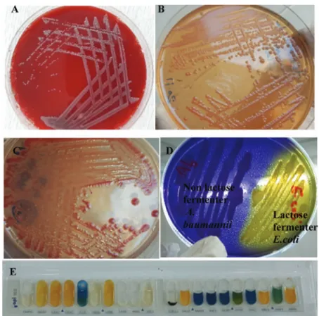
Of the 350 clinical specimens and 50 soil samples, 52 and 22 isolates were identified as A. baumannii, respectively. All isolates werefirst recognized phenotypically on blood agar, MacConkey agar, CHROMagar Acinetobacter, and Herellea agar (Figure1A–D), then confirmed by API 20E (Figure1E).
Analysis for the presence ofblaOXA-51-likegene presented that all identified isolates were positive andA. baumanniiwas confirmed (Figure 2).
The clinical isolates ofA. baumanniiwere recovered from tracheal aspirate (32.69%), sputum (28.85%), burned wound (21.15%), and urine (9.61%), and the lowest numbers of isolates 2 (3.85%) were from both blood and cerebrospinal fluid (Figure 3).
Antibiotic susceptibility
Distribution of antimicrobial resistance properties within the A. baumanniiisolates from clinical and soil samples is shown in Table I and Figure 4. As observed, all clinical and soil isolates (100%) were resistance to AMC acid. All clinical isolates were resistant to CIP, CAZ, and PRL, but the rate of resistance in soil isolates for these antibiotics was 9.09%, 63.64%, and 90.01%, respectively. IMP and MEM resistance were 96.15 vs. 0% and 92.31 vs. 0% in clinical and soil isolates, respectively. All isolates (clinical and soil) were sensitive to polymyxin B. Of the 74A. baumannii isolates, 14 (18.92%) were classified as MDR, 38 (51.35%) were classified as XDR, and 22 (29.73%) of the isolates were non-MDR. As such,
∼70.0% of the isolates were either MDR or XDR (Figure4B).
All clinical isolates grew well on CHROMagar Acinetobacter supplemented with MDR selective supplement, whereas all soil isolates were not culturable on this medium.
Genotypic and phenotypic determination of MBL
Although MBL enzymes were detected phenotypically in some clinical isolates, MBL genes (blaVIM,blaIMP, andblaSIM) were not detected neither in clinical nor in soil isolates (data not shown).
Bio fi lm production assay
In CRA method, out of eight positive isolates, 7 (9.46%) displayed black colonies with dry crystalline morphology.
The remaining 66 (89.19%) isolates exhibited pink colonies
(as a non-biofilm producer), when cultured on CRA medium (Figure 5A), but most of these isolates produce biofilms in tube and TCP methods.
In the TM, the rate of the non-biofilm producer was 27 (36.49%) isolates and biofilm producers were 47 (63.51%) (TableII and Figure5B).
The results of biofilm formation in the TCP method showed that 64 (86.49%) isolates could produce biofilms and only 10 (13.51%) isolates were not able to produce
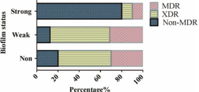
biofilms (Table II). The soil isolates produced higher rate (36.36%) of strong biofilms than the clinical isolates (3.84%) as showed in Table III.
The results of biofilm formation by the TCP method were used to study if there is any relation between biofilm production and drug resistance in clinical and soil isolates.
The mean OD490value for the reference strain (A. bauman- niiATCC 19606) was 0.472±0.024. The mean OD490value for the negative control (wells inoculated with sterile broth were used as negative control) was 0.121±0.036 and this value was used as the ODc. The classification of biofilm based on ODc revealed that OD490≤0.121=non-biofilm; 0.121<
OD490≤0.242=weak biofilm, 0.242<OD490≤0.484= moderate biofilm; OD490>0.484=strong biofilm. The median OD490and IQR value for non-biofilm formers was 0.112 (0.104, 0.119), for weak biofilm formers was 0.183 Figure 1. Identification of A. baumannii isolates. (A) A. baumannii colonies on blood agar. (B) Non-lactose-fermenting colonies of A. baumanniion MacConkey agar. (C)A. baumanniimetallic reddish colonies on CHROMagar Acinetobacter. (D) Non-lactose fermenterA.
baumanniicolonies appear lavender in color on Herellea agar. (E) Biochemical identification ofA. baumanniiby API-20E system with 7-digit number (0004042) according to analytical index
Figure 2.PCR products of theblaOXA-51-likegene inA. baumannii. Lane 1: 100-bp ladder; Lanes 2–6 were related to the clinical and soil isolates; Lane 7: positive control (A. baumanniiATCC 19606);
and Lane 8: negative control (sterile distilled water)
Figure 3.Clinical sources of A. baumanniiisolates
Acta Microbiologica et Immunologica Hungarica –170
164 67 (2020) 3, 161
(0.142, 0.230), for moderate biofilm formers was 0.349 (0.311, 0.365), and for strong biofilm formers 0.498 (0.490, 0.572). All carbapenems (MEM and IMP) susceptible A. baumanniiisolates were from soil isolates, and 36.36% of them tended to form stronger biofilms than resistant strains (4.16%) of the clinical isolates (Figure6).
To investigate whether there is any relationship between the formation of biofilm and antibiotic resistance, we first evaluated the composition of the biofilm-producing groups with resistance phenotypes. Among the 10 strong biofilm
formers, 80% were non-MDR isolates and 20% were MDR/
XDR ones. The 16 weak biofilm formers consisted of 12.5%
non-MDR, 56.25% XDR, and 31.25% MDR isolates. Ten strains that were negative for biofilm formation consisted of 20% non-MDR and 80% MDR/XDR isolates (Figure7). As observed, the population that revealed more robust biofilm formation expected to contain larger proportion of non-MDR isolates.
The correlation between biofilm and resistance to the six antibiotics was analyzed. For four of them (MEM, IMP, Table I.Antibiotic susceptibility pattern of clinical and soil A. baumanniiisolates
Antibiotic disks
Sensitive Intermediate Resistance
Clin. no. (%) Soil no. (%) Clin. no. (%) Soil no. (%) Clin. no. (%) Soil no. (%)
AK 4 (7.69) 8 (36.36) 7 (13.46) 8 (36.36) 41 (78.85) 6 (27.28)
MEM 0 (0) 22 (100) 4 (7.69) 0 (0) 48 (92.31) 0 (0)
IMP 0 (0) 22 (100) 2 (3.85) 0 (0) 50 (96.15) 0 (0)
PB 52 (100) 22 (100) 0 (0) 0 (0) 0 (0) 0 (0)
CAZ 0 (0) 8 (36.36) 0 (0) 0 (0) 52 (100) 14 (63.64)
AMC 0 (0) 0 (0) 0 (0) 0 (0) 52 (100) 22 (100)
TMP 1 (1.92) 0 (0) 3 (5.77) 0 (0) 48 (92.31) 22 (100)
CIP 0 (0) 20 (90.1) 0 (0) 0 (0) 52 (100) 2 (9.09)
PRL 0 (0) 2 (9.09) 0 (0) 0 (0) 52 (100) 20 (90.91)
CN 2 (3.85) 22 (100) 0 (0) 0 (0) 50 (96.15) 0 (0)
ATM 0 (0) 0 (0) 3 (5.77) 0 (0) 49 (94.23) 22 (100)
Note:AK: amikacin; MEM: meropenem; IMP: imipenem; PB: phosphate buffer; CAZ: ceftazidime; AMC: amoxicillin/clavulanic acid; TMP: trimethoprim;
CIP: ciprofloxacin; PRL: piperacillin; CN: gentamicin; ATM: aztreonam.
Figure 4.Antibiotic-resistant phenotypes ofA. baumanniiisolates examined in this study. (A) The antibiotic susceptibility patterns of clinical (cli) and soil (so)A. baumanniiisolates to tested antibiotics. (B) Approximately 70% of the isolates exhibited multidrug and extensively drug resistance, and approximately 30% revealed as non-MDR and all of them from soil isolates. S: sensitive; I: intermediate; R: resistance, Amo/
Clavu: amoxicillin/clavulanicacid
Figure 5.Detection of biofilm producerA. baumanniiisolates. (A) Growth on CRA medium. (A –1): Black-colored colonies with dry crystalline consistency is indicative of strong biofilm forming phenotype. (B) Tube method: Tubes 1, 2, and 7 with moderate biofilm formation; Tube 3 for positive control ATCCA. baumanniistrain; Tube 4 for negative control; and Tubes 5 and 6 strong biofilm former isolates. (C) The tissue culture plate method: 1–negative control, 2 and 4–strong biofilm former isolates, 3 and 7–moderate, 5 forA.
baumanniiATCC 19606 strain, and 6 weak biofilm producer isolate
Table II.Screening ofA. baumanniiisolates for biofilm formation by tissue culture plate (TCP), tube method (TM), and Congo red agar (CRA)
Biofilm formation TCPM [n(%)] TM [n(%)] CRA [n (%)]
No. of isolates (74) High 10 (13.51) 9 (12.16) 7 (9.46)
Moderate 38 (51.35) 28 (37.84) 1 (1.35)
Weak 16 (21.62) 10 (13.51) 0 (0)
None 10 (13.51) 27 (36.49) 66 (89.19)
Table III.Characterization of biofilm status in clinical and soil isolates ofA. baumannii
Biofilm production Clinical isolates (CRAB) [n(%)] Soil isolates [n (%)] Total no. (%)
Non 8 (15.38) 2 (9.09) 10 (13.51)
Weak 14 (26.92) 2 (9.09) 16 (21.62)
Moderate 28 (53.84) 10 (45.46) 38 (51.35)
Strong 2 (3.84) 8 (36.36) 10 (13.51)
Total 52 (100) 22 (100) 74 (100)
Note:CRAB: carbapenem-resistantAcinetobacter baumannii.
Acta Microbiologica et Immunologica Hungarica –170
166 67 (2020) 3, 161
CIP, and GEN), susceptible isolates could form stronger biofilms than non-susceptible ones (p<0.05; Mann–
WhitneyU test; Figure 8A–D). While for TMP and AK, no significant difference in biofilm formation between sensitive and resistance isolates was observed (Figure 8E and F). We presume this was due to huge sample size difference, 70 isolates were non-susceptible to CAZ and only 4 were susceptible. For AK, 47 isolates were non- susceptible and only 12 were susceptible. These results were also confirmed to compare the median (IQR) of OD490
between antimicrobial drug-resistant and susceptible groups (TableIV).
DISCUSSION
In the past few years, nosocomial infections caused byA.
baumanniihave become a major challenge for healthcare systems and treatments of infections caused by MDR strains are major concerns [17, 18]. In this study 350 clinical samples were collected, and altogether 52 (14.86%) isolates were A. baumannii that represent importance of this bacterium as a nosocomial pathogen in our hospitals.
These results were concurred with other studies conducted in Iraq [19,20] and Iran [21]. The high prevalence of this bacteria observed in this study is probably related to non- obedience with the recommendations for control of the hospital environment [22], lack of hand hygiene, and misuse of antibiotics [23]. Some studies have shown that this microorganism, which has emerged worldwide as a pathogen causing severe infections in hospitalized patients, can persist in the environment for a long period of time, colonize patients or healthy individuals, and can at any time cause infection [24].
In the current research,blaOXA-51-likegene was detected in all isolates, this gene is known as omnipresent and intrinsic to A. baumanniispecies [8, 25].
In this study, most isolates were recovered from tracheal aspirate (32.69%) followed by sputum (28.85%) and burned wound (21.15%). Amudhan et al. [26] found that the highest percentage of isolation was from respiratory secretions fol- lowed by blood and wounds. Furthermore, Martins et al. [27]
found that the majority of isolates (66.4%) was obtained from the respiratory tract, followed by blood (9.67%) and urine (8.4%) cultures.
In this study, antibiotic resistance rates ofA. baumannii clinical isolates from hospitalized patients were very high, especially against AMC, PRL, CIP, CAZ, IMP, GEN, ATM, and MEM. These results disagree with results obtained by Jabour [28], who has reported a low resistant rate of A.
baumanniiisolates to PRL (20%), AK and GEN (40%), CIP (30%) and IMP (30%). Furthermore, our results are rela- tively in accordance with Ayan et al. [29], who reported that from 52 clinical isolates, all strains were resistant to PRL, CAZ, GEN, and ATM. The results of this study also showed high resistant rate of clinical isolates to IMP and MEM, similarfindings exhibited by a study conducted on Arab League countries [30], which showed that the highest percentage of carbapenem-resistant A. baumannii was recorded in Iraq (89%).
Analysis of antibiotic resistance patterns showed that all the 52 clinical isolates ofA. baumanniiwere MDR and XDR.
Such resistance ascribed to the extensive use of broad- spectrum antibiotics in clinical settings [31, 32].
Our results indicated that soil A. baumannii isolates exhibited 100% resistance to AMC, TMP, and ATM and 90.91% resistance to PRLPRL. However, AMC, ATM, and PRL areβ-lactam antibiotics and all soil strains in this study showed negative forblaOXA-23and MBL enzymes (blaVIM, blaIMP, andblaSIM). The increased resistance of soil strains to AMC, ATM, and PRL was not intriguing, as these antibiotics belong to the family of β-lactams, and most of them originate from natural drugs that are present in the environment. For this reason, several reports confirm that antibiotics are produced at adequately high concentrations in soil to inhibit bacterial growth in the area of the pro- ducers [33–35]. These results suggest that soil habitants should be expected to impose a selective pressure to drive the development of antibiotic resistance mechanisms as an essential survival strategy to defend against antibiotic- producing strains.
Figure 6. Characterization of biofilm production in 48 clinical carbapenem-resistant (CR) and 22 soil carbapenem-sensitive (CS) isolates ofA. baumannii(AB). Each individual bar represents the proportion that contain different biofilm status
Figure 7. A percentage stacked bar graph displaying the distribution of non-susceptible phenotypes among different A.
baumanniibiofilm production capacities. Population that showed strong biofilm formation expected larger proportion of non-MDR isolates
Figure 8.Relationship between biofilm formation and the resistance of clinical and soilA. baumanniiisolates to six antibiotics. Optical density at 490 nm represents biofilm-forming capacity. (A–D) For meropenem (MEM), imipenem (IMP), ciprofloxacin (CIP), and gentamicin (GEN), susceptible isolates tended to form stronger biofilms (higher OD values) than non-susceptible ones. (E–F) For trimethoprim (TMP) and amikacin (AK), no significant difference in biofilm production among susceptible and resistant isolates was observed (Mann–WhitneyU test)
Table IV. Correlation between the biofilm value (OD490) and drug-resistance phenotype in all (clinical and soil)A. baumannii strains
Antimicrobial group Antimicrobial
Median (IQR) OD490
p value
Susceptible Resistant
Carbapenems Imipenem 0.408 (0.290, 0.498) 0.299 (0.152, 0.349) <0.05*
Meropenem 0.408 (0.290, 0.498) 0.299 (0.152, 0.349) <0.05*
Aminoglycosides Gentamicin 0.386 (0.290, 0496) 0.299 (0.172, 0.349) <0.05*
Amikacin 0.261 (0.195, 0.393) 0.334 (0.182, 0.365) 0.54*
Fluoroquinolones Ciprofloxacin 0.408 (0.320, 0.498) 0.299 (0.162, 0.349) <0.05*
Cephems Ceftazidime 0.433 (0.375, 0.482) 0.311 (0.182, 0.360) <0.05*
Penicillins Piperacillin – 0.313 (0.183, 0.365) –
Penicillins+β-lactamase inhibitors
Amoxicillin/clavulanic acid – 0.313 (0.183,0.365) –
Folate pathway inhibitor Trimethoprim 0.311 (0.162, 0.358) 0.315 (0.183, 0.370) 0.6165
Monobactam Aztreonam – 0.313 (0.183, 0.365) –
Lipopeptide Polymyxin B 0.313 (0.183, 0.365) – –
Note:Optical density at 490 nm (OD490) data shown in median [interquartile range (IQR)]. The values ofprepresent the comparison of median OD490 of bacterial isolates between two groups (Mann–WhitneyUtest). An asterisk (*) indicates the significance (pvalue).
Acta Microbiologica et Immunologica Hungarica –170
168 67 (2020) 3, 161
Of the 11 antimicrobials tested, the most powerful one was polymyxin B (100%) for all clinical and environmental isolates. In agreement with Mak et al. [31] who requested, the most effective drug in controlling A. baumannii is polymyxin B.
Only 8 out of the total 74 isolates that were cultured on the CRA medium produced black colony. However, in the TM, 47 (63.51%) isolates were biofilm producers. The results of biofilm formation in the TCP method showed that 86.49% of the isolates were biofilm producer. In accordance with the previous studies, CRA method cannot be suggested as a general screen- ing test to detect biofilm-producing isolates [36,37].
The TM harmonized well with TCP for identifying strong biofilm producers, but it was difficult to differentiate between moderate and weak because of the variability in the results detected by different observers. Hence, based on the previous reports, TCP is a quantitative, reliable and gold standard method to identify biofilm-producing isolate [14, 37].
The prevalence of biofilm-producing strains among the soilA. baumanniiisolates were higher than 90%, in which 100% of non-MDR and 80% of all strong biofilm former were non-MDR strains. As observed, more non-resistant isolates intrinsically tended to produce stronger biofilms.
Qi et al. [38] reported in their study that biofilm acts as a mechanism for bacteria to obtain a better survival, particu- larly in cases when resistance level is low. The relationship between the biofilm value and individual drug resistance of allA. baumanniiisolates (clinical and soil) indicates that the isolates susceptible to MEM, IMP, CIP, CAZ, and GEN tend to form stronger biofilms (higher OD490 value) than the resistant strains (p<0.05; Mann–Whitney U test).
Rodriguez-Bano et al. [39] reported that A. baumannii biofilm-forming isolates were (less resistant to IMP and CIP) unlike non-biofilm-forming strains; these strains are not dependent on antimicrobial resistance for survival, which is comparable with our results.
However, in contrast to our results, Thummeepak et al.
[40] concluded that high-level GEN-resistant strains show larger biofilm biomass compared with those strains, which have low-level resistance.
CONCLUSIONS
The prevalence of antibiotic resistance pattern in A. bau- mannii of soil isolates is less than clinical isolates. These differences may be due to excessive use of drugs in human beings, an uncompleted period of antibiotic therapy and incorrect use of detergents and disinfectants that all these lead to increased mutagenesis in bacteria recently. In addi- tion, this study showed that most clinical and soil isolates of A. baumanniihave the ability to produce biofilms. However, the soil isolates produce more robust biofilms (help them to persist in new environments) than clinical isolates and consequently could lead to a change of soil isolates from non-MDR to MDR when transmitted to the hospital envi- ronment. Finally, the non-MDR strains tend to form stronger biofilms than the MDR and XDR strains.
Conflict of Interest:The authors declare no conflict of interest.
REFERENCES
1. Bergogne-Berezin, E., Towner, K. J.: Acinetobacter spp. as nosocomial pathogens: Microbiological, clinical, and epidemio- logical features. Clin Microbiol Rev9, 148–165 (1996).
2. Zarrilli, R., Crispino, M., Bagattini, M., Barretta, E., Di Popolo, A., Triassi, M., Villari, P.: Molecular epidemiology of sequential outbreaks ofAcinetobacter baumanniiin an intensive care unit shows the emergence of carbapenem resistance. J Clin Micro- biol42, 946–953 (2004).
3. Harding, C. M., Hennon, S. W., Feldman, M. F.: Uncovering the mechanisms of Acinetobacter baumannii virulence. Nat Rev Microbiol16, 91–102 (2018).
4. Hall-Stoodley, L., Costerton, J. W., Stoodley, P.: Bacterial biofilms: From the natural environment to infectious diseases.
Nat Rev Microbiol2, 95–108 (2004).
5. Singhai, M., Malik, A., Shahid, M., Malik, M. A., Goyal, R.: A study on device-related infections with special reference to biofilm production and antibiotic resistance. J Glob Infect Dis 4, 193–198 (2012).
6. Dunne, W. M.: Bacterial adhesion: Seen any good biofilms lately? Clin Microbiol Rev15, 155–166 (2002).
7. Hoiby, N., Ciofu, O., Johansen, H. K., Song, Z. J., Moser, C., Jensen, P. O., Molin, S., Givskov, M., Tolker-Nielsen, T., Bjarnsholt, T.: The clinical impact of bacterial biofilms. Int J Oral Sci3, 55–65 (2011).
8. Turton, J. F., Woodford, N., Glover, J., Yarde, S., Kaufmann, M. E., Pitt, T. L.: Identification ofAcinetobacter baumanniiby detection of theblaOXA-51-likecarbapenemase gene intrinsic to this species. J Clin Microbiol44, 2974–2976 (2006).
9. Woodford, N., Ellington, M. J., Coelho, J. M., Turton, J. F., Ward, M. E., Brown, S., Amyes, S. G., Livermore, D. M.:
Multiplex PCR for genes encoding prevalent OXA carbapene- mases in Acinetobacter spp. Int J Antimicrob Agents 27, 351–353 (2006).
10. Clinical and Laboratory Standards Institute: Performance Standards for Antimicrobial Susceptibility Testing, 27th Edi- tion. CLSI Supplement M100. Clinical and Laboratory Stan- dards Institute, Wayne, PA, 2017.
11. Manchanda, V., Sanchaita, S., Singh, N. P.: Multidrug resistant Acinetobacter. J Global Infect Dis2, 291–304 (2010).
12. Christensen, G. D., Simpson, W. A., Bisno, A. L., Beachey, E. H.: Adherence of slime-producing strains ofStaphylococcus epidermidis to smooth surfaces. Infect Immun 37, 318–326 (1982).
13. Christensen, G. D., Simpson, W. A., Younger, J. A., Baddor, M. A., Barrett, F. F., Melton, M. D., Beachy, H. E.: Adherence of coagulase negative Staphylococci to plastic tissue cultures: A quantitative model for the adherence of Staphylococci to medical devices. J Clin Microbiol22, 996–1006 (1985).
14. Mathur, T., Singhal, S., Khan, S., Upadhyay, D. J., Fatma, T., Rattan, A.: Detection of biofilm formation among the clinical isolates of Staphylococci: An evaluation of three different screening methods. Indian J Med Microbiol24, 25–29 (2006).
15. Zhang, D., Xia, J., Xu, Y., Gong, M., Zhou, Y., Xie, L., Fang, X.:
Biological features of biofilm-forming ability ofAcinetobacter baumannii strains derived from 121 elderly patients with hospital-acquired pneumonia. Clin Exp Med16, 73–80 (2016).
16. Freeman, D. J., Keane, C. T.: New method for detecting slime production by coagulase negative staphylococci. J Clin Pathol 42, 872–874 (1989).
17. Tang, S. S., Apisarnthanarak, A., Hsu, L. Y.: Mechanisms of beta-lactam antimicrobial resistance and epidemiology of ma- jor community- and healthcare-associated multidrug-resistant bacteria. Adv Drug Deliv Rev78, 3–13 (2014).
18. Peleg, A. Y., Seifert, H., Paterson, D. L.: Acinetobacter bau- mannii: Emergence of a successful pathogen. Clin Microbiol Rev21, 538–582 (2008).
19. Al Sehlawi, A. M., Al Thahab, A. A.: Isolation and identification of Acinetobacter baumannii clinical isolates using novel methods. J Pure Appli Sci22, 1041–1050 (2014).
20. Al-Kadmy, I. M. S., Ali, A. N. M., Salman, I. M. A., Khazaal, S. S.: Molecular characterization of Acinetobacter baumannii isolated from Iraqi hospital environment. New Microbes New Infect21, 51–57 (2018).
21. Babapour, E., Haddadi, A., Mirnejad, R., Angaji, S. A., Amirmozafari, N.: Biofilm formation in clinical isolates of nosocomialAcinetobacter baumanniiand its relationship with multidrug resistance. Asian Pac J Trop Biomed 6, 528–533 (2016).
22. Huang, C. H., Lee, C. L., Lin, A. C. M., Chen, W. Y., Teng, P. C., Lee, S. H., Hsieh, Y. J., Jang, T. N.: Different strains of Acinetobacter baumanniispreading in an intensive care unit.
J Acute Med1, 5–10 (2011).
23. Fishbain, J., Peleg, A. Y.: Treatment ofAcinetobacterinfections.
Clin Infect Dis51, 79–84 (2010).
24. Jung, J. Y., Park, M. S., Kim, S. E., Park, B. H., Son, J. Y., Kim, E. Y., Lim, J. E., Lee, S. K., Lee, S. H., Lee, K. J., Kang, Y. A., Kim, S. K., Chang, J., Kim, Y. S.: Risk factors for multi-drug resistant Acinetobacter baumanniibacteremia in patients with coloni- zation in the intensive care unit. BMC Infect Dis 10, 1–11 (2010).
25. Khorsi, K., Messai, Y., Hamidi, M., Ammari, H., Bakour, R.:
High prevalence of multidrug-resistance inAcinetobacter bau- manniiand dissemination of carbapenemase-encoding genes blaOXA-23-like,blaOXA-24-likeandblaNDM-1in Algiers hospitals.
Asian Pac J Trop Med8, 438–446 (2015).
26. Amudhan, S. M., Sekar, U., Arunagiri, K., Sekar, B.: OXA beta-lactamase-mediated carbapenem resistance in Acineto- bacter baumannii. Indian J Med Microbiol29, 269–274 (2011).
27. Martins, A. F., Kuchenbecker, R. S., Pilger, K. O., Pagano, M., Barth, A. L.: CMCIES-PMPA/SMS Task Force: High endemic levels of multidrug-resistantAcinetobacter baumanniiamong hospitals in southern Brazil. Am J Infect Control40, 108–112 (2012).
28. Jabur, M. H.: Isolation of Acinetobacter baumannii from different clinical source and study some antibiotic resistant
and β-lactamase production. Med J Babylon 11, 456–464 (2014).
29. Ayan, M., Durmaz, R., Aktas, E., Durmaz, B.: Bacteriological, clinical and epidemiological characteristics of hospital- acquired Acinetobacter baumannii infection in a teaching hospital. J Hosp Infect54, 39–45 (2003).
30. Moghnieh, R. A., Kanafani, Z. A., Tabaja, H. Z., Sharara, S. L., Awad, L. S., Kanj, S. S.: Epidemiology of common resistant bacterial pathogens in the countries of the Arab League. Lancet Infect Dis18, e379–e394 (2018).
31. Mak, J. K., Kim, M. J., Pham, J., Tapsall, J., White, P. A.:
Antibiotic resistance determinants in nosocomial strains of multidrug-resistant Acinetobacter baumannii. J Antimicrob Chemother63, 47–54 (2009).
32. Liu, S., Wang, Y., Xu, J., Li, Y., Guo, J., Ke, Y., Yuan, X., Wang, L., Du, X., Wang, Z., Huang, L., Zhang, N., Chen, Z.: Genome sequence of an OXA23-producing, carbapenem-resistantAci- netobacter baumanniistrain of sequence type ST75. J Bacteriol 194, 6000–6001 (2012).
33. Anukool, U., Gaze, W. H., Wellington, E. M.: In situ monitor- ing of streptothricin production byStreptomyces rocheiF20 in soil and rhizosphere. Appl Environ Microbiol70, 5222–5228 (2004).
34. Hansen, L. H., Ferrari, B., Sorensen, A. H., Veal, D., Sorensen, S. J.: Detection of oxytetracycline production byStreptomyces rimosusin soil microcosms by combining whole-cell biosen- sors andflow cytometry. Appl Environ Microbiol67, 239–244 (2001).
35. Li, D. M., Alexander, M.: Factors affecting co-inoculation with antibiotic-producing bacteria to enhance rhizobial colonization and nodulation. Plant Soil129, 195–201 (1990).
36. Ruzicka, F., Hola, V., Votava, M., Tejkalova, R., Horvat, R., Heroldova, M., Woznicova, V.: Biofilm detection and the clinical significance of Staphylococcus epidermidis isolates.
Folia microbiol49, 596–600 (2004).
37. Hassan, A. U., Kaleem, F., Omair, M., Khalid, A., Iqbal, M.:
Evaluation of different detection methods of biofilm forma- tion in the clinical isolates. Braz J Infect Dis 15, 305–311 (2011).
38. Qi, L., Li, H., Zhang, C., Liang, B., Li, J., Wang, L., Du, X., Liu, X., Qiu, S., Song, H.: Relationship between antibiotic resistance, biofilm formation, and biofilm-specific resistance inAcineto- bacter baumannii. Front Microbiol7, 483 (2016).
39. Rodriguez-Bano, J., Marti, S., Soto, S., Fernandez-Cuenca, F., Cisneros, J. M., Pachon, J., Pascual, A., Martinez-Martinez, L., McQueary, C., Actis, L. A., Vila, J., Spanish Group for the Study of Nosocomial Infections: Biofilm formation inAcinetobacter baumannii: Associated features and clinical implications. Clin Microbiol Infect14, 276–278 (2008).
40. Thummeepak, R., Kongthai, P., Leungtongkam, U., Sitthisak, S.: Distribution of virulence genes involved in biofilm forma- tion in multi-drug resistantAcinetobacter baumanniiclinical isolates. Int Microbiol19, 121–129 (2016).
170 Acta Microbiologica et Immunologica Hungarica 67 (2020) 3, 161–170


![maltophilia ;nevertheless,otherlessfrequentlyisolatedgenerashouldalsobetakeninto functions[ , ].NFGNBincludesomecommonlyisolatedgenera,suchas Pseudomonas , inanoxidativefashion)rods.Inaddition,theyareallsimilarinthattheyareunableto Burkholderiamallei , an](data:image/gif;base64,R0lGODlhAQABAIAAAP///wAAACH5BAEAAAAALAAAAAABAAEAAAICRAEAOw==)