Epidemiological analysis of carried Streptococcus pneumoniae among healthy children attending
communities
Ph.D. thesis
Adrienn Tóthpál
Semmelweis University
Doctoral School of Pathological Sciences
Supervisor: Dr. Orsolya Dobay Ph.D.
Official reviewers:
Dr. Zsófia Mészner, MD, Ph.D.
Dr. Endre Ludwig MD, D.Sc.
Head of the Final Examination Committee:
Dr. György Bagdy D.Sc.
Members of the Final Examination Committee:
Dr. Levente Emődy MD, D.Sc.
Dr. Barna Vásárhelyi MD, D.Sc.
Budapest 2014
Introduction
Streptococcus pneumoniae -the pneumococcus- affects children and adults worldwide.
According to WHO, pneumonia and invasive pneumococcal disease (IPD), including meningitis and bacteraemia, has been associated annually to the deaths of nearly one million children. The pneumococcus is also a significant contributor to other mucosal infections such as acute otitis media and sinusitis. Though pneumococcal infections can occur at any age, persons at greatest risk include children younger than 2 years of age and adults aged 65 years or older.
The normal habitat for pneumococci is the nasopharynx, mostly of small children.
Colonization precedes pneumococcal disease and colonized individuals serve as a reservoir for horizontal spread of the bacterium in the community.
The polysaccharide capsule is the major virulence factor of pneumococci, which helps in the inactivation of the immune system. So far 94 different capsular polysaccharides (CPSs) are known, which are distinguished by using a set of antisera that recognise the antigenic differences in the capsules, therefore they are called serotypes.
Since the discovery of penicillin, it has been the first choice of antibiotic against pneumococcal infections, but its efficacy is threatened by the rapid dissemination of penicillin-nonsusceptible clones worldwide. Also resistance to other antibiotics appeared, therefore the treatment of pneumococcal infections has become more difficult and expensive.
To prevent pneumococcal infections, the most reliable way is vaccination. Two types of vaccines are currently available, those which contain a cocktail of unconjugated purified capsular polysaccharides and those in which the capsular polysaccharides are conjugated to a carrier protein to enhance immunogenicity. The vaccines in current use are the pneumococcal polysaccharide vaccine 23 (PPV23), which contains pneumococcal capsular polysaccharides from 23 different serotypes, and the conjugate vaccines, PCV7, PCV10 and the most recently developed PCV13. PCV7 contains 7 purified capsular antigens (4, 6B, 9V, 14, 18C, 19F and 23F) conjugated to an attenuated, bacterial protein carrier. When PCV7 was licensed, these serotypes caused the majority of invasive pneumococcal infections and were also associated with antibiotic resistance. PCV7 was included in the National Immunization Programmes (NIP), or was recommended for routine vaccination, most European countries, between
2006 and 2008. In Hungary, PCV7 was introduced in 2005, but in the first 3 years only very few children received vaccination due to a high price and a low awareness. In October 2008, PCV7 became freely available for children under the age of 2 years in the framework of a pneumococcus surveillance programme and the vaccination uptake increased rapidly. Almost 90% of the children born in 2009 received the first two doses.
In April 2009 PCV7 was included in the NIP as a recommended, but not obligatory vaccine. A 2 + 1 schedule has been used, with immunizations administered at 2, 4 and 15 months of age. In September 2010, PCV7 was replaced by PCV13. This vaccine contains 6 additional capsular antigens (1, 3, 5, 6A, 7F, and 19A) compared to PCV7.
From 1st July 2014, PCV13 was made a mandatory childhood vaccine.
PCV7 has greatly reduced the incidence of diseases caused by the serotypes present in the vaccine both in vaccinated young children and among non-vaccinated groups due to herd immunity and has led to public health benefits. Unfortunately following the widespread introduction of PCV7, the pneumococcal population has completely changed. The prevalence of some non-PCV7 serotypes (e.g. 3, 6A and 19A) has increased among asymptomatic carriers and also increased as causes of invasive diseases.
The introduction of PCV13 has broaden the serotype coverage and was expected to have a positive and substantial impact on further reduction of pneumococcal carriage, as well as otitis media, pneumonia and IPD caused by NVT serotypes mostly by serotype 19A and 3. Parallel the emergence of non-PCV13 serotypes was predicted, and observed in many countries.
Therefore the objective of the present study was to provide an epidemiological insight into the dynamism of carried pneumococci before and after the vaccination of PCV7 and PCV13 among healthy children attending communities, for the first time in Hungary.
Objectives
To collect samples from healthy carriers, small children attending communities, since they are the major source of pneumococcal infections.
To calculate carriage rate and reveal other factors which can influence the carriage: gender, age, number of siblings, vaccination status and recurrent otitis media observed in the past.
To determinate the antimicrobial resistance, serotype distribution, genetic diversity and clonal spread of the carried pneumococci.
To follow the serotype replacement and epidemiology of the newly prevailing serotypes after the introduction of PCV7 and PCV13 and compare it to the non- vaccinated population.
To compare our isolates to the internationally wide-spread resistant pneumococcal clones and examine their presence in the country.
Additionally, with this study we tried to predict which serotypes will be dominant after PCV13 vaccination. Based on the serotypes and genetic background we can prognose the invasiveness of a certain serotype.
Methods
Study population
All together 2485 children were screened from 48 different nurseries and day care centres (DCCs), from 15 different villages and cities around the country, between March 2009 and April 2013. Informed consent obtained from the parents was a condition for enrolment.
Specimen collection and identification of pneumococci
Nasal samples were taken from both nostrils with sterile swab (Transwab, Medical Wire
& Equipment, Corsham, UK), transported to the laboratory within a few hours and inoculated onto Columbia blood agar plates. After incubating the cultures overnight at 37°C, in the presence of 5% CO2, suspected colonies showing typical colony morphology of Streptococcus pneumoniae (α-haemolysis and umbilicus colonies) were subcultured and tested for optochin sensitivity (5 μg discs, Mast Diagnostica, Bootle, UK). The identity of the strains was confirmed by detecting the lytA (autolysin) gene by PCR in every case and confirmed strains were stored at -80°C on cryobeads (Mast Diagnostica).
Serotyping
To determine the serotypes of the isolates, the Pneumotest Latex Kit (Statens Serum Institut, Copenhagen, Denmark) was used. Factor determination was done either by PCR using primers published by CDC or others, or by the Quellung method at the Hungarian National Pneumococcal Reference Centre in OEK and the German National Reference Centre for Streptococci (GNRCS).
Genotyping
To demonstrate the clonality of the strains, pulsed-field gelelectrophoresis (PFGE) was used. Complete bacterial genome was digested with SmaI restriction enzyme. Multi- locus sequence typing (MLST) was also performed for selected isolates, based on the PFGE dendrogram. Well defined sections of seven housekeeping genes (aroe, gdh, gki, recP, spi, xpt, ddl) were amplified by PCR, using the primers provided on the MLST
website (http://spneumoniae.mlst.net/). The products were purified and sent for sequencing to the BIOMI Ltd., Gödöllő, Hungary. The allele sequences were compared to the MLST database and the sequence types identified.
Whole genome sequencing
Whole genome sequencing was done in case of two strains where so far only the MLST results were extracted from the next generation sequencing data.
Antibiotic susceptibility testing
The antibiotic sensitivity of the strains to penicillin, cefotaxime, imipenem, erythromycin, telithromycin, clindamycin, levofloxacin, moxifloxacin and vancomycin was determined by the agar dilution method using an A400 multipoint inoculator (AQS Manufacturing Ltd., Southwater, UK), and by MIC test strips (Liofilchem, Roseto, Italy), on Müller-Hinton blood agar plates, supplemented with horse blood and 20 mg/L NAD. Incubation was done at 37°C in 5% CO2 and ATCC 49619 was used as control strain. The susceptibility and resistance rates were determined using the breakpoints suggested by the EUCAST guidelines.
Determination of the macrolide resistance mechanisms
The strains were tested for the presence of ermB, ermA, ermTR and mef genes by PCR and the distinction between mefA and mefE was carried out by BamHI digestion, which generates two fragments in mefA, but none in mefE.
Detection the presence of pili in pneumococcus
PCR amplification was used to detect the presence of pili.
Statistical analysis
We used the Chi-squared distribution and the Fisher exact test (for small sample size).
We used the p value as a significance level for rejecting the null hypothesis of no correlation if it was < 0.05.
Results
Carriage
All together 838 carried pneumococci were isolated from the 2485 children, thus the overall carriage rate was 33.7%. In three cases we could detect double carriage with 2 different serotypes.
Carriers were arranged into 3 groups based on the time when the samples were collected, which correlates with the children’s vaccination status. DCC groups with low-level vaccination (GR1) were screened before October 2010 and the average PCV7 vaccination rate was 14.0%. DCC groups with high-level PCV7 vaccination (44.0% in average, GR2) were screened between October 2010 and June 2012. Nursery groups (GR3) were screened in March and April 2013. These children (aged 1-3 years) were vaccinated already with PCV13 and the vaccination rate was 84.5%.
Carriage in GR1
From the 631 children, screened in this population, 216 were carrying pneumococcus, thus the carriage rate was 34.2%.
Carriage in GR2
From the 1627 children, screened in this population, 528 were carrying pneumococcus, thus the carriage rate was 32.5%.
Carriage in GR3
From the 227 children, screened in this population, 94 were carrying pneumococcus, thus the carriage rate was 41.4%.
Serotype distribution
Since the vaccination rate has significant effect on the serotype distribution, it has to be taken into consideration during data analysis. GR1 had low vaccination rate and the children received PCV7. GR2 had high vaccination rate, also with PCV7. GR3 had very high vaccination rate, but they were vaccinated with the new PCV13.
Serotype distribution in GR1
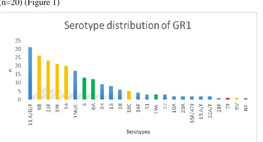
From the 218 isolated strains 14.2% (n=31) belonged to serotype 11A/D/F and was followed by the so called „paediatric” serotypes, 6B (n=25), 23F (n=22), 19F (n=21), 14 (n=20) (Figure 1)
Figure 1. Yellow column: PCV7 serotypes, red column: additional serotypes in PCV10, green column: additional serotypes in PCV13, blue column: non-PCV7 serotypes, NT:
non typeable isolates. n= number of isolates
The vaccine coverage of PCV7 in GR1 was 44.5%. It means that 44.5% of the carried strains could have been prevented by PCV7. The coverage of PCV10 would be only slightly higher, 45.0% and for PCV13 it would be 57.8%.
Serotype distribution in GR2
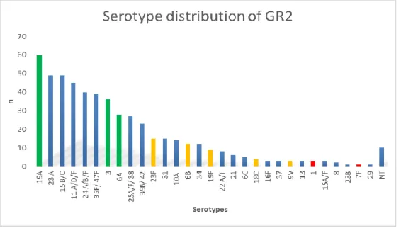
The prevalence of the most frequent serotypes in ranking order among the 529 strains in GR2 was the following: 19A (n=60), 23A (n=49), 15B/C (n=49), 11A/D/F (n=45), 24A/B/F (n=40), 35F/47F (n=39), 3 (n=36), 6A (n=28) and altogether 30 different serotypes were identified (Figure 2).
Figure 2. Yellow column: PCV7 serotypes, red column: additional serotypes in PCV10, green column: additional serotypes in PCV13, blue column: non-PCV7 serotypes, NT:
non typeable isolates, n= number of isolates
The calculated vaccine coverage for the 529 strains is 8.1% for PCV7, 8.9% for PCV10 and 32.3% for PCV13.
Serotype distribution in GR3
The prevalence of serotypes in GR3 in ranking order was the following: 11A/D/F (n=18), 15B/C (n=15), 35F/47F (n=15), 23B (n=14), 23A (n=13), 10A (n=6), 6C (n=5), 19A (n=2), 24 A/B/F (n=2), 8 (n=1), 19F (n=1), 16F (n=1) and 21 (n=1) (Figure 3).
Figure 3. Yellow column: PCV7 serotypes, green column: additional serotypes in PCV13, blue column: non-PCV7 serotypes, n= number of isolates
The calculated vaccine coverage of PCV7 was 1.1% (n=1), of PCV10 the same and of PCV13 it was 3.2% (n=3).
Antibiotic susceptibility and resistance genes
Antibiotic susceptibility in GR1
Generally, antibiotic susceptibility to the tested antibiotics was very high in GR1, except for macrolides and clindamycin, where we could detect 21.4% and 14.2%
resistance, respectively. None of the isolates were resistant to penicillin or to other β- lactams, but intermediate resistance to penicillin was observed in 24.2% of the cases.
The resistant serotypes here belonged to the PCV7 vaccine types (mostly 23F, 19F, 6B).
In 24 cases we could find the ermB gene resulting high-level resistance to macrolides and lincosamides. In 16 strains the mefE genes were detected resulting low level resistance.
Antibiotic susceptibility in GR2
Almost the same observation was done in case of antibiotic susceptibility in GR2. Only one strain was found to be penicillin resistant, and 21.9% showed intermediate resistance. Among macrolides where to erythromycin 21.8% resistance was detected, and 19% of the strains was resistant to clindamycin. The majority of the resistant strains belonged to serotype 19A. Among macrolide resistant strains we detected the ermB gene in 91 cases, in 16 strains the mefE gene was observed, where 5 strains carried ermB and mefE genes simultaneously.
Antibiotic susceptibility in GR3
The measured antimicrobial susceptibility in GR3 was also relatively low; the isolates were generally sensitive to the tested drugs except to macrolides again where we found 21.7% resistance to erythromycin and 17.4% to clindamycin. We didn’t find any penicillin resistant strain, but intermediate resistance was found to be 20.7%. In this group the most resistant strains were found to be serotype 23A. Among macrolide resistant strains, 8 had the ermB gene and none of the strains carried the mefE gene.
Pilus results
Pilus results in GR1
Out of the 218 strains, we could detect pilus in 25 cases (11.5%) by PCR. Sixteen isolates showed intermediate resistance to penicillin, 14 isolates were highly resistant to erythromycin and clindamycin.
Pilus results in GR2
Out of 529 strains, we could detect pilus in 102 cases (19.3%). In 27 cases the strains had intermediate resistance to penicillin and also 27 isolates were highly resistant to erythromycin and clindamycin (25 ermB positive and 3 mefE positive). We found a strong association between serotype 24 A/B/F and pilus positivity.
PFGE pattern of certain distinguished serotypes
The clonality of certain serotypes was examined with PFGE. Gels were analysed using the Bionumerics software (Bio-Rad), with the Jaccard coefficient and the unweighted
pair group method with arithmetic mean (UPGMA) dendrogram type. Among all serotypes, 19A was the most interesting due to its emergence after PCV7 vaccination and also its average antibiotic resistance.
PFGE of serotype 19A
The digested genome patterns of 19A showed that although all isolates were related to one another, sub-clones could be identified. These PFGE clusters shared similar penicillin susceptibility rates. In order to compare the 19A strains of the current study to the previously frequent PMEN (Pneumococcal Molecular Epidemiology Network) clone Hungary19A-6, which typically was resistant to penicillin, we have included one representative isolate of that clone (strain 21646, isolated in 2001) in the PFGE analysis.
As we detected the new 19A isolates differ greatly from this older clone. In GR3 out of the 94 isolates, only two were serotype 19A. These 2 isolates had identical PFGE pattern to some of those obtained from the nurseries in GR2.
PFGE of serotype 19F
Digestion pattern on 19F indicates that a few isolates from two different nurseries in Szeged and Győr were identical to the PMEN-Taiwan19F-14 clone, which is another worldwide spread famous clone.
PFGE of serotype 3
In case of serotype 3 we could detect identical PFGE patterns from different nurseries in Miskolc, Szeged, Debrecen.
PFGE of other serotypes
Among other serotypes (15B/C, 11 A/D/F, 37) mostly we could detect clonality within the same nursery but rarely similarities between the strains deriving from different nurseries.
MLST results of representative serotype 19A isolates
For MLST analysis, a few isolates were selected from each PFGE cluster of 19A serotype. Four isolates (all penicillin sensitive) with identical PFGE pattern belonged to
the same sequence type (ST) 8430. Four of the penicillin-intermediate isolates, belonging to another PFGE cluster, were all ST-319. ST-97, ST-1611, ST-1757, ST- 320, ST-268 represented itself as well. These MLST data correlate well with the PFGE pattern.
Discussion
1.
The average carriage rate throughout the whole study was 33.8%. The detected carriage rates in GR1, GR2 and GR3 (34.2%, 32.5% and 41.4%, respectively) correlate well to the literature data reported from different countries. We detected double carriage only in 3 occasions. This low co-colonisation rate might be the result of the culture-base technique we applied. We didn’t find any correlation between carriage and acute otitis media in any of the 3 groups.2.
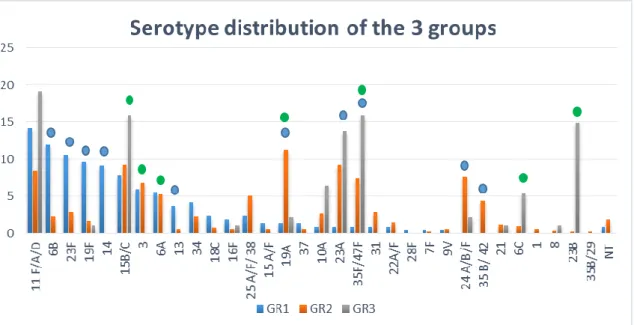
The majority of the strains in GR1 belonged to the usual so called “paediatric serotypes” (6A/B, 14, 19F, 23F). Their disease causing potential is very high, these serotypes caused 70%–88% of IPD in young children all over the word before PCV7 vaccination (Figure 4).3.
In GR2 we found striking differences in serotype distribution. The previously dominant „paediatric” serotypes were rapidly replaced by others. For example, although serotype 14 had been the leading type before, it fully disappeared as soon as children became vaccinated with PCV7. The frequency of 19F, 23F and 6B also decreased significantly but on the other hand, the previously absent serotypes 19A and 23A significantly emerged, competing for the second position.Unfortunately serotype 19A, which became a leading serotype, was shown to have high invasive disease potential and antibiotic resistance capacity and has often been reported worldwide as an emerging serotype after the implementation of PCV7 (Figure 4).
4. In GR3 a decline in the carriage of PCV13 serotypes was detected. 19A, which became a dominant serotype after PCV7 vaccination in GR2, were detected only in 2 cases in GR3, however, these were clonally identical to some of the GR2 isolates. Significantly emerging serotypes were 15B/C, 35F/47F, 6C and 23B. The same situation was observed in other countries after the replacement of PCV7 by PCV13, in case of both IPD cases and carriage (Figure 4)
Figure 4. Serotype distribution in GR1, GR2 and GR3. Blue circle shows significant changes between GR1 and GR2, after PCV7 vaccination (p< 0.05). Green circle shows significant changes between GR2 and GR3 after PCV13 vaccination (p< 0.05).
5. Our results found in the three groups suggest that although the average resistance did not change significantly due the vaccinations, the serotypes changed, which carry the same resistance genes.
6. In GR1, decreased penicillin susceptibility was shared among the previously prevalent “paediatric’ serotypes. In GR2, after PCV7 vaccination, the serotype distribution changed markedly and 19A became dominantly responsible for resistance. In GR3, after PCV13 vaccination, 19A decreased, meanwhile mostly serotype 23A replaced it.
7. The same observation was done in case of macrolide resistance. In GR1, the
“paediatric’ serotypes were mostly responsible for macrolide resistance. In GR2, after PCV7 vaccination, the serotype distribution changed markedly and 19A became most dominant. In GR3, after PCV13 vaccination, 19A decreased and serotype 23A replaced it.
8. In GR1, we could detect a strong association between pilus positivity and antibiotic resistance, high-level erythromycin resistance measured in 64% of the piliated strains. In GR2, we could detect a strong association between serotype 24 A/B/F and pilus positivity, despite that these strains were sensitive to all tested
antibiotics. Serotype 6A was the second most common piliated serotype and 8 out of the 15 strains were resistant to erythromycin and 5 showed intermediate resistance to penicillin. These results need more investigations since recent studies could demonstrate that native pilus could be dedicated to DNA transformation in pneumococci.
9. Based on the PFGE patterns, it was obvious that the same serotypes within a DCC group often belonged to the same clones, providing evidence for strong clonal spread between the children in the same group. This can be easily explained by the close contact between children during the time spent together in the given community.
10. On several occasions we could detect the same clone in different DCCs in different parts of Hungary. For example in case of serotype 19F, we could detect the same clone in Szeged and in Győr and interestingly these clones were identical to the PMEN Taiwan 19F-14 clone, which has been a widespread clone on the earth and resistant to erythromycin and non-susceptible to penicillin, just like our strains. This strain was found already earlier among invasive isolates in Hungary.
11. The most important PFGE examination was conducted among serotype 19A isolates. Since this serotype became highly frequent after vaccination and was highly resistant to macrolides, we wanted to know whether they belong to any worldwide spread PMEN clone, especially the famous resistant Hungary19A-6 clone. We found that all 19A strains were more or less clonal, but PFGE could identify some smaller clusters, which showed strong relation to penicillin sensitivity levels. It is clear from the PFGE patterns and MLST results that none of them is identical to the previously dominant, highly resistant and virulent Hungarian 19A clone.
12. Among our 19A isolates with the highest level of penicillin non-susceptibility and macrolide resistance, possessing the ermB + mefE genes together, with MLST we identified ST-320, which is a famous multi-resistant pneumococcal clone circulating all over the world.
Conclusions
Vaccinations with PCV7 lead to a dramatic re-arrangement in pneumococcal serotypes. The rate of previously prevalent ‘paediatric’ types decreased or vanished (e.g. serotype 14) and other, formerly absent serotypes emerged.
The most important emerging serotype after PCV7 was 19A, which is multi- resistant and has high invasive capacity and caused worries in most countries in the post-PCV7 era.
After the introduction of PCV13, serotype 19A seems to decline again and the other PVC13-PCV7 serotype, serotype 3 was not detected any more among the youngest children.
The overall carriage rate decreased slightly, but not significantly after PCV vaccination.
The overall antibiotic resistance of the carried strains did not change during the 3 examined period, but different serotypes were involved in resistance; while the
‘paediatric’ serotypes dominated in GR1, 19A alone took over in GR2 and it was replaced by 23A in GR3.
Based on the PFGE and MLST data, the highly prevalent serotype 19A isolates were genotypically unrelated from the world-wide prevalent PMEN Hungary19A-6 clone, however, other PMEN clones were detected among our 19F strains.
PFGE is a useful tool in the determination of the genetic relatedness of the pneumococcal isolates within a country of the size of Hungary; further refinement of the clustering was possible with the MLST method.
The PFGE data showed that clonal spread of the isolates as well as horizontal transfer of resistance genes together contribute to the dynamic serotype changes observed in the post-PCV era.
Very strong correlation could be established between pilus positivity and antibiotic resistance (penicillin and macrolide non-susceptibility) and/or serotypes (especially serotype 24 A/B/F).
Both PCV7 and PCV13 proved to be very successful in the elimination of the vaccine-types. We suggest that vaccination with PCV13 should definitely be continued in Hungary.
Based on the results of many studies like ours, pharmacies should extend the conjugate vaccines covering more serotypes, or producing totally different composition like whole cell vaccine, protein based vaccine or others.
List of publications
Related to the thesis Papers
Tóthpál A, Kardos S, Laub K, Nagy K, Tirczka T, van der Linden M, Dobay O. (2014) Radical serotype rearrangement of carried pneumococci in the first 3 years after intensive vaccination started in Hungary. Eur J Pediatr. September 2014, DOI:
10.1007/s00431-014-2408-1 IF: 1.983
Tóthpál A, Laub K, Kardos S, Nagy K., Dobay O. (2014) Invazív és hordozott Streptococcus pneumoniae izolátumok vakcina lefedettsége Magyarországon. Lege Artis Medicinae, 24:(4) pp. 180-185.
Tóthpál A, Laub K, Kardos S, Nagy K, Dobay O. (2012) Changes in the serotypes of Hungarian pneumococci isolated mainly from invasive infections: a review of all available data between 1988 and 2011. Acta Microbiol Immunol Hung, 59(3): 423-433.
IF: 0.646
Tóthpál A, Kardos S, Hajdú E, Nagy K, Linden M, Dobay O. (2012) Nasal carriage of Streptococcus pneumoniae among Hungarian children before the wide use of the conjugate vaccine. Acta Microbiol Immunol Hung, 59(1): 107-118. IF: 0.646
Tóthpál A, Dobay O. (2012) [Drastic changes in serotypes of carried pneumococci due to an increased vaccination rate in Hungary]. Orv Hetil, 153(26): 1031-1034.
Tóthpál A, Ordas A, Hajdú E, Kardos S, Nagy E, Nagy K, Dobay O. (2011) A marked shift in the serotypes of pneumococci isolated from healthy children in Szeged, Hungary, over a 6-year period. Acta Microbiol Immunol Hung, 58(3): 239-246.
IF: 0.787