Epidemiological analysis of carried Streptococcus pneumoniae among healthy children attending
communities
Ph.D. thesis
Adrienn Tóthpál
Semmelweis University
Doctoral School of Pathological Sciences
Supervisor: Dr. Orsolya Dobay Ph.D.
Official reviewers:
Dr. Zsófia Mészner, MD, Ph.D.
Dr. Endre Ludwig MD, D.Sc.
Head of the Final Examination Committee:
Dr. György Bagdy D.Sc.
Members of the Final Examination Committee:
Dr. Levente Emődy MD, D.Sc.
Dr. Barna Vásárhelyi MD, D.Sc.
Budapest 2014
1
TABLE OF CONTENTS
TABLE OF CONTENTS ... 1
1 LIST OF THE FREQUENTLY USED ABBREVIATIONS ... 6
2 INTRODUCTION ... 7
2.1. About Streptococcus pneumoniae in general ... 7
2.1.1. Historical outlook ... 7
2.1.2. Morphology and culivation ... 8
2.1.3. Identifiation ... 8
2.1.4. Most important virulence factors ... 9
2.1.4.1. Capsule ... 10
2.1.4.2. The presence of pilus... 13
2.1.4.3. Surface proteins ... 14
2.1.4.4. Most important enzymes ... 15
2.1.4.4.1. Pneumolysin ... 15
2.1.4.4.2. Autolysin ... 15
2.1.4.4.3. IgA protease... 16
2.1.4.4.4. Other enzymes ... 17
2.2. Genetic background of Streptococcus pneumoniae ... 17
2.2.1. The position of pneumococcus in the Streptococcaceae family ... 17
2.2.1.1. Evolution of Streptococcus pneumoniae ... 19
2.2.2. Genotyping of pneumococcus ... 19
2.2.3. Serotyping of pneumococcus ... 21
2.3. Clinical aspects ... 21
2.3.1. Carriage ... 23
2.3.1.1. The impact of carriage... 23
2.3.2. Diseases caused by Streptococcus pneumoniae ... 24
2.3.2.1. Otitis media ... 24
2.3.2.2. Eye infections ... 24
2.3.2.3. Pneumonia ... 25
2.3.2.4. Invasive pneumococcal diseases (meningitis, sepsis) ... 26
2.4. Prevention of pneumococcal diseases ... 26
2.4.1. History of vaccination ... 26
2
2.4.2. Available vaccines nowadays ... 29
2.4.2.1. Polysaccharide vaccine ... 29
2.4.2.2. Conjugate vaccines (PCV) ... 29
2.4.2.3. New perspectives in vaccination ... 32
2.4.2.3.1. Protein-based pneumococcal vaccines... 32
2.4.2.3.2. Whole cell vaccine ... 32
2.4.2.3.3. DNA vaccines ... 33
2.5. Antibiotic treatment ... 34
2.5.1. Antimicrobial resistance of Streptococcus pneumoniae ... 34
2.5.1.1. Development of resistance ... 34
2.5.1.2. Resistance mechanism... 35
2.5.1.2.1. β-lactam resistance ... 35
2.5.1.2.2. Macrolide resistance ... 36
2.5.1.2.3. Resistance to other antibiotics... 36
2.5.1.2.4. Successful pneumococcal clones ... 37
2.6. Pneumococcus data from Hungary... 38
2.6.1. Serotyping data of pneumococci in Hungary ... 38
2.6.2. Antibiotic resistance data of pneumococci in Hungary ... 38
2.6.3. Vaccine situation in Hungary ... 39
3 OBJECTIVES ... 41
4 MATERIALS AND METHODS ... 42
4.1 Permissions ... 42
4.1.1 Parents’ permission ... 42
4.1.2 Ethical permission ... 42
4.2 Bacterial strains ... 42
4.2.1 Origin of the strains ... 42
4.2.2 Specimen collection and growth media ... 43
4.3 Identification ... 44
4.3.1 Conventional microbiological methods for identification ... 44
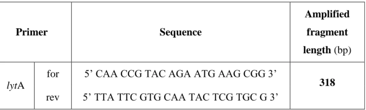
4.3.2 LytA PCR ... 44
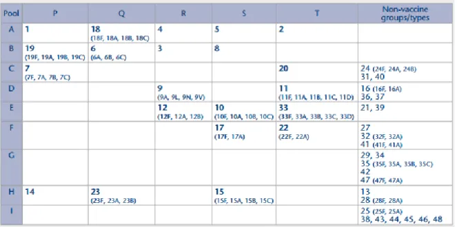
4.4 Serotyping ... 45
4.4.1 Serotyping by latex agglutination ... 45
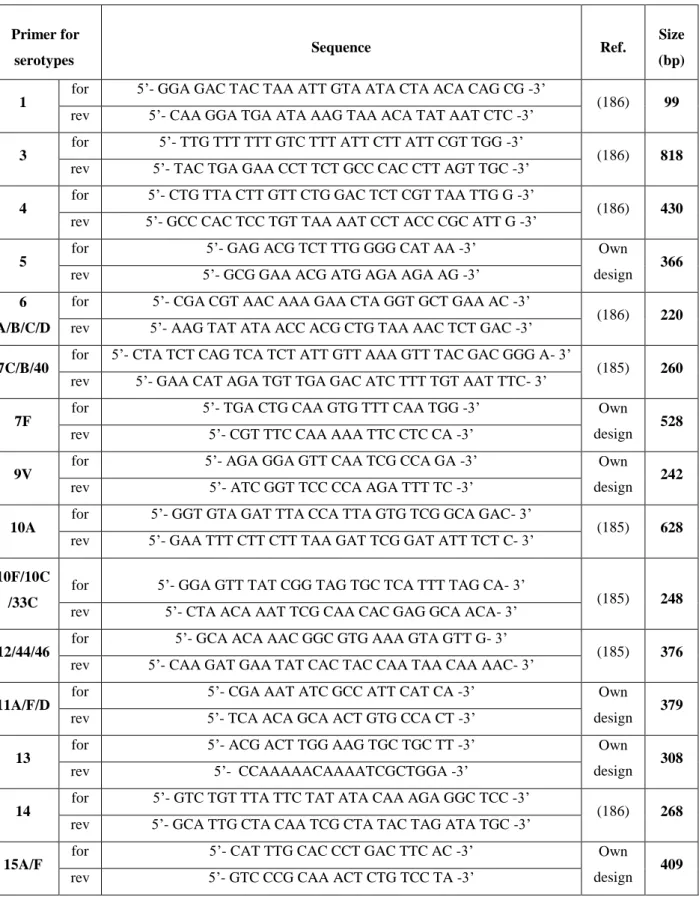
4.4.2 Serotyping by PCR ... 46
4.5 Polymerase chain reaction ... 48
3
4.5.1 DNA template preparation... 49
4.5.2 PCR mix ... 49
4.5.3 PCR cycle ... 50
4.5.4 Gel running and analysis ... 50
4.6 Antimicrobial susceptibility testing... 51
4.6.1 Susceptibility testing by disc diffusion and E-test ... 51
4.6.2 Susceptibility testing by agar dilution ... 52
4.7 Determination of the macrolide resistance mechanisms ... 53
4.7.1 Detection of the presence of erm and mef genes by PCR ... 53
4.7.2 Distinction between mefE and mefA ... 54
4.8 Pulsed-field gel electrophoresis (PFGE) ... 54
4.8.1 Preparation of chromosomal DNA ... 54
4.8.2 Digestion ... 55
4.8.3 Gel running ... 56
4.8.4 Gel analysis ... 56
4.9 Multi-locus sequence typing (MLST) ... 57
4.10 Whole genome sequencing (WGS) ... 58
4.11 Detection of the presence of pili in pneumococcus ... 59
4.12 Statistical analysis ... 59
4.13 Solutions ... 60
5 RESULTS ... 61
5.1 General information about the isolates ... 61
5.1.1 Carried isolates ... 61
5.2 GR1 in general... 61
5.3 GR2 in general... 63
5.4 GR3 in general... 65
5.5 Serotype distribution of the carried isolates ... 67
5.5.1 Serotype distribution in GR1 ... 67
5.5.1.1 Vaccine coverage ... 68
5.5.2 Serotype distribution in GR2 ... 68
5.5.2.1 Vaccine coverage ... 69
5.5.3 Serotype distribution in GR3 ... 69
5.5.3.1 Vaccine coverage ... 70
5.6 Antibiotic susceptibility ... 70
5.6.1 Antibiotic susceptibility in GR1 ... 71
4
5.6.2 Antibiotic susceptibility in GR2 ... 72
5.6.3 Antibiotic susceptibility in GR3 ... 73
5.7 Macrolide resistance mechanisms ... 74
5.7.1 Erm and mef genes in GR1 ... 74
5.7.2 Erm and mef genes in GR2 ... 75
5.7.3 Erm and mef genes in GR3 ... 76
5.8 Pilus positive strains ... 77
5.8.1 Pilus positive strains in GR1 ... 77
5.8.2 Pilus positive strains in GR2 ... 78
5.9 PFGE pattern of certain distinguished serotypes ... 79
5.9.1 PFGE pattern of serotype 19A... 79
5.9.2 PFGE pattern of serotype 19F ... 82
5.9.3 PFGE pattern of serotype 11 A/D/F ... 83
5.9.4 PFGE pattern of serotype 15 B/C ... 84
5.9.5 PFGE pattern of serotype 3 and 37 ... 84
5.10 MLST results of representative serotype 19A isolates ... 85
6 DISCUSSION ... 87
6.1 Carriage rate ... 87
6.1.2 Carriage rate in GR1 ... 87
6.1.3 Carriage rate in GR2 ... 87
6.1.4 Carriage rate in GR3 ... 88
6.2 Serotype distribution and vaccine coverage ... 89
6.2.2 Serotype distribution in GR1 ... 89
6.2.3 Serotype distribution in GR2 ... 89
6.2.4 Serotype distribution in GR3 ... 91
6.3 Antibiotic susceptibility and resistance genes ... 91
6.3.2 Antibiotic susceptibility in GR1 ... 91
6.3.3 Antibiotic susceptibility in GR2 ... 91
6.3.4 Antibiotic susceptibility in GR3 ... 92
6.3.5 Summarised antibiotic susceptibility ... 92
6.4 Pilus positive strains in the 3 groups ... 95
6.5 PFGE results of the 3 groups ... 96
6.6 MLST results of representative serotype 19A isolates ... 97
6.7 Strengths and limitations of this thesis ... 98
7 CONCLUSIONS ... 100
5
8 SUMMARY ... 104
9 ÖSSZEFOGLALÁS ... 105
10 BIBLIOGRAPHY ... 106
11 LIST OF PUBLICATIONS ... 129
12 APPENDIX ... 130
13 AKNOWLEDGEMENT ... 132
6
1 LIST OF THE FREQUENTLY USED ABBREVIATIONS
AOM - Acute otitis media
CAP - Community acquired pneumonia CBP - Choline-binding protein
CDC- Centers for Disease Control and Prevention CPS - Capsular polysaccharide
DCC - Day care centre
EUCAST - European Committee on Antimicrobial Susceptibility Testing GNRCS - German National Reference Centre for Streptococci
GR1 – Group-1; DCC groups with low-level vaccination rate
GR2 - Group-2; DCC groups with high-level vaccination rate (with PCV7) GR3 - Group-3; toddlers groups with high-level vaccination rate (with PCV13) IPD - Invasive pneumococcal disease
LytA- Autolysin
MDR - Multidrug resistant
MIC - Minimal inhibitory concentration MLST - Multilocus sequence typing NCSP - Non-classical surface protein NIP - National Immunization Programme NT- Non-typeable (serotypes)
OEK - National Epidemiological Centre in Hungary PBP - Penicillin-binding protein
PCR - Polymerase chain reaction
PCV10 – 10-valent pneumococcal conjugate vaccine, Synflorix PCV13 – 13-valent pneumococcal conjugate vaccine, Prevenar-13 PCV7 – 7-valent pneumococcal conjugate vaccine, Prevenar-7 PFGE - Pulsed-field gel electrophoresis
PMEN - Pneumococcal Molecular Epidemiology Network PPS23 – 23-valent polysaccharide vaccine, Pneumovax 23 ST - Sequence type
WHO - World Health Organization
7
2 INTRODUCTION
2.1. About Streptococcus pneumoniae in general 2.1.1.
Historical outlookThe bacterium itself was discovered in 1880 by Sternberg, a US Army physician and by Louis J. Pasteur (1822–1895) independently, Pasteur named the bacteria Microbe septicemique du salive and Sternberg named it Micrococcus pasteuri than the first name Diplococcus pneumoniae was given (2-4). In 1886 Fränkel identified it as the causative agent of lobar bacterial pneumonia and not so long afterwards its pathogenic role was proved also in meningitis, endocarditis, arthritis and otitis media (5).
In the turn of the 20th century realms of immunology and vaccinology started using Streptococcus pneumoniae (pneumococcus) for fundamental researches. In 1909 Neufeld and Händel described different serotypes of pneumococci and observed that protection by immune serum was type-specific (2, 6). With the help of this finding they recognized the specific antisera and its role in therapy which resulted in the first effective treatment in the pre-antibiotic era against pneumococcal infections. This experience helped Oswald T. Avery and his colleague Michael Heidelberger in 1923 to determine the pneumococcal antigens that induced protective immunity as carbohydrates (2, 7).
The basements of molecular biology could be owed to S. pneumoniae as well. In 1928 Griffiths proved that pneumococci are capable of taking up large foreign DNA fragments from the surroundings (4).
In 1944 Avery together with Colin MacLeod and Maclyn McCarty demonstrated their favourite experiment with the help of different virulence of R (rough, non-capsulated) and S (smooth, capsulated) colonies and proved the chemical nature of the principle of transforming substance as DNA (2, 8). After the introduction of penicillin in 1940s, treatment of the pneumococcal infections seemed to be solved. Nevertheless, the first penicillin resistant strains appeared in 1960s, followed by multidrug resistant (MDR) strains, which still remained a problem (9).
8
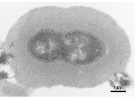
Figure 1. Microscopic picture of S.
pneumoniae by A Tóthpál
2.1.2.
Morphology and culivationS. pneumoniae is a Gram-positive coccus, sized 0.8- 1.5 m, forming pairs or short chains (Figure 1).
Chain formation is more frequent when grown on solid medium. The bacterium can have pili and are encapsulated, with a large polysaccharide capsule.
S. pneumoniae is a fastidious, facultative anaerobe, bacterium, growing best on media supplemented with 5% blood and requires 5% to 10% CO2 for incubation at 35-37C. On blood agar it shows - haemolysis and the colonies have special umbilicus morphology (Figure 2), due to autolytic activity (10).
2.1.3.
IdentifiationIt is not an easy task to identify S. pneumoniae. As a respiratory pathogen, it can be found in the nasopharynx and in the sputum sample during infection, but sometimes the less fastidious normal microbiota members, such as the genetically related viridans Streptococci (for example Streptococcus mitis) can overgrow the pneumococci leading to problems in identification.
The classical diagnostic identification of pneumococci is based on colony morphology (-haemolysis and characteristic colonies), optochin sensitivity - although optochin resistant strains were described (11) - and some biochemical activities, such as the lack of catalase enzyme, bile solubility (when the surface active bile salts provide full activity for the autolytic enzymes) or inulin degradation, for separation from the viridans Streptococci (12). Antigen detection is possible from sterile body fluids (e.g. in case of meningitis) with latex agglutination. Urinary antigen tests are also available.
These are rapid immunochromatographic tests which detect the C polysaccharide cell Figure 2. S. pneumoniae umbilicated colonies on blood agar by A Tóthpál and Á Ghidán
9
wall antigen common to all strains of S. pneumoniae and can be positive for weeks to several months after the infection(13).
For serotyping pneumococci, monovalent antisera and latex agglutination kits are also available (14, 15). The most reliable way for identification is using molecular biological methods, such as PCR for pneumococcal specific genes like lytA gene (1) ply gene (16) pspA gene (17), or detection of a conserved gene such as 16S rRNA (18).
2.1.4.
Most important virulence factorsProperties that explain the pathogenic potential of S. pneumoniae include polysaccharide capsule, pili, different enzymes such as IgA1 protease, pneumolysin, autolysin and several surface-exposed proteins that mediate contact with components of host tissues and secretions (Table 1).
Table 1. List of virulence factors, only selected examples are shown (19)
Pneumococcal virulence factors and disease
Main role in colonization
Upper-airway colonization
Capsule
Prevents entrapment in the nasal mucus, thereby allowing access to epithelial surfaces. Also inhibits effective opsonophagocytosis.
Phosphorylcholine Binds to the epithelial surface of the human nasopharynx.
Choline-binding protein A Binds to human secretory component on a polymeric Ig receptor during the first stage of translocation across the epithelium.
Neuraminidase
Act sequentially to cleave terminal sugars from human glycoconjugates, which might reveal receptors for adherence.
Hyaluronate lyase
Breaks down hyaluron-containing extracellular matrix components.
10 Pneumococcal adhesion and
virulence A Binds to fibronectin.
Enolase
Binds to plasminogen.
Competition in upper airway
Bacteriocin (pneumocin) Small antimicrobial peptide that targets members of the same species.
Respiratory-tract infection and pneumonia
Pneumolysin
Cytolytic toxin that also activates complement. An important determinant of virulence in in vivo models of disease. Wide range of effects on host immune
components at sub-lytic concentrations.
Pneumococcal surface protein
A Prevents binding of C3 onto pneumococcal surface. Also binds lactoferrin.
Autolysin A
Digests the cell wall, which results in the release of Ply.
Pneumococcal surface antigen Component of the ABC transport system, which is involved in resistance to oxidative stress.
PiaA: pneumococcal iron
acquisition Component of the ABC transport system.
IgA1 protease Cleaves human IgA1 immuneglobulin.
2.1.4.1. Capsule
Capsule (Figure 3) is the major virulence factors of pneumococci and it was the first non-protein substances shown to be antigenic in humans. Fred Neufeld described first the process to differentiate pneumococci into serotypes with the help of type-specific antisera and he also discovered that these type-specific antigens were carbohydrates, which are the basement of the capsule (2).
11
Figure 3. Immunoelectron microscopy of pneumococcal capsules showing an increased zone of capsular material in serotype 6B (20)
So far we know 94 different capsular polysaccharides (CPSs), which are distinguished by using a set of antisera that recognise the chemical differences in the capsules, therefore they are called serotypes (21). Expression of a capsule is important for survival in the blood and is associated with the ability of pneumococci to cause invasive disease. CPSs are built up from repeating oligosaccharide units and antibodies against these saccharides provide protection against pneumococcal disease. The most frequent monosaccharides in CPSs in different combinations are α/β-D-glucose, α/β-D-galactose, α/β-L-rhamnose, N-acetyl-α/β-D-glucosamine, N-acetyl-α/β-D-galactosamin, N-acetyl- β-D-mannosamine, N-acetyl- α-L-fucosamine and α/β-D-glucuronic acid (21).
There are significant differences between different serotypes. In addition to the above mentioned monosaccharides, α-L-fucose (serotype 19A), β-D-ribose (serotype 7, 19B, 19C), 2-acetamindo-4-amino-2,4,6-trideoxy-α-D-galactose (serotype 1) can be present.
Comparison of the CPSs of serotype 6A and 6B shows that the only difference is the presence of 3-substituted D-ribitol in 6A and 4-substituted D-ribitol in 6B.
The capsular gene (cps) locus, responsible for the expression of CPSs - with the exception of serotypes 3 and 37 (22) - is located between dexB and aliA on the pneumococcal chromosome (23) and the total size of alternative coding DNA at this
12
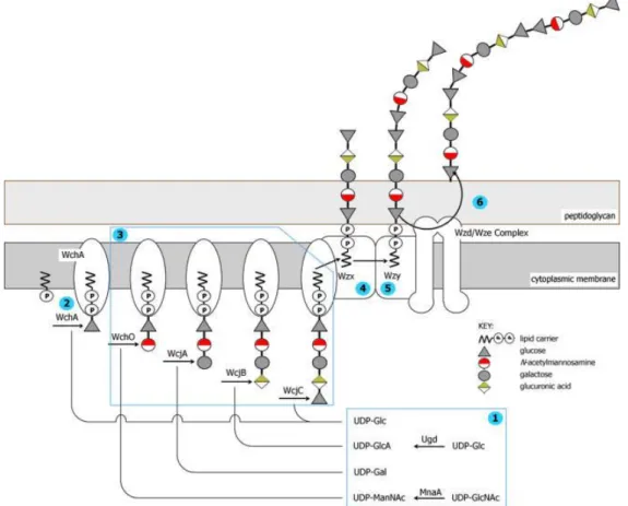
one locus exceeds 1.8 Mbp. The regulatory and processing genes wzg, wzh, wzd and wze (also known as cpsABCD) are conserved with high sequence identity in all cases and are almost always in this gene order at the 5′ end of the cps locus (23). In most cps clusters, the fifth gene encodes the initial glucose phosphate transferase, WchA (also known as cpsE), responsible for linkage of an activated glucose phosphate to the lipid carrier. The polysaccharide polymerase (wzy) and flippase (wzx) genes are always present downstream together with a varying set of genes for glycosyl transferases, acetyl transferases, nucleotide diphosphate sugar biosynthesis and modifying enzymes (Figure 4). In the regions between the cps genes and the flanking dexB and aliA genes, there is almost always evidence of mobile genetic elements.
Figure 4. Pneumococcal capsular biosynthesis: Schematic representation of the biosynthesis of CPS by the Wzy-dependent pathway. The biosynthesis of the CPS of serotype 23F is represented (24)
13
Due to the natural transformability of the pneumococcus, horizontal recombination allows that one serotype can belong to different genotypes and a single genotype can express different capsular genes, i.e. different serotypes. This phenomenon is known as capsular switching (25).
S. pneumoniae capsule can affect several aspects of complement activity. These include preventing binding of both IgG (Immunoglobulin G) and CRP (C-reactive protein) to S.
pneumoniae and thereby inhibiting classical pathway activity, reducing alternative pathway activity through unexplained mechanisms and decreasing the degradation of C3b bound to the bacterial surface to iC3b. The effects on C3b/iC3b deposition prevent phagocytosis of encapsulated bacteria, but data also suggest that the capsule inhibits phagocytosis mediated directly by IgG and by nonopsonic phagocytic receptors. The results clarify some of the mechanism by which the S. pneumoniae capsule could mediate immune evasion (26).
2.1.4.2. The presence of pilus
S. pneumoniae, like many other Gram-positive bacteria, has long filamentous pili extending on their surface through which they adhere to host cells. This may be the first of those virulence factors which are responsible for initial adhesion of the bacteria to host tissues during colonization and biofilm formation. The pilus of S. pneumoniae was first identified in 2006 (27).
Pilus genes can be found in a 12 Kb pathogenicity island called Pilus islet-1 (PI-1), encoding a positive transcriptional regulator (rlrA), the pilus-1 structural subunits (rrgA, rrgB and rrgC) and three pili-specific sortases. RrgB encodes the major component RrgB (28) which is strictly necessary for the pilus formation while the other two (RrgA, RrgC encoded by rrgA and rrgC) are ancillary proteins (29). PI-1 is present in about one-third of the clinical isolates (30) and its prevalence is higher among antibiotic non- susceptible strains (31).
Pilus subunits are immunogenic in humans (32) and were able to elicit a protective response when tested in mouse models of infection (33). Thus pili could be a good target to develop a new vaccine.
14
Recent studies demonstrated that pilus can be dedicated to DNA transformation in pneumococci (34). According to Laurenceau et al. the transformation pilus act as a
“DNA-trap” to capture DNA in the environment(35). Their data clearly establish the existence and function of a transformation pilus on the surface of competent pneumococci.
2.1.4.3. Surface proteins
Many of the surface proteins are virulence factors that contribute to the pathogenesis of this organism. Three main groups of them have been identified in S. pneumoniae:
around 50 lipoproteins, up to 18 peptidoglycan binding (LPxTG) consensus sequence- carrying proteins that are covalently linked via sortase to the cell wall peptidoglycan and up to 16 choline-binding proteins (CBPs). In addition to the three main groups of surface proteins, the cell wall of pneumococci is decorated with another cluster of proteins that lack classic leader peptide and membrane-anchoring motifs (Figure 5).
These proteins are termed non-classical surface proteins (NCSPs) and they could also play a relevant role in subverting the physiological function of host-derived proteins (36).
Figure 5. Schematic model of the pneumococcal outer cell wall and surface-exposed proteins. The pneumococcal cell wall consists of a phospholipid membrane (LM), peptidoglycan (PG) and teichoic and lipoteichoic acids (TA, LTA). An unusual component of the cell wall is phosphorylcholine (PCho), which anchors the choline- binding proteins (CBPs) non-covalently on the cell wall. Virulence proteins of the different classes of pneumococcal surface proteins are depicted above (36)
15
Cell wall components
The cell wall of S. pneumoniae contains an unusually complex wall TA, which has identical repeating units as the membrane-anchored LTA (37). Although the structure of the pneumococcal peptidoglycan was found to resemble that of other characterized Gram-positive bacteria, the wall TA is complex and unusual and contains choline as a structural component. Thus pneumococcus is unique among prokaryotes due to an absolute requirement for choline in growth, which is incorporated as phosphorylcholine into the cell wall TA and the membrane LTA (38).
2.1.4.4. Most important enzymes 2.1.4.4.1. Pneumolysin
A key virulence factor of pneumococcus in colonizing the upper respiratory tract is the pneumococcal thiol-activated, membrane pore forming toxin, called pneumolysin (39).
Pneumolysin is a 53 kDa protein composed of 471 amino acids. It is common to all serotypes and it can be thought of as a multi-effective factor for virulence following pneumococcal infection. At high levels it is lytic to all cells with cholesterol in the membrane. The lytic activity of this toxin can be inhibited by preincubation with cholesterol, consistent with the suggestion that membrane cholesterol is the receptor for this toxin (40). At lower, sublytic concentrations, which exist in the early stages of infection, the toxin also may cause a range of effects, including induction of apoptosis, activation of host complement and induce proinflammatory reactions in immune cells.
At higher lytic concentrations, which may exist in the later stage of infection, the toxin may cause widespread direct cellular and tissue damage by virtue of its membrane pore forming properties (40-42).
2.1.4.4.2. Autolysin
S. pneumoniae has a special autolytic response that leads to the excessive lysis of cultures in vitro and causes the characteristic colony morphology called umbilicated shaped colonies. The main autolysin in the pneumococcus is N-acetyl-muramoyl-1- alanine amidase, commonly known as LytA, which encoded by the lytA gene. LytA causes lysis by cleaving the lactyl-amide bond that links the stem peptides and the
16
glycan strands of the peptidoglycan, resulting in hydrolysis of the cell wall. LytA orthologs are now known to be conserved throughout eubacteria and in many bacteriophages (43-45).
The in vivo function of the suicide LytA enzyme remains controversial. One hypothesis is that LytA mediates lysis to release other virulence factors such as pneumolysin (46).
Another theory suggests that LytA is released to lyse neighbouring non-competent pneumococcal cells (47). This would potentially facilitate genetic exchange between naturally competent pneumococcal populations that easily take up and incorporate DNA by homologous recombination.
A third possibility is that LytA mediates lysis to release proteins involved in immune evasion or cell wall components that may interfere with the host immune response (48).
From diagnostic point of view, lytA gene can be used for specific identification of S.
pneumoniae (1).
It is also interesting that there is a highly polymorphic region in the lytA gene where two different families of alleles can be differentiated by PCR and restriction digestion.
Morales et al proved that this polymorphic region arose from recombination events with homologous genes of pneumococcal temperate phages (45).
2.1.4.4.3. IgA protease
Antibodies of the immunoglobulin A (IgA) class react with capsular polysaccharides of S. pneumoniae and support complement-dependent opsonophagocytosis of the organism by phagocytes. IgA may provide both local defence against mucosal infection and activity in local tissues to prevent dissemination of the infection (49).
The IgA1 protease is one of the two to four large zinc metalloproteinase present in the pneumococcal genome (50). This protease is a polypeptide of about 1900 amino acids associated to the bacterium via N-terminal anchoring. The enzyme specifically cleaves the hinge regions of human IgA1, which dominates most mucosal surfaces and is the major IgA isotype in serum. This protease is expressed in all of the known pneumococcal strains and plays a major role in pathogen's resistance to the host immune response(51).
17
2.1.4.4.4. Other enzymes
Most of the human cell surfaces and secreted molecules are glycosylated. This glycosylation is often complex and involves a number of different sugar residues.
Glycosylation serves a number of functions including recognition processes, cell-to-cell interactions and the binding and transport of positively charged molecules (52).
Pneumococcus expresses a variety of enzymes: surface attached exoglycosidases:
neuraminidase, β-galactosidase and N-acetylglucosaminidase, which sequentially remove terminal sugars common to many human glycoconjugates. Deglycosylation of host molecules may expose cell-surface receptors, inhibit mechanisms of clearance that require these glycoproteins, or provide nutrition for the organism (53).
2.2. Genetic background of Streptococcus pneumoniae 2.2.1. The position of pneumococcus in the Streptococcaceae family
Streptococci were first recorded in 1683 in van Leeuwenhoek's drawings of microscope images of the material removed from between his teeth (54). The main entry of streptococci into history was in 1879, when Louis Pasteur was studying puerperal fever.
This was causing high mortality rates in maternity wards. Within the bodies of diseased women, he found rounded granules (microorganisms) arranged in the form of chains or strings of beads. He was convinced and it was later proven, that this was the cause of infections in women after childbirth (54).
This famous meaningful event belongs to Ignác Semmelweis, who was working at the largest maternity hospital in the world: the Vienna Maternity Hospital, which was divided into two clinics. Almost all the maternal deaths there were due to puerperal fever. The excess deaths in the first clinic were due to the routine procedures carried out in the courses attended by doctors and medical students. Each day started with postmortem examinations of women who had died of puerperal fever. Then, without washing their hands, the pupils went straight to the maternity wards to perform vaginal examinations. By contrast, the pupil midwives in the second clinic did not undertake either post-mortem examinations, nor routine vaginal examinations (55). In 1847 he introduced a system whereby the students were required to wash their hands in chloride of lime before entering the maternity ward. The result was dramatic. In 1848, the
18
maternal mortality rate in the first clinic fell compared to the rate in the second (midwives) clinic. Thus, streptococci were one of the first microbes to be identified as causing contagious disease and their existence led to the introduction of hygiene and aseptic practices into hospital wards (55).
There are now over 100 recognized species of streptococci. Historically, the classification of streptococci was based on the Lancefield scheme, which groups streptococcal strains according to the carbohydrate composition of cell wall antigens (56, 57). Such antigens, known as group-specific antigens or C substances, are either polysaccharides (as in groups A, B, C, E, F and G), teichoic acids (as in groups D and N), or lipoteichoic acid (as in group H). This approach has proved successful for the more pathogenic streptococci, but its widespread application is hindered by the fact that group-specific antigens for other species may be absent or shared between distinct taxa.
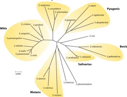
The streptococci may also be organized into six groups (Figure 6) based on 16S rRNA gene sequences (10).
Figure 6. The pyogenic group of Strepotococci. It includes Streptococcus pyogenes (Lancefield group A), Streptococcus agalactiae and Streptococcus uberis (group B) and Streptococcus dysgalactiae (group C, G, or L). The mitis group comprises species almost all of which are isolated from the human oral cavity or nasopharynx.
Streptococcus oralis, Streptococcus mitis, Streptococcus gordonii and Streptococcus pneumoniae are highly related and because of extensive horizontal gene transfer, the delineation of strains into these species is often blurred (10)
19
Although identification of streptococci is based on the current taxonomic standards using a combination of 16S rRNA gene sequence analyses, DNA-DNA hybridization, serologic and phenotypic data, 16S rRNA gene sequences of S. mitis and S. oralis are almost identical (> 99%) to S. pneumoniae, making the use of this information alone insufficient to distinguish these species.
2.2.1.1. Evolution of Streptococcus pneumoniae
Based on Kilian et al. research it is supposed that the immediate common ancestor of S.
pneumoniae, S. mitis and S. pseudopneumoniae (pneumoniae-mitis-pseudopneumoniae) cluster was a bacterium with resemblance to the present-day pneumococcus with all the properties associated with virulence(58). One of these properties, the IgA1 protease evolved by gene duplication in response to emergence of the immunoglobulin A1 (IgA1) subclass in the common ancestor of man, chimpanzees and gorillas (59), which according to recent calculations existed 6 to 7 million years ago (60). While the pneumococcus lineage conserved the expression of both capsule production and IgA1 protease activity to ensure their ability to colonize in the presence of IgA1 antibodies (61), lineages evolving into a commensal life style with a more subtle relationship with the mucosal immune system and the host in general gradually lost both characters and achieved the colonization advantage of the capsule-deficient phenotype (62). This evolutionary model proposing that the pneumoniae-mitis-pseudopneumoniae cluster arose from a pneumococcus-like organism pathogenic to the immediate ancestor of hominoids is consistent with our inability to isolate S. mitis-like bacteria from a range of mammals including old and new world monkeys, pigs, dogs, sheep, cattle, rats and mice, while there is evidence of pneumococci causing infections in chimpanzees and other mammals (63-65).
2.2.2. Genotyping of pneumococcus
For a better taxonomic resolution 16S rRNA gene sequence analysis, multilocus sequence analysis (MLSA) (66), average amino acid identity (AAI) (67), Genome-to- genome distances (GGD) (68) and codon usage analysis (69) can be rarely used (70).
20
Currently, pulsed-field gel electrophoresis (PFGE) (71, 72) and multilocus sequence typing (MLST) (66) are the gold standards for genotyping of pneumococci.
Advantages of PFGE are that it has good typeability, reproducibility and resolving power. In addition, the costs for materials and equipment are relatively low and handling of the equipment is easy. However, it is laborious and time consuming and may yield ambiguous results if not performed by a well-trained technician. PFGE is quite useful for local epidemiology and it has also been used for global epidemiology once standardized (73).
MLST is a DNA sequence-based method that relies on PCR amplification and sequencing of internal fragments of 7 housekeeping genes [aroE (shikimate dehydrogenase), gdh (glucose-6-phosphate dehydrogenase), gki (glucose kinase), recP (transketolase), spi (signal peptidase I), xpt (xanthine phosphoribosyltransferase), ddl (D-alanine-D-alanine ligase)] (73). For allele assignment, each sequence is compared to all known alleles which are available at an online database (http://spneumoniae.mlst.net/). Different sequences are assigned different allelic numbers. The 7 assigned allele numbers form an allelic profile or sequence type (ST).
MLST is expensive; therefore many laboratories cannot afford to use it routinely.
However, it has the advantages of being reproducible, unambiguous, portable allowing intra-laboratory comparisons and suited to create international databases. For S.
pneumoniae, MLST has a good resolving power being useful for local and global epidemiology. Furthermore, in contrast to PFGE, MLST does not always require a culture and can sometimes be directly performed on samples containing bacterial DNA such as cerebrospinal fluid (74).
In 1992 conserved repeated sequences, named BOX elements, were identified in the genome of the pneumococcus (75). The genome contain 115 and 127 BOX elements, respectively. BOX elements consist of 3 different subunits, BoxA, BoxB and BoxC.
The function and origin of BOX elements are unknown; however, they may be involved in regulating the expression of virulence-associated genes (75). A multiple-locus variable number tandem repeat analysis (MLVA) scheme based on BOX typing was introduced in 2005 (76). It analyses 16 BOX loci that are PCR-amplified in single PCR reactions and products are analysed by agarose gel electrophoresis. A website
21
(www.mlva.eu) providing a database in which profiles can be compared has been created (76).
2.2.3. Serotyping of pneumococcus
S. pneumoniae serotyping was developed at the beginning of the 20th century using panels of specific anti-sera produced in animals and directed against polysaccharides of the pneumococcal capsule (14). For several decades the traditional capsular swelling test, the Quellung reaction was the gold standard method (77). To perform traditional agglutination assays, growth of S. pneumoniae on culture media is required. However, culture is often negative if patients received antibiotics before sampling of blood, cerebrospinal fluid or other biological fluids. In recent years, immunological assays based on ELISA (Enzyme-linked immunosorbent assay) or latex agglutination have been shown to work directly on clinical specimens (78). PCR-based serotyping using primers that amplify serotype-specific sequences are also widely used method for serotyping (79, 80).
2.3. Clinical aspects
S. pneumoniae is a major cause of morbidity and mortality worldwide, particularly in young children, individuals with chronic cardiopulmonary disease, the elderly and immunocompromised such as HIV+. In splenectomised patients, where the encapsulated organisms are the most virulent pathogens, pneumococcus is the most important, but Haemophilus influenzae and Neisseria meningitidis are also significant (81). S. pneumoniae causes wide variety of infections including mucosal infections, sinusitis and otitis media, eye infections, pneumonia, arthritis, pericarditis, peritonitis and severe invasive infections such as meningitis and septicaemia (Figure 7).
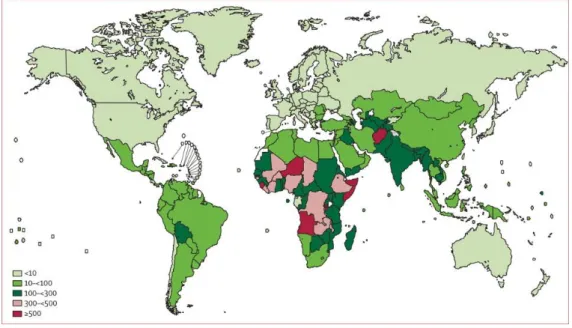
According to CDC, ∼1 million children below 5 years of age die yearly in pneumococcal infections (Figure 8) (82-84). The normal habitat for pneumococci is the nasopharynx, mostly of small children (85). Colonization precedes pneumococcal disease and colonized individuals serve as a reservoir for horizontal spread of the bacterium in the community (85).
22
Figure 7. Pathogenic route for S. pneumoniae infection. Organs infected through the airborne and haematogenic routes are depicted in blue and red, respectively (85).
Figure 8. Pneumococcal mortality rate. Pneumococcal deaths in children aged 1–59 months per 100000 children younger than 5 years (HIV-negative pneumococcal deaths only). Colours show different incidences (84)
23
2.3.1. Carriage
The main biological niche for S. pneumoniae is the upper respiratory tract of humans where it colonizes the mucosal surfaces lining the nasopharynx (86). Disease occurs when resident organisms from the upper respiratory tract gain access to normally sterile spaces in the middle ear, lung, or bloodstream and therefore colonization is the initial step in the pathogenesis of all pneumococcal disease (85, 87). The carriage rate is highest in young children, who most likely carry pneumococci in the nasopharynx at least one time and are the primary source for its spread within a community (86).
Before the wide use of conjugate vaccines four serogroups 6, 14, 19 and 23 were referred to as ‘pediatric serotypes’ and they were among the most commonly carried ones in children and at the same time caused the most cases of pneumococcal acute otitis media (88, 89).
Although conjugate vaccines against S. pneumoniae reduce carriage of the serotypes contained in the vaccine, there is little or no impact in most populations on the overall prevalence of pneumococcal carriage as other serotypes become more common (90, 91).
This process, known as serotype replacement is exemplified by increased carriage and disease prevalence caused by serotype 19A, observed in several populations following the introduction of seven-valent pneumococcal conjugate vaccine (PCV7) (91), (92).
2.3.1.1. The impact of carriage
Gray et al. investigated carriage and disease in 82 infants followed from birth up to 2 years of age. In this study they sampled the nasopharynx of children monthly for the first 6 months of life and at 2–3-month intervals thereafter until 2 years of age and collected information about any disease episodes experienced by the children. By correlating acquisition episodes and carriage periods of pneumococcus with culture- confirmed pneumococcal disease, they concluded that pneumococcal infection, most of which (28/31) was acute otitis media (AOM), is mainly associated with a newly acquired serotype. In particular, although prolonged duration of carriage was common, 74% of infections were caused by serotypes found less than a month before the illness.
Carriage itself was not found to pose risk for disease and the authors hypothesized the
24
role of prolonged carriage as one of the protective factors against pneumococcal disease (93).
McCool et al. performed an experimental pneumococcal colonization in humans. Many of the participants developed a mild rhinorrhea without other symptoms. Together, these observation provide suggest that initiation of pneumococcal colonization is an inflammatory rather than a quiescent process (94).
2.3.2. Diseases caused by Streptococcus pneumoniae
2.3.2.1. Otitis media
Otitis media (OM) is the most frequently reported paediatric bacterial infection, with approximately 80% of children experiencing an episode by the age of three years (95).
At the beginning of the 20th century, group A Streptococcus (GAS) was the most common pathogen leading to complications in AOM, but it is now rare in the Western world. A ‘new’ triad of AOM pathogens has emerged in the last century – S.
pneumoniae, non-encapsulated H. influenzae and Moraxella catarrhalis (96, 97) - with the ability to cause long-term hearing damage (98). AOM can be caused also by a number of different viruses (99).
Some pneumococcal serotypes (i.e., serotypes 3, 5, 1, 12F, 19F, 19A) seem to have a higher AOM disease potential once carriage is established, since in countries where the pneumococcal conjugate vaccines (PCV7, and later PCV13) is widely used, non- vaccine serotypes account for a more significant proportion of this disease. Among them an increase in serotypes 6C, 22F, 23B, 35B and nontypeables (NT) was observed after PCV13 vaccination (88, 100).
2.3.2.2. Eye infections
S. pneumoniae is found rarely in normal conjunctivae (0–0.3%) (101), but obstruction of the nasolacrimal duct predisposes to ascendant colonisation and infection of the lacrimal system and conjunctivae with the normal nasopharyngeal commensals including this bacterium (102). Therefore pneumococcus is typically among the top three most commonly isolated species from cases of bacterial keratitis, an infection of the cornea of the eye (103, 104). Pneumococcal keratitis can be a sight-threatening
25
infection if left untreated or if treatment is delayed. Corneal ulceration occurs during the course of the infection and often results in an opaque scarification of the corneal surface after the infection is cleared. The clinical picture of pneumococcal keratitis is a central yellowish or greyish white ulcer associated with infiltrates, folds in Descemet's membrane and hypopyon. Spread in an irregular fashion produces a ‘serpiginous ulcer’
(Ulcus serpens corneae) with the advancing edge of the ulcer being undermined (103).
In case of endophthalmitis S. pneumoniae is an infrequent causative agent, being isolated from 2.2–13.6% of cases (102, 105).
2.3.2.3. Pneumonia
S. pneumoniae remains the leading microbial aetiology of community acquired pneumonia (CAP) (106). It occurs most frequently in patients least well equipped to handle the effects of the disease, namely, the very young or the very old, the immunosuppressed or chronically ill. The development of pneumococcal pneumonia results from translocation of pneumococci from the nasopharynx to the lung through aspiration and possibly blood-borne dissemination (107) (Figure 7). In adult invasive and non-invasive pneumococcal CAP, a study from UK (between 2008 and 2010) showed that the most common serotypes implicated were 14, 1, 8, 3 and 19A (108).
According to CDC the use PCV7 since 2000 and PCV13 since 2010 among children in the United States has reduced pneumococcal infections directly and indirectly among children, and indirectly among adults. Approximately, 20%–25% of IPD cases and 10%
of community-acquired pneumonia cases in adults aged ≥65 years are caused by PCV13 serotypes and are potentially preventable with the use of PCV13 in this population (109).
A new French study from the post PCV13 era demonstrated 32% decrease in CAP cases in children younger than 2 years. These data suggest a strong impact of PCV13 on CAP, pleural effusion and documented pneumococcal pneumonia, particularly cases due to PCV13 serotypes (110).
26
2.3.2.4. Invasive pneumococcal diseases (meningitis, sepsis)
World Health Organization (WHO) estimates that approximately 1 million children die each year of invasive pneumococcal disease (IPD) such as meningitis and sepsis (84).
The seven most common serotypes causing IPD in the era prior to PCV introduction included 1, 5, 6A, 6B, 14, 19F and 23F (111). Serotype 14 was the most common serotype accounting for 12%–29% of IPD (111). After the introduction of PCV7 vaccine the rate of all IPD cases dropped by almost 70%, as it happened in the US (112). The highest incidence of IPD generally occurs among children aged 6–11 months, at the very same age when the incidence of pneumococcal acquisition is high (113). It is also shown that serotypes 3, 6B, 14, 23F can cause more severe meningeal inflammation than serotypes 1, 5, 9 and 7F (114). A recently published study from Israel conclude the effect of PCV7 and PCV13 vaccination on IPD (115). In total, a 63% reduction of all-serotype IPD episodes was observed. They found that the rate of IPD caused by serotype 1, 3, 5, 7F and 19A increased by 47% in children <5 years after PCV7 vaccination and after PCV13 vaccination an overall 70% reduction of IPD caused by these serotypes was observed. On the other hand the rate of IPD caused by non- PCV13 serotypes increased by 54% when comparing the PCV7 and the pre-PCV periods in children <5 years. Of all non-PCV13 disease, serotypes 12F, 15B/C and 33F were found to be the most common serotypes.
Other studies from Europe observed almost the same, that PCV13 introduction significantly decreased the cases of IPD, pneumonia and otitis media despite potential non-PCV13 serotype replacement (116, 117).
2.4. Prevention of pneumococcal diseases 2.4.1. History of vaccination
The first anti-pneumococcal serum appeared in the H.K. Mulford catalogue (where it remained into the 1940s) in 1895 (118). Early studies of antiserum therapy of pneumococcal infections came in the 1910s (6). The first specific record is about a whole-cell heat-treated pneumococcal vaccine (‘Pneumo-Bacterin’) licensed in the USA in 1909, with manufacturers such as H.K. Mulford Co., Eli Lilly & Company and Parke, Davis & Co (3). From 1942 to 1945, Heidelberger and MacLeod took advantage of the
27
preceding developments in polysaccharide technology to develop and test a pneumococcal polysaccharide vaccine at the US Army Air Force Technical School (119). With this finding it was proved that purified CPS can be used as active immunogens in adult humans and clinical efficacy against pneumonia was shown but the approach was abandoned with the availability of antibiotics. As the limitations of antibiotics therapy were realized, vaccination with pneumococcal CPS was revived in the 1960s through the efforts of Robert Austrian (120). A 14-type mixture of the most prevalent types was selected for vaccination in 1977 and increased to 23 types in 1983 (Table 2). This vaccine is licensed and recommended for adults >65 y and younger subjects with conditions, such as asplenia, that predispose to pneumococcal infection.
Pneumococcal polysaccharide vaccines are poorly immunogenic in children younger than 2 years of age, who are at high risk of invasive pneumococcal disease (121). To improve the immune response to the capsular polysaccharide in young children, third- generation vaccines in which capsular polysaccharides are conjugated to one of several different proteins were developed and tested (122). However, the technology limits the number of serotypes that can be included and infections caused by non-included types are a substantial problem in some populations (91).
28
Table 2. Pneumococcal vaccines distributed since 1977 (3)
MANUFACTURERS (TRADE NAMES)
VALENCE: SEROTYPES INCLUDED
DATE INTRODUCED, INITIAL REGION
PNEUMOCOCCAL POLYSACCHARIDE VACCINES
Merck Sharp & Dohme (MSD) (Pneumovax™) Lederle Laboratories (Pnu- Imune™) Institut Mérieux (Imovax
Pneumo 14™)
14-valent: 1, 2, 3, 4, 5, 6A, 7F, 8, 9N, 12F, 18C, 19F, 23F, 25F
November 1977, USA August 1979, USA February 1981, France
Smithkline Beecham (Moniarix™)
17-valent: 1, 2, 3, 4, 6A, 7F, 8, 9N, 11A, 12F, 14, 15F, 17F, 18C, 19F, 23F, 25
1980s, Europe
Merck Sharp & Dohme (MSD) (Pneumovax™ 23) Lederle Laboratories (Pnu-Imune™
23) Institut Mérieux (Pneumo 23™) Chengdu Institute Of
Biological Products
23-valent: 1, 2, 3, 4, 5, 6B, 7F, 8, 9N, 9V, 10A, 11A, 12F, 14, 15B, 17F, 18C, 19F, 19A, 20, 22F, 23F, 33F
USA July 1983, USA 1987, Europe 2005, China
PNEUMOCOCCAL CONJUGATE VACCINES
Wyeth Laboratories (Prevnar™ Or Prevenar™) Now Pfizer
7-valent: 4, 6B, 9V, 14, 18C, 19F, 23F
February 2000 for infants, USA and Europe
Glaxo Smith Kline (GSK) (Synflorix™)
10-valent: 1, 4, 5, 6B, 7F, 9V,
14, 18C, 19F, 23F March 2009 for infants: Europe
Pfizer (Prevenar™ 13) 13-valent: 1, 3, 4, 5, 6A, 6B, 7F, 9V, 14, 18C, 19A, 19F, 23F
February 2010 for infants, late 2011 for adults, Europe, Australia, USA
29
2.4.2. Available vaccines nowadays
2.4.2.1. Polysaccharide vaccine
Pneumovax®23 (PPV23, MSD) has been recommended since the mid-1980s for use in adults ≥65 years and in younger adults who have any risk conditions (asplenia or splenic dysfunction, various malignancies, immunosuppression, asthma etc.) (123). There is considerable debate regarding the efficacy of PPV23 in preventing non-invasive pneumococcal pneumonia, although the general belief is that there is evidence of protection against IPD, at least in healthy young adults and in the healthy older population (123). Its use in children is limited as polysaccharides are poorly immunogenic in infants and children under the age of 2 years (124).
2.4.2.2. Conjugate vaccines (PCV)
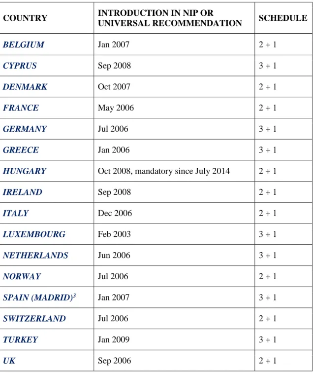
Lack of efficacy of PPV23 in neonates and infants <2 years of age led to the development of PCVs. While PPV23 elicits a T-cell independent, humoral immune response, covalent conjugation of capsular polysaccharides to a carrier protein activates a T-cell dependent antibody response in the setting of mucosal immunity and immunological memory (125) (126). Mucosal immunity mediates nasopharyngeal carriage, thus being associated with, among other factors, herd protection. This has been confirmed in many studies in which the use of childhood PCV was associated not only with a sustained decrease in IPD and declines in US hospitalisations for pneumonia in children, but also significant decreases in IPD in older adults (125) (127). Prevenar-7 (PCV7) contains capsular polysaccharides from seven S. pneumoniae serotypes conjugated to CRM197, a non-toxic mutant of diphtheria toxin. When PCV7 was licensed, those serotypes (4, 6B, 9 V, 14, 18C, 19 F and 23 F) caused the majority of invasive pneumococcal infections in the US (128) and were also associated with antibiotic resistance (129). PCV7 was included in the National Immunization Programmes (NIP), or was recommended for routine vaccination, in a number of European countries, between 2006 and 2008 (Table 3) (87).
30
Table 3. PCV7 adoption in NIP or universal vaccination recommended by health authorities in European countries (87)
1 Limited recommendation in 2003 with 3 + 1 administration schedule
2. PCV7 was recommended regionally prior to the date of inclusion in the NIP
3. PCV vaccination recommendation is regional
COUNTRY INTRODUCTION IN NIP OR
UNIVERSAL RECOMMENDATION SCHEDULE
BELGIUM Jan 2007 2 + 1
CYPRUS Sep 2008 3 + 1
DENMARK Oct 2007 2 + 1
FRANCE May 2006 2 + 1
GERMANY Jul 2006 3 + 1
GREECE Jan 2006 3 + 1
HUNGARY Oct 2008, mandatory since July 2014 2 + 1
IRELAND Sep 2008 2 + 1
ITALY Dec 2006 2 + 1
LUXEMBOURG Feb 2003 3 + 1
NETHERLANDS Jun 2006 3 + 1
NORWAY Jul 2006 2 + 1
SPAIN (MADRID)3 Jan 2007 3 + 1
SWITZERLAND Jul 2006 2 + 1
TURKEY Jan 2009 3 + 1
UK Sep 2006 2 + 1
31
PCVs are effective not only in small children, but also in elderly. CAPiTA study (Community-Acquired Pneumonia Immunization Trial in Adults) shows that adults 65 years and older who received PCV13 had 45.56% fewer first episodes of vaccine-type CAP, 45% fewer episodes of non-bacteraemic vaccine-type CAP, and 75% fewer episodes of vaccine-type IPD compared to the placebo group (http://www.empr.com/detailed-data-from-prevnar-13-capita-study-
released/article/337871/). Therefore vaccination in this group is also suggested.
Some emerging serotypes are associated with increased resistance to antibiotics and higher propensity for invasive disease, especially serotype 19A. Additionally, the efficacy of PCV7 in preventing pneumonia or mucosal disease such as AOM is less pronounced than its ability to reduce bacteremic disease. To address these issues, several expanded valency (so-called 2nd generation, Table 4) PCVs have been licensed (13-valent Prevenar®, Pfizer; 10-valent with 3 different protein carriers Synflorix™, GSK Biologicals), or are currently under investigation (15-valent PCV, Merck). The expanded serotype coverage of these vaccines may further reduce IPD rates in the US and Western Europe, but the complexity and cost of these vaccines will undoubtedly continue to rise.
Table 4. Pneumococcal serotypes included in the licensed pneumococcal conjugate vaccines (87)
VACCINE MANUFACTURER SEROTYPES INDICATED
AGE
PCV7 (PREVENAR) Pfizer 4, 6B, 9V, 14, 18C,19F, 23F < 5 years
>60 years PCV10 (SYNFLORIX) GSK 4, 6B, 9V, 14, 18C,19F, 23F,
1, 5, 7F,
< 5 years
PCV13 (PREVENAR 13)
Pfizer 4, 6B, 9V, 14, 18C,19F, 23F, 1, 3, 5, 6A, 7F, 19A
< 5 years
>50 years
32
2.4.2.3. New perspectives in vaccination 2.4.2.3.1. Protein-based pneumococcal vaccines
Polyhistidine triad protein D (PhtD) has been described as a promising vaccine candidate for use against pneumococcal infections (130). These surface-exposed antigens are characterised by the presence of five to six histidine triad motifs and are highly conserved amongst pneumococcal strains and could elicit protective immunity in mouse models of pneumococcal disease against a number of pneumococcal strains (131).
Other strategy involves the use of one or more pneumococcal protein antigens common to all serotypes, to provide cheap, non-serotype-dependent protection. In one study, the protective efficacy of immunization of mice was evaluated with PdB (a pneumolysin toxoid), PspA, PspC (CbpA), PhtB and PhtE in an invasive-disease model (132).
Based on several in vitro assays Giefing et al. preselected 18 novel candidates for animal studies and 4 of them showed significant protection against lethal sepsis. Two vaccine candidates, protein, required for cell wall separation of group B streptococcus (PcsB) and serine/threonine protein kinase (StkP), were found to be exceptionally conserved among clinical isolates. Therefore a vaccine containing PcsB and StkP is intended for the prevention of infections caused by all serotypes of pneumococcus in the elderly and in children (133).
Alexander et al. immunized mice with a genetically engineered toxoid version of pneumolysin, which was derived from a serotype 2. Immunized mice had significantly increased levels of anti-pneumolysin antibodies. And they have shown protection against nine tested pneumococcal serotypes too (134).
2.4.2.3.2. Whole cell vaccine
Malley and Anderson investigated killed cells of a noncapsulated strain, which expose many such common antigens. Given to mice intranasally, this vaccine elicits antibody- independent, CD4+ T lymphocyte-dependent accelerated clearance of pneumococci of various serotypes from the nasopharynx mediated by the cytokine IL-17A. Given by injection, the killed cell vaccine induces bifunctional immunity: plasma antibodies
33
protective against fatal pneumonia challenge, as well as IL-17A–mediated nasopharyngeal clearance (135).
Recent study shows, that live and heat-killed Lactobacillus casei enhanced the antigen- specific immune response when administered nasally conjugated with a pneumococcal antigen (pneumococcal protective protein A: PppA). This type of vaccines can be a safe and effective strategy for the prevention of pneumococcal infections and opens new possibilities of application of dead lactobacilli as adjuvants in vaccine formulations against other pathogens (136). It is also proved that this vaccine is safety in liver, kidney, pulmonary and systemic levels (137).
In a study by Lu et al. whole-cell pneumococcal vaccine was examined as an injectable vaccine using the currently approved aluminum adjuvant. A pneumococcal strain was engineered to be capsule negative, autolysin negative and to express a nontoxic mutant pneumolysoid. The adjuvant Al(OH)3 strongly increased immune responses in mice injected with the chemically inactivated cells. The immunized mice were protected against nasal colonization and sepsis with different pneumococcal serotypes (138).
Roche et al. constructed live attenuated mutants of S. pneumoniae containing deletions in genes encoding three of major virulence determinants including capsular polysaccharides, pneumolysin and pspA. The attenuated strains were not able to cause disease but retained the ability to colonize the upper respiratory tract. Nasal colonization by live attenuated vaccine strains was used to immunize mice. A single intranasal administration of live attenuated vaccine without adjuvant induced both systemic and mucosal protection from intranasal challenge with a high dose of the parent strain.
Colonization by live attenuated S. pneumoniae is an effective vaccine strategy that may offer broad protection against pneumococci (139).
2.4.2.3.3. DNA vaccines
DNA vaccines are easy to manufacture, have a low cost and are stable in transportation.
These properties make DNA vaccines ideal for implementation in developing countries.
Vectors encoding the complete N-terminal regions of PspAs elicited humoral responses in mice and cross-reactivity was mainly restricted to the same family (140).
Nasal delivery of naked plasmid DNA induces only weak immune responses, which may be due to the degradation of naked DNA at mucosal surfaces (141). Therefore, for
34
construction of effective DNA vaccines, an appropriate DNA delivery system is demanded. Chitosan is a natural biodegradable polysaccharide derived from chitin that possesses biocompatibility and mucoadhesion properties. Mice were immunized intranasally with chitosan-DNA nanoparticles expressing PsaA (chitosan-psaA).
Compared to levels in control mice immunized with naked DNA containing psaA gene or chitosan-backbone vector without psaA gene, systemic and mucosal immune responses against PsaA were elevated in mice immunized with chitosan-psaA. In addition, fewer pneumococci were recovered from the nasopharynx of mice vaccinated with chitosan-psaA than for the control groups following intranasal pneumococcal challenge. These findings suggested that chitosan-DNA nanoparticles expressing pneumococcal major antigens could be developed to prevent pneumococcal infections (142).
2.5. Antibiotic treatment
The discovery of penicillin by Alexander Fleming initiated a success story of antimicrobial compounds unmatched by any other antibiotic. This β-lactam antibiotic has long been the mainstay against pneumococcal infections, but its efficacy is threatened by the rapid dissemination of penicillin-nonsusceptible clones worldwide (4, 143).
2.5.1. Antimicrobial resistance of Streptococcus pneumoniae
2.5.1.1. Development of resistance
The emergence of penicillin-resistant and multidrug-resistant pneumococcal strains has become a global concern. Since 1960s, penicillin-resistant strains have been found in various parts of the world with increasing frequency. Resistance to non-β-lactam antibiotics such as chloramphenicol, tetracycline, erythromycin, clindamycin, rifampin and trimethoprim-sulfamethoxazole has been reported (9, 144). Penicillin-resistant and multidrug-resistant pneumococci are known to be restricted worldwide to a few serogroups, namely 23, 6, 19, 9 and serotype 14, which were particularly associated with carriage and disease in children in the pre-vaccination era (9, 145).
35
Molecular studies have shown that the penicillin-resistant and multidrug-resistant pneumococcal populations are highly dynamic and that resistance is a combination of the spread of resistant clones, the acquisition and loss of resistance genes within those clonal lineages and the spread of resistance genes to new lineages (73).
2.5.1.2. Resistance mechanism 2.5.1.2.1. β-lactam resistance
Resistance to β-lactam antibiotics in clinical isolates of S. pneumoniae is mediated by mosaic genes encoding altered penicillin-binding proteins (PBPs; a family of enzymes involved in peptidoglycan metabolism) with lower antibiotic binding affinities than their native versions (146, 147). While S. pneumoniae contains six PBPs, variants of PBP2x, PBP2b and PBP1a are considered the most relevant for resistance and the acquisition of low-affinity PBP2x and PBP2b variants is a necessary first step for the acquisition of PBP1a variants that confer high-level resistance to β-lactams (148).
Pneumococci have a dedicated system for the acquisition of exogenous DNA from the environment and the mosaic gene structure of low-affinity PBPs is the result of interspecies gene transfer events involving closely related streptococcal species (149).
The presence of other, non-PBP contributors has also been reported. For example, the cell wall of penicillin-nonsusceptible isolates is often highly enriched in branched-chain muropeptides, a phenomenon that has been linked to mosaic alleles of the murM gene (150, 151). Furthermore, mutations in a peptidoglycan N-acetylglucosamine (GlcNAc) deacetylase (152), a peptidoglycan O-acetyltransferase (153), a putative glycosyltransferase (154), a serine threonine kinase (155), a histidine protein kinase that is part of a two-component signal-transducing system (156) and a phosphate ABC transporter (157) have all been implicated in resistance to β-lactams. Finally, the selection of a nonsense mutation in a putative iron permease in penicillin-resistant S.
pneumoniae has recently been shown to decrease susceptibility to bactericidal antibiotics, including penicillin (158).
36
2.5.1.2.2. Macrolide resistance
Resistance to erythromycin in S. pneumoniae was first detected in 1967 in the United States and subsequently worldwide (9, 159, 160). Macrolide resistance is mediated by two main mechanisms: target modification due to a ribosomal methylase encoded by the erm (erythromycin ribosome methylase) gene, which confers high-level resistance to macrolides, lincosamides and streptogramin B (MLSB types) usually carried by transposable elements (160) and the second one the efflux transport system associated with the mef gene (160).
The widely predominant erm gene is ermB gene although this is not the only representative of the erm gene class in pneumococci. The presence of an ermA gene has been reported in some strains where it conferred cross-resistance to erythromycin and clindamycin (161).
Resistance with the help of efflux pump appears expressed at moderate levels, with erythromycin MICs of between 1 and 64 μg/ml (generally between 8 and 32 μg/ml).
Because the 16-membered macrolides, the lincosamides and the streptograminB antibiotics are not substrates of the pump, these antimicrobial agents remain active, even after induction with erythromycin. Resistance to erythromycin combined with susceptibility to clindamycin, whether the cells are induced or not induced by erythromycin, defines the M resistance phenotype. The mef gene is also transferable among pneumococci (162). Of the two variants of the mef gene, mefA was originally found in Streptococcus pyogenes (163) and mefE was originally found in S.
pneumoniae (164). MefA and mefE are 90% identical at the nucleotide level.
2.5.1.2.3. Resistance to other antibiotics
The genetic basis of sulfonamide resistance in S. pneumoniae was demonstrated to be 3- or 6-bp duplications within sulA, the chromosomal gene encoding dihydropteroate synthase (165).
The tetracycline resistance is a result of the acquisition of one of the two genes, tetM and tetO, both of which encode ribosome protection proteins (166, 167).
Pneumococcal resistance to erythromycin and tetracycline is frequently associated with