METHICILLIN-RESISTANT STAPHYLOCOCCUS AUREUS COLONIZATION IN HIV PATIENTS OF
ARBA MINCH PROVINCE, ETHIOPIA:
CARRIAGE RATES, ANTIBIOTIC RESISTANCE, AND BIOFILM FORMATION
ASEERMANILAL1*, MISGUNSHEWANGIZAW2, MOHAMMEDAMAN MAMA3, TIGISTGEZMU1and BEHAILU MERDEKIOS2
1Department of Medical Laboratory Science, College of Medicine and Health Sciences, Arba Minch University, Arba Minch, Ethiopia
2Department of Public Health, College of Medicine and Health Sciences, Arba Minch University, Arba Minch, Ethiopia
3Department of Medical Laboratory Science, Madda Walabu University Goba Referral Hospital, Bale-Goba, Ethiopia
(Received: 19 January 2019; accepted: 6 March 2019)
Methicillin-resistantStaphylococcus aureus(MRSA) has emerged as a signifi- cant opportunistic pathogen among human immunodeficiency virus (HIV) patients of Ethiopia. This study aimed at delineating the prevalence, antimicrobial resistance, and biofilm-forming potentials of nasally colonized MRSA among HIV patients in the Arba Minch province of Ethiopia. A cross-sectional study was performed in HIV patients who visit anti-retroviral therapy clinic of the Arba Minch Hospital between February and April 2017. Nasal samples were collected and inspected forStaphylo- coccusfollowing standard procedures. MRSA was identified using cefoxitin disk and antibiotics sensitivity test was performed as per Kirby–Baur disk diffusion method.
The formation of biofilm was inspected using both qualitative and quantitative methods. A total of 307 HIV patients were examined. The overall prevalence of S. aureuswas found to be 39.7%. The prevalence of MRSA was 20.8%. The rate of nasal colonization of MRSA was relatively higher among females. In bivariate analysis, MRSA colonization was statistically significant in patients with CD4count
≤350 (pvalue=0.002) and co-trimoxazole prophylaxis (pvalue=0.003). Concomi- tant resistance to erythromycin, tetracycline, and co-trimoxazole were 48.4%, 45.3%, and 39.0%, respectively. Invariably, all MRSA isolates were 100% sensitive to vancomycin. Of the 64 MRSA isolates, 18.7% were considered as multidrug-resistant.
The rate of biofilm formation was 34.3%. The results revealed a high prevalence rate in the nasal colonization of MRSA in HIV patients.
*Corresponding author; E-mail:aseermanilal@gmail.com
Keywords: methicillin-resistant Staphylococcus aureus, HIV patients, nasal colonization, biofilm formation
Introduction
Methicillin-resistantStaphylococcus aureus(MRSA) is a superbug resistant to multiple antibiotics such as methicillin, oxacillin, penicillin, and amoxicillin owing to its genetic plasticity [1]. It is recognized as one of the most resistant bacteria that has riveted intense scientific and political interest in global magnitude due to the limited choice of antibiotics for its effective treatment [2]. Recently, WHO has enlisted MRSA as a high priority pathogen that immediately require new class of antibiotics. It is a formidable pathogen capable of deploying a battery of virulence factors to inflict serious life-threatening diseases in the healthcare settings and also in the community [3].
MRSA has been implicated as an opportunistic pathogen in patients with HIV infection [4]. Epidemiological studies proved that HIV patients have 6 to 18-fold higher risk of acquiring MRSA infections as compared to the general population [5].
It is evident that MRSA is responsible for causing invasive and often fatal infections in HIV patients since the time of its emergence [4]. It has been evidenced from an erstwhile study that nasal colonization with bothS. aureusand MRSA is relatively common among HIV patients and appears to be associated with an increased risk of subsequent MRSA infections by the same colonizing strain [6]. Several prior studies from various regions of the world showed a diverse range in the prevalence of nasal colonization ofS. aureusand MRSA among HIV patients [7–9]. Moreover, strains of MRSA are attaining high resistance to the existing antibiotics, thereby under- scoring an immediate need to develop novel antibiotics.
Recent estimates of WHO showed that the prevalence of HIV among the Ethiopian adult population is∼2.4% [10] and infection is slowly increasing in the past few years [11]. Nevertheless, nationally, there is only limited number of studies on the prevalence of MRSA colonization among diverse population of HIV-infected patients [12,13]. Moreover, in the Arba Minch province of Southern Ethiopia, no such studies have been reported hitherto. It is suggested that an accurate susceptibility testing and screening of patients for MRSA colonization are important tools in limiting the propensity of the spread of this organism. For that, regional studies to elucidate the prevalence and pattern of antibiotic sensitivity are pivotal. For this reason, this study is initiated to determine the prevalence, associated factors, antimicrobial resistance profile, and biofilm-forming potency of nasally colonized MRSA among HIV patients attending antiretroviral drug therapy clinic at the Arba Minch General Hospital, Arba Minch, Ethiopia.
Materials and Methods Study area, design, period, and study population
This study was carried out at the Arba Minch Hospital, Arba Minch province and situated 505 km southwest of Addis Ababa, Ethiopia. The study protocol was ethically approved by review board of Arba Minch University, College of Medicine and Health Sciences. Informed verbal consent was obtained from each study participant. A cross-sectional study was conducted to assess the prevalence and associated factors of nasally colonized MRSA among HIV-infected patients who attended the antiretroviral therapy (ART) clinic of the Arba Minch Hospital between February and April 2017. The inclusion criteria for the study were all HIV-positive patients over the age of 18 years who were routinely receiving therapeutic and medical care at ART clinic and volunteered to participate in the study. The exclusion criteria for the study were (1) patients who were severely sick and (2) patients who underwent recent (4 weeks prior) nasal decolonization with antibiotics.
Sample size determination and sampling technique
The sample size was computed using a sample size determination formula for the estimation of single population proportion. It was ascertained by adopting the prevalence of MRSA in HIV patients as 13.6% from a study conducted once [7].
After considering 95% of confidence interval (z=1.96) and 4% of marginal error (d=0.04), the initial sample size was found to be 284 and then by calculating a 10%
(∼28 subjects) of non-response rate, thefinal sample size was consolidated as 310.
Systematic random sampling technique was opted to recruit the study units.
Data and specimen collection
Data and specimen collections were conducted with the help of two well- trained nurses working in the ART clinic of Arba Minch Hospital. Prior to the data collection, purpose of the study was explained to all participants and consents were obtained. In order to analyze the risk factors of the nasal colonization of MRSA, data pertaining to sociodemographic characteristics and clinical history of the patients were retrieved from the patients’registration book. Nasal samples were aseptically collected from both the anterior nares using sterile cotton swabs wetted with normal saline (0.85% NaCl). The samples were procured by inserting the swab 2 cm into the nasal vestibule and circulating it six times against the anterior nasal mucosa.
Afterward, swabs were placed in sterile-labeled sleeves and transported at room temperature and stored at 5 °C until processing on the same day.
Culture and isolation of S. aureus
The isolation and identification ofS. aureuswas carried out at the Microbi- ology and Parasitology Laboratory, Department of Medical Laboratory Science, College of Medicine and Health Sciences, Arba Minch University, Arba Minch. All the samples were processed immediately to avoid contamination. Briefly, each sample was directly inoculated onto Mannitol Salt Agar (Oxiod, Hampshire, UK).
The inoculated plates were incubated face up for 24 h at 35–37 °C. After incubation, yellowish colonies from the plates were subcultured to nutrient agar (Oxiod). The pure cultures of bacterial isolates were subsequently subjected to species identification and confirmation. Biochemical (positive catalase and coagu- lase test), morphological, and physiological characteristics of isolated bacteria were ascertained by adopting standard laboratory methods including Gram staining, colonial morphology on different media, and growth on selective media as described elsewhere [14]. Corresponding American Type Culture Collection strains were utilized as reference standards to validate the biochemical identification of S. aureus.
Identification of MRSA
MRSA identification test was performed in accordance with the criteria of Clinical Laboratory Standard Institute (CLSI) [15] using cefoxitin disk diffusion assay. Bacterial suspension (5 ml) of 0.5 McFarland (1×10⁸ CFU/ml) was prepared from eachS. aureus isolate and swabbed on to the surface of Muller- Hinton agar (Hi-media, India). After incubation for 24 h at 35 °C, zone of inhibitions was measured. The strains showing zone of inhibition≤21 mm were extrapolated as MRSA.
Antimicrobial susceptibility testing
Antibiotic susceptibility profile of all MRSA isolates was determined by Kirby–Bauer disk diffusion technique according to the criteria set by CLSI using Hi-media antibiotic disks. For the assay, inoculums were prepared by picking parts of similar test organism with a sterile wire loop and suspended in sterile normal saline. The density of suspension to be inoculated was determined by comparing
with opacity standard on McFarland 0.5 barium sulfate solution. The test organisms were uniformly swabbed over the Mueller–Hinton agar (Hi-Media, Mumbai) surface and exposed to a concentration gradient of antibiotic diffusion from antibiotic-impregnated paper disk into the agar medium, and then incubated face up at 37 °C for 24–48 h. Diameters of the zones of inhibition around the disks were measured to the nearest millimeter using a ruler and categorized as sensitive, intermediate, and resistant according to the standardized table described in CLSI [15]. S. aureus showing resistance to three classes of antibiotics was considered as multidrug resistant (MDR). The following antibiotic disks in respective concentrations (in μg/ml) [tetracycline (30 μg), doxycycline (30 μg), gentamicin (10 μg), erythromycin (15 μg), ciprofloxacin (5 μg), clindamycin (30μg), chloramphenicol (30μg), amikacin (10μg), co-trimoxazole (25μg), and vancomycin (30μg)] were used to determine antibiogram.
Assay of biofilm formation
Qualitative and quantitative assay of biofilm-forming capacity of all MRSA isolates was carried out as per the methodology described in our previous study with suitable modifications [16]. Briefly, the MRSA isolates (final concentration of 1×10⁶ colony forming units) were inoculated in culture tubes containing trypticase soy broth (supplemented with 3.0% NaCl and 0.5% dextrose) and incubated overnight at 37 °C. Culture tubes containing only trypticase soy broth were used as control. Biofilm-producing MRSA were identified visually based on the appearance of slime formation on the air–liquid interface and the bottom of culture tubes. To evaluate the biofilm production, the culture tubes adhered with dense matt of biofilm were stained using 0.1% crystal violet solution (w/v).
A positive result was considered as the visible presence of crystal violet-stained biofilm matrix adhered to the inner wall of the culture tubes by direct observation.
The positive results were noted after comparing with the negative control (uninoculated culture tubes). For the quantification of adhered biofilm matrix, the same experiment was replicated in sterile polystyrene 96-well flat bottom microtiter plates. Each well was inoculated with 200μl of fresh overnight culture of MRSA isolates. The plate was covered with lid and incubated at 37 °C for 24 h.
After incubation, wells were emptied by micropipette aspiration and gently rinsed thrice with sterile phosphate-buffered saline (PBS) in order to remove non- adherent cells. The adhered biofilm-forming isolates were subsequently stained with 0.1% crystal violet. Excess stain was removed by rinsing thrice with sterile PBS and left for drying. Furthermore, the biofilm formations were quantified by resolubilization of the wells containing crystal violet stain in 200 μl of 95%
ethanol. The optical density was determined at 595 nm in a microplate reader (MR 9602). The uninoculated wells containing broth alone were considered as the blank control. In this study, MRSA isolates with optical density greater than that of the negative control were considered positive for biofilm production. According to the degree of biofilm formation, isolates were ranked in to three groups: weakly or non-adherent, optical density ≤0.111; moderately adherent, optical density
≥0.111 or equal to or <0.222; strongly adherent, optical density >0.222 [16].
The whole tests were carried out in triplicates and the data are expressed as mean±standard deviation in order to validate the findings statistically.
Statistical analysis
The data were analyzed using Statistical Package for Social Services (Windows, version 20, Chicago, IL, USA). The odds ratio, 95% confidence intervals, and p value were calculated using this software. Descriptive statistics was conducted. Logistic regression analyses were used to examine the association among different variables and the outcome variable. The pvalue of ≤0.05 was considered statistically significant.
Results Sociodemographic and clinical data
A total of 307 HIV-infected patients were included in this study with the response rate of 99.03%. The majority of the participants were females 176 (57.3%). The age of participants ranged between 18 and 70 years with a median age of 39.8 years. During the study period, the majority of the patients (90.2%) was receiving highly active antiretroviral drug treatment for more than 1 year. The mean CD4 count was 591.01 cells/mm and 59 (19.2%) patients were under co-trimoxazole prophylaxis.
Prevalence ofS. aureusand MRSA
During the study period, totally 122 (39.7%) isolates of S. aureus were retrieved from the 307 HIV-positive patients. Among the 122S. aureus, 64 isolates with zone of inhibition≤21 mm in cefoxitin disk diffusion assay were identified as MRSA. Therefore, the prevalence of MRSA amongS. aureus-positive HIV patients was calculated as 52.45% (64/122) and the overall prevalence of MRSA
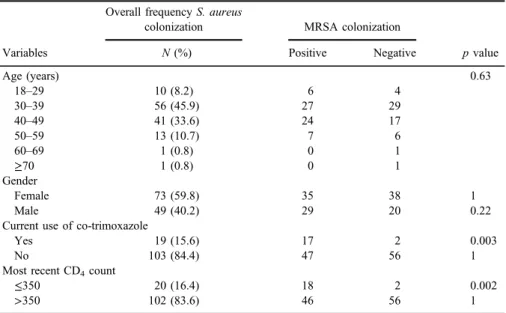
among HIV patients was detected as 20.8% (64/307). The rate of nasal colonization was relatively higher among females 35 (54.6%). Regarding the age, the highest number of MRSA was observed in the age group of 30–39 (27; 42.1%), followed by 40–49 (24; 37.5%), 50–59 (7; 10.9%), 18-29 (6; 9.3%), and≥60 (1; 1.5%). It was found that 17 (26.6%) of the MRSA-positive patients were currently under co-trimoxazole prophylaxis and the most recent CD4count of 18 (28.1%) patients were≤350. Details of sociodemographic and clinical data of the participants are shown in Table I.
Risk factors for nasal colonization of MRSA
Different risk factors were analyzed for a possible association with nasal colonization of MRSA among HIV-infected patients. It was observed that patients in the age group of 30–39 years were more colonized by MRSA (Table I). In addition, MRSA colonization rate was slightly higher among females (35; 54.8%).
No significant association of MRSA colonization with gender (pvalue=0.63) of the patient was noted in this study (Table I). In bivariate analysis, MRSA colonization was statistically significant in patients with CD4 count ≤350 (pvalue=0.002) and co-trimoxazole prophylaxis (pvalue=0.003). Risk factors
Table I.Details of age, gender, antibiotic usage, and the recent CD4count of theS. aureus-positive study participants
Variables
Overall frequencyS. aureus
colonization MRSA colonization
N(%) Positive Negative pvalue
Age (years) 0.63
18–29 10 (8.2) 6 4
30–39 56 (45.9) 27 29
40–49 41 (33.6) 24 17
50–59 13 (10.7) 7 6
60–69 1 (0.8) 0 1
≥70 1 (0.8) 0 1
Gender
Female 73 (59.8) 35 38 1
Male 49 (40.2) 29 20 0.22
Current use of co-trimoxazole
Yes 19 (15.6) 17 2 0.003
No 103 (84.4) 47 56 1
Most recent CD4count
≤350 20 (16.4) 18 2 0.002
>350 102 (83.6) 46 56 1
Note:MRSA: methicillin-resistantStaphylococcus aureus.
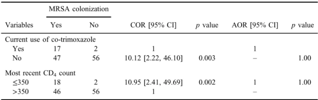
involved in the colonization of MRSA with statistical significance in bivariate analysis were further analyzed using the method of multivariate analysis and were found to be devoid of any prominent association among themselves (TableII).
Antimicrobial susceptibility pattern of MRSA
Antibiotic susceptibility profile of all MRSA isolates was confirmed using 10 antibiotics. The results revealed that all the MRSA isolates were susceptible to the antibiotics tested to a varied extent. Moderate degree of resistance was manifested against four antibiotics. Isolates that are resistant to erythromycin were 48.4%, followed by tetracycline (45.2%), co-trimoxazole (39.0%), doxycy- cline (32.8%), and gentamicin (23.4%). However, a lower degree of resistance was displayed against ciprofloxacin (9.3%), chloramphenicol (9.3%), clindamycin (3.1%), and amikacin (9.3%). Invariably, all MRSA isolates were 100% sensitive to vancomycin (TableIII). The multidrug resistance in this study was extrapolated as resistance to three or more groups of the 10 antibiotics tested. A notable result obtained from this study is that 18.75% (n=12) of the MRSA isolates could be considered as MDR (TableIV). All the MRSA strains were further taken up for elucidating the biofilm-forming potential.
Biofilm-forming potential of MRSA
In this study, 22 MRSA isolates were found to be positive for biofilm formation. The rate of biofilm formation in qualitative tube assay was 34.3%. The results of the quantitative analysis revealed that the biofilm formation varied
Table II.Analysis of risk factors for nasal colonization of MRSA in HIV-positive individuals attending ART clinic
Variables
MRSA colonization
COR [95% CI] pvalue AOR [95% CI] pvalue
Yes No
Current use of co-trimoxazole
Yes 17 2 1 1
No 47 56 10.12 [2.22, 46.10] 0.003 – 1.00
Most recent CD4count
≤350 18 2 10.95 [2.41, 49.69] 0.002 1 1.00
>350 46 56 1 –
Note:MRSA: methicillin-resistantStaphylococcus aureus; CI: confidence interval; ART: antiretroviral therapy; COR: crude odds ratio; AOR: adjusted odds ratio.
among the isolates. It was found that 7 of the isolates exhibited strong biofilm- forming potential and 11 of the isolates showed moderate potency, whereas the rest of them were weak biofilm producers. Among the biofilm-forming MRSA isolates, 54.5% of them were MDR. In this study, all the MDR isolates showed biofilm formation in varied degrees. The statistical analysis of biofilm-forming MRSA and MDR isolates showed that the MDR isolates were significantly associated with the formation of biofilm (p≤0.05).
Table III.Antibacterial susceptibility profile of MRSA isolates against 10 antibiotics
Antibiotics
Susceptibility pattern (n=64)
R I S
Erythromycin 31 (48.4%) 14 (21.8%) 19 (29.6%)
Gentamicin 15 (23.4%) 10 (15.6%) 39 (60.9%)
Tetracycline 29 (45.3%) 10 (15.6%) 25 (39.0%)
Doxycycline 21 (32.8%) 4 (6.2%) 39 (60.9%)
Ciprofloxacin 6 (9.3%) 4 (6.2%) 54 (84.3%)
Clindamycin 2 (3.1%) 2 (3.1%) 60 (93.7%)
Co-trimoxazole 25 (39.0%) 19 (29.6%) 20 (31.2%)
Chloramphenicol 6 (9.3%) 2 (3.1%) 56 (87.5%)
Amikacin 6 (9.3%) 0 58 (90.6%)
Vancomycin 0 0 64 (100%)
Note:MRSA: methicillin-resistantStaphylococcus aureus; S: susceptible; R: resistant; I: intermediate.
Table IV.Multidrug co-resistant profile of MRSA isolates
Antibiotics Resistant strains (n)
E, TET, and COT 1
E, TET, and GEN 1
E, TET, GEN, and COT 1
E, TET, DOX, and GEN 2
E, TET, DOX, and CIP 1
E, TET, DOX, and COT 1
E, TET, COT, and CIP 1
E, TET, DOX, COT, and CH 1
E, TET, DOX, COT, CLI, GEN, and AM 1
E, TET, DOX, COT, GEN, CH, and AM 1
E, TET, DOX, COT, GEN, CLI, CH, and AM 1
Total 12
Note: MRSA: methicillin-resistant Staphylococcus aureus; E: erythromycin; TET: tetracycline; DOX:
doxycycline; COT: co-trimoxazole; GEN: gentamicin; CIP: ciprofloxacin; CLI: clindamycin; CH:
chloramphenicol; AM: amikacin.
Discussion
The findings from this study provides a baseline data pertaining to the prevalence, risk factors, antimicrobial susceptibility profiles, and biofilm-forming potentials of nasally colonized MRSA among HIV-infected patients in the Arba Minch province of Ethiopia. In this study, totally 307 patients were included.
It was noticed that females in the age group of 30–39 years were found to be more affected and this was in accordance with an earlier study conducted at another prefecture of Ethiopia [12]. The reason for predominance of female patients could be due to high prevalence of HIV among them. Accordingly, more females consult the ART clinic than males. It has been previously reported that sociodemographic factors could cause higher MRSA colonization rates among HIV patients [4].
The prevalence rate in this study was higher than that reported in Ethiopia (37.3%), India (36.11%), and South Africa (36.3%) [17–19]. Moreover, the results of prior meta-analysis study showed that the pooled prevalence of MRSA among symptomatic study population in Ethiopia was 32.5%, corresponding to diverse clinical specimens [20]. For instance, studies reported from Bahirdar, Debre Markos, Harar, and Jimma showed prevalence rates of MRSA as 54.9% (in and out patients), 49.7% (patients with surgical site infection), and 49% (in patients), respectively [21–23]. Furthermore, nasal carriage rates of MRSA in this study are compared with various studies in general population of Ethiopia and found to be in range between 5.2% and 48%. It has been reported that nasal carriage rates of MRSA in food handlers, school children, janitors, medical students, healthcare workers, and prisoners were 5.2%, 9.8%, 34.7%, 37.8%, 44.1%, and 48%, respectively [16,24–28]. On the whole, the estimates of WHO revealed that the combined prevalence of MRSA in diverse population of African region is between 12% and 80% [29]. However, the prevalence rate found in this study is considerably higher than that observed in certain other region of Ethiopia (16.8%) and also Brazil (21.8%) in HIV patients [13, 30]. Moreover, a recent meta-analysis conducted on the combined prevalence of MRSA among HIV patients from different regions of the world is found to be much lower (6.9%) than ourfindings [31]. The possible reason for the high prevalence rate observed in this study can be attributed to the routine visits of HIV patients to the hospital and healthcare facilities. Due to its easy mode of transmission, a majority of HIV patients carry MRSA unknowingly and asymptomatically; hence, they too serve as silent reservoirs. It is corroborated that MRSA is endemic in most hospitals and healthcare facilities and is a major risk factor leading to the colonization [4]. Since the majority of MRSA infections is acquired from hospitals or healthcare setups, vigilant screening of incoming patients for MRSA as well as precautions with
MRSA-positive patients is needed to prevent the risk of self-infection and for attaining a reduced transmission rate.
In contrast with our results, the rate of prevalence reported from USA was comparatively higher (82%) in pediatric HIV patients [32]. The discrepancy in the result of prevalence rate of MRSA may be attributed to the sociodemographic variations in the study population, sample size, and laboratory techniques.
Regarding the risk factors, relationship between the low CD4 count and nasal colonization of MRSA was not significant in this study and is consistent with the previous study suggesting that the colonization of MRSA in HIV patients might be independent of CD4T-lymphocyte counts [33].
Prompt detection of MRSA and elucidation of its susceptibility profile have a pivotal role from a treatment point of view due to limited therapeutic options.
Published literature pertaining to the antibiogram of MRSA obtained from HIV patients in Ethiopia is scanty. In this study, totally 10 commonly used antibiotics were selected to inspect the resistance profile of MRSA. A varied level of antibiotic resistance was detected in all the MRSA isolates. Higher extent of resistance was particularly exhibited against four antibiotics, such as erythro- mycin, tetracycline, co-trimoxazole, and doxycycline, which are in line with the prior studies conducted in Ethiopia [12, 13]. The higher rate of resistance exhibited by MRSA could be attributed to the presence of genes conferring resistance. Multidrug resistance of MRSA is currently considered as a global threat by the WHO. The results of this study showed that the emergence of high multiresistant MRSA could be due to the erroneous or over use of those antibiotics in the study area. Invariably, a majority of the MRSA isolates were highly susceptible to clindamycin, amikacin, chloramphenicol, and vancomycin.
Similar pattern of susceptibilities was noticed in an erstwhile study [12].
Therefore, the overall results of antimicrobial susceptibility profile could be helpful for judiciously choosing antibiotics for the effective management of MRSA in HIV patients.
It is evident that biofilm production is an inherent pathogenic mechanism exhibited by the species of genusStaphylococci. The biofilm-forming capacity of Staphylococci is well documented under natural and laboratory conditions [34].
The ability of biofilms to safeguard S. aureus from the host immunity is well- known; however; the precise mechanisms for this intricate process are not yet clearly elucidated [35]. Invariably, all the MDR isolates showed biofilm formation and 31.81% of them were found to be strong biofilm producers and these results obtained are in agreement with earlier studies [16,36,37]. In addition, biofilm- forming MRSA isolates were more resistant than non-biofilm-forming MRSA.
Further statistical analysis of biofilm-forming MRSA and MDR isolates revealed that biofilm formation in MRSA is positively skewed toward the MDR isolates. It
is acknowledged that biofilm-producing bacteria are highly resistant to many broad spectrum antibiotics [38].
Limitation of the study
The sample size was small and the information pertaining to the socio- demographic and clinical data of the study population was limited. For example, history of hospitalization, surgery in the prior year, viral load, prior skin and soft tissue infections, hygiene status, comorbidities, drinking/smoking habits, and persistent or intermittent status of MRSA were not included. In addition, molecular detection of resistance and biofilm-encoding genes was not carried out. However, this study is exclusively focusing on the prevalence, antimicrobial susceptibility profile, and biofilm-forming potentials of MRSA among HIV-infected patients.
Conclusions
This study revealed a high prevalence rate in the nasal colonization of MRSA in HIV-infected patients. Higher rate of nasal colonization of MRSA in the HIV patients implies the possibilities of cross transmission and infection. Of the 64 MRSA isolates, 18.75% (n=12) were considered as MDR. The results revealed that biofilm formation in MRSA is positively skewed toward the MDR isolates. With regard to antimicrobial susceptibility tests, notably 80%–100% of the MRSA isolates were susceptible to clindamycin, chloramphenicol, amikacin, and vancomycin. Our preliminary results on antibiogram envisage the feasibility of using these antibiotics as empirical treatment in the study area. Screening of HIV patients for colonization with MRSA, accurate susceptibility testing, and decolonization efforts are critical parts of an effective strategy to minimize the alarming spread of this organism. Therefore, an MRSA control program that includes a regular laboratory-based surveillance, hygiene education, and standard and contact precautions to stop transmission and decolonization with topical or systemic treatment are imperative in the study area. Finally, further in-depth researches examining more risk factors and molecular surveillance to evaluate possible outbreaks, emergence, and evolution of MRSA strains are warranted.
Acknowledgements
AM would like to acknowledge the research grant (GOV/AMU/TH.3.2/
CMHS/MLS/01/09) by Research Directorate, Arba Minch College of Medicine
and Health Sciences, Arba Minch University. He would also like to thank ethical review board of Arba Minch University for giving ethical clearance. The authors are also indebted to the staffs of ART clinic, Arba Minch Hospital, and study participants.
Conflict of Interest
All authors declare no conflict of interest.
References
1. Weigelt, J. A.: MRSA. In Weigelt, J. A. (ed): Informa Healthcare USA Inc. CRC Press, New York, 2007.
2. Easton, P. M., Marwick, C. A., Williams, F. L., Stringer, K., McCowan, C. D., Nathwani, D.: A survey on public knowledge and perceptions of methicillin resistantStaphylococcus aureus. J Antimicrob Chemother63, 209–214 (2009).
3. WHO: Report on Antibiotic Resistance, 2017. Available athttp://www.who.int/mediacen- tre/news/releases/2017/bacteria-antibiotics-needed/en/
4. Hidron, A. I., Kempker, R., Moanna, A., Rimland, D.: Methicillin-resistantStaphylococcus aureusin HIV-infected patients. Infect Drug Resist3, 73–86 (2010).
5. Popovich, K. J., Weinstein, R. A., Aroutcheva, A., Rice, T., Hota, B.: Community- associated methicillin-resistantStaphylococcus aureus and HIV: Intersecting epidemics.
Clin Infect Dis50, 979–987 (2010).
6. Ahuja, D., Albrecht, H.: HIV and community-acquired MRSA. J Acquir Immune Defic Syndr40, 155 (2005).
7. Kotpal, R., Prakash, K. S., Bhalla, P., Dewan, R., Kaur, R.: Incidence and risk factors of nasal carriage ofStaphylococcus aureusin HIV-infected individuals in comparison to HIV- uninfected individuals: A case-control study. J Int Assoc Provid AIDS Care15, 141–147 (2016).
8. Reinato, L. A. F., Pio, D. P. M., Lopes, L. P., Pereira, F. M. V., Lopes, A. E. R., Gir, E.:
Nasal colonization withStaphylococcus aureusin individuals with HIV/AIDS attended in a Brazilian teaching hospital. Rev Lat Am Enfermagem21, 1235–1239 (2013).
9. Reid, M. J. A., Steenhoff, A. P., Mannathoko, N., Muthoga, C., McHugh, E., Brown, E. L., Fischer, R. S. B.:Staphylococcus aureusnasal colonization among HIV-infected adults in Botswana: Prevalence and risk factors. AIDS Care29, 961–965 (2017).
10. WHO: Ethiopia: Analytical Summary, HIV/AIDS. Available at http://www.aho.afro.
who.int/profiles_information/index.php/Ethiopia:Analytical_summary_HIV/AIDS 11. Girum, T., Wasie, A., Worku, A.: Trend of HIV/AIDS for the last 26 years and predicting
achievement of the 90-90-90 HIV prevention targets by 2020 in Ethiopia: A time series analysis. BMC Infect Dis18, 320 (2018).
12. Gebremedhn, G., Tewelde, G. T. T., Wasihun, A. G., Dejene, T. A., Saravanan, M.:
Prevalence and risk factors of methicillin-resistant Staphylococcus aureus colonization among HIV patients in Mekelle, Northern Ethiopia. Springer Plus5, 877 (2016).
13. Lemma, M. T., Zenebe, Y., Tulu, B., Mekonnen, D., Mekonnen, Z.: Methicillin resistant Staphylococcus aureus among HIV infected pediatric patients in Northwest Ethiopia:
Carriage rates and antibiotic co-resistance profiles. PLoS One,10, e0137254 (2015).
14. Collee, J. G., Marmion, B. P., Fraser, A. G., Simmons, A. (eds): Mackie and McCartney Practical Medical Microbiology, 14thEdition. Elsevier, Edinburgh, Churchill Livingstone, 2007, pp. 131–148.
15. Clinical and Laboratory Standards Institute: Performance Standards for Antimicrobial Susceptibility Testing; Twenty-Fifth Informational Supplement. CLSI document M100- S25. Clinical and Laboratory Standards Institute, Wayne, PA, 2015.
16. Mama, M., Alemu, G., Manilal, A., Seid, M., Idhayadhulla, A.: Prevalence and biofilm forming potency of multi-drug resistant Staphylococcus aureusamong food handlers in Arba Minch University, South Ethiopia. Acta Microbiol Hellen 63, 51–64 (2018).
17. Zenebe, Y., Tibebu, M., Tulu, B., Mekonnen, D., Mekonnen, Z.: Methicillin-resistant Staphylococcus aureus with genotyping method among human immunodeficiency virus positive pediatric patients in Northwest Ethiopia: A cross-sectional study design. Ethiop J Health Dev32, 3 (2018).
18. Kumar, S., Bandopadhyay, M., Banerjee, P., Laskar, S.: Nasal methicillin-resistant Staphylococcus aureus colonization in HIV-infected patients from Eastern India. Saudi J Health Sci2, 14–17 (2013).
19. Lisa, M., Jeena, P. M., Gajee, K., Sturm, A. W., Tomkins, A. M., Coovadia, H. M., Goldblatt, D.: Lack of association between the nasopharyngeal carriage ofStreptococcus pneumoniaeandStaphylococcus aureusin HIV-1 infected South African Children. J Infect Dis194, 385–390 (2006).
20. Eshetie, S., Tarekegn, F., Moges, F., Amsalu, A., Birhan, W., Huruy, K.: Methicillin resistant Staphylococcus aureusin Ethiopia: A meta-analysis. BMC Infect Dis16, 689 (2016).
21. Abera, B., Alem, A., Bezabih, B.: Methicillin-resistant strains ofStaphylococcus aureus and coagulase-negative staphylococus from clinical isolates at Felege Hiwot Refferal Hospital, North West Ethiopia. Ethiop Med J46, 149–154 (2008).
22. Kahsay, A., Mihret, A., Abebe, T., Andualem, T.: Isolation and antimicrobial susceptibility pattern ofStaphylococcus aureusin patients with surgical site infection at Debre Markos Referral Hospital, Amhara Region, Ethiopia. Arch Public Health72, 1–7 (2014).
23. Rasheed, M., Ahmed, Z.: Phenotypic methods of greater accuracy to detect themecAgene product for the recognition of MRSA in resource constraint settings. Asian Pac J Trop Med 3, 741–744 (2010).
24. Tigabu, A., Tiruneh, M., Mekonnen, F.: Nasal carriage rate, antimicrobial susceptibility pattern, and associated factors ofStaphylococcus aureuswith special emphasis on MRSA among urban and rural elementary school children in Gondar, Northwest Ethiopia:
A comparative cross-sectional study. Adv Prev Med2018, 9364757 (2018).
25. Kahsay, A. G., Hagos, D. G., Abay, G. K., Mezgebo, T. A.: Prevalence and antimicrobial susceptibility patterns of methicillin-resistant Staphylococcus aureus among janitors of Mekelle University, North Ethiopia. BMC Res Notes11, 294 (2018).
26. Efa, F., Alemu, Y., Beyene, G., Gudina, E.K., Kebede, W.: Methicillin-resistantStaphylo- coccus aureuscarriage among medical students of Jimma University, Southwest Ethiopia.
Heliyon5, e01191 (2019).
27. Shibabaw, A., Abebe, T., Mihret, A.: Antimicrobial susceptibility pattern of nasal Staphylococcus aureus among Dessie Referral Hospital health care workers, Dessie, Northeast Ethiopia. Int J Infect Dis25, 22–25 (2014).
28. Kejela, T., Bacha, K.: Prevalence and antibiotic susceptibility pattern of methicillin- resistantStaphylococcus aureus (MRSA) among primary school children and prisoners in Jimma Town, Southwest Ethiopia. Ann Clin Microbiol Antimicrob12, 11 (2013).
29. World Health Organization: Antimicrobial Resistance: Global Report on Surveillance.
World Health Organization, Geneva, 2014.
30. Reinato, L. A., Pio, D. P., Lopes, L. P., Pereira, F. M., Lopes, A. E., Gir, E.: Nasal colonization with Staphylococcus aureus in individuals with HIV/AIDS attended in a Brazilian teaching hospital. Rev Lat Am Enfermagem21, 1235–1239 (2013).
31. Fainareti, N., Ioannis, Z. M., Panayiotis, Z. D., Josiah, Z. D., Rich, R. E.: Prevalence of and risk factors for methicillin-resistantStaphylococcus aureuscolonization in HIV Infection:
A meta-analysis. Clin Infect Dis59, 1302–1311 (2014).
32. McNeil, J. C., Hulten, K. G., Kaplan, S. L., Schwarzwald, H. L., Mason, E. O.:
Staphylococcus aureusinfections in HIV-positive children and adolescents. Pediatr Infect Dis J31, 284–286 (2012).
33. Shet, A., Mathema, B., Mediavilla, J.R., Kishii, K., Mehandru, S., Jeane-Pierre, P., Laroche, M., Willey, B. M., Kreiswirth, N., Markowitz, M., Kreiswirth, B. N.: Colonization and subsequent skin and soft tissue infection due to methicillin-resistantStaphylococcus aureusin a cohort of otherwise healthy adults infected with HIV type 1. J Infect Dis200, 88–93 (2009).
34. Ji, Y.: Methicillin-Resistant Staphylococcus aureus (MRSA) Protocols, 2nd Edition.
Humana Press, New Jersey, 2007, p. 197.
35. Watkins, R. R., David, M. Z., Salata, R. A.: Current concepts on the virulence mechanisms of meticillin-resistantStaphylococcus aureus. J Med Microbiol61, 1179–1193 (2012).
36. Desouky, S. E., El-Gamal, M. S., Mohammed, A. F., Abu-Elghait, M. A.: Determination of some virulence factors inStaphylococcusspp. isolated from clinical samples of different Egyptian patients. World Appl Sci J32, 731–740 (2014).
37. Suhyla, T., Osman, K.: Determination of some virulence factors inStaphylococcusspp.
isolated from various clinical isolates. Turk J Vet Anim Sci30, 127–131 (2006).
38. Bryers, J. D.: Medical biofilms. Biotechnol Bioeng100, 1–18 (2008).