STUDY OF MAZEF, SAM, AND PHD-DOC PUTATIVE TOXIN – ANTITOXIN SYSTEMS IN
STAPHYLOCOCCUS EPIDERMIDIS
SADEGHIKALANIBEHROOZ1, LOTFOLLAHILIDA2, SHIVAEE ALI1, MOGHADAMPOURMEHDI3, MIRZAEIRASOUL1, OHADI ELNAZ1,
BIDEROUNI TAHVILDARFARID4 and IRAJIAN GHOLAMREZA1*
1Department of Microbiology, School of Medicine, Iran University of Medical Sciences, Tehran, Iran
2Faculty of Medicine, Department of Microbiology, Urmia University of Medical Sciences, Urmia, Iran
3Department of Bacteriology and Virology, Isfahan University of Medical Sciences, Isfahan, Iran
4Faculty of medicine, Department of Parasitology and Mycology, Shahid Beheshti University of Medical Sciences, Tehran, Iran
(Received: 20 July 2017; accepted: 21 November 2017)
Today, to replace the antibacterial targets to overcome antibiotic resistance, toxin–antitoxin (TA) system is noticeable, where the unstable antitoxin neutralizes the stable toxin and protects the bacteria against the toxic effects. The presence and expression of TA genes in clinical and non-clinical strains of Staphylococcus epidermidis were investigated in this study. After identification of three TA pairs (mazEF, sam, and phd-doc) via existing databases (earlier, there has been no information in the case ofS. epidermidis isolates), the presence and expression of these pairs were investigated by PCR and q-PCR, respectively. We detected three TA modules in all antibiotic sensitive and resistant isolates. In addition, q-PCR analysis revealed that the transcripts were produced from the three TA modules. This study showed the significant prevalence of these systems in pathogenic bacteria and they were equally found in both oxacillin-resistant and oxacillin-susceptible bacteria. The high prevalence of three systems can make them suitable as potential targets for antibiotic therapy.
Keywords: S. epidermidis, toxin–antitoxin system, antibiotic resistance, real-time PCR
*Corresponding author; E-mail:dr.irajian@gmail.com
Introduction
Toxin–antitoxin (TA) systems are small, bicistronic genetic elements that are found on plasmids or chromosomes of bacteria. These systems have two parts:
a stable part called toxin protein that targets an essential cellular process and an unstable part called antitoxin that acts as a direct inhibitor or controls toxin production [1–4].
TA loci are often associated with pathogenic bacteria and most of them have been found on plasmids containing antibiotic resistance genes [5].
Thefirst TA system was discovered inEscherichia coli. Over the years, TA systems have been identified in many bacteria. Nowadays, based on the mode of action and types of the antitoxin (can be a protein or RNA but toxins are always proteins), six different types of TA systems have been identified. In type I systems, the antitoxin suppresses the activity of the toxin protein by binding to mRNA toxin. In types II and III, protein antitoxins and the RNA antitoxin directly bind to the toxin proteins and block the activity of toxin proteins, respectively. In type IV, antitoxin protein prevents the activity of the toxin by binding to its substrate. Type V, identified as an endoribonuclease, cuts the toxin mRNA specifically. Type VI, which is the last-type antitoxin molecules, acts as a proteolytic adapter, promoting the degradation of the toxin protein [2,6–8]. Importantly, these systems have not only been reported to be associated with bacterial persistence but also have been reported to have roles in biofilm formation. These systems are proposed to be considered as novel bacterial targets for antimicrobial therapy in pathogenic bacteria [9].
One of the pathogenic bacteria that has a role in nosocomial infections is S. epidermidis. This bacterium, as a member of human skin flora [10], parti- cipates in wide spectrum of infections by residing in medical devices [10, 11].
Unfortunately, in recent decades, antibiotic consumption in hospital wards and development of different resistance mechanisms in S. epidermidis have led to the situation of emergence as limited number of antibacterial agents (e.g., vancomycin and linezolid) are available for treatment in severe hospital- acquired infections [12].
If one of these TA systems is identified in all pathogenic clinical strains, the TA systems would be a good target for antibiotic therapy for pathogenic bacteria. For example, small molecules could be used to inter- rupt the TA system interaction that would free the toxin to kill the host cell [13–15].
Therefore, it is necessary to study the prevalence of TA systems and evaluate the TA systems as new targets in clinical pathogenicS. epidermidis.
Methods and Methods Bacterial isolates and identification
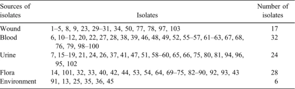
In total, 107S. epidermidisisolates obtained from clinical [including blood (n=32), wound (n=17), human flora (n=28), and urine (n=24)], and non-clinical samples [collected from laboratory environment (n=6)] (Table I) were collected during 5-month period from different hospitals and laboratories in Tehran, Iran. The strains were identified asS. epidermidisby standard laboratory tests, such as catalase, coagulase tests, resistance to polymyxin B and bacitracin disks, sensitivity to novobiocin disk, and mannitol fermentation test.
Antimicrobial susceptibility testing
Susceptibility to six antibacterial agents including vancomycin (30 μg), cefoxitin (30 μg) (as marker to detect of methicillin resistance or oxacillin resistance), linezolid (30 μg), tetracycline (30 μg), erythromycin (15 μg), and clindamycin (2μg) was determined by disk diffusion method as CLSI guideline.
Staphylococcus aureus strain ATCC 25923 used for quality control of disk diffusion method [16].
Identification of TA genes in S. epidermidis
To find TA genes inS. epidermidis, we obtained the complete genome of S. epidermidisATCC 12228 andS. epidermidisisolate BPH0662 from NCBI and via different databases [17, 18] and we managed tofind the common TA loci (type II) in these bacteria (mazEF, sam, and phd-doc) (Figure1). Finally, specific primers were designed using oligo software version 7.56 (TableII) [19].
Table I.The source ofS. epidermidisisolates Sources of
isolates Isolates
Number of isolates Wound 1–5, 8, 9, 23, 29–31, 34, 50, 77, 78, 97, 103 17 Blood 6, 10–12, 20, 22, 27, 28, 38, 39, 46, 48, 49, 52, 55–57, 61–63, 67, 68,
76, 79, 98–100
32 Urine 7, 15–19, 21, 24, 26, 37, 41, 47, 51, 58–60, 65, 66, 75, 80, 81, 94, 96,
95, 102
24 Flora 14, 101, 32, 33, 40, 42, 44, 53, 54, 64, 69–75, 82–90, 92, 93, 43 28
Environment 91, 13, 25, 35, 36, 45 6
Table II.Characteristics of design primers and related genes used in the study TA
system Sequence (5′→3′) TM
Amplicon (bp)
Chromosome
locationa Reference
mazF F CAAGGGGGAGTAAGACCTGT
58 95 1934159–
1934521 This
study R TTAATCCTACCAGTAATCGCAGC
mazE F GAAGGTTATGCACAAATGGC
56 83 1934518–
1934688 This
study R TTGCAATCACAAGCTTCACA
doc F GCTCAAAAGCACGCATTCCAT
60 110 1807207– 1807602
This study R ACCGCTTCATCTTGAGTGCAT
phd F GCACTCGGAATATCAAATGGTG
57 93 1807602–
1807769 This
study R AAACTCCTCGTCCTCTCTTAC
sam-T F TTGCTCGTTGATGGAATGGCTT
61 181 1226550– 1227428
This study R GCCCCATATCCCAACAAACTGC
sam-AT F GAAGTCAGCGCAGAAGCACAAT
61 231 1227447– 1230884
This study R AGTGGTCGCCTCATTAAGTCGT
Note:aOnS. epidermidisisolate BPH0662.
Figure 1.Genomic location of TA system in the chromosome ofS. epidermidisisolate BPH0662
Detection of TA genes in S. epidermidis isolates
PCR assay was used to depict the presence of TA modules in theS. epidermidis isolates. PCR was performed in a DNA thermal cycler (Bio-Rad, USA) in a volume of 25μl. The program consisted of an initial denaturation step at 94 °C for 4 min, 35× 94 °C for 30 s, annealing (annealing Tm for each primer is indicated in TableII) for 30 s, and extension at 72 °C for 20 s. No template control was used as negative control. Finally, PCR products were sequenced (Macrogen, South Korea).
Quantitative real-time polymerase chain reaction
Bacteria were pelleted by centrifugation at 2,500×gfor 15 min. Total RNA was isolated using the QIAGEN RNeasy Mini kit. Extracted RNA was analyzed using a nanodrop ND1000 and running on a denaturing 1.5% Tris-acetate-EDTA- agarose gels (80 V for 1 h) to assess RNA concentration, quality, and integrity.
The RNA was DNase treated with Promega RNase-free DNase (at 37 °C for 1 h).
RNA was precipitated with 1 volume isopropanol and 0.1 volume of 3 M NaOAc (pH 4.6). The suspension was incubated on ice for 20 min and centrifuged at high speed for 30 min at 4 °C. The RNA was pellet, dried, and resuspended with RNase-free MilliQ H2O. According to the manufacturer’s instructions, 500 ng– 1μg RNA was converted into cDNA using AccuPower CycleScript RT PreMix (Bioneer, Korea). Quantitative real-time PCR was performed in a Rotor-Gene thermal cycler (Corbett 6000, Australia) using SYBR Green method (AccuPower Green Star qPCR Master Mix, Bioneer, Korea). A total volume of 20μl reaction containing 2μl of cDNA, 12.5μl SYBR Green master mix, 4.5 μl nuclease-free water, and 1μl of each primer (5 pmol) was run according to following program:
an initial activation step at 94 °C for 4 min, 35 cycles of denaturation at 94 °C for 30 s, annealing at (indicated in TableII) for 30 s, and extension at 72 °C for 20 s.
Statistical analysis
We used SPSS version 16 and chi-square program to assess the correlation between the presence of TA genes and antibiotic-resistance pattern inS. epidermidis isolates. APvalue ≤0.05 was considered as significant.
Results
The antibiotic susceptibility pattern in all 107S. epidermidisisolates showed 39.2%, 60.8%, 32%, 70.1%, 1%, and 1% resistance to clindamycin, methicillin,
tetracycline, erythromycin, linezolid, and vancomycin, respectively. The highest levels of resistance were seen to erythromycin and oxacillin, 70.1% (n=68) and 60.8% (n=59), respectively. The highest and lowest resistance to methicillin were observed in blood (79.3%), environmental (77.8%), urine (72%), wound (60%), and humanflora isolates (20.8%).
Surprisingly, urine, blood, and environmental isolates also had the highest resistance to erythromycin (84%, 79.3%, and 77.8%, respectively) and the highest sensitivity was seen in humanflora isolates (58.3%).
Fortunately, all isolates were sensitive to linezolid and vancomycin except one vancomycin-resistant blood isolate and one linezolid-resistant environmental isolate.
The highest resistance to tetracycline and clindamycin was shown in wound isolates, whereas the highest sensitivity was seen in humanflora isolates (in both antibiotics) and blood isolates (on the subject of tetracycline).
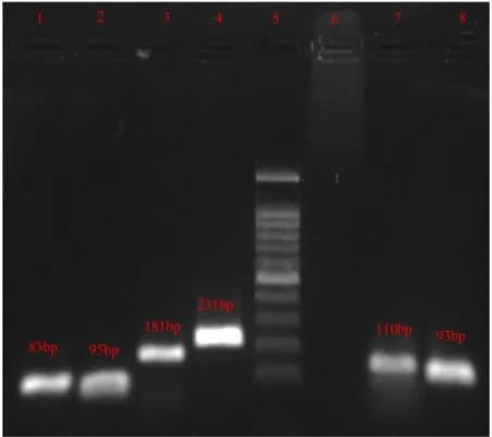
An attractive observation was the prevalence of three TA genes in these isolates (Figure2). Notably, mazEF,sam, andphd-doc loci were presented in all 107 S. epidermidis isolates. These results demonstrated that there is no difference between clinical and non-clinical and also resistant and sensitive isolates to antibiotics in presence of TA genes. Given to qPCR results, it could be concluded that the identified genes are transcriptionally activated and might be translated to the proteins or functionally active RNAs in the different strains ofS. epidermidis.
Discussion
Nowadays, the consumption of common antibiotics in treatment of S. epidermidisinfections has enhanced emergence of resistant isolates and has increased use of glycopeptide antibiotics (vancomycin). The high rate of resistance to methicillin (60.8%) was despairing, but the sensitivity of majority of the isolates (99%) to vancomycin and linezolid is promising. Therefore, vancomycin can be the best choice for S. epidermidis infections. Since, the rate of resistance to vancomycin is increasing, to meet this problem (one blood isolate in this study) and tofind the new antibacterial targets that can be useful in treatment. TA system can be introduced as an antibacterial target [20]. The distribution of TA loci in different species is shown according to the analyses in databases and studies of prokaryotic genome [20].
However, there have been few studies about the existence and performance of TA loci in different bacterial species. Current studies on TA systems showed the possible roles of this system in antibiotic tolerance, biofilm formation, niche colonization, persistence, cell programmed death, chronic infections, and patho- genicity [21–26].
S. epidermidis is one of the common biofilm-producing bacteria and the cause of related infections [27]. Therefore, it is important to study factors involved in biofilm formation and increased tolerance to antibiotics in this bacteria as an important pathogen. As different types of TA systems have roles in biofilm formation, pathogenesis, and tolerance to antibiotics, and there have been no studies or analyses on these bacteria, it is important to study the prevalence and existence of these systems in this pathogenic bacteria.
Type II system is the most common among TA systems according to previous studies [28].
MazEF is one of the major groups of type II systems, which today is considered as one of the important TA systems in biofilm formation and tolerance to antibiotics [29]. PCR and qPCR used in this study showed that the prevalence of these systems in pathogenic bacteria is significant and found in both oxacillin-resistant and oxacillin-sensitive bacteria equally. The results showed that all the oxacillin-resistant and oxacillin-sensitive bacteria in this
Figure 2.PCR detection of TA genes. Lane1,mazE; lane2,mazF; lane3,samT; lane4,samAT;
lane5, ladder; lane6, negative control; lane7,doc; lane8,phd.(T: toxin, AT: antitoxin)
study had maz TA system with the prevalence of 100%. A similar prevalence of this TA system was reported by Jain et al. [29]. They studied 101 MRSA strains and reported that 100% of biofilm-forming MRSAs had mazEF gene. Their study shows the same prevalence of this TA system as this study. Another study was conducted on the plasmid of 75 vancomycin-resistant enterococci (VRE) samples by Moritz and Hergenrother [30] and same results were reported. They showed in a study that all the plasmids had maz TA system.
Maz system was mostly on the plasmid containingvanAgene. With the results of their study and high prevalence of mazEF in VRE plasmid, mazEF is expected to be found on plasmids of similar species, such as staphylococci and streptococci.
Phd-doc is another important type II TA system. This system is derived from P1 bacteriophage and is involved in stress responses, plasmid stability resistance, and essential performances, such as pathogenicity [31, 32]. The results of this study, using PCR and real-time PCR, were the same as the results of maz TA and all theS. epidermidisstrains resistant and sensitive to oxacillin had this system and also same prevalence of this system was observed in oxacillin-resistant and oxacillin-sensitive strains.
It is also noteworthy that there are no evidences about samgene in other bacteria so far.
As this bacterium is known as a problem in hospitals for its ability to form biofilms and high prevalence and antibiotic resistance,finding new approaches to fight against pathogens is considered a worldwide priority in healthcare system.
Therefore, if one of these TA systems is found in all clinical species, it could be a suitable target for antibiotic therapy.
In this study, which was based on PCR and qPCR techniques, all three TA systems includingmazEF,sam, andphd-docwere found in all 107S. epidermidis isolates. The high prevalence of these three systems can make them suitable targets for antibiotic therapy.
Acknowledgements
All authors appreciated Department of Microbiology, Iran University of Medical Sciences. IG designed, drafted, analyzed, and supervised this study.
Funding Sources
This study was supported by Iran University of Medical Sciences, Tehran, IR Iran.
Conflict of Interest None.
References
1. Yamaguchi, Y., Park, J.-H., Inouye, M.: Toxin-antitoxin systems in bacteria and archaea.
Annu Rev Genet45, 61–79 (2011).
2. Ghafourian, S., Raftari, M., Sadeghifard, N., Sekawi, Z.: Toxin-antitoxin systems:
Classification, biological function and application in biotechnology. Curr Issues Mol Biol 16, 9–14 (2013).
3. Sadeghifard, N., Soheili, S., Sekawi, Z., Ghafourian, S.: Is the mazEF toxin-antitoxin system responsible for vancomycin resistance in clinical isolates ofEnterococcus faecalis?
GMS Hyg Infect Control9, (2014).
4. Savari, M., Rostami, S., Ekrami, A., Bahador, A.: Characterization of toxin-antitoxin (TA) systems inPseudomonas aeruginosaclinical isolates in Iran. Jundishapur J Microbiol9, e26627 (2016).
5. Mine, N., Guglielmini, J., Wilbaux, M., Van Melderen, L.: The decay of the chromo- somally encodedccdO157toxin-antitoxin system in theEscherichia colispecies. Genetics 181, 1557–1566 (2009).
6. Schuster, C. F., Bertram, R.: Toxin-antitoxin systems are ubiquitous and versatile modulators of prokaryotic cell fate. FEMS Microbiol Lett340, 73–85 (2013).
7. Wang, X., Lord, D. M., Cheng, H.-Y., Osbourne, D. O., Hong, S. H., Sanchez-Torres, V., Quiroga, C., Zheng, K., Herrmann, T., Peti, W.: A new type V toxin-antitoxin system where mRNA for toxin GhoT is cleaved by antitoxin GhoS. Nat Chem Biol8, 855–861 (2012).
8. Hall, A. M., Gollan, B., Helaine, S.: Toxin-antitoxin systems: Reversible toxicity.
Curr Opin Microbiol36, 102–110 (2017).
9. Mutschler, H., Meinhart, A.: ε/ζ systems: Their role in resistance, virulence, and their potential for antibiotic development. J Mol Med89, 1183–1194 (2011).
10. Chaudhry, V., Patil, P. B.: Genomic investigation reveals evolution and lifestyle adaptation of endophyticStaphylococcus epidermidis. Sci Rep6, 19263 (2016).
11. Vandecandelaere, I., Coenye, T.: Microbial composition and antibiotic resistance of biofilms recovered from endotracheal tubes of mechanically ventilated patients.
In Gianfranco, D. (ed): Biofilm-Based Healthcare-Associated Infections. Springer, Cham, Switzerland, 2015, pp. 137–155.
12. Morgenstern, M., Erichsen, C., Hackl, S., Mily, J., Militz, M., Friederichs, J., Hungerer, S., Bühren, V., Moriarty, T. F., Post, V.: Antibiotic resistance of commensalStaphylococcus aureus and coagulase-negative staphylococci in an international cohort of surgeons:
A prospective point-prevalence study. PLoS One11, e0148437 (2016).
13. Engelberg-Kulka, H., Sat, B., Reches, M., Amitai, S., Hazan, R.: Bacterial programmed cell death systems as targets for antibiotics. Trends Microbiol12, 66–71 (2004).
14. Gerdes, K., Christensen, S. K., Løbner-Olesen, A.: Prokaryotic toxin-antitoxin stress response loci. Nat Rev Microbiol3, 371 (2005).
15. DeNap, J. C., Hergenrother, P. J.: Bacterial death comes full circle: Targeting plasmid replication in drug-resistant bacteria. Org Biomol Chem3, 959–966 (2005).
16. Drew, W. L., Barry, A., O’Toole, R., Sherris, J. C.: Reliability of the Kirby-Bauer disc diffusion method for detecting methicillin-resistant strains of Staphylococcus aureus.
Appl Microbiol24, 240–247 (1972).
17. Sevin, E. W., Barloy-Hubler, F.: RASTA-Bacteria: A web-based tool for identifying toxin-antitoxin loci in prokaryotes. Genome Biol8, 1 (2007).
18. Shao, Y., Harrison, E. M., Bi, D., Tai, C., He, X., Ou, H.-Y., Rajakumar, K., Deng, Z.:
TADB: A web-based resource for Type 2 toxin-antitoxin loci in bacteria and archaea.
Nucleic Acids Res39, D606–D611 (2011).
19. Rychlik, W.: OLIGO 7 Primer Analysis Software. In Yuryev, A. (ed): PCR Primer Design.
Methods in Molecular Biology™. Humana Press, Totowa, NJ, 2007, Vol.402.
20. Makarova, K. S., Wolf, Y. I., Koonin, E. V.: Comprehensive comparative-genomic analysis of type 2 toxin-antitoxin systems and related mobile stress response systems in prokaryotes.
Biol Direct4, 19 (2009).
21. Kim, Y., Wang, X., Ma, Q., Zhang, X.-S., Wood, T. K.: Toxin-antitoxin systems in Escherichia coliinfluence biofilm formation through YjgK (TabA) andfimbriae. J Bacteriol 191, 1258–1267 (2009).
22. Fineran, P. C., Blower, T. R., Foulds, I. J., Humphreys, D. P., Lilley, K. S., Salmond, G. P.:
The phage abortive infection system, ToxIN, functions as a protein-RNA toxin-antitoxin pair. Proc Natl Acad Sci U S A,106, 894–899 (2009).
23. Hazan, R., Engelberg-Kulka, H.:Escherichia colimazEF-mediated cell death as a defense mechanism that inhibits the spread of phage P1. Mol Genet Genomics 272, 227–234 (2004).
24. Norton, J. P., Mulvey, M. A.: Toxin-antitoxin systems are important for niche-specific colonization and stress resistance of uropathogenic Escherichia coli. PLoS Pathog 8, e1002954 (2012).
25. Ren, D., Walker, A. N., Daines, D. A.: Toxin-antitoxin loci vapBC-1 and vapXD contribute to survival and virulence in nontypeableHaemophilus influenzae. BMC Microbiol12, 263 (2012).
26. Rowe-Magnus, D. A., Guerout, A.-M., Biskri, L., Bouige, P., Mazel, D.: Comparative analysis of superintegrons: Engineering extensive genetic diversity in the Vibrionaceae.
Genome Res13, 428–442 (2003).
27. Namvar, A. E., Bastarahang, S., Abbasi, N., Ghehi, G. S., Farhadbakhtiarian, S., Arezi, P., Hosseini, M., Baravati, S. Z., Jokar, Z., Chermahin, S. G.: Clinical characteristics ofStaphylococcus epidermidis: A systematic review. GMS Hyg Infect Control 9, (2014).
28. Yamaguchi, Y., Inouye, M.: Toxin-antitoxin systems in bacteria and archaea. In de Bruijn, Frans J. (ed): Stress and Environmental Regulation of Gene Expression and Adaptation in Bacteria. John Wiley & Sons Inc., Hoboken, NJ, 2016, pp. 97–107.
29. Jain, S., SenGupta, M., Sinha, A., Sarkar, S.: Identification of MazEF toxin-antitoxin system and biofilm formation in clinical isolates of MRSA isolated from Eastern India.
Al Ameen J Med Sci9, 53–57 (2016).
30. Moritz, E. M., Hergenrother, P. J.: Toxin-antitoxin systems are ubiquitous and plasmid- encoded in vancomycin-resistant enterococci. Proc Natl Acad Sci U S A104, 311–316 (2007).
31. Lehnherr, H., Maguin, E., Jafri, S., Yarmolinsky, M. B.: Plasmid addiction genes of bacteriophage P1: Doc, which causes cell death on curing of prophage, and phd, which prevents host death when prophage is retained. J Mol Biol 233, 414–428 (1993).
32. Garcia-Pino, A., Christensen-Dalsgaard, M., Wyns, L., Yarmolinsky, M., Magnuson, R. D., Gerdes, K., Loris, R.: Doc of prophage P1 is inhibited by its antitoxin partner Phd through fold complementation. J Biol Chem283, 30821–30827 (2008).