IDENTIFICATION OF AN INTESTINAL MICROBIOTA SIGNATURE ASSOCIATED WITH
HOSPITALIZED PATIENTS WITH DIARRHEA
NIMA MOHAMMADZADEH1, BEHROOZSADEGHIKALANI1, SHAHIN BOLORI2, AZADEH AZADEGAN3, AFSANEHGHOLAMI4, ROKHSAREH MOHAMMADZADEH1
and FARAMARZMASJEDIANJAZI1*
1Department of Microbiology, School of Medicine, Iran University of Medical Sciences, Tehran, Iran
2Department of Microbiology, Faculty of Medicine, Shahid Beheshti University of Medical Sciences, Tehran, Iran
3Department of Microbiology, School of Medicine, Tehran University of Medical Sciences, Tehran, Iran
4Department of Microbiology, Karaj Branch, Islamic Azad University, Karaj, Iran
(Received: 11 July 2018; accepted: 22 September 2018)
As an important global health challenge, diarrhea kills nearly two million people each year. Postinfectious irritable bowel syndrome (IBS) usually manifests itself as the diarrhea-predominant subtype. Small intestinal bacterial overgrowth has been observed more frequently in patients with IBS compared to healthy controls.
However, the pathophysiology of IBS is not fully understood, and based on recent evidences, altered gut microbiota is involved in the pathogenesis of IBS. Therefore, we aimed to compare the microbiome in hospitalized patients with diarrhea and healthy individuals. Thirty patients and 10 healthy controls were included into this case– control study. Microbial count was performed using quantitative real-time polymerase chain reaction method using bacterial 16S rRNA gene. Clostridium cluster IVand Bacteroideswere significantly more frequent in the patients compared with the healthy individuals (p=0.02 and 0.023, respectively). However, the quantity ofEnterococcus and Bifidobacterium groups were significantly higher in healthy controls than in diarrheal group (p=0.000076 and 0.001, respectively). The results showed that the number of bacteria in all bacterial groups was significantly different between healthy individuals and diabetic group, whereas the difference between the healthy group and IBS was not significant forBifidobacteriumgroup. Thefindings of this study outlined the relationship between diarrhea, IBS, and diabetes disease and bacterial composition.
It could be concluded that modifying the bacterial composition by probiotics can be helpful in the control and management of the mentioned disease.
*Corresponding author; E-mail:fmasjedian@yahoo.com
Keywords:diarrhea, irritable bowel syndrome, microbiota, diabetes Introduction
Recent reports focused on the reality of a positive relationship between human health and the microbialflora that colonize the human gut. Among the gut bacteria, which behave as a functional human organ, probiotics represent the Holy Grail because they are related to a broad spectrum of positive effects on host health, including positive effects on host longevity [1]. As a functional gastroin- testinal (GI) disorder, irritable bowel syndrome (IBS) is characterized by abdomi- nal pain and changes in the pattern of bowel movements associated with bowel habit change such as diarrhea and constipation without any evidence of underlying damage [2,3]. Although IBS is not known as a serious disorder, 10%–15% of the adult population in the developed world has been observed affected with the disorder; however, the pooled prevalence of IBS varies considerably, both by geographical location and by applied diagnostic criteria [4–6]. It is more common in South America and less common in Southeast Asia and the high prevalence of IBS is joined by vast societal monetary burdens and negative impacts on the quality of life in the afflicted individuals with the disorder [7,8]. IBS does not lead to dangerous conditions in most patients, but can be associated with increased side effects such as chronic pain and fatigue and reduced work productivity [9,10].
Researchers have reported that the high incidence of IBS together with the associated comorbidities can increase social costs and can also greatly affect the patient’s quality of life [8,11].
Although the pathophysiology of IBS is not fully understood, several hypotheses have been proposed. Acute GI infections increase the risk of developing and expanding IBS. Prolonged fever, anxiety, and depression are the other factors that increase the likelihood of developing the syndrome. The biochemical signaling taken place between the GI tract and the central nervous system (the brain–gut axis) has been accepted as the major pathogenetic mechanism of IBS, which suggests that IBS occurs due to disturbances in the interoperability of the brain-intestinal axis and is correlated with a dysfunction of the GI autonomic nervous system [4]. Other theories including gut motility disorders, pain sensitivity, infections including small intestinal bacterial overgrowth, neurotransmitters, genetic factors, and food sensi- tivity have also known as the causes of the disorder [12].
Despite the considerable burden of IBS, treatment alternatives stay restricted and look into the etiology, and pathophysiology of this multifactorial disorder is continuous [13]. The growing evidences have indicated that IBS might present due to other potential mechanisms including gut microbiota and low-grade inflamma- tion/immune activation [14, 15]. Comparisons of IBS patients with healthy
participant in several lines of literatures have shown that the microbiota differs significantly between groups, which demonstrate the putative role of gut micro- biota in IBS [16,17]. Postinfectious IBS; colonic fecal microbiota transplantation;
and therapeutic effects of probiotics, prebiotics, synbiotics, and non-systemic anti- microbials have also supported the contribution of the gut microbiota to the pathophysiology of IBS [18–23].
Given the evidence that approximately 10% of IBS patients’ symptoms began following an episode of infectious diarrhea and based on our knowledge of the relationship between alteration of gut microbiota and inflammation of gut, we encouraged to compare the microbiome in hospitalized patients with diarrhea and healthy individuals.
Materials and Methods Participant and sampling
Thirty affected hospitalized participants with diarrhea (age: 56±8 years) who referred to Institute of Endocrinology and Metabolism Research and Training Center, Iran University of Medical Sciences in Tehran, Iran were recruited into the study as the case group. Twenty non-diarrheal individuals, matched for age, gender, and their current living environment were recruited as the healthy participants. Stool samples from the patients and healthy individuals were collected. Sterile cups were used to instant stool sampling after defecation and brought to the laboratory within 2 h. Collected stool samples were instantly stored in microbiology laboratory at– 70 °C upon arrival.
Pregnancy, lactation, organic GI disease, severe systematic disease, major or complicated abdominal surgery, severe endometriosis, and dementia were the exclusion criteria for patients, and intestinal disturbances (including lactose intolerance and celiac disease), ongoing antibiotic treatment, and all exclusion criteria of the patients were considered as the exclusion criteria for controls.
This study was approved by research ethics committees of Iran University of Medical Sciences and according to Declaration of Helsinki. A signed informed consent form was received from each of the participant and they were ensured anonymity of all information.
DNA extraction
An amount of 200 mg of each fecal sample was used for bacterial total DNA extraction. Extraction of total DNA from all stool specimens was performed using
QIAamp® DNA Stool Mini Kit (Qiagen Retsch GmbH, Hannover, Germany) according to the manufacturer’s instruction. Quality and quantity of the extracted DNA was measured by Nanodrop spectrophotometer (Nanodrop Technologies, Wilmington, DE, USA) and the DNA integrity was assessed by 1% agarose gel-electrophoresis. Finally, entire extracted DNA samples were immediately transferred into−20 °C storage.
Microbial quantification by real-time polymerase chain reaction (PCR)
Briefly, all bacterial 16S rRNA sequences were extracted from SILVA High Quality Ribosomal RNA database [30], then converted into 16S rDNA, and specific probe and primer sequences were designed in several steps using various databases including NCBI, probebase, IDT and EMBL-EBI, and also AlleleID software (version 7.5, Humana Press, USA). Characteristics of primers and TaqMan probes are demonstrated in TableI.
Using 16S rDNA gene-specific primers and probes, amplification of target 16s rDNA was performed in Rotor-Gene 6000 real-time PCR cycler (Qiagen Corbett, Hilden, Germany) by real-time TaqMan quantitative PCR (qPCR). Each reaction mixture in a total volume of 20μl, which contained 0.5μl of forward primer, 0.5μl of reverse primer, 0.5μl of TaqMan probe, 12μl of Probe Ex Taq (probe qPCR) Master Mix (Takara Bio, Shiga, Japan), 1μl of template DNA, and 5.5μl sterilized ultrapure water, was run to amplify the target region under following real-time qPCR cycling condition: an initial holding at 95 °C for 30s, followed by 40 cycles of denaturation at 95 °C for 5s, and annealing/extension at 60 °C for 30s. No template reaction was used as the negative control.Bifidobacterium bifidumATCC 29521, Lactobacillus acidophilus ATCC 4356, and Fusobacterium nucleatum ATCC 25586 were provided from the American Type Culture Collection (ATCC) and were used as the bacterial standard strains. All qPCR runs were carried out in triplicate, and averaged numbers were used for calculation and analysis.
Bacterial count
Determination of the numberof Lactobacillus, Bifidobacterium, Fusobac- terium, andPrevotellagroups in each sample was performed after construction of standard curves based on 10-fold serial dilutions of bacterial standard strains genomic DNA of known concentration from pure cultures, corresponding to 101–1010 copies per gram feces. According to Applied Biosystems tutorials, standard curves were created and were normalized to the copy number of the 16S rRNA gene for each species. If there was a copy number-unknown species of
TableI.Characteristicsof16SrDNAgene-targetedprimersandprobes TargetbacteriaPrimer/probe*Sequence(5′→3′)Oligonucleotidesize(bp)Productsize(bp)Ref. EnterococcusgroupPrimer-FTGGGTAGCGGAGAAATTCCA20103Thisstudy Primer-RACAGTGCTCTACCTCCATCA20 ProbeCCGAGGCTAGCCCTAAAGCTATTTCGG27 BifidobacteriumgroupPrimer-FAAGCGATGGACTTTCACACC2087Thisstudy Primer-RTACGTAGGGTGCAAGCGTTA20 ProbeCGCGACGAACCGCCTACGAGC21 BacteroidesgroupPrimer-FGTATGTCRCAAGCGTTATCC20100Thisstudy Primer-RAACGCAATACRGAGTTGAGC20 ProbeTAGACGCGCTTTACGCCCAAT21 ClostridiumclusterIVPrimer-FCGAACAGGATTAGATACCC19134Thisstudy primerRCTTTGAGTTTCACCGTTG18 ProbeAAACGATGGATGCCCGC17 Note:*PrimersF(forward),R(reverse),andprobestargetingthe16SrDNAgene.
16S rRNA operon, average operon numbers of the closest bacterial taxa obtained from the ribosomal RNA database rrnDB was used as the operon copy number.
The threshold cycle values (Ct) obtained from the standard curves were applied to determine bacterial copy number per gram stool.
Sample size and statistical analysis
SPSS version 20.0 software (SPSS Inc., Chicago, IL, USA) and Minitab version 16.2.0 (State College, PA, USA) were used for statistical analysis.
According to a predicted difference of at least 2×10⁵copies per gram of feces in the mean bacterial numbers between the healthy individuals and patients, the sample size of 20 subjects provided the sufficient power (80%), considering a type I error of 0.05 and effect size of 0.4176. For group comparison, independent sample t-test was used and the Pearson’s correlation was assessed for linear correlation analysis. A p<0.05 was considered statistically significant. All the descriptive data have been expressed as mean±standard deviation. The Kolmo- gorov–Smirnov test was applied to test for a normal distribution.
Results Participants
Thirty participants with diarrhea with the mean age of 56±8 years and 10 healthy individuals with the mean age of 51±4 years were recruited to the study;
14 participants with diarrhea were males and 16 were females. Healthy individuals included 4 males and 7 females.
qPCR analysis of bacterial groups
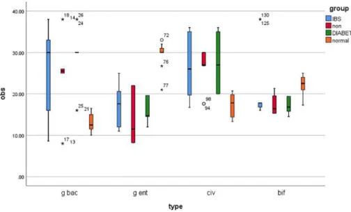
This case–control study qPCR analysis was aimed to assess the differences in composition of fecal microbiota in patients with diarrhea and healthy participant for four groups of bacteria includingBacteroides,Clostridium cluster IV,Enterococcus, andBifidobacteriumgroups. As indicated in TableII, the results showed that fecal microbiota in diarrheal cases and healthy individuals were significantly different for all the studied bacterial groups. Clostridium cluster IV and Bacteroides were significantly more frequent in the patients compared with the healthy individuals (p=0.02 and 0.023, respectively). However, the quantity of Enterococcus and Bifidobacteriumgroups was significantly higher in healthy controls than in diarrheal
TableII.Correlationsbetweenconcentrationofthebacterialspeciesinhealthy–diarrheal,healthy–diabetic,andhealthy–IBSgroups Bacterialspecies ControlIBSDiarrheaDiabetic Copies/gram offecal (N=10) Copies/gram offecal (N=10)Zp Copies/gram offecal (N=10)zp
Copies/gram offecal (N=10)zp Bacteroidesgroup53E9±18E570E5±61E3−2.120.03587E5±15E3−2.270.02344E4±79E2−3.680.000 Enterococcusgroup28E4±35E225E8±48E6−3.640.00070E10±56E2−3.850.00079E7±E4−3.800.000 ClostridiumclusterIV21E8±28E321E5±87E3t=3.42,df=10.800.00639E5±24E1−3.030.02066E4±71E2t=4.74,df=11.580.001 Bifidobacteriumgroup58E6±41E412E7±83E4−1.970.05216E8±21E53.190.00126E8±26E4t=−5.59,df=18.00.001 Note:IBS:irritablebowelsyndrome.
group (p=0.000076 and 0.001, respectively). Differences of intestinal bacterial genera in the study groups were represented in Table IIand Figure 1.
The correlations between concentration of the bacterial species in healthy–diabetic and healthy–IBS groups
Some of the participants in this study had background diseases of diabetes and IBS; therefore, the correlation or, in other words, the difference in bacterial groups between healthy individuals and those with any of the mentioned diseases was investigated. The results showed that the number of bacteria in all bacterial groups was significantly different between healthy individuals and diabetic group, whereas the difference between the healthy group and IBS was not significant forBifido- bacteriumgroup. The mean quantity of Bacteroides,Clostridium cluster IV, and Bifidobacterium groups was significantly higher in diabetic group than in non- diabetic individuals (p=0.000043,p=0.001, andp<0.001, respectively).Bacter- oides, Clostridium cluster IV, and Bifidobacterium were also significantly more frequent in the IBS patients compared with the healthy individuals (p=0.035, 0.006, and 0.052, respectively). Bacteroides concentration was higher both in
Figure 1.Bacterial groups quantified by real-time PCR and expressed as copy number of bacterial groups per gram stool in human adults with diarrhea (red boxes;N=10), IBS (blue boxes;N=10), diabetes (green boxes;N=10), and healthy controls (orange boxes;N=10). Boxes show the upper (75%) and the lower (25%) percentiles of the data. Whiskers indicate the highest and the smallest
values. *Outlier points
diabetic group and IBS patients than in healthy group (p=0.000011 and 0.000042, respectively). The correlations between concentration of the bacterial species in healthy–diarrheal, healthy–diabetic, and healthy–IBS groups have been indicated.
Discussion
Correlation between imbalance normal microbial community and GI con- ditions such as inflammatory bowel disease and IBS, and wider systemic manifestations of disease such as obesity, type 2 diabetes, and atopy have previously been reported in several reports [6,13,14]. Although intensive research over the past two decades has been carried to find the etiology of IBS, its pathophysiology remains poorly understood [19]. Diarrhea, constipation, or mixed type are the symptoms of IBS. Based on this knowledge, we hypothesized that intestinal microbiota composition in patients with diarrhea is different from healthy control participant. In this study, the number of four groups of bacteria includingBacteroides,Clostridium cluster IV,Enterococcus, andBifidobacterium was measured using qPCR in patients and healthy participants. Our data showed that fecal microbiota in diarrheal cases and healthy individuals were significantly different for all the studied bacterial groups.
In comparison to healthy controls, our data showed that the copy number of BacteroidesandClostridium cluster IV group was significantly higher in partici- pants with diarrhea. In contrast with this study, Swidsinski et al. [24] using fluorescence in situ hybridization analysis (FISH) demonstrated that the fecal microbiota in idiopathic diarrhea was markedly different, marked by reduction in concentrations of habitualEubacterium rectale,Bacteroides, andFaecalibacterium prausnitzii groups. Using FISH analysis, a study conducted on patients by Soko et al. also showed that a reduction of a major member of Firmicutes, F. prausnitzii, is associated with a higher risk of postoperative recurrence of ileal Crohn’s disease (CD). They found thatF. prausnitziiexhibits anti-inflammatory effects, partly due to secreted metabolites able to block NF-κB activation and IL-8 production; therefore, counterbalancing dysbiosis usingF. prausnitzii as a probiotic can be a promising strategy in CD treatment [25].
In addition to the diarrhea, which is designated as the principal diagnosis, some patients also have underlying diseases of diabetes and IBS. Therefore, we also decided to evaluate the association of each of the studied bacterial group with the underlying diseases. In agreement with findings of Vrakas et al. [26], Maukonen et al. [27], and Rajili´c-Stojanovi´c et al. [28], our observation showed that the concentrations ofClostridium cluster IVandFusobacteriumgroups were relatively higher in the IBS group compared to the healthy participant.
Consistently, Kerckhoffs et al. [29] showed thatbifidobacterialevels in both fecal and duodenal brush samples of IBS patients were significantly lower (6±0.6 vs.
19±2.5,p<0.001) compared to healthy participants, which indicate a role for microbiotic composition in IBS pathophysiology. They analyzed fecal samples for the composition of the total microbiota using FISH and analyzed both fecal and duodenal brush samples for the composition of bifidobacteria using qPCR.
Similarly, Rajili´c-Stojanovi´c et al. [28] and Si et al. [30] showed meaningful lower concentration ofBifidobacteriumin IBS patients compared to the controls.
In this study, we found that the level of Enterococcus group was significantly lower in the patients with IBS compared to the healthy group. This observation was in contrast with thefindings of Zhuang et al. [31].
In line with our results, the study conducted by Rajili´c-Stojanovi´c, the intestinal microbiota including a twofold increased ratio of Bacteroidetes (p=0.0002), approximately 1.5-fold increase in numbers of Clostridium spp.
(p=0.005); a twofold decrease in the number ofBacteroidetes (p=0.0001); a 1.5-fold decrease in numbers of Bifidobacterium and Faecalibacterium spp.
(p=0.05), of IBS patients differed significantly (p=0.0005) from that of controls [28]. Characterization of the fecal microbiota using high-throughput sequencing of the 16S rRNA gene showed a significant increase of Bacteroidetes andProteo- bacteriain the IBS group compared to the healthy participants [32]. Other report also confirmed our data and demonstrated that the mucosa-associated microbiota in patients with IBS is significantly different from healthy controls with increases in bacteroides and clostridia and a reduction in bifidobacteria in patients with IBS-D [33]. However, global and deep molecular analysis of microbiota signa- tures in fecal samples from patients with IBS disclosed a twofold decrease in the number ofBacteroidetes[33]. In this study, the level ofBifidobacteriumwas lower in IBS patients than controls, but this difference was not statistically significant.
This study revealed that the level of Bacteroides, Bifidobacterium, and Clostridium cluster IV in patients with underlying disease of diabetes was significantly higher compared with those in their healthy counterparts. Micro- biome profile showed a high level ofEnterococcusgroup in healthy participant than individuals with diabetes. On the contrary to this study, Remely et al. [34] and Sedighi et al. [6] found no significant differences in copy number of genus Bifidobacterium between the case and control groups; nonetheless, Murri et al.
[35] reported a significant decrease in the number of Bifidobacterium in the children with diabetes. Consistent with ourfindings, Larsen et al. [36] found that the ratio of Bacteroidetes was increased in diabetes’ cases; however, Lambeth et al. [37] did not observe differences in the abundances of phyla Bacteroidetes.
On the contrary to this study, data reported by Remely et al. [34] and Larsen et al.
[36] showed no significant difference in the level of Clostridium cluster IV
between patients with diabetes and healthy individuals. It is previously reported that total cholesterol and low-density lipoprotein cholesterol reduction are associated with dairy products enriched withEnterococcus faeciumin participant having a normal lipid profile and participant with medium to moderate hypercholesterolemia [38].
Although participants included in this study were matched for age, gender, race, living environment, and non-interventions of medications and foods, which may affect the outcomes (such as different antibiotics, prebiotics, and probiotics), ourfindings were not completely consistent with previous reports, and controver- sial results regarding the dysbiosis of the gut microbiota in hospitalized patients with diarrhea as well as contradictoryfindings on the relationship between various bacterial groups and IBS and diabetes were observed. This discrepancy might be due to heterogeneity in various factors such as genetic background, ethnicity, geographical location, environmental and occupational exposures, medical history, possible underlying diseases/disorders, lifestyle, and diet habits of participant across studies. Non-significance in some of our results may have been related to a relatively small sample size.
On the whole, the results of this study indicate that diarrhea and the underlying disorders of IBS and diabetes in humans are associated with compo- sitional changes in intestinal microbiota; however, we cannot conclude about the causality of this dysbiosis. Further studies are suggested to determine if microbial imbalance causes these medical conditions or changes in microbiota profile are a reflection of the disease state. Overall, it could be concluded that modifying the bacterial composition by probiotics can be helpful in the control and management of the mentioned disease.
Acknowledgements
The authors are grateful for the support provided by Iran University of Medical Sciences (Tehran, Iran).
Conflict of Interest The authors declare no conflict of interest.
References
1. Ayala, F. R., Bauman, C., Cogliati, S., Lenini, C., Bartolini, M., Grau, R.: Microbial˜ flora, probiotics,Bacillus subtilisand the search for a long and healthy human longevity. Microb Cell4, 133–136 (2017).
2. Lee, B. J., Bak, Y.-T.: Irritable bowel syndrome, gut microbiota and probiotics.
J Neurogastroenterol Motil17, 252–266 (2011).
3. Farahani, N. N., Jazi, F. M., Nikmanesh, B., Asadolahi, P., Kalani, B. S., Amirmozafari, N.:
Prevalence and antibiotic susceptibility patterns ofSalmonellaandShigellaspecies isolated from pediatric diarrhea in Tehran. Arch Pediat Inf Dis6, e57328 (2018).
4. Ohman, L., Simren, M.: New insights into the pathogenesis and pathophysiology of irritable bowel syndrome. Dig Liver Dis39, 201–215 (2007).
5. Lovell, R. M., Ford, A. C.: Global prevalence of and risk factors for irritable bowel syndrome: A meta-analysis. Clin Gastroenterol Hepatol10, 712–721.e4 (2012).
6. Sedighi, M., Razavi, S., Navab-Moghadam, F., Khamseh, M. E., Alaei-Shahmiri, F., Mehrtash, A., Amirmozafari, N.: Comparison of gut microbiota in adult patients with type 2 diabetes and healthy individuals. Microb Pathog 111, 362–369 (2017).
7. Drossman, D. A., Camilleri, M., Mayer, E. A., Whitehead, W. E.: AGA technical review on irritable bowel syndrome. Gastroenterology123, 2108–2131 (2002).
8. Chey, W. D., Kurlander, J., Eswaran, S.: Irritable bowel syndrome: A clinical review.
JAMA313, 949–958 (2015).
9. Simrén, M., Axelsson, J., Gillberg, R., Abrahamsson, H., Svedlund, J., Björnsson, E. S.:
Quality of life in inflammatory bowel disease in remission: The impact of IBS-like symptoms and associated psychological factors. Am J Gastroenterol97, 389–396 (2002).
10. Bercik, P., Verdu, E. F., Collins, S. M.: Is irritable bowel syndrome a low-grade inflammatory bowel disease? Gastroenterol Clin North Am34, 235–245 (2005).
11. Simrén, M., Svedlund, J., Posserud, I., Björnsson, E. S., Abrahamsson, H.: Health-related quality of life in patients attending a gastroenterology outpatient clinic: Functional disorders versus organic diseases. Clin Gastroenterol Hepatol4, 187–195 (2006).
12. Ohman, L., Simrén, M.: Pathogenesis of IBS: Role of inflammation, immunity and neuroimmune interactions. Nat Rev Gastroenterol Hepatol7, 163–173 (2010).
13. Dupont, H.: Review article: Evidence for the role of gut microbiota in irritable bowel syndrome and its potential influence on therapeutic targets. Aliment Pharmacol Ther39, 1033–1042 (2014).
14. Ghoshal, U. C., Kumar, S., Mehrotra, M., Lakshmi, C., Misra, A.: Frequency of small intestinal bacterial overgrowth in patients with irritable bowel syndrome and chronic non- specific diarrhea. J Neurogastroenterol Motil16, 40–46 (2010).
15. Karantanos, T., Markoutsaki, T., Gazouli, M., Anagnou, N. P., Karamanolis, D. G.: Current insights in to the pathophysiology of irritable bowel syndrome. Gut Pathog2, 3 (2010).
16. Kassinen, A., Krogius-Kurikka, L., Mäkivuokko, H., Rinttilä, T., Paulin, L., Corander, J., Malinen, E., Apajalahti, J., Palva, A.: The fecal microbiota of irritable bowel syndrome patients differs significantly from that of healthy subjects. Gastroenterology133, 24–33 (2007).
17. Posserud, I., Stotzer, P.-O., Björnsson, E. S., Abrahamsson, H., Simrén, M.: Small intestinal bacterial overgrowth in patients with irritable bowel syndrome. Gut56, 802–808 (2007).
18. Carroll, I. M., Ringel-Kulka, T., Keku, T. O., Chang, Y.-H., Packey, C. D., Sartor, R. B., Ringel, Y.: Molecular analysis of the luminal-and mucosal-associated intestinal microbiota in diarrhea-predominant irritable bowel syndrome. Am J Physiol Gastrointest Liver Physiol 301, G799–G807 (2011).
19. Carroll, I. M., Ringel-Kulka, T., Siddle, J. P., Ringel, Y.: Alterations in composition and diversity of the intestinal microbiota in patients with diarrhea-predominant irritable bowel syndrome. Neurogastroenterol Motil24, 521–530 (2012).
20. Halvorson, H. A., Schlett, C. D., Riddle, M. S.: Postinfectious irritable bowel syndrome– A meta-analysis. Am J Gastroenterol101, 1894–1899 (2006).
21. Ortiz-Lucas, M., Tobias, A., Saz, P., Sebastián, J. J.: Effect of probiotic species on irritable bowel syndrome symptoms: A bring up to date meta-analysis. Rev Esp Enferm Dig105, 19–36 (2013).
22. Behrooz, S. K., Lida, L., Ali, S., Mehdi, M., Rasoul, M., Elnaz, O., Farid, B. T., Gholamreza, I.: Study of MazEF, sam, and phd-doc putative toxin–antitoxin systems in Staphylococcus epidermidis. Acta Microbiol Immunol Hung65, 81–91 (2018).
23. Kalani, B. S., Irajian, G., Lotfollahi, L., Abdollahzadeh, E., Razavi, S.: Putative type II toxin-antitoxin systems inListeria monocytogenesisolated from clinical, food, and animal samples in Iran. Microb Pathog122, 19–24 (2018).
24. Swidsinski, A., Loening-Baucke, V., Verstraelen, H., Osowska, S., Doerffel, Y.: Bios- tructure of fecal microbiota in healthy subjects and patients with chronic idiopathic diarrhea. Gastroenterology135, 568–579 (2008).
25. Sokol, H., Pigneur, B., Watterlot, L., Lakhdari, O., Bermúdez-Humarán, L. G., Gratadoux, J. J., Blugeon, S., Bridonneau, C., Furet, J. P., Corthier, G., Grangette, C., Vasquez, N., Pochart, P., Trugnan, G., Thomas, G., Blottière, H. M., Doré, J., Marteau, P., Seksik, P., Langella, P.:Faecalibacterium prausnitziiis an anti-inflammatory commensal bacterium identified by gut microbiota analysis of Crohn disease patients. Proc Natl Acad Sci U S A 105, 16731–16736 (2008).
26. Vrakas, S., Mountzouris, K. C., Michalopoulos, G., Karamanolis, G., Papatheodoridis, G., Tzathas, C., Gazouli, M.: Intestinal bacteria composition and translocation of bacteria in inflammatory bowel disease. PLoS One12, e0170034 (2017).
27. Maukonen, J., Satokari, R., Mättö, J., Söderlund, H., Mattila-Sandholm, T., Saarela, M.:
Prevalence and temporal stability of selected clostridial groups in irritable bowel syndrome in relation to predominant faecal bacteria. J Med Microbiol55, 625–633 (2006).
28. Rajili´c-Stojanovi´c, M., Biagi, E., Heilig, H. G., Kajander, K., Kekkonen, R. A., Tims, S., de Vos, W. M.: Global and deep molecular analysis of microbiota signatures in fecal samples from patients with irritable bowel syndrome. Gastroenterology141, 1792–1801 (2011).
29. Kerckhoffs, A. P., Samsom, M., van der Rest, M. E., de Vogel, J., Knol, J., Ben-Amor, K., Akkermans, L. M.: LowerBifidobacteriacounts in both duodenal mucosa-associated and fecal microbiota in irritable bowel syndrome patients. World J Gastroenterol15, 2887–2892 (2009).
30. Si, J.-M., Yu, Y.-C., Fan, Y.-J., Chen, S.-J.: Intestinal microecology and quality of life in irritable bowel syndrome patients. World J Gastroenterol10, 1802–1805 (2004).
31. Zhuang, X., Xiong, L., Li, L., Li, M., Chen, M.: Alterations of gut microbiota in patients with irritable bowel syndrome: A systematic review and meta-analysis. J Gastroenterol Hepatol32, 28–38 (2017).
32. Carroll, I. M., Ringel-Kulka, T., Siddle, J. P., Klaenhammer, T. R., Ringel, Y.: Characteri- zation of the fecal microbiota using high-throughput sequencing reveals a stable microbial community during storage. PLoS One7, e46953 (2012).
33. Parkes, G., Rayment, N. B., Hudspith, B. N., Petrovska, L., Lomer, M. C., Brostoff, J., Whelan, K., Sanderson, J. D.: Distinct microbial populations exist in the mucosa-associated microbiota of sub-groups of irritable bowel syndrome. Neurogastroenterol Motil24, 31–39 (2012).
34. Remely, M., Dworzak, S., Hippe, B., Zwielehner, J., Aumüller, E., Brath, H., Haslberger, A.: Abundance and diversity of microbiota in type 2 diabetes and obesity. J Diabetes Metab 4, 2 (2013).
35. Murri, M., Leiva, I., Gomez-Zumaquero, J. M., Tinahones, F. J., Cardona, F., Soriguer, F., Queipo-Ortuno, M. I.: Gut microbiota in children with type 1 diabetes differs from that in˜ healthy children: A case-control study. BMC Med11, 46 (2013).
36. Larsen, N., Vogensen, F. K., van den Berg, F. W., Nielsen, D. S., Andreasen, A. S., Pedersen, B. K., Al-Soud, W. A., Sørensen, S. J., Hansen, L. H., Jakobsen, M.: Gut microbiota in human adults with type 2 diabetes differs from non-diabetic adults. PLoS One 5, e9085 (2010).
37. Lambeth, S. M., Carson, T., Lowe, J., Ramaraj, T., Leff, J. W., Luo, L., Bell, C. J., Shah, V. O.: Composition, diversity and abundance of gut microbiome in prediabetes and type 2 diabetes. J Diabetes Obes2, 1–7 (2015).
38. Rebolledo, C., Cuevas, A., Zambrano, T., Acuna, J. J., Jorquera, M. A., Saavedra, K.,˜ Martínez, C., Lanas, F., Ser´on, P., Salazar, L. A., Saavedra, N.: Bacterial community profile of the gut microbiota differs between hypercholesterolemic subjects and controls. BioMed Res Int2017, 8127814 (2017).