1Synthetic and Systems Biology Unit, Institute of Biochemistry, Biological Research Centre, Hungarian Academy of Sciences, Szeged, Hungary.
2Biomolecular Electronics Research Group, Bionanoscience Unit, Institute of Biophysics, Biological Research Centre, Hungarian Academy of Sciences, Szeged, Hungary. 3Biological Barriers Research Group, Institute of Biophysics, Biological Research Centre, Hungarian Academy of Sciences, Szeged, Hungary. 4Institute of Clinical Microbiology, Albert Szent-Györgyi Medical and Pharmaceutical Center, Faculty of Medicine, University of Szeged, Szeged, Hungary. 5Institute of Pharmaceutical Analysis, University of Szeged, Szeged, Hungary. 6SeqOmics Biotechnology Ltd, Mórahalom, Hungary.
7Sequencing Platform, Institute of Biochemistry, Biological Research Centre of the Hungarian Academy of Sciences, Szeged, Hungary. 8Present address:
Faculty of Biology, Technion – Israel Institute of Technology, Haifa, Israel. 9These authors contributed equally to this work: Viktória Lázár and Ana Martins.
*e-mail: pappb@brc.hu; cpal@brc.hu
E volution of resistance towards antibiotics, or any other drug, can simultaneously increase (cross-resistance) or decrease (col- lateral sensitivity) fitness to multiple other drugs1–5. The molec- ular mechanisms driving cross-resistance are well-described
2,4,5. In contrast, it remains unclear how frequently genetic adaptation to a single drug increases bacterial sensitivity to other drugs and what the underlying molecular mechanisms of collateral sensitivity are.
This issue is important as collateral sensitivity could direct future multidrug therapeutic strategies
6. However, such strategies are lim- ited by the scarcity of available drug pairs showing collateral sensi- tivity that could also be used in clinical settings. Clearly, the concept of collateral sensitivity needs to be expanded by studying a broader scope of antimicrobial agents.
Here, we systematically study the effect of antibiotic-resistance mechanisms on susceptibility to antimicrobial peptides, a promis- ing class of new antibacterial compounds. Antimicrobial peptides are short peptides with a broad spectrum of antibacterial activities
7. Such peptides are found among all classes of life, and are part of the defence mechanisms against microbial pathogens
7. Because antimi- crobial peptides have diverse chemical features and cellular targets, they are promising antibacterial agents
8,9. However, the degree of similarity between the resistance mechanisms to peptides and to
small-molecule antibiotics remains disputed
10. This is relevant as some peptides have now reached advanced stages in clinical trials
8.
We had previously initiated laboratory evolutionary experi- ments starting with a single clone of Escherichia coli K-12
2,3. Parallel evolving populations were exposed to gradually increasing concen- trations of 1 of 12 clinically relevant antibiotics (Supplementary Table 1), leading to up to 328-fold increases in their minimum inhibitory concentrations (MICs) relative to the wild type
2. The resistance levels were equal to or above the European Committee on Antimicrobial Susceptibility Testing (EUCAST) clinical break- points, and 52% of the antibiotic-resistant strains showed resistance to multiple antibiotics
2. Here, we focus on a representative set of 60 antibiotic-resistant strains (5 strains per antibiotic) that have been subjected to whole-genome sequence analysis in order to identify the molecular mechanisms underlying antibiotic resistance
2. Many of the observed mutations have also been detected in clinical drug- resistant isolates, and are known to target cellular systems involved in inner and outer membrane transport and permeability (for example, efflux pumps, porins) and cell envelope biogenesis
2.
We hypothesized that such membrane-altering antibiotic-resis- tance mutations not only influence susceptibility to other antibiot- ics, but to antimicrobial peptides as well. Why should this be so?
Antibiotic-resistant bacteria show widespread collateral sensitivity to antimicrobial peptides
Viktória Lázár
1,8,9, Ana Martins
1,9, Réka Spohn
1, Lejla Daruka
1, Gábor Grézal
1, Gergely Fekete
1, Mónika Számel
1, Pramod K Jangir
1, Bálint Kintses
1, Bálint Csörgő
1, Ákos Nyerges
1, Ádám Györkei
1, András Kincses
2, András Dér
2, Fruzsina R Walter
3, Mária A Deli
3, Edit Urbán
4, Zsófia Hegedűs
5, Gábor Olajos
5, Orsolya Méhi
1, Balázs Bálint
6, István Nagy
6,7, Tamás A Martinek
5, Balázs Papp
1* and Csaba Pál
1*
Antimicrobial peptides are promising alternative antimicrobial agents. However, little is known about whether resistance to
small-molecule antibiotics leads to cross-resistance (decreased sensitivity) or collateral sensitivity (increased sensitivity) to
antimicrobial peptides. We systematically addressed this question by studying the susceptibilities of a comprehensive set of
60 antibiotic-resistant Escherichia coli strains towards 24 antimicrobial peptides. Strikingly, antibiotic-resistant bacteria show
a high frequency of collateral sensitivity to antimicrobial peptides, whereas cross-resistance is relatively rare. We identify
clinically relevant multidrug-resistance mutations that increase bacterial sensitivity to antimicrobial peptides. Collateral sen-
sitivity in multidrug-resistant bacteria arises partly through regulatory changes shaping the lipopolysaccharide composition of
the bacterial outer membrane. These advances allow the identification of antimicrobial peptide–antibiotic combinations that
enhance antibiotic activity against multidrug-resistant bacteria and slow down de novo evolution of resistance. In particular,
when co-administered as an adjuvant, the antimicrobial peptide glycine-leucine-amide caused up to 30-fold decrease in the
antibiotic resistance level of resistant bacteria. Our work provides guidelines for the development of efficient peptide-based
therapies of antibiotic-resistant infections.
The action of most antimicrobial peptides relies on the interaction between the positively charged peptide and the negatively charged membrane components, for two reasons. First, the structural and physicochemical properties of antimicrobial peptides (for example, net positive charge, hydropathicity) and their capacity to adopt an amphipathic conformation upon membrane binding influence this interaction
8,11,12. Second, the insertion of antimicrobial peptides into the hydrophobic core of the membrane depends on the general properties of the bacterial outer membrane
11,12. For instance, lipo- polysaccharide (LPS) composition has a major impact on the killing efficiency of cationic antimicrobial peptides
13. For these reasons, it is plausible that membrane-affecting antibiotic-resistance mutations shape genetic susceptibility to antimicrobial peptides. However, no systematic study has been devoted to address this problem in detail.
Here, we first measured the susceptibility of the 60 antibiotic- resistant strains
2,3against a set of 24 antimicrobial peptides of diverse origin and various modes of action. We found widespread bacterial collateral sensitivity towards antimicrobial peptides. Analysis of the mutational and transcriptome profiles of the antibiotic-resistant strains revealed that antibiotic-resistance mutations increase sensi- tivity to peptides via regulatory changes in LPS biosynthesis. The consequent alteration in the surface charge presumably strengthens the interaction of cationic antimicrobial peptides with the outer membrane, and thus enhances the killing efficiency of these pep- tides. Finally, we demonstrated that the antimicrobial peptide gly- cine-leucine-amide (PGLA), when co-administered as an adjuvant, restores antibiotic activity against resistant bacteria and slows down antibiotic-resistance evolution.
Results
Widespread collateral sensitivity to antimicrobial peptides. To
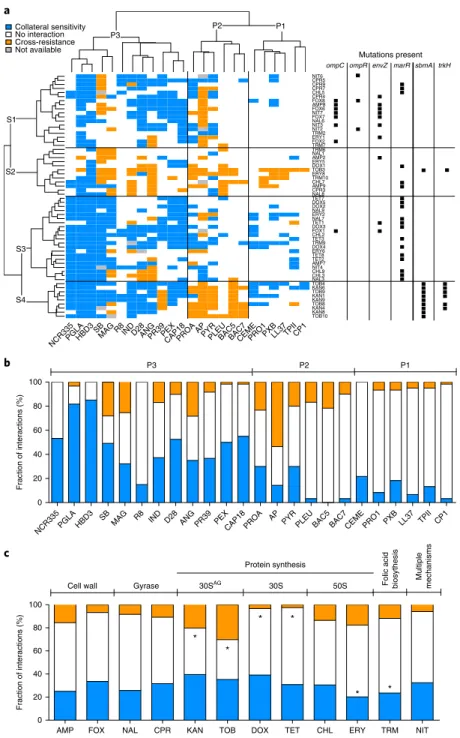
study whether antibiotic resistance in E. coli leads to cross-resis- tance or collateral sensitivity towards antimicrobial peptides, we measured the changes in the susceptibilities of 60 antibiotic-resis- tant strains to a set of 24 peptides. Peptides were chosen based on the following criteria: diverse sources (synthetic/natural), different putative mechanisms of action, structural diversity, and clinical rel- evance (Supplementary Table 2). The obtained results allowed us to chart the map of cross-resistance/collateral sensitivity of the anti- biotic-resistant strains towards the 24 antimicrobial peptides, and identify several general patterns (Fig. 1a, Supplementary Table 3).
First, cross-resistance to antimicrobial peptides was relatively rare: only 12% of all possible antibiotic-resistant strain and pep- tide pairs showed cross-resistance, whereas 31% showed collateral sensitivity. The observed strength of cross-resistance and collateral sensitivity was usually a twofold change in MIC relative to the wild type (Supplementary Fig. 1, Supplementary Table 4). In typical clin- ical settings, drug concentrations well above the wild-type MIC are applied
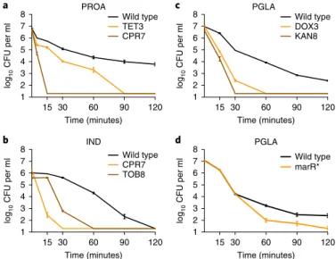
14. Therefore, we asked how collateral sensitivity shapes the killing kinetics with peptides applied at a concentration 10–15-fold above the wild-type MIC. We focused on five resistant strains that showed collateral sensitivity to at least one of the peptides studied (Fig. 2a–c). Upon antimicrobial peptide stress, collateral-sensitive populations showed a far more rapid decline in size than the wild type. For example, 15 minutes of protamine (PROA) exposure nearly completely eradicated ciprofloxacin (CPR)-resistant bacte- ria, whereas the size of the wild-type population declined only by tenfold (Fig. 2a).
Second, collateral sensitivity and cross-resistance patterns clus- tered the antimicrobial peptides into three main groups (Fig. 1) with major differences in their physicochemical properties and modes of action. Antimicrobial peptides belonging to the P1 and P3 groups generally insert themselves into membrane bilayers to form pores and thus induce cell lysis (pore formers). However, these two groups differ both in their physicochemical properties and their interactions with antibiotic-resistant strains: P1 peptides
have lower isoelectric point and hydropathicity index compared to P3 peptides (see Supplementary Fig. 2) and are depleted in cross-resistance and collateral sensitivity interactions, whereas collateral sensitivity towards P3 peptides is prevalent (Fig. 1a, Supplementary Table 5).
On the contrary, members of the P2 group typically penetrate into the cell and have intracellular targets (Supplementary Table 2).
Unlike P1 and P3, the P2 group consists of proline-rich peptides that are characterized by unstable secondary peptide structure, high propensity for aggregation in aqueous solutions and relatively low aliphatic and hydropathicity indices (Supplementary Fig. 3, Supplementary Table 6). Notably, cross-resistance towards P2 pep- tides is frequent compared to the other peptide groups (Fig. 1a, Supplementary Table 5).
Third, not all peptides were equally effective against antibiotic- resistant bacteria (Fig. 1b). Most notably, 82% of the antibiotic- resistant strains showed collateral sensitivity to PGLA, a member of the magainin family
15(Fig. 1c). Consistent with their conserved evolutionary roles, human peptides, such as human beta defensin 3 (HBD3) and LL-37 cathlecidin (LL37), also showed few, if any, cross-resistance interactions (Fig. 1b, Supplementary Table 3). In contrast, various antibiotic-resistant strains showed cross-resistance to the proline-rich peptide apidaecin IB (AP). This result could be of clinical relevance, as AP is currently being investigated for therapeutic usage
16.
We next tested the evolutionary conservation of collateral sen- sitivity by measuring the susceptibility of clinically-derived E. coli strains towards nine relevant antimicrobial peptides. Three clinical isolates were previously allowed to adapt to three different antibiot- ics (gentamicin, nalidixic acid (NAL) and ampicillin (AMP)) in the laboratory
1. The peptide susceptibility profiles of these antibiotic- resistant strains showed good agreement with those presented in Fig. 1, indicating that collateral sensitivity is partly conserved across multiple genetic backgrounds (Supplementary Table 7).
Aminoglycoside resistance induces cross-resistance to anti- microbial peptides. Whereas cross-resistance to antimicrobial
peptides was generally much less common than collateral sensitiv- ity, the distribution of cross-resistance interactions was far from random (Fig. 1c). Specifically, strains resistant to aminoglyco- sides (tobramycin (TOB) and kanamycin (KAN)) showed cross- resistance to proline-rich peptides (P2 group, see Fig. 1a). These strains are reminiscent of aminoglycoside-resistant small-colony variants observed in clinical settings, as they accumulated mem- brane potential-altering mutations
17. Moreover, they uniquely car- ried a loss-of-function mutation in the gene encoding the inner membrane transport protein sbmA
2(Fig. 1a).
sbmA is commonlymutated in response to aminoglycoside stress
18, and is involved in the uptake of proline-rich peptides
10,19.
Here, we asked whether a loss-of-function mutation in sbmA contributes to the observed cross-resistance pattern of the ami- noglycoside-resistant strains against P2 peptides. As expected, deletion of this gene in E. coli BW25113 and in the clinical isolate
E. coli ATCC 25922 conferred mild, but significant, resistance to bothaminoglycosides and proline-rich peptides (Table 1, Supplementary Text 1, Supplementary Fig. 4a–f). For a possible contribution of other mutations to cross-resistance between aminoglycosides and proline-rich peptides, see Supplementary Text 2.
Multidrug-resistance mutations confer collateral sensitivity to antimicrobial peptides. We next explored the mutations underly-
ing collateral sensitivity. To this end, we first clustered the antibi- otic-resistant strains based on their peptide susceptibility profiles and compiled the set of mutations shared among them (Fig. 1a).
This procedure revealed four main groups of resistant strains
(S1–S4), each of them carrying distinct sets of mutations.
S1
b
NCR335PGLA HBD3 SBMAG R8 IND D28 ANG PR39 PEXCAP18 PROA AP PYRPLEU BAC5 BAC7CEME PRO1 PXB LL37 TPII CP1 0
20 40 60 80 100
P3 P2 P1
c
AMP FOX NAL CPR KAN TOB DOX TET CHL ERY TRM NIT
0 20 40 60 80 100
Cell wall
Protein synthesis
Gyrase 30SAG 30S 50S Multiple mechanisms
Folic acid biosythesis
Fraction of interactions (%) * *
*
*
* *
a
NCR335PGLAHBD3SB
MAGR8INDD28ANGPR39PEXCAP18PROAAPPYRPLEUBAC5BAC7CEMEPRO1PXBLL37TPIICP1
TOB10 KAN8KAN4 TOB8KAN9 KAN1TOB9 KAN6TOB4 NAL3CHL3 CHL9NIT4 AMP7TET2 TET8ERY6 DOX4TRM9 TET3CHL2 FOX1DOX3 TET1NAL7 ERY2NAL9 DOX2DOX5 TET7NAL8 CPR3AMP9 CHL7TRM10 ERY8TOB3 DOX1ERY5 AMP2NAL1 TRM6TRM7 FOX2ERY1 TRM2NIT2 NIT3NAL6 FOX7NIT7 FOX6AMP8 FOX8CPR4 CHL5CPR7 CPR9CPR5 NIT6
P3
P2 P1
S2
S3
S4
Mutations present ompC ompR envZ marR sbmA trkH No interaction
Cross-resistance Not available Collateral sensitivity
Fraction of interactions (%)
Fig. 1 | Susceptibility profiles of 60 laboratory-evolved antibiotic-resistant E. coli strains. a, Hierarchical clustering of 60 antibiotic-resistant strains (rows) and a set of 24 antimicrobial peptides (columns) based on the cross-resistance and collateral sensitivity interactions between them (for abbreviations of antibiotics and antimicrobial peptides see Supplementary Tables 1 and 2, respectively). Hierarchical clustering was performed separately on rows and columns, using Ward’s method67. Black squares on the right side of each antibiotic-resistant strain denote previously identified mutations in antibiotic-resistance genes2,3 that were significantly enriched in one or more strain clusters (P < 0.05, two-sided Fisher’s exact test). S1 strains were enriched in envZ, ompR and ompC mutations, whereas S3 strains were enriched in marR mutations (P < 0.05 for all cases, two-sided Fisher’s exact test).
While S3 strains show widespread collateral sensitivity to antimicrobial peptides, especially to P1 and P3 peptides, aminoglycoside-resistant strains (S4) show extensive cross-resistance to proline-rich peptides (P2) (P < 0.0001, two-sided Fisher’s exact test). b, Efficiency of antimicrobial peptides against antibiotic-resistant bacteria expressed as the percentage of strains showing collateral sensitivity (blue), no interaction (white) or cross-resistance (orange) against each peptide. A total of 56–60 strains per antimicrobial peptide were employed for the analysis. c, Relative frequency of collateral sensitivity and cross-resistance interactions towards antimicrobial peptides upon adaptation to single antibiotics (n = 5 strains per antibiotic). The frequency of interactions for each peptide was calculated by counting the number of cross-resistance (orange), no interaction (white) and collateral sensitivity (blue) interactions displayed by all strains adapted to a given antibiotic. Antibiotic modes of action are shown on the top of the figure. 30SAG refers to aminoglycosides. Asterisks (*) mark significant deviations from hypergeometric distribution models calculated from all the interactions of all peptide-strain combinations separately for cross-resistance and collateral sensitivity, respectively. Strains adapted to DOX and TET were depleted, whereas strains adapted to TOB and KAN were enriched in cross-resistance interactions towards peptides (P = 0.005, P < 0.008, P = 0.003 and P < 0.001, respectively, two-sided Fisher’s combined probability test). Furthermore, strains adapted to ERY and TRM were significantly depleted in collateral sensitivity interactions (P = 0.003 and P < 0.001, respectively, two-sided Fisher’s combined probability test).
Strains belonging to group S1 were adapted to a range of dif- ferent antibiotics, including cefoxitin (FOX), a cell-wall inhibitor, and nitrofurantoin (NIT) that targets several biochemical processes within the bacterial cell. However, they mostly exhibited collateral sensitivity to P3 peptides (Fig. 1a) and accumulated mutations in a similar set of genes. Enrichment analysis revealed that they typi- cally carry mutations in the EnvZ/OmpR two-component regula- tory system and in the outer membrane porin C (ompC)
2; for details and statistics, see Fig. 1a. The EnvZ/OmpR system, through regu- lating the outer membrane porin genes ompC and ompF, mediates bacterial defence against antimicrobials by reducing the uptake of hydrophilic antibiotics
20,21. Consistent with a causal role in collat- eral sensitivity, inserting an ompC loss-of-function mutation into wild-type
E. coli conferred resistance to NIT and cell-wall inhibi-tor antibiotics, and simultaneously increased sensitivity to multiple antimicrobial peptides (see Table 1 and Supplementary Text 3 for a proposed mechanism).
We further identified a group of strains (S3, see Fig. 1a) that typi- cally showed collateral sensitivity to pore-forming peptides (clusters P1 and P3). These strains carry mutations in marR, a transcriptional repressor of antibiotic stress response
22(Fig. 1a). MarR represses the mar regulon that controls genes involved in membrane permeabil- ity and efflux of toxic chemicals
23. Elevated expression of the mar regulon is frequently found in multidrug-resistant clinical isolates
23. Insertion of a recurrently observed marR mutation
2either into wild-type
E. coli BW25113 or into the antibiotic-sensitive clinicalisolate E. coli ATCC 25922 conferred mild, but significant, resis- tance to several antibiotics, and simultaneously enhanced sensitivity
to P3 antimicrobial peptides (Table 1, Fig. 2d, Supplementary Text 1, Supplementary Fig. 4g–j).
Overall, these findings demonstrate that collateral sensitivity to antimicrobial peptides is induced by multidrug-resistant mutations that appeared repeatedly in response to various antibiotic stresses (for further examples, see Table 1).
Gene expression changes in LPS biosynthesis contribute to col- lateral sensitivity. We next hypothesized that changes in the outer
membrane composition of antibiotic-resistant bacteria underlie the observed collateral sensitivity to antimicrobial peptides.
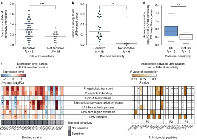
To test this possibility, we started by determining the sensitivity of the antibiotic-resistant strains to bile acid, a membrane-damag- ing agent. As expected, 75% of the antibiotic-resistant strains were more susceptible to bile acid than the wild type, and the bile acid- sensitive strains showed collateral sensitivity to an especially high number of antimicrobial peptides (Fig. 3a). To explore the common mechanistic basis of this pattern, we compared the transcriptome profiles of the wild type and 24 antibiotic-resistant strains covering all 12 investigated antibiotics (see Methods). All strains were grown in antibiotic-free medium to ensure comparability between strains that display vastly different resistance levels.
Several lines of evidence support the hypothesis that antibiotic resistance leads to alterations in outer membrane composition.
In 75% of the analysed antibiotic-resistant strains, transcriptional changes are significantly enriched in LPS- and outer membrane- related genes (Fig. 3c, left panel), including genes involved in LPS biosynthesis and phospholipid binding and transfer. Importantly, strains with an especially high number of upregulated LPS-related genes (Supplementary Tables 8 and 9) display an enhanced sus- ceptibility to bile acid (Fig. 3b). Finally, strains with upregulated genes in membrane-related functions were more likely to show collateral sensitivity to specific antimicrobial peptides (Fig. 3c and Supplementary Fig. 5).
Chemogenomic analysis of collateral sensitivity. To directly
investigate the causality between upregulation of membrane-related genes and collateral sensitivity, we carried out a chemogenomic screen to identify genes that, when overexpressed, sensitize E. coli to the membrane-interacting 18-kDa cationic antimicrobial peptide (CAP18). CAP18 is an ideal choice for two reasons. First, it dis- places divalent cations in the LPS layer and thereby permeabilizes the outer membrane
24. Second, strains that are sensitive to CAP18 displayed upregulation of genes involved in LPS biosynthesis, and phospholipid binding and transport (Fig. 3c, Supplementary Tables 8 and 9).
The chemogenomic screen of CAP18 was carried out by apply- ing an established plasmid collection overexpressing all E. coli open reading frames (ORFs)
25in a pooled fitness assay with a deep sequencing readout (for details see Supplementary Fig. 6).
Following growth of the pooled collection both in the presence and absence of CAP18, we identified genes that produce a growth defect when overexpressed in CAP18 treatment only (see Supplementary Fig. 6c). Such growth defects indicate increased CAP18 sensitivity upon overexpression of single genes. As a control, we performed the same assay on cecropin P1 (CP1), a peptide to which antibi- otic-resistant strains rarely show collateral sensitivity (Fig. 1b).
Our chemogenomic screen revealed 624 and 258 sensitizing genes for CAP18 and CP1, respectively (see Supplementary Table 10).
As expected, LPS-related genes were highly enriched among genes that sensitize to CAP18, but not to CP1, when overexpressed (Supplementary Table 11). For example, LPS biosynthesis genes were especially likely to sensitize the bacteria to CAP18 when overexpressed (odds ratio
= 4.4, P = 0.004, two-sided Fisher’s exacttest). Furthermore, CAP18-sensitizing LPS biosynthesis genes, as inferred by chemogenomics, showed higher average expression
a 87
log10 CFU per mllog10 CFU per ml log10 CFU per ml
6 5 4 3
15 30 60
Time (minutes) PROA
IND
PGLA
PGLA Wild type
TET3 CPR7
Wild type marR*
Wild type CPR7 TOB8
Wild type DOX3 KAN8
90 120
15 30 60
Time (minutes)
90 120
15 30 60
Time (minutes)
90 120
2 1
8 7 6 5 4 3 2 1
15 30 60
Time (minutes)
90 120
8 7 6 5 4 3 2 1
c
b d
8 7
log10 CFU per ml 6 5 4 3 2 1
Fig. 2 | Survival of collateral-sensitive antibiotic-resistant strains under lethal antimicrobial peptide stress. a–c, The wild-type and antibiotic- resistant strains were exposed to high concentrations of antimicrobial peptides: the TET-resistant TET3 and the CPR-resistant CPR7 strains were exposed to 15-fold (3,000 μ g ml−1) MIC of PROA (a); the CPR-resistant CPR7 and the TOB-resistant TOB8 were exposed to 10-fold MIC (125 μ g ml−1) of IND (b); and the DOX-resistant DOX3 and the KAN- resistant KAN8 were exposed to 15-fold MIC (1,500 μ g ml−1) of PGLA (c).
All antibiotic-resistant strains exhibited collateral sensitivity towards the applied peptide. d, A strain containing a single point mutation in marR (marR*) was also exposed to 15-fold MIC of PGLA. This strain exhibits resistance to multiple antibiotics and collateral sensitivity to many of the peptides tested, including PGLA (Table 1). Cells were incubated with the particular peptide for 120 minutes. Samples were taken at defined time points and plated in lysogeny broth agar plates. Percentage of survival was calculated by counting the colony forming units (CFUs). Each data point shows the mean ± s.e.m. of three biological replicates.
levels in those antibiotic-resistant strains that are sensitive to CAP18 compared to the rest (P = 0.008, two-sided Wilcoxon rank-sum test, Fig. 3d). We note that, although also frequently upregulated in CAP18-sensitive strains (Fig. 3c), genes with phospholipid-related functions were not enriched among sensitizing genes in the che- mogenomic screen (Supplementary Table 11). Taken together, these analyses indicate that altered LPS biosynthesis plays a causal role in the widespread collateral sensitivity of antibiotic-resistant strains to antimicrobial peptides.
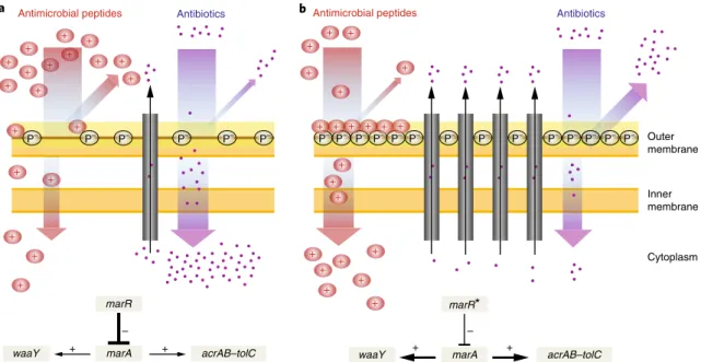
A marR mutation induces collateral sensitivity. We next deci-
phered a mechanistic link between a specific regulatory mutation and its impact on antibiotic resistance and collateral sensitivity. We focused on marR, as mutation in this gene affects bacterial response to antimicrobials in two opposite ways: it increases resistance to multiple antibiotics, but simultaneously sensitizes to membrane- interacting peptides (Table 1). Why is this so?
LPS is a major component of the bacterial outer membrane that stabilizes the membrane structure, regulates its permeability and contributes to its negative charge
26. The mar regulon is known to mediate LPS modification by positively regulating WaaY, a kinase responsible for phosphorylation of the inner core of LPS, lead- ing to an increased negative surface charge of the bacterial outer membrane
27,28(Fig. 4). As an increased negative surface charge of the membrane generally promotes antimicrobial peptide killing efficiency
9,27, upregulation of waaY is expected to enhance bacterial susceptibility to peptides.
Based on these facts, we hypothesized that mutations in marR increase antimicrobial peptide susceptibility through upregulation of waaY. Such upregulation should lead to an increase in phosphory- lation of LPS and, as a consequence, elevated negative surface charge of the bacterial outer membrane (Fig. 4). Several lines of evidence support this hypothesis. First, the antimicrobial peptide susceptibil- ity profiles of the marR single-mutant and the waaY-overexpressing strain showed substantial overlap (Table 1). Second, gene expression analysis using reverse-transcription quantitative PCR (RT–qPCR)
confirmed that waaY is nearly fourfold upregulated in the marR single-mutant strain when compared to the wild type (fold change:
3.88 ± 0.026 s.e.m., see Methods). Third, we carried out zeta poten- tial measurements and demonstrated that both the marR mutant and the waaY-overexpressing strain display relatively high negative surface charge compared to the wild type (Supplementary Fig. 7).
Finally, inactivation of waaY abolished collateral sensitivity to P3 peptides in marR mutants (Supplementary Fig. 8). To summarize, these results indicate that collateral sensitivity of the marR mutant to peptides occurs via modulation of the LPS phosphorylation path- way. We propose that the consequent altered outer membrane com- position facilitates the interaction of antimicrobial peptides with the cell membrane and thereby enhances their killing efficiency (Fig. 4).
Peptide PGLA restores antibiotic activity in antibiotic-resistant bacteria. The above results have important implications for future
development of drug combinations. Recently, two complementary mechanisms were proposed to contribute to the selective eradica- tion of drug-resistant bacteria
29. First, resistance to one drug can induce hypersensitivity to the other (collateral sensitivity, see also Supplementary Text 4)
1,2and, second, interactions can become more synergistic with the evolution of resistance, making the mutant more sensitive to the combination (resistance mutation- induced synergy)
30.
We focused on PGLA for two reasons. First, it shows an excep- tionally high number of collateral sensitivity interactions (Fig. 1).
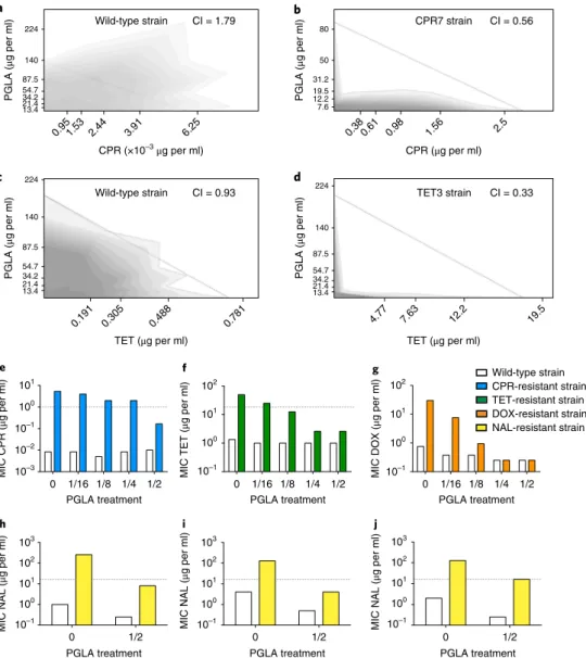
Second, when used in combination, antibiotic-PGLA pairs show strong synergism in antibiotic-resistant, but not in the corre- sponding wild-type, bacteria (Fig. 5a–d, Supplementary Table 12).
Remarkably, strong synergism is prevalent in antibiotic-resistant strains carrying mutations in marR, envZ or ompF. As these genes influence membrane permeability, we speculate that PGLA may interfere with the activity of multidrug efflux systems and/or porins responsible for the observed antibiotic resistance in these strains.
We then tested whether PGLA could be used as an adjuvant and selectively potentiate antibiotic activity against antibiotic-resistant
Table 1 | Selected individual mutations and their susceptibility profiles across antimicrobial peptides and antibioticsRelative MIC change
P3 P2 P1
Gene Mutation PGLA HBD3 SB IND D28 PR39 CAP18 PROA AP PyR BAC5 CEME LL37 TPII CP1
ompC Met1Ile 0.7a 0.6a 1.0c 1.0c 0.7a 1.0c 0.8a 0.8a 0.8a 0.8a 1.0c 0.8a 1.0c 1.0c 1.0c trkH Thr350Lys 0.7a 2.5b 1.0c 0.8a 0.8a 1.2b 1.0c 1.4b 0.6a 0.6a 1.2b 1.0c 1.0c 1.0c 1.0c sbmA Deletion 0.8a 0.8a 0.8a 0.8a 1.0c 1.3b 1.0c 1.3b 2.0b 3.6b 1.8b 1.2b 1.0c 1.2b 1.0c marR Val84Glu 0.6a 0.7a 0.7a 0.8a 1.0c 1.0c 0.8a 0.8a 1.4b 1.0c 1.0c 0.8a 1.0c 1.2b 1.0c waaY Overexpression 1.0c 0.7a 0.8a 0.8a 0.8a 1.0c 0.8a 1.0c 2.0b 1.4b 1.0c 0.8a 1.0c 1.0c 1.0c
Relative MIC change
Cell wall Gyrase Multiple 50 s 30 s Folic
acid Aminoglycoside
Gene Mutation AMP FOX CPR NAL NIT CHL ERy DOX TET TRM TOB KAN
ompC Met1Ile > 5b 1.2b 0.7a 0.8a 1.2b 0.7a 1.0c 1.0c 1.0c 1.0c 1.0c 1.0c
trkH Thr350Lys 0.5a 0.8a 0.6a 0.3a 0.8a 0.6a 0.9a 0.5a 0.5a 0.6a 3.4b 2.1b
sbmA Deletion 1.0c 1.0c 1.0c 1.0c 1.0c 0.7a 1.0c 1.2b 1.2b 1.0c 1.3b 1.5b
marR Val84Glu 2.0b 3.3b 1.9b 2.1b 1.0c 2.2b 2.0b 1.9b 1.8b 1.3b 1.0c 1.0c
waaY Overexpression 1.4b 1.0c 1.2b 1.2b 0.5a NA 1.2b 1.0c 1.0c 1.4b 1.7b 2.0b
The selected mutations repeatedly occurred in different antibiotic-resistant strains. Each mutation was inserted back into the wild-type strain separately. Relative change in MIC was calculated as the ratio between the MIC of the compound on the mutant strain and on the wild-type strain. Values lower, higher and equal to one represent acollateral sensitivity, bcross-resistance or cno interaction, respectively.
Data on antibiotic susceptibility are based on results previously published in case of the envZ, marR and trkH mutants2. NA, not available. For antibiotic and antimicrobial peptide abbreviations, see Supplementary Tables 1 and 2.
bacteria. We tested CPR-, NAL-, tetracycline (TET)- and doxycy- cline (DOX)-resistant E. coli, including laboratory-evolved strains (Fig. 5e–g) and clinical isolates (Fig. 5h–j, Supplementary Fig. 9).
The dataset was also augmented with NAL-resistant Klebsiella
pneumoniae and Shigella flexneri isolates (Supplementary Fig. 10).PGLA was administered at subinhibitory concentrations; that is, it allowed growth of the wild-type and the antibiotic-resistant strains alike. Strikingly, when used in combination, PGLA caused up to 30-fold decrease in the antibiotic resistance level of the resistant strains (Fig. 5e–g, Supplementary Figs. 9 and 10, and Supplementary Table 13). We conclude that PGLA can restore anti- biotic susceptibility against resistant bacteria when administered as an adjuvant.
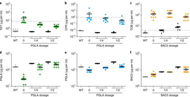
PGLA inhibits de novo evolution of resistance to antibiotics.
Finally, we asked whether concurrent administration of antibiotics
with subinhibitory dosages of PGLA hinders de novo resistance evolution in the laboratory (Fig. 6). To this end, we focused on two antibiotic-peptide combinations, CPR-PGLA and TET-PGLA. We evolved the wild-type E. coli strain in the presence of these anti- biotic-PGLA combinations and their corresponding single drug components. The protocol aimed to maximize the level of antibiotic resistance in the evolving populations during a fixed time period.
For each of the eight drug conditions, ten parallel bacterial lineages were evolved (Methods).
After only 160 generations, bacterial populations evolving in the presence of a single antibiotic reached 40- and 390-fold increases in TET and CPR MIC levels relative to their ancestor, respectively (Fig.
6a,b). Reassuringly, collateral sensitivity to PGLA arose inthese lineages (Fig. 6d,e). In contrast, antibiotic-PGLA co-treat- ment significantly slowed down the evolution of antibiotic resis- tance. In the presence of subinhibitory dosages of PGLA, the level of
cBile acid sensitivity
CHL2KAN1ERY2TOB8FOX8CPR4DOX2NAL6CPR9KAN4NAL8TRM7FOX1TOB9TET7DOX5NIT6ERY6AMP7NIT7AMP9TET1CHL9TRM6
Sensitive Not sensitive
a
Sensitive N = 45
Not sensitive N = 15 0.0
0.2 0.4 0.6 0.8
Fraction of collateral sensitivity interactions
Bile acid sensitivity
***
NCR335PGLAHBD3SBMAGR8INDD28ANGPR39PEXCAP18PROAAPPYRPLEUBAC5BAC7CEMEPRO1PXBLL37TPIICP1
Expression level
Average log2(FC)
−1 0 1
P value Expression level across
antibiotic-evolved strains
P value of association
Association between upregulation and collateral sensitivity
Antimicrobial peptides Evolved strains
0.01 0.05 0.1
b **
Not sensitive N = 6 Sensitive
N = 18
Bile acid sensitivity 0.0
0.1 0.2 0.3 0.4
Fraction of upregulated LPS-related genes
CS N = 12
Collateral sensitivity Not CS N = 12 –0.5
0.0 0.5 1.0 1.5 2.0
Average expression (log2(FC)) of CAP18-sensitizing LPS biosynthesis genes
d **
– – – – – – –
– – – – – – – – – – – – – –
– – – – – – –
– – – – – – – – – – – – – – –
– – – – – – –
– – – – – –
– – – – – – –
* * * * * * *
*
*
*
*
* *
*
* * *
*
* *
*
*
*
* * *
*
*
*
*
*
*
* *
*
*
*
* * *
* * * *
LPS transport LPS core region synthesis
LPS biosynthetic process Extracellular polysaccharide synthesis
Lipid-A biosynthesis Phospholipid transport
Phospholipid binding
P1 P2
P3
Fig. 3 | Altered membrane composition in antibiotic-resistant bacteria contributes to increased sensitivity to antimicrobial peptides. a, Antibiotic- resistant strains sensitive towards a membrane-damaging agent (bile acid) show especially large numbers of collateral sensitivity interactions with antimicrobial peptides. For antibiotic abbreviations see Supplementary Table 1. Strains sensitive to bile acid show significantly more collateral sensitivity interactions to peptides than those showing wild-type bile acid sensitivity (not sensitive group) (P < 10-3, two-sided GLMM with binomial response distributions, see Methods section). Relative frequency was calculated by dividing the number of the collateral sensitivity interactions by the number of all tested peptides (N = 24). b, Resistant strains with sensitivity to bile acid have significantly more LPS-related synthesis genes being transcriptionally upregulated than those showing wild-type bile acid sensitivity (P < 10-3, two-sided GLMM with binomial response distributions). Relative frequency was calculated by dividing the number of upregulated genes by the number of all LPS-related genes (N = 100). c, Left heatmap shows the average log2(fold change (FC)) of genes related to selected membrane-associated gene ontology processes. Many antibiotic-resistant strains are enriched in significantly up- or downregulated genes (fold change > 2 or < 0.5, false discovery rate (FDR)-corrected P-value < 0.05, two-sided Fisher’s exact test) associated with membrane-related functions. Significant enrichments (P < 0.05) are marked with an asterisk (*). Strains sensitive to a given peptide show significant upregulations in specific gene ontology groups compared to non-sensitive strains (right heatmap, two-sided Student’s t-test; for further details, see Supplementary Fig. 5). Peptides with either too few or too many collateral sensitivity interactions (n < 4 or n > 21, respectively) were excluded from the statistical analysis based on sample size calculation with alpha = 0.1, power = 0.8, delta = 2, s.d. = 1, and are indicated with a minus sign (− ). Sample size used in this analysis is provided in Supplementary Table 3. d, Upregulation of LPS-related genes sensitizes to CAP18. CAP18-sensitive antibiotic-resistant strains (CS, n = 12) have significantly higher expression levels of CAP18-sensitizing genes within the ‘LPS biosynthetic process’ gene ontology category than non-sensitive strains (not CS, n = 12) (P = 0.008, two-sided Wilcoxon rank-sum test). Boxplots show the median, first and third quartiles, with whiskers showing the 5th and 95th percentile. Significant differences are marked with asterisks (**P < 0.01 and ***P < 0.001).
antibiotic resistance reached during the course of laboratory evolu- tion was tenfold lower than in the absence of PGLA, and it remained consistently below the suggested EUCAST clinical breakpoints (Fig. 6a,b). In addition, evolution of resistance to PGLA was mar- ginal (Fig. 6d,e). Reassuringly, no substantial cross-resistance was observed between PGLA and the antibiotics CPR or TET (Fig. 6).
These patterns are not due to variations in population size across treatments (Supplementary Fig. 11).
We suspect that the efficiency of the antibiotic-PGLA co-treat- ment reflects an elevated fitness cost of antibiotic resistance muta- tions under such conditions. In other words, collateral sensitivity to PGLA may reduce the number of available resistance-conferring mutations under CPR-PGLA or TET-PGLA co-treatment (see also Supplementary Fig. 12). A full answer to this question will require detailed molecular and phenotypic characterization of laboratory- evolved bacteria. As a preliminary test, we used the drug pair TOB-bactenecin 5 (BAC5), as TOB-resistant strains showed cross- resistance rather than collateral sensitivity to the peptide BAC5 (Fig. 1). As expected, the TOB-BAC5 combination did not reduce the rate of antibiotic resistance evolution (Fig. 6c,f).
Discussion
How do mutations conferring antibiotic resistance change the sus- ceptibility to antimicrobial peptides? This question is all the more relevant as antimicrobial peptides are promising antibacterial alter- natives to antibiotics currently used in the clinics
8,9. In this work, we applied an integrated approach to study the susceptibilities of antibiotic-resistant E. coli strains towards antimicrobial peptides.
First, we found that antibiotic-resistant bacteria generally exhibited collateral sensitivity (increased susceptibility) to antimicrobial pep- tides, while cross-resistance was relatively rare. Several prior works have investigated cross-resistance and collateral sensitivity interac- tions between conventional antibiotics
1–3,31,32. Despite substantial
differences in the protocols, these systematic studies agreed that cross-resistance is generally 2–3 times more frequent than collateral sensitivity. By contrast, we see nearly three times more collateral sen- sitivity than cross-resistance towards antimicrobial peptides (Fig. 1).
Second, although we studied bacteria adapted to a diverse set of antibiotics with major differences in their mechanisms of action (Supplementary Table 1), bacterial susceptibilities to antimicro- bial peptides revealed several general trends. This is partly due to mutations in canonical resistance genes that emerged repeatedly in response to various antibiotic stresses. By introducing these muta- tions into a wild-type genetic background, we showed that muta- tions conferring resistance to one or more antibiotics simultaneously increase sensitivity to several antimicrobial peptides (Table 1).
The most noteworthy example is the transcriptional repressor of the mar regulon (marR). Mutations in this gene are frequently observed both in the laboratory and in clinical settings, and increase resis- tance to multiple unrelated antibiotics
2,22. Here, we showed that a mutation in this gene increases the negative surface charge of the bacterial outer membrane (Supplementary Fig. 7) and, eventually, leads to elevated susceptibility to several antimicrobial peptides.
More generally, our findings indicate that susceptibility to anti- microbial peptides arises as a by-product of genomic expression changes in antibiotic-resistant bacteria (Fig. 3), presumably because these alterations modify the chemical structure of the bacterial outer membrane. Importantly, these conclusions do not hold for all combinations of antimicrobial peptides and resistant bacteria stud- ied. Most notably, aminoglycoside-resistant bacteria accumulated a distinct set of mutations showing practically no overlap with other laboratory-evolved antibiotic-resistant bacteria
2. They uniquely carried deleterious mutations in the gene encoding the inner mem- brane transport protein sbmA
2,3. This mutation delivered resis- tance to proline-rich peptides and aminoglycosides as well (Fig. 1 and Table 1).
marR
marA –
waaY + + acrAB–tolC
marR*
marA – waaY +
acrAB–tolC +
Outer membrane
Inner membrane
Cytoplasm Antibiotics
a b
+
+ + +
+ +
+ +
+ + + +
+
+
Antibiotics
P– + + + +
+ +
+ +
+
+ + + + + +
+ + +
+ +
+ + +
+ +
+ +
+
+ + +
+ +
+ +
+
Antimicrobial peptides Antimicrobial peptides
P– P– P– P– P–P– P– P– P– P– P– P– P– P– P– P– P– P– P–
Fig. 4 | A putative mechanism underlying collateral sensitivity of antibiotic-resistant bacteria to cationic antimicrobial peptides. a, The wild-type MarR represses marA, which leads to the reduced expression of the AcrAB–TolC efflux pump and hence the cytosolic accumulation of antibiotics. b, Upon a canonical resistance mutation in the marR gene (marR*), repression of marA is substantially decreased leading to the upregulation of the AcrAB–TolC efflux pump, and hence an increased resistance to multiple antibiotics. On the other hand, MarA simultaneously promotes the upregulation of WaaY, a kinase responsible for phosphorylation of the inner core of LPS, which increases the net negative surface charge of the bacterial outer membrane. This in turn enhances susceptibility to membrane-interacting cationic antimicrobial peptides (+ ). In addition, phosphorylation of the LPS core may also enhance the permeability barrier of the outer membrane by cross-linking of neighbouring LPS molecules. Such changes promote both (1) a decreased uptake and increased efflux of antibiotics, thereby contributing to antibiotic resistance, and (2) a higher affinity of the positively charged antimicrobial peptides to the negative phosphoryl groups (P–) of the LPS core, leading to enhanced sensitivity to antimicrobial peptides.
These considerations could be important for the development of combination therapies. The fact that antibiotic-resistant strains showed extensive collateral sensitivity to certain antimicrobial peptides led us to study one of them, PGLA, in more detail. We identified two important properties of PGLA. First, antibiotic resistance generally resulted in collateral sensitivity to this pep- tide (Fig. 1) and, second, antibiotic-PGLA combinations induced synergism in certain antibiotic-resistant bacteria (resistance muta- tion-induced synergy, Fig. 5a–d). Based on these properties, we hypothesized that PGLA could be used for the eradication of drug- resistant bacteria as well as the inhibition of de novo resistance evolution. When used in combination, a subinhibitory dose of
PGLA caused up to 30-fold increase in susceptibility in antibiotic- resistant bacteria (Fig. 5e–j). Furthermore, co-administration of the same subinhibitory dose of PGLA efficiently slowed down the evolution of antibiotic resistance and kept resistance levels under the EUCAST breakpoints (Fig. 6). Future works should examine whether such a combination strategy can be maintained over lon- ger time scales without the appearance of resistance mutations that diminish the antibiotic-PGLA synergy
33. Needless to say, we only consider PGLA as a first step towards the development of an adju- vant therapy. By studying the structural and functional properties of promising antimicrobial peptides, peptidomimetic molecules could be developed that are less prone to resistance, stable against
100 10–1 101
102 101 103
100 10–1
102 101 103
100 10–1
102 101 103
100 10–1 10–2
10–3
101 102
100 10–1
101 102
100 10–1
0 1/2
0 1/16 1/8 1/4 1/2
0 1/16 1/8 1/4 1/2 0 1/16 1/8 1/4 1/2
0 1/2 0 1/2
224 Wild-type strain
a b
c d
e f g
h i j
CI = 1.79
Wild-type strain CI = 0.93 TET3 strain CI = 0.33
CPR7 strain CI = 0.56 80
50 31.2 19.512.2 7.6 140
87.5 54.734.2 21.413.4
0.95 1.53
2.44 3.91
6.25
CPR (×10–3μg per ml) CPR (μg per ml)
0.191 0.305 0.488 0.781
TET (μg per ml)
PGLA treatment PGLA treatment PGLA treatment
PGLA treatment Wild-type strain CPR-resistant strain TET-resistant strain DOX-resistant strain NAL-resistant strain
PGLA treatment PGLA treatment
4.77 7.63 12.2 19.5
TET (μg per ml)
0.380.61 0.98 1.56 2.5
PGLA (μg per ml) PGLA (μg per ml)
224
140
87.5 54.7 34.221.4 13.4
224
140
87.5 54.7 34.221.4 13.4
PGLA (μg per ml)MIC CPR (μg per ml) MIC TET (μg per ml) MIC DOX (μg per ml)MIC NAL (μg per ml)
MIC NAL (μg per ml)
MIC NAL (μg per ml) PGLA (μg per ml)
Fig. 5 | Interaction of PGLA and antibiotics when applied in combination. Antibiotic–PGLA interactions were determined in E. coli K-12 BW25133 wild- type and corresponding antibiotic-resistant strains. For antibiotic abbreviations see Supplementary Table 1. a–d, The combination effect of PGLA and CPR or TET on the wild-type strain (a and c), CPR-resistant strain (CPR7) (b) and TET-resistant strain (TET3) (d). While the combination shows strong antagonism (a) or no interaction (c) in the wild-type strain, the interaction shifted to strong synergism in the resistant strain (b and d). Dashed line represents no interaction calculated based on the Loewe additivity model (see Methods). Growth rate is represented in the combination space by the shade of the grey colour with darker shades denoting higher growth rates. e–j, The effect of subinhibitory concentrations of PGLA on antibiotic activity.
CPR-resistant CPR7 (e), TET-resistant TET3 (f) and DOX-resistant DOX3 (g) strains, derived from E. coli K-12 BW25133, were treated with subinhibitory concentrations of PGLA, while measuring the MIC for the given antibiotic to which they were adapted. The concentrations of PGLA used were 1/16, 1/8, 1/4 and 1/2 of its MIC against the wild-type strain. The MIC of NAL was measured in E. coli clinical isolates 0370 (h), 3539 (i) and CFT073 (j), and their corresponding NAL-resistant strains in the presence of 1/2 of the MIC for PGLA. None of the PGLA concentrations, when applied alone, affected the growth of the wild-type or the resistant strains (the only exception being the 40% growth rate reduction of the TET-resistant strain in response to 1/2 MIC PGLA). Dashed lines represent the clinical breakpoints for the antibiotics in E. coli (not available for DOX). Data in this figure are representative of at least two biological replicates. CI, combination index.
human proteases
8,34and efficient against a broad range of drug- resistant pathogens.
An important unresolved issue is whether collateral-sensitive peptide–antibiotic combinations could also be employed sequen- tially to select against resistance. This strategy, termed collateral sensitivity cycling, has remained controversial
1,35. Our preliminary results indicate that the two key ingredients of this strategy are pres- ent. First, evolving resistance to an antimicrobial peptide (CAP18) results in collateral sensitivity to several conventional antibiotics, and this collateral sensitivity is reciprocal (Supplementary Fig. 13).
Second, both antibiotic- and CAP18-resistant bacteria can be selec- tively eradicated by deploying the collateral-sensitive drug partner (Supplementary Fig. 14).
Last, we need to emphasize the limitations of our work. The molecular mechanisms underlying collateral sensitivity need to be addressed in more detail. It also remains to be established whether antimicrobial peptide–antibiotic combinations generally outper- form conventional antibiotic combinations in their ability to hinder resistance evolution. At best, we made the first step in these direc- tions. An important objection to clinical usage of antimicrobial pep- tides is that acquisition of resistance against synthetic antimicrobial peptides could drive cross-resistance towards antimicrobial peptides of the host innate immune system
36,37. While this is certainly a real- istic danger—at least in laboratory settings—two notes must be made. First, the diverse susceptibility profiles of antibiotic-resistant strains towards antimicrobial peptides (Fig. 1) confirm major dif- ferences both in the mechanisms of action of antimicrobial peptides and the potential routes to antimicrobial peptide resistance. Second, our data indicate that certain peptide–antibiotic combinations could select against the de novo evolution of resistance against both agents (Fig. 6).
In summary, our work establishes that antibiotic-resistant bacte- ria frequently show collateral sensitivity to antimicrobial peptides, a finding that can be utilized to identify peptide–antibiotic combina- tions that effectively eradicate resistant bacteria and slow down the de novo evolution of resistance to antibiotics.
Methods
Medium, antimicrobial agents and strains used in the study. Antimicrobial peptides. Twenty-four cationic antimicrobial peptides were used in this study:
SB006, HBD3, PROA, PR-39, PGLA, pexiganan (PEX), indolicidin (IND), BAC5 and bactenecin 7 (BAC7), NCR335, magainin 2 (MAG), R8, D28, anginex (ANG), apidaecin IB, rabbit CAP18, pyrrhocoricin (PYR), pleurocidin (PLEU), synthetic cecropin-melittin hybrid (CEME), protegrin-1 (PRO1), polymyxin B (PXB), LL37, tachyplesin II (TPII) and CP1 (see also Supplementary Table 2). Antimicrobial peptides were custom synthesized by ProteoGenix, except for PROA and PXB, which were purchased from Sigma-Aldrich. The antimicrobial peptide solutions were prepared in sterile water and kept at − 80°C until usage.
Antibiotics. The following antibiotics were used in this study (see also Supplementary Table 1): chloramphenicol (CHL), TET, AMP, FOX, CPR, erythromycin (ERY), DOX, trimethoprim (TRM), TOB, KAN, NIT and NAL.
Most of the antibiotics were purchased from Sigma-Aldrich, except for ERY (AMRESCO) and DOX (AppliChem). Fresh antibiotic solutions were prepared from powder stocks on a weekly basis, kept at − 20°C and filter sterilized before use.
Strains. E. coli K-12 BW25113 was used as the wild-type strain. The 60 laboratory- evolved resistant strains, adapted to 12 different clinically relevant antibiotics (5 evolved lines per antibiotic with the exception of AMP and NAL, for which 4 and 6 replicates were used, respectively) were established in our previous work3. Briefly, the parallel-evolved populations reached up to 328-fold increases in the MICs relative to the ancestor. In all cases, the resistance levels were equal to or above the current clinical breakpoints according to EUCAST or the European Reference Laboratory for Antimicrobial Resistance. The evolution of multidrug resistance (resistance to two or more drugs) was frequent under a single antibiotic pressure:
on average, 52% of all investigated antibiotic pairs showed cross-resistance in WT
a b c
d e f
0 1/4 1/2
PGLA dosage
WT 0 1/4 1/2
PGLA dosage
WT 0 1/4 1/2
BAC5 dosage
WT 0 1/4 1/2
PGLA dosage
WT 0 1/4 1/2
PGLA dosage
WT 0 1/4 1/2
BAC5 dosage 101
102 103 10–1 100 101 102 103
TET (µg per ml)PGLA (µg per ml) PGLA (µg per ml) BAC5 (µg per ml)
CPR (µg per ml) TOB (µg per ml)
101 102 103 10–3 10–2 10–1 100 101 102
100 101 102 100 101 102 103
Evolved to TET + PGLA combination Evolved in the absence of antibiotic
Wild-type (WT) strain
Evolved to CPR + PGLA combination Evolved to TOB + BAC5 combination
Fig. 6 | MICs of laboratory-evolved lines adapted to antibiotics in the absence and the presence of subinhibitory dosage of antimicrobial peptides.
a–f, MIC was measured following a laboratory evolution of the wild-type E. coli strain to TET (green), CPR (blue) and TOB (orange) in the absence or in the presence of 1/4 or 1/2 of the MIC of the antimicrobial peptides PGLA (a, b, d, e) or BAC5 (c, f) against the wild-type strain. MICs of the wild type and both PGLA and BAC5 evolved lines (in the absence of antibiotic) are represented by grey and white coloured bars, respectively. Each data point represents the MIC value of one of each ten parallel-evolved lines. Error bars represent the mean ± s.e.m. for each experimental condition. Dashed lines represent clinical breakpoints for TET, CPR or TOB in E. coli. Both the CPR-PGLA and the TET-PGLA combinations, which are representatives of collateral-sensitive interactions (Fig. 1), significantly slowed down the evolution of resistance towards the given antibiotic when administered together. Reassuringly, the control combination (BAC5-TOB), representing a cross-resistance interaction, did not reduce the rate of TOB resistance evolution (P = 0.0834, one-way analysis of variance).