Sinorhizobium meliloti Functions Required for Resistance to Antimicrobial NCR Peptides and Bacteroid Differentiation
Quentin Nicoud,aQuentin Barrière,aNicolas Busset,a*Sara Dendene,a Dmitrii Travin,b,aMickaël Bourge,aRomain Le Bars,a Claire Boulogne,aMarie Lecroël,aSándor Jenei,c Atilla Kereszt,c Eva Kondorosi,cEmanuele G. Biondi,aTatiana Timchenko,a
Benoît Alunni,a Peter Mergaerta
aUniversité Paris-Saclay, CEA, CNRS, Institute for Integrative Biology of the Cell (I2BC), Gif-sur-Yvette, France
bCenter of Life Sciences, Skolkovo Institute of Science and Technology, Moscow, Russia
cInstitute of Plant Biology, Biological Research Centre, Szeged, Hungary
Quentin Nicoud, Quentin Barrière, and Nicolas Busset contributed equally. Author order was determined by age, from youngest to oldest.
ABSTRACT Legumes of the Medicago genus have a symbiotic relationship with the bacterium Sinorhizobium meliloti and develop root nodules housing large numbers of intracellular symbionts. Members of the nodule-specific cysteine-rich peptide (NCR) family induce the endosymbionts into a terminal differentiated state. Individual cationic NCRs are antimicrobial peptides that have the capacity to kill the symbiont, but the nodule cell environment prevents killing. Moreover, the bacterial broad-specificity peptide uptake transporter BacA and exopolysacchar- ides contribute to protect the endosymbionts against the toxic activity of NCRs.
Here, we show that otherS. melilotifunctions participate in the protection of the endosymbionts; these include an additional broad-specificity peptide uptake trans- porter encoded by the yejABEFgenes and lipopolysaccharide modifications medi- ated by lpsB and lpxXL, as well as rpoH1, encoding a stress sigma factor. Strains with mutations in these genes show a strain-specific increased sensitivity profile against a panel of NCRs and form nodules in which bacteroid differentiation is affected. The lpsB mutant nodule bacteria do not differentiate, the lpxXL and rpoH1 mutants form some seemingly fully differentiated bacteroids, although most of the nodule bacteria are undifferentiated, while theyejABEFmutants form hypertrophied but nitrogen-fixing bacteroids. The nodule bacteria of all the mutants have a strongly enhanced membrane permeability, which is dependent on the transport of NCRs to the endosymbionts. Our results suggest thatS. meliloti relies on a suite of functions, including peptide transporters, the bacterial enve- lope structures, and stress response regulators, to resist the aggressive assault of NCR peptides in the nodule cells.
IMPORTANCEThe nitrogen-fixing symbiosis of legumes with rhizobium bacteria has a predominant ecological role in the nitrogen cycle and has the potential to provide the nitrogen required for plant growth in agriculture. The host plants allow the rhi- zobia to colonize specific symbiotic organs, the nodules, in large numbers in order to produce sufficient reduced nitrogen for the plants’needs. Some legumes, includ- ingMedicagospp., produce massively antimicrobial peptides to keep this large bac- terial population in check. These peptides, known as NCRs, have the potential to kill the rhizobia, but in nodules, they rather inhibit the division of the bacteria, which maintain a high nitrogen-fixing activity. In this study, we show that the tempering of the antimicrobial activity of the NCR peptides in theMedicago sym- biont Sinorhizobium meliloti is multifactorial and requires the YejABEF peptide transporter, the lipopolysaccharide outer membrane, and the stress response regu- lator RpoH1.
CitationNicoud Q, Barrière Q, Busset N, Dendene S, Travin D, Bourge M, Le Bars R, Boulogne C, Lecroël M, Jenei S, Kereszt A, Kondorosi E, Biondi EG, Timchenko T, Alunni B, Mergaert P. 2021.Sinorhizobium meliloti functions required for resistance to antimicrobial NCR peptides and bacteroid differentiation. mBio 12:e00895-21.https://doi .org/10.1128/mBio.00895-21.
EditorJoerg Graf, University of Connecticut Copyright© 2021 Nicoud et al. This is an open-access article distributed under the terms of theCreative Commons Attribution 4.0 International license.
Address correspondence to Peter Mergaert, peter.mergaert@i2bc.paris-saclay.fr.
*Present address: Nicolas Busset, Plant Health Institute, Montpellier, France.
Received29 March 2021 Accepted21 June 2021 Published
® 27 July 2021
involving hundreds of peptides, has been described for the rhizobium-legume symbio- sis (2, 5–9). Legumes form a symbiosis with phylogenetically diverse nitrogen-fixing soil bacteria, collectively called rhizobia. This nutritional symbiosis provides reduced nitrogen to the plants. The symbiosis implies the formation of nodules, specific symbi- otic organs, on the roots of the plants. These nodules house the nitrogen-fixing rhizo- bia, which transfer their produced ammonia to the plant in return for the exclusive niche in the nodules, where they multiply massively from a single bacterium or very few infecting bacteria to a population of millions.
After endocytic uptake by the symbiotic nodule cells, the multiplied bacteria reside intracellularly in vesicles called symbiosomes. The nodule cells and symbiosomes es- tablish the optimal conditions for nitrogenfixation and metabolic exchange with the endosymbionts and, at the same time, keep them in check. The low oxygen levels pre- vailing in the symbiotic nodule cells transform the rhizobia into a differentiated physio- logical state, called the bacteroid, which is adapted for nitrogenfixation. Moreover, in certain legume clades, like the Inverted Repeat-Lacking Clade (IRLC) and the Dalbergioids, the physiological transition of the bacteroids is accompanied by a re- markable differentiation process that is manifested in an irreversible loss of the capacity of bacteroids to divide (10, 11). These terminally differentiated bacteroids have a partially permeabilized cell membrane. They are giant bacterial cells. A switch in the bacterial cell cycle, from a regular succession of replication and division to a se- ries of repeated genome replications without divisions, drives this cell enlargement, resulting in polyploid bacteroids.
The terminal differentiation is triggered by a family of nodule-specific cysteine-rich peptides (NCRs), produced by the symbiotic nodule cells (11–13). Over 600NCRgenes were identified in theMedicago truncatulagenome, while theNCRrepertoire in other species of the IRLC and the Dalbergioids ranges from a few to several hundred (11, 12, 14). TheNCRgenes in M. truncatulaare specifically expressed in the symbiotic cells (15). They are activated in waves during the differentiation of the bacteroids, including sets ofNCRgenes activated at the onset and others at the intermediate orfinal stages of the differentiation. These temporal profiles indicate that theNCRgenes have specific functions during the bacteroid formation process.
The NCR peptides have structural features shared with AMPs, and at least some NCRs, in particular, the cationic ones, can kill or inhibitin vitrothe growth of not only the rhizobium symbionts but also many other bacteria and even fungi (16). Their major antibacterial activity results from their capacity to disturb the integrity of the inner and outer membranes of bacteria (13, 17), although some NCRs also have activities inhibi- ting essential intracellular machineries (18). However, bacteroids remain metabolically active for a very long time, despite the high burden of NCRs. Possibly, the environment of the symbiotic nodule cells and symbiosomes contributes to tempering the antimi- crobial activities of the peptides. Importantly, also specific functions of the bacteria themselves are NCR resistance determinants in the bacteroids.
Sinorhizobium meliloti, the symbiont ofMedicagoplants, requires the peptide trans- porter BacA to counter the NCR peptides inside the symbiotic nodule cells and estab- lish a chronic infection (19).S. meliloti bacAmutants are hypersensitive to the antimi- crobial NCRs. They induce nodules and infect plant cells in a seemingly normal fashion,
but the mutants die rapidly after their release in the symbiotic cells. This death can be avoided by blocking NCR transport to the infecting rhizobia in theM. truncatula dnf1 mutant (19). BacA proteins are broad-specificity peptide uptake transporters (20–22).
They can promote the uptake of NCR peptides, suggesting that BacA provides resist- ance by redirecting the peptides away from the bacterial membrane, thereby limiting membrane damage. Exopolysaccharide (EPS) is another known factor ofS. melilotithat helps the endosymbionts to withstand the NCRs (14, 23). This negatively charged extracellular polysaccharide traps the cationic AMPs, reducing their effective concen- tration in the membrane vicinity.
Bacterial resistance to AMPs is usually multifactorial (24), suggesting that besides BacA and EPS, additional functions ofS. melilotibacteroids contribute to resisting the NCRs in the symbiotic nodule cells. The literature onS. melilotiis rich in the description of bacterial genes that are required for symbiosis. However, the reporting on these mutants often lacks precise information on their bacteroid phenotype and/or on their sensitivity to NCRs. Moreover, transcriptome sequencing (RNA-seq) and transposon sequencing (Tn-seq) analyses of NCR-treated cells and NCR-protein interaction studies identified a whole suite of additional candidate NCR-responsive functions inS. meliloti (18, 25–27). Together with BacA and EPS, some of theseS. melilotifunctions may con- tribute to alleviate the NCR stress on the bacteroids. To test this hypothesis, we have selected candidate genes and analyzed the phenotype of the corresponding mutants in NCR resistance and bacteroid formation.
RESULTS
Sinorhizobium melilotimutants with enhanced sensitivity to NCR peptides.The S. melilotifunctions selected in this study include a broad-specificity peptide uptake transporter encoded by theyejABEFgenes (SMc02829 to SMc02832) and lipopolysac- charide (LPS) modifications mediated by lpsB (SMc01219) and lpxXL (SMc04268), as well asrpoH1(SMc00646), encoding a stress sigma factor. The YejABEF ABC transporter was selected on the basis of a genetic screen by Tn-seq ofS. meliloti, revealing that mutants have an increased sensitivity to the peptide NCR247 (25). Moreover, another Tn-seq study revealed that the transporter is essential for symbiosis (28). The LPS struc- ture is one of the major determinants of AMP resistance and sensitivity in Gram-nega- tive bacteria (24, 29). The selected geneslpsBandlpxXLencode a glycosyltransferase involved in the synthesis of the LPS core and a very-long-chain fatty acid acyltransfer- ase involved in the biosynthesis of lipid A, respectively. Strains with mutations in these genes are affected in their resistance to AMPs and in symbiosis (30, 31). Finally, the rpoH1gene is a global stress response regulator inS. meliloti(32). This gene, as well as its target genes, is upregulated in NCR247-treated cells (26, 27). AnrpoH1mutant is also affected in symbiosis (33). A second gene,rpoH2, has no apparent function under free-living conditions or during symbiosis (34, 35). These selected genes are expressed in all nodule zones but have peak expression in different regions of the nodule, where bacteria infect plant cells, undergo the differentiation process, orfix nitrogen (36) (see Fig. S1 in the supplemental material).
The sensitivities of strains with mutations in these candidate genes (Table S1) were tested with a small panel of cationic NCR peptides that were previously shown to have antimicrobial activities (14). The tested peptides, NCR169 (isoelectric point, 8.45), NCR183 (10.10), NCR247 (10.15), and NCR280 (9.80), displayed three different expres- sion patterns in the nodule tissues (15). TheNCR280gene is expressed in the younger nodule cells and theNCR169gene in the older cells, whileNCR183andNCR247have an intermediate expression pattern (Fig. S1). The selected mutants were tested along with the wild-type strain and thebacAmutant, which was previously shown to be hypersen- sitive to NCRs (19). The four tested NCR peptides had strong antimicrobial activities against the wild-type strain, which displayed a survival rate ranging from 8% to 0.03%, depending on the tested peptide (Table S2). In agreement with previous results, the bacAmutant was hypersensitive to the four peptides (Table S2; Fig. 1). Interestingly,
the newly analyzed mutants all displayed a higher sensitivity to at least one of the pep- tides than the wild type (Table S2; Fig. 1). ThelpxXLmutant was more sensitive to the four peptides, and thelpsB, yejE, and yejF mutants were more sensitive to NCR183, NCR247, and NCR280. TheyejAmutant was more sensitive to NCR280, and therpoH1 mutant was more sensitive to NCR247. The differential responses to peptide NCR247 of theyejAmutant, on one hand, and of theyejE andyejFmutants, on the other hand, correspond to results of the previously described Tn-seq screen with this peptide (25).
In contrast to the other mutants, the strains with mutations in the YejABEF transporter genes were newly constructed and not characterized before. Therefore, we confirmed that the NCR247 sensitivity phenotype was directly attributable to the inactivation of the transporter by complementation of theyejF mutant phenotype with a plasmid- borne copy of theyejABEFgenes (Table S2). Taken together, our analysis indicated that each mutant displayed a sensitivity profile specific to the panel of tested peptides.
Inside the nodules, bacteroids experience (besides exposure to the NCR peptides) additional stress factors, such as elevated hydrogen peroxide levels (37), an acidic pH formed in the peribacteroid space (38), and a microaerobic environment (39). Almost all the mutants behaved very similarly to the wild type during unstressed growth in culture or in response to hydrogen peroxide, acid, and low-oxygen stress (Fig. S2).
Only therpoH1mutant was much less resistant to acid, in agreement with previous reports (33, 40), as well as to anaerobic stress, and it had a reduced growth rate (Fig. S2). On the other hand, the mutants were more sensitive than the wild type to the exposure to the detergent sodium dodecyl sulfate (SDS) (Fig. S2), indicating that these mutants have a reduced ability to cope with membrane-permeabilizing stresses, in agreement with their increased NCR sensitivity. Thus, except for the rpoH1mutant, which has a general defect in growth and stress management, the selected mutants seem to be, among the known stress factors in the nodule environment, specifically sensitive to the NCRs.
Nodule formation by NCR-sensitiveSinorhizobium melilotimutants.Next, the phenotypes of these mutants in symbiosis with M. truncatula were compared with those of the wild-type strain and thebacAmutant. Measurement of the nitrogenfixa- tion activity and macroscopic inspection of the root systems of plants inoculated with the wild type and the seven mutants (Fig. S3) revealed that, besides the wild type, strains with mutantyejA,yejE,yejF, orlpxXL genes formed functional nodules (Fix1), although theyejE,yejF, andlpxXLmutants had a reduced nitrogenfixation activity. On FIG 1 Sensitivity profile ofSinorhizobium melilotistrains to a panel of NCR peptides. The heatmap shows the survival of the mutant strains, expressed as percentages of that of the wild type (WT), set at 100%, for each peptide treatment. Nonparametric Kruskal-Wallis and post hocDunn tests were used to assess the significance of differences, which is indicated by asterisks (**,P,0.05;*,P,0.1).
The results of one representative experiment out of two are shown.
the other hand, the bacA,lpsB, andrpoH1mutants formed nonfunctional (Fix–) and abnormal-looking nodules that were small and white, in agreement with previous descriptions (19, 30, 33).
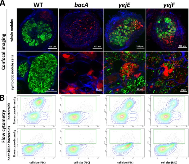
The histological organization of the nodules formed by the mutants, the formation of infected symbiotic cells, and the viability of the bacteria that they contain were ana- lyzed using confocal microscopy (Fig. 2A). The used staining procedure highlights the nodule bacteria with a greenfluorescence signal (SYTO 9) when their membranes are well preserved and with a redfluorescence signal (propidium iodide) when their mem- branes are highly permeable. As previously reported, wild-type nodules formed symbi- otic cells infected with green-labeled elongated bacteroids, while the nodules infected with thebacAmutant contained symbiotic cells carrying small undifferentiated bacte- ria stained red (19).
In contrast to what we expected from the macroscopic inspection of the nodules and their ability tofix nitrogen, theyejA,yejE, andyejFmutant bacteroids were substantially altered compared to wild-type bacteroids. A high proportion of them were stained red, indicating that their membranes were strongly permeabilized. Nevertheless, other host cells contained bacteroids stained green by the SYTO 9 dye. LpxXL is known to be impor- tant, but not essential, toS. melilotiduring symbiosis with alfalfa (Medicago sativa) (41).
The mutant forms hypertrophied bacteroids (31). In our experiments, thelpxXLmutant displayed elongated bacteroids that were mostly permeable to propidium iodide (stained red). This observation confirmed thatS. melilotiLpxXL is also essential for the normal bac- teroid differentiation process inM. truncatulabut that it is not crucial for infection and
FIG 2 Symbiotic phenotypes ofSinorhizobium melilotimutants during symbiosis with wild-typeMedicago truncatulaor thednf1mutant. (A) Membrane permeability in bacteroids of theS. melilotiSm1021 wild type and mutants in wild-typeM. truncatulanodules, determined by live-dead staining of nodule sections and confocal microscopy. (B) Membrane permeability of theS. melilotiSm1021 wild type and mutants in nodules of theM. truncatula dnf1mutant, determined by live-dead staining of nodule sections and confocal microscopy. Nodule phenotype at 21 days post inoculation. (Top row) Full nodule sections; (bottom row) enlarged images of symbiotic cells. Scale bars are indicated in each panel. The staining procedure of nodule sections with a mixture of the dyes propidium iodide, SYTO 9, and calcofluor white highlights the bacteroids and nodule bacteria with a greenfluorescence signal (SYTO 9) when their membranes are well preserved and with a redfluorescence (propidium iodide) when their membranes are highly permeable. Plant cell walls are stained blue (calcofluor white). Representative images are shown, originating from at least two independent nodulation assays and from 5 to 10 nodules analyzed per condition.
nitrogenfixation. TherpoH1mutant formed elongated bacteroids that nevertheless were strongly stained by propidium iodide, in agreement with the Fix–phenotype of the nodules and the described phenotype of the mutant in alfalfa (33). Inside the small bumps elicited by thelpsBmutant, no cell seemed to be colonized by bacteria, as revealed by confocal microscopy. Thus, theS. meliloti lpsBmutant failed to colo- nize the host cells and was defective at a stage before plant cell infection. It was pre- viously reported that thelpsBmutant colonizes nodule cells inM. sativa, suggesting a more severe defect inM. truncatula(42, 43). Therefore, we tested the phenotype of all the selected mutants also onM. sativa(Fig. S4). ThelpsB,rpoH1, and theyejA mutants indeed had milder defects on this host, while the other mutants had similar phenotypes on the two hosts.
Can the increased membrane permeability of the NCR-sensitiveSinorhizobium melilotimutants be attributed to the NCRs in nodules?The high membrane perme- ability of thebacAmutant nodule bacteria is the result of the action of the NCR pep- tides in the nodule cells of this NCR-hypersensitive mutant (19). Possibly, the same is true for the other mutants. To test this hypothesis, we made use of theM. truncatula dnf1mutant, which is defective in a nodule-specific subunit of the signal peptidase complex (44). This mutant cannot transport NCR peptides to the symbiosomes (13). As a result, the wild-typeS. melilotinodule bacteria in this mutant are not differentiated.
ThebacAmutant, which is strongly permeabilized and stained by propidium iodide in nodules of wild-typeM. truncatulaplants, is not so in the dnf1mutant nodule cells because the bacteria are not challenged anymore with the NCRs (Fig. 2B) (19). Similarly to thebacAmutant, theyejA,yejE,yejF,lpxXL, andrpoH1mutants did not display mem- brane permeability, as revealed by the absence of propidium iodide staining, in the infected nodule cells of the dnf1 mutant (Fig. 2B). This thus suggests that these mutants become membrane permeabilized by the actions of the NCRs and that their symbiotic defects are at least in part due to their hypersensitivity to the NCRs. ThelpsB mutant did not form detectable nodule-like structures on thednf1roots. Therefore, we cannot draw conclusions about the involvement of the NCR peptides in the symbiotic phenotype of this mutant.
Bacteroid differentiation of the NCR-sensitiveSinorhizobium melilotimutants.
A very strong cell enlargement and an increase in the ploidy level of the bacteria char- acterize the differentiated bacteroids inMedicagonodules. These parameters can read- ily be measured by DAPI (49,6-diamidino-2-phenylindole) staining andflow cytometry (10). The wild-type bacteroids fromM. truncatula nodules had a high DNA content, over 20-fold higher than the DNA content in free-livingS. meliloti(Fig. 3), and increased light-scattering parameters reflecting the cell enlargement (Fig. S5). As previously reported, the nodules infected by thebacAmutant did not contain differentiated bac- teria (Fig. 3; Fig. S5) (21). The bacteria in nodules induced by thelpsBmutant, probably
FIG 3 DNA contents of nodule bacteria inMedicago truncatula. Flow cytometry analysis of the DNA contents of bacteria in culture or isolated from nodules infected with the indicated strains and stained with 49,6-diamidino-2-phenylindole (DAPI). The cell counts (yaxes) are represented as a function of the DAPIfluorescence intensity (xaxes). The arrow in each graph indicates the mean DNA content of wild-type bacteroids, as in the upper left panel. The results of one representative experiment out of two are shown.
located in infection threads, had a profile that confirmed the complete absence of dif- ferentiated bacteria (Fig. 3; Fig. S5). Nodules infected with the rpoH1 mutant had mostly undifferentiated bacteria, although a small amount of fully differentiated cells was detected, as were cells at an intermediate stage. Also, the nodules of thelpxXLmu- tant contained many undifferentiated bacteria as well as fully differentiated ones (Fig. 3; Fig. S5). In contrast, theyejA,yejE, andyejF mutant nodules contained large numbers of fully differentiated bacteria (Fig. 3; Fig. S5).
It was reported previously that thelpxXLmutant formed nodules containing hyper- trophied and larger bacteria than the wild-type bacteroids (31). In our study, this differ- ence was detectable in the flow cytometry measurements, which showed a higher DAPIfluorescence and light scattering for the small portion of differentiated bacteria in these nodules (Fig. 3; Fig. S5). Moreover, we noticed that the bacteroids in the nod- ules infected with theyejA,yejE, andyejFmutants displayed similar higher levels of DNAfluorescence and light scattering (Fig. 3; Fig. S5), suggesting that these bacteroids also have abnormal morphologies.
These patterns inM. truncatula nodules were overall similar to those inM. sativa, although therpoH1mutant showed a higher number of intermediate and fully differ- entiated bacteroids and theyejAmutant had a profile similar to that in nodules of the wild type at 21 days post inoculation (dpi), while larger-than-normal bacteroids were detected only at 32 dpi (Fig. S6). These differences corresponded well with the macro- scopic and microscopic differences in the nodules between the two host plants (Fig. 2;
Fig. S3 and S4).
Defective bacteroid differentiation of yejE and yejFmutants. To confirm the altered bacteroid morphologies of theyejmutants suggested by the cytometry analy- sis, the yejE and yejF mutants were observed at high magnification by microscopy.
Confocal and transmission electron microscopy (TEM) of M. sativa nodule sections showed that the nodule cells infected with theyejEoryejF mutant contained a very heterogeneous population of abnormal bacteroid morphs, including elongated, spheri- cal, club-shaped, or irregular blob-like cells (Fig. 4A; Fig. S7). These cells contrasted strongly with the narrow, elongated wild-type bacteroids. This aberrant bacteroid phe- notype of the mutants was restored to a wild-type phenotype by the introduction of a plasmid-borne copy of theyejABEFgenes (Fig. S7; Table S1). The difference between wild-type and mutant bacteroids was also obvious byfluorescence microscopy obser- vations of purified nodule bacteria (Fig. 4B). The quantification of cell morphology pa- rameters by image analysis with MicrobeJ of free-living bacteria and the purified nod- ule bacteria confirmed the irregular forms ofyejEandyejFbacteroids, notably showing that these bacteroids are broader and more spherical than the elongated wild-type bacteroids (Fig. 4C). Transmission electron microscopy further showed that the cyto- plasm and inner membranes of many of theyejF bacteroids were retracted, leaving very large intermembrane spaces, which in some cases even developed into vacuoles entirely surrounded by cytoplasm. Some cells had multiple small and large vacuoles (Fig. S7A). Unlike with the bacteroids, in the cultured bacteria, no differences were observed in the ultrastructure of the wild-type andyejFmutant bacteria (Fig. S7B).
The formation of strongly abnormal bacteroids of theyejmutants isa prioricontra- dictory with the nitrogenfixation activity of these nodules. Expression of thenifHgene is a marker for nitrogen-fixing bacteroids (45). A greenfluorescent protein (GFP) gene under the control of thenifHpromoter was introduced into the wild-type strain and thebacA,yejE, andyejFmutants (Table S1). Analysis of the GFPfluorescence in nodule bacteria ofM. truncatula(Fig. 5) and M. sativa(Fig. S8) by confocal microscopy and flow cytometry showed that despite their aberrant morphology, theyejEandyejFbac- teroids were mostly functional, although some nodule cells contained highly permea- ble bacteroids lacking the GFP signal, suggesting that they were nonfunctional.
NCR247 uptake by the YejABEF transporter.InEscherichia coli, both YejABEF and the BacA homolog SbmA mediate the transport of microcin C peptide-nucleotide anti- biotics. Therefore, we tested whether the overlap in the substrates of YejABEF and BacA can be extended to NCR peptides, which are known substrates of BacA (20, 21).
FIG 4 Bacteroid morphology ofyejEandyejFmutants inMedicago sativanodules. (A) Sections of nodules infected with the wild type and theyejEoryejFmutant were stained with a mixture of the dyes propidium iodide, SYTO 9, and calcofluor white and observed by confocal (Continued on next page)
We tested the impact of the mutations in the YejABEF transporter genes on NCR247 uptake and compared it with a mutation inbacA. With aflow cytometry-based assay and a fluorescent derivative of the NCR247 peptide, we found that while NCR247 uptake is completely abolished in thebacA mutant, as expected, its uptake is also reduced but not completely abolished in theyejA,yejE, andyejFmutants (Fig. 6). This suggests that the YejABEF transporter contributes to NCR uptake.
FIG 4Legend (Continued)
microscopy. Scale bars (10mm) are indicated in each panel. Representative images are shown, originating from at least two independent nodulation assays and from 5 to 10 nodules analyzed per condition. (B) Preparations of cultured bacteria or purifiedM. sativanodule bacteria of the wild type and theyejFmutant were observed byfluorescence microscopy. The panels are composite images, and the shown individual cells were cut from original images and recombined in a single panel. Each panel is at the same magnification, and the scale bar (10mm) is indicated in the left panel. (C) Cell shape parameters of free-living bacteria and bacteroids determined by MicrobeJ.
Dot plots show the mean widths and lengths of cells of the indicated strains. Each point represents the values of one bacterium. Numbers of analyzed cells (n) are indicated.
FIG 5 Nitrogenase expression in the yejEand yejFmutant bacteroids inMedicago truncatulanodules. (A) Confocal microscopy of sections of nodules infected with S. melilotiSm1021.pHC60-pnifH::GFP(WT), Sm1021.DbacA.pHC60-pnifH::GFP(bacA), Sm1021.DyejE.pHC60-pnifH::GFP(yejE), or Sm1021.DyejF.
pHC60-pnifH::GFP (yejF) and stained with propidium iodide (red stain). Green-stained bacteroids are functional, while red-stained bacteroids are nonfunctional. (B) Flow cytometry determination of GFP levels in nodule bacteria (upper panels) and heat-killed nodule bacteria (lower panels). The green square shows the position of the GFP-positive bacteroids. FSC, forward scatter.
DISCUSSION
Multiple functions ofSinorhizobium meliloticontribute to NCR resistance and are required for bacteroid formation and persistence.The NCR peptides are a two- edged sword. On the one hand, they maneuver the rhizobial endosymbionts into a termi- nally differentiated state via a multitude of activities on the bacteria, of which membrane permeabilization, cell cycle perturbation (polyploidization), and cell enlargement are the most visible ones. On the other hand, many NCRs, notably the cationic ones, have antimi- crobial activities that potentially kill the endosymbionts (13, 14). Therefore, rhizobia have to defend themselves to be able to establish a chronic infection in the NCR-producing symbiotic cells of the nodules. Previous work has identified the BacA peptide transporter and the EPS barrier in the bacterial envelope as defenses ofSinorhizobiumstrains against the NCRs of theirMedicagohosts (14, 19, 23).
Here, we defined three new functions inS. meliloti, the LPS, the sigma factor RpoH1, and the YejABEF peptide transporter, as additional determinants in bacteroids required to cope with NCR peptides. We show that mutants with the corresponding genes knocked out are more sensitive to a panel of antimicrobial NCRs and that this hyper- sensitivity is correlated with a strongly enhanced membrane permeability of the nod- ule bacteria and abnormalities in their morphology and ploidy levels. It is striking, how- ever, that the different mutants have markedly different bacteroid phenotypes, ranging from undifferentiated to hyper-differentiated. This can be attributed to several factors. Each mutant has a specific NCR sensitivity profile when tested against a small panel of peptides, and the“NCR landscape”present in the developing symbiotic cells is continuously changing because of the expression of the NCR genes in different waves during symbiotic cell differentiation (15). Accordingly, the mutants may accumu- late NCR-induced damage at different rates and reach the breaking point at different stages over the course of the bacteroid differentiation process (Fig. S1). Moreover, the expression patterns of the bacterial genes in the nodule zones are different, suggesting that their principal impact is realized at distinct stages of the symbiotic cell develop- ment and bacteroid differentiation.
Blocking the targeting of the NCR peptides to the nodule bacteria by the use of the M. truncatula dnf1mutant prevents the membrane permeabilization of the nodule bac- teria in the mutants and results in the formation of very similar aberrant nodules by
FIG 6 Uptake of NCR247 mediated by the BacA and YejABEF transporters. Flow cytometry measurement offluorescein isothiocyanate (FITC)fluorescence inS. meliloticells before (time zero [T0]) and after 30 min of incubation (T30) of the bacteria with the NCR247-FITC peptide.“FITC”on thexaxis is FITC- derived fluorescence, and “FSC” on the y axis is forward scatter. In the wild type, two subpopulations of bacteria were observed, an FITC-negative population and an FITC-positive one, which has taken up the NCR247-FITC peptide. The green rectangle in the different panels shows the FITC-positive subpopulation in the wild type and the corresponding region in the analyzed mutants. In thebacAmutant, the FITC-positive subpopulation is absent, and in theyejA,yejE, andyejFmutants, it is strongly reduced compared to that of the wild type. The analysis was performed in quadruplicate, and the results of a representative example are shown.
the wild-type and all mutant strains (except for thelpsBmutant [see below]). This ob- servation places the symbiotic role of these bacterial genes, at least in part, down- stream of the peptide targeting the symbiosomes and is in agreement with the impor- tant role of these genes in responding to the NCRs. It should be noted that besides the antimicrobial NCR peptides, other stress factors, such as high H2O2levels, a low pH, and low oxygen, are present in the nodule environment (37–39). We found that, except for therpoH1mutant, strains with mutations in the studied genes were not affected by these stresses differently from the wild type, making it unlikely that they are the main cause of the bacteroid phenotypes in these mutants. However, our analyses do not exclude the possibility that these genes also contribute in the bacteroids to other, less- well-characterized stress factors in the nodule environment.
The functions ofS. melilotidescribed here and before probably picture only part of the full toolkit of this symbiont to survive the NCR challenge. Many additionalS. meli- lotimutants are described with symbiotic phenotypes that are suggestive of a similar contribution to NCR resistance (46–57). In addition, other genes that may be important for symbiosis were discovered in a Tn-seq screen with the peptide NCR247 (25). This screen identified here-described functions, like those of the BacA and YejABEF trans- porters and LPS and EPS biosynthesis, but also identified never-analyzed functions. It would be of major interest to explore the functions of these genes in relation to the NCR response of bacteroids. Note that some of these genes might have escaped identification in previous genetic screens for symbiosis mutants because mutations might provoke only subtle phenotypes with a weak overall effect on nitrogenfixation under standard labora- tory conditions but with bacteroid alterations in morphology and persistence, as illus- trated here with theyejABEFmutants.
Response to NCR-induced stress regulated by the alternative sigma factor RpoH1.Bacteria deal with different types of stress conditions by global transcriptional responses mediated by alternative sigma factors. Among them, the RpoE and RpoH sigma factors are known to respond to periplasmic and membrane stressors, including AMPs (58–60). Given the membrane damage provoked by NCRs, a role of the ortholo- gous regulators ofS. melilotiin bacteroid differentiation and the NCR response maya priorihave been expected.S. melilotihas 11 RpoE-like sigma factors. Remarkably, de- spite the fact that some RpoEs have a considerable effect on gene transcription, all sin- gle mutants, all possible double mutants, and even a mutant lacking all 11 genes showed no detectable phenotypic difference from the wild type in symbiosis or during many tested free-living growth conditions, including growth in the presence of mem- brane stresses (61, 62). Thus, the 11S. melilotiRpoEs do not have the expected role in regulating the envelope stress response. This role seems to be taken up by RpoH1 inS.
meliloti, in agreement with the observation that the growth of therpoH1mutant is affected in the presence of various membrane-disrupting agents (33). We propose that the here-uncovered role of RpoH1 in NCR resistance is connected to its regulation of membrane stress. In addition, RpoH1 is crucial to various cytoplasmic stresses, includ- ing heat and osmotic and acidic stress, as well as anoxia or microoxia (33–35, 40). Thus, besides providing resistance to NCRs, RpoH1 might also be crucial for handling these additional stress factors in bacteroids. As RpoH1 controls directly and indirectly the expression of several hundreds of genes, a future challenge will be to dissect this mas- sive response into specific stress adaptations (32, 40).
The lipopolysaccharide barrier against NCR membrane damage.In Gram-nega- tive bacteria, LPS constitutes thefirst point of attack of cationic AMPs. In a two-stage process, AMPs make thefirst electrostatic interactions with the negatively charged LPS, allowing the AMPs to approach the membrane lipids and subsequently to insert into the lipid bilayer, perforate it, and translocate into the periplasm (63). The chemical composition of LPS can influence the efficiency of the AMP attack, and pathogens have evolved mechanisms to recognize the presence of AMPs and to modify in response the composition of the LPS to lower the potency of the AMP attack (64).
S. meliloti lpsBencodes a glycosyltransferase that participates in the biosynthesis of the LPS core (65). The mutation of the gene affects both the core structure and
mutant before the release of bacteria into nodule cells suggests that the LPS may pro- vide protection against another stressor produced very early on in the infection pro- cess inM. truncatula. On the other hand, it was recently reported that someNCRgenes are expressed in infected root hairs or Nod factor-stimulated root epidermal cells (66, 67). Thus, it is possible that the challenge with these early NCR peptides is already det- rimental to the mutant, blocking any further progress in the infection process.
LpxXL ofS. melilotiis a specific acyltransferase that introduces in the lipid A moiety of LPS the very-long-chain fatty acid 27-OHC28:0 (41). This LpxXL-dependent acylation is expected to make the lipid A hydrophobic, forming the biophysical basis for LpxXL- dependent NCR resistance. Indeed, it is known that the increased hydrophobicity of lipid A in bacteria increases the thickness of the outer layer of the outer membrane and reduces the membranefluidity, which in its turn prevents or delays the insertion of diverse AMPs and subsequent membrane damage. Increasing lipid A hydrophobicity by introducing additional acyl chains is a well-known mechanism used bySalmonella to enhance its resistance against host AMPs during infection (64).
The YejABEF peptide transporter provides resistance to membrane-damaging peptides and sensitizes bacteria to AMPs with intracellular targets.A Tn-seq study has shown that transposon insertions in theS. meliloti yejBgene provoke a moderate growth impairment (68). Moreover, in a strain cured of the two symbiotic plasmids but not in the wild type, insertions in the four genes of the transporter resulted in a strong growth defect, signifying that a function redundant to YejABEF is encoded on one of the symbiotic plasmids (68). Additionally, the expression of theyejAgene, encoding the periplasmic binding protein of the transporter, was found in a systematic analysis of periplasmic binding proteins inS. melilotito be induced by taurine, valine, isoleu- cine, and leucine, indicating that these amino acids may be substrates of YejA (69).
Together, these data suggest that the YejABEF transporter ofS. melilotihas a transport role that is important for growth in free-living bacteria. However, this ABC transporter has never been analyzed before in detail in the context of the rhizobium-legume sym- biosis. Furthermore, even though this transporter is highly conserved among proteo- bacteria, its physiological role in whichever bacterium has been characterized in only a few instances. One of them is the uptake of microcin C inE. coli. This translation-inhibi- ting peptide-nucleotide of bacterial origin has no action on the bacterial membrane but has an intracellular target, the Asp aminoacyl-tRNA synthase (70). A strain with a mutation in the YejABEF transporter cannot take up microcin C and is resistant to it (71, 72). Conversely, theSalmonellaandBrucella yejABEFmutants are more sensitive to peptides with membrane-damaging activities, such as defensins, polymyxin B, prota- mine, and melittin. Consequently, these mutants have reduced pathogenicity because their capacity to survive in macrophage cell lines or in mice is diminished (73, 74).
Thus, the increased sensitivity of theyejA,yejE, andyejFmutants ofS. melilotitoward NCRs is consistent with these previousfindings.
The characteristics of the YejABEF peptide transporter toward membrane-damaging peptides versus peptides with intracellular actions are intriguingly parallel to the features of the BacA peptide transporter (named SbmA inE. coliandSalmonella). These transport- ers inE. coli,Salmonella, orS. melilotiare required for the import of diverse peptides with intracellular targets (microcins B17 and J25, Bac7, Bac5, and bleomycin), and mutants are
therefore resistant to them, while the same mutants are hypersensitive to membrane- active peptides (defensins, NCRs) (19, 75–78). Moreover, the ranges of peptides that can be imported overlap between these two transporters. Both, YejABEF and SbmA inE. coli can take up microcin C derivatives (71). Furthermore, we show that inS. meliloti, both BacA and YejABEF contribute to NCR247 uptake. Intriguingly, our genetic analysis sug- gests that both transporters cooperate to import this peptide, since inactivation of one or the other abolishes or strongly reduces uptake. How these two transporters can physically interact and cooperate is an issue of interest for future research. Nevertheless, BacA can function (partially) in the absence of YejABEF, while the inverse is not the case. This may be the basis for the markedly different symbiotic phenotype of thebacAandyejmutants.
While thebacAmutation is detrimental from the earliest contact with NCRs, and the mu- tant bacteria die as soon as they are released from infection threads in the symbiotic cells, theyejmutants cope with the NCR peptides much longer and show abnormalities only at the end of the bacteroid differentiation process and symbiont life span. TheyejAmutant had a different NCR sensitivity profile than theyejEandyejFmutants, and this was corre- lated with a different symbiotic phenotype in theM. sativahost. This suggests that the periplasmic binding protein YejA contributes to the interaction with only a subset of the NCR peptides. Nevertheless, YejA seems to contribute to NCR247 uptake to an extent sim- ilar to that of YejE and YejF, despite the fact that, unlike with theyejEandyejFmutants, the sensitivity of theyejAmutant to the NCR247 peptide is not affected compared to that of the wild type.
How do the YejABEF and BacA transporters contribute to resistance to NCRs and other membrane-damaging peptides? The most straightforward model that has been proposed before for BacA (19–21), as well as for the unrelated SapABCDF peptide uptake transporter inSalmonella(79), is the reduction of the AMP concentration in the vicinity of the inner membrane below a critical threshold. Alternatively, the presence or activity of the transporters might indirectly affect the bacterial envelope structure, rendering it more robust against AMPs. The higher sensitivity of theyejmutants to- ward SDS is in agreement with this possibility. Similarly, membrane alterations were reported in thebacAmutant ofS. meliloti(80).
Conclusions.The multifaceted NCR resistance required for symbiosis and chronic infection of the nodule cells mirrors the multitude of AMP resistance mechanisms in animal pathogens, which collectively contribute to the pathogenicity of these bacteria (2, 3, 81). However, one dimension of this strategy in pathogens is not known inS. meli- loti and consists of the direct recognition of host AMPs by receptors triggering an adaptive response. Probably the best-studied AMP receptor is the two-component reg- ulator PhoPQ inSalmonella, which adjusts the LPS composition in response to the pres- ence of host peptides (64). In this respect, it is of interest to note that the rhizobial LPS structure changes strongly in bacteroids of NCR-producing nodules (82, 83). Perhaps theS. melilotiExoS-ChvI or FeuP-FeuQ two-component regulators, which are upregu- lated by NCR treatment and essential for symbiosis (26, 46, 47), form such a regulatory module, recognizing NCRs and controlling an appropriate response in the bacteroids.
Strikingly, theyejApromoter is a proposed direct target of ChvI (84).
As shown here, rhizobia have to defend themselves to be able to establish a chronic infection in the NCR-producing symbiotic cells of the nodules. On the other hand, the profile of NCR peptides produced in the nodule cells is also determinant for the out- come of the symbiosis, and some M. truncatulastrains with mutations in individual NCRgenes orM. truncatulastrains expressing specificNCRalleles display incompatibil- ity withS. melilotistrains (85–88). Thus, afine balance must be established in the sym- biotic nodule cells between the NCR landscapes and matching multifactorial bacterial countermeasures. Perturbations in the host or in the endosymbiont, like the ones described here, affecting this equilibrium lead to a breakdown of the symbiosis.
MATERIALS AND METHODS
Bacterial strains, plant growth and nodulation assays, and analysis.The procedures for the growth of theS. melilotiSm1021 strain and its derivatives (see Table S1 in the supplemental material)
ing construct, pSRK-yejABEF(Table S1), was confirmed by restriction analysis and sequencing. The plas- mid and the empty pSRKGm vector were conjugated to wild-type Sm1021 or theyejEandyejFdeletion mutants by triparental mating. In the pSRK-yejABEFplasmid, theyejABEFgenes are downstream of the lacpromoter, providing a low basal expression and an isopropyl-b-D-thiogalactopyranoside (IPTG)-in- ducible expression. To ensure sufficient expression of the plasmid-derivedyejABEFgenes in the nodules infected with strains carrying the pSRK-yejABEFplasmid, plants were watered twice per week with 50 ml 1 mM IPTG-containing nutrient solution.
Derivatives of theS. melilotiwild-type strain and thebacA,yejE, andyejFmutants, carrying aGFP gene under the control of thenifHpromoter on the broad-host-range, low-copy-number IncP plasmid pHC60-pnifH::GFP, were obtained by triparental mating (Table S1).
Thein vitrosensitivity assays (Fig. 1 and 6; Fig. S2) were performed with theyejA,yejE, andyejFplas- mid insertion mutants. Initial nodulation experiments were performed with both theyejA,yejE, andyejF plasmid insertion mutants and the corresponding deletion mutants. Since both types of mutants dis- played identical phenotypes in symbiosis, all subsequent nodulation experiments, including all experi- ments shown here, were performed with the deletion mutants.
Quantitative analysis of the shape of free-living bacteria and bacteroids was performed with the MicrobeJ plugin of ImageJ (92). Bacteroid extracts and exponential-phase cultures were stained with 2.5 nM SYTO 9 for 10 min at 37°C and mounted between slides and coverslips. Bacterial imaging was performed on an SP8 laser-scanning confocal microscope (Leica Microsystems) equipped with hybrid detectors and a 63oil immersion objective (Plan Apo, numerical aperture [NA], 1.4; Leica). For each condition, multiple z-stacks (2.7-mm width, 0.7-mm step) were automatically acquired (excitation, 488 nm; collection offluorescence, 520 to 580 nm). Stacks were transformed as maximum intensity pro- jections using ImageJ software (93). Bacteria in the image stacks were automatically detected with MicrobeJ using an intensity-based threshold method with a combination of morphologicalfilters. To ensure high data quality, every image was manually checked to remove false-positive objects (mainly plant cell debris). Morphological parameters were directly extracted from MicrobeJ, andfigures were created with Excel or ggplot2 in R.
In vitrosensitivity and peptide uptake assays.NCR sensitivity assays were carried out essentially as described previously (20). Triplicate measurements were performed in at least two independent experiments per treatment. Nonparametric Kruskal-Wallis andpost hocDunn tests were performed to assess the significance of differences between the sensitivities of the wild type and the mutant strains.
To measure the resistance of strains to SDS, H2O2, and HCl stress, overnight cultures of the wild type and mutants were diluted to an optical density at 600 nm (OD600) of 0.2. A total of 100ml of these suspen- sions was added to 3 ml soft agar (0.7% agar) and poured onto 1.5% agar plates. After solidification of the soft agar,filter paper disks (5-mm diameter) were placed on the center of the plate, and 5ml of 10%
(wt/vol) SDS, 2 M HCl, or 1% H2O2was added to the disks. Plates were incubated at 28°C for 3 days, and the diameter of the clearing zone was measured. For microaerobic and anaerobic treatments, cultures of the wild type and mutants were diluted to an OD600of 0.1, and 5-fold dilution series of each were pre- pared. The dilution series were subsequently spotted (5ml per spot) on agar plates. One series of plates was grown under aerobic conditions. A second series of plates was placed in a 2.5-liter airtight jar con- taining an AnaeroPack-MicroAero (Mitsubishi Gas Chemical) bag for microaerobic growth (6 to 12% O2, according to the product specifications). The third series of plates was placed in a 2.5-liter jar containing an AnaeroGen 2.5-liter (Thermo Scientific) bag for anaerobic growth (,0.1% O2, according to the prod- uct specifications). All plates were grown at 28°C, and after 3 days, the plates were removed from the jars for the microaerobic and anaerobic conditions. After 3 days, colonies were sufficiently grown under the aerobic condition for counting. Growth was observed under the microaerobic conditions but was less strong than under the aerobic condition, and these plates were further incubated for 1 day before colony counting. No growth was observed after 3 days under the anaerobic conditions, and plates were incubated for 4 days under aerobic conditions before colony counting. Colony counting was done using a binocular. Quadruplicate measurements and two independent experiments per treatment were per- formed. Nonparametric Kruskal-Wallis andpost hocDunn tests were performed to assess significance of differences in all the assays. For growth curves, precultures were diluted to an OD600of 0.04 in YEB me- dium (89). Cultures were dispatched in microtiter plates with 200ml of culture per well. The plates were incubated in a SPECTROstar Nano (BMG Labtech) plate reader for 100 h at 28°C with 100-rpm shaking, and OD600measurements were taken every 30 min. Doubling times were calculated from the obtained growth curves (n= 10), and nonparametric Kruskal-Wallis andpost hocDunn tests were performed to
assess the significance of differences between growth rates. Peptide uptake assays were performed in quadruplicate as described previously (20, 94).
TEM.OD600bacterial suspensions of 6- or 21-day-old nodule samples were incubated infixative (3%
glutaraldehyde, 1% paraformaldehyde in 0.1 M cacodylate, pH 6.8) for 1 to 3 h and washed with 0.1 M cacodylate buffer, pH 6.8. Samples were then incubated for 1 h in 1% osmium tetroxide, 1.5% potassium ferrocyanide in water. After being washed, bacterial suspensions were pelleted in 2% low-melting-point agarose to facilitate their manipulation. Samples were dehydrated by incubation in increasing concen- trations of ethanol (nodule samples for a total of 4 h in 10, 20, 30, 50, 70, 90, and 100% absolute ethanol- propylene oxide; bacterial pellets for a total of 2 h in 10, 30, 50, 70, 90, and 100% absolute ethanol), followed by infiltration with epoxy resin (low-viscosity Premix kit medium; Agar Scientific) (for nodule samples, 3 days of infiltration; for bacterial pellets, 24 h of infiltration) and polymerization for 24 h at 60°C. Ultrathin sections (80 to 70 nm) were obtained with an ultramicrotome EM UC6 (Leica Microsystems) and collected on Formvar carbon-coated copper grids (Agar Scientific). Sections were stained with 2% uranyl acetate (Merck) and lead citrate (Agar Scientific) before being observed with a JEOL JEM-1400 transmission electron microscope operating at 120 kV. Images were acquired using a post-column chromatography high-resolution (11-megapixel) high-speed camera (SC1000 Orius; Gatan) and processed with Digital Micrograph (Gatan).
SUPPLEMENTAL MATERIAL
Supplemental material is available online only.
FIG S1, PDFfile, 0.2 MB.
FIG S2, PDFfile, 0.8 MB.
FIG S3, PDFfile, 0.7 MB.
FIG S4, PDFfile, 1 MB.
FIG S5, PDFfile, 0.8 MB.
FIG S6, PDFfile, 1.1 MB.
FIG S7, PDFfile, 0.8 MB.
FIG S8, PDFfile, 1 MB.
TABLE S1, PDFfile, 0.2 MB.
TABLE S2, PDFfile, 0.6 MB.
ACKNOWLEDGMENTS
We are grateful to Corinne Foucault for assistance with plant experiments and Cynthia Dupas for help with the TEM experiments.
Q.N. and S.D. were supported by Ph.D. fellowships from the Paris-Saclay University, and Q.B. and N.B. were supported by postdoc grants from the Agence Nationale de la Recherche. The present work has benefited from the Imagerie-Gif core facility, supported by the Agence Nationale de la Recherche (grant ANR-11-EQPX-0029/
Morphoscope, grant ANR-10-INBS-04/FranceBioImaging; grant ANR-11-IDEX-0003-02/
Saclay Plant Sciences). The E.K. laboratory is supported by the NKFIH Frontline Research project KKP129924 and a Balzan research grant. The work in the P.M. laboratory was supported by the LabEx Saclay Plant Sciences-SPS and grants ANR-17-CE20-0011 and ANR-16-CE20-0013 from the Agence Nationale de la Recherche.
Q.N., Q.B., and N.B. jointly initiated this work. Q.N., Q.B., N.B., S.D., D.T., M.L., E.G.B., and T.T. constructed strains and performed nodulation assays. Q.N., Q.B., S.D., R.L.B., C.B., and T.T. performed microscopy andflow cytometry. Q.B., S.J., A.K., and E.K. performed thein vitro experiments and provided reagents. Q.N., Q.B., N.B., E.G.B., T.T., B.A., and P.M.
conceived the study and analyzed the data. P.M. wrote the manuscript with input from all authors.
REFERENCES
1. Hanson MA, Lemaitre B. 2020. New insights onDrosophilaantimicrobial peptide function in host defense, and beyond. Curr Opin Immunol 62:22–30.https://doi.org/10.1016/j.coi.2019.11.008.
2. Mergaert P. 2018. Role of antimicrobial peptides in controlling symbiotic bacterial populations. Nat Prod Rep 35:336–356.https://doi.org/10.1039/
c7np00056a.
3. Maróti G, Kereszt A, Kondorosi E, Mergaert P. 2011. Natural roles of antimicro- bial peptides in microbes, plants and animals. Res Microbiol 162:363–374.
https://doi.org/10.1016/j.resmic.2011.02.005.
4. Mergaert P, Kikuchi Y, Shigenobu S, Nowack ECM. 2017. Metabolic inte- gration of bacterial endosymbionts through antimicrobial peptides.
Trends Microbiol 25:703–712.https://doi.org/10.1016/j.tim.2017.04.007.
5. Kondorosi E, Mergaert P, Kereszt A. 2013. A paradigm for endosymbiotic life: cell differentiation ofRhizobiumbacteria provoked by host plant fac- tors. Annu Rev Microbiol 67:611–628.https://doi.org/10.1146/annurev -micro-092412-155630.
6. Alunni B, Gourion B. 2016. Terminal bacteroid differentiation in the legume-rhizobium symbiosis: nodule-specific cysteine-rich peptides
ume symbiosis. Proc Natl Acad Sci U S A 103:5230–5235.https://doi.org/
10.1073/pnas.0600912103.
11. Czernic P, Gully D, Cartieaux F, Moulin L, Guefrachi I, Patrel D, Pierre O, Fardoux J, Chaintreuil C, Nguyen P, Gressent F, Da Silva C, Poulain J, Wincker P, Rofidal V, Hem S, Barrière Q, Arrighi J-F, Mergaert P, Giraud E.
2015. Convergent evolution of endosymbiont differentiation in Dalber- gioid and Inverted Repeat-Lacking Clade legumes mediated by nodule- specific cysteine-rich peptides. Plant Physiol 169:1254–1265.https://doi .org/10.1104/pp.15.00584.
12. Mergaert P, Nikovics K, Kelemen Z, Maunoury N, Vaubert D, Kondorosi A, Kondorosi E. 2003. A novel family inMedicago truncatulaconsisting of more than 300 nodule-specific genes coding for small, secreted polypep- tides with conserved cysteine motifs. Plant Physiol 132:161–173.https://
doi.org/10.1104/pp.102.018192.
13. Van de Velde W, Zehirov G, Szatmari A, Debreczeny M, Ishihara H, Kevei Z, Farkas A, Mikulass K, Nagy A, Tiricz H, Satiat-Jeunemaître B, Alunni B, Bourge M, Kucho K, Abe M, Kereszt A, Maroti G, Uchiumi T, Kondorosi E, Mergaert P. 2010. Plant peptides govern terminal differentiation of bacte- ria in symbiosis. Science 327:1122–1126.https://doi.org/10.1126/science .1184057.
14. Montiel J, Downie JA, Farkas A, Bihari P, Herczeg R, Bálint B, Mergaert P, Kereszt A, KondorosiE. 2017. Morphotype of bacteroids in different legumes correlates with the number and type of symbiotic NCR peptides.
Proc Natl Acad Sci U S A 114:5041–5046. https://doi.org/10.1073/pnas .1704217114.
15. Guefrachi I, Nagymihaly M, Pislariu CI, Van de Velde W, Ratet P, Mars M, Udvardi MK, Kondorosi E, Mergaert P, Alunni B. 2014. Extreme specificity ofNCRgene expression inMedicago truncatula. BMC Genomics 15:712.
https://doi.org/10.1186/1471-2164-15-712.
16. Lima RM, Kylarová S, Mergaert P, KondorosiE. 2020. Unexplored arsenals of legume peptides with potential for their applications in medicine and agriculture. Front Microbiol 11:1307.https://doi.org/10.3389/fmicb.2020 .01307.
17. Mikuláss KR, Nagy K, Bogos B, Szegletes Z, Kovács E, Farkas A, Váró G, KondorosiE, Kereszt A. 2016. Antimicrobial nodule-specific cysteine-rich peptides disturb the integrity of bacterial outer and inner membranes and cause loss of membrane potential. Ann Clin Microbiol Antimicrob 15:43.https://doi.org/10.1186/s12941-016-0159-8.
18. Farkas A, Maróti G, Durgo†H, Györgypál Z, Lima RM, Medzihradszky KF, Kereszt A, Mergaert P, KondorosiE. 2014.Medicago truncatulasymbiotic peptide NCR247 contributes to bacteroid differentiation through multi- ple mechanisms. Proc Natl Acad Sci U S A 111:5183–5188.https://doi.org/
10.1073/pnas.1404169111.
19. Haag AF, Baloban M, Sani M, Kerscher B, Pierre O, Farkas A, Longhi R, Boncompagni E, Hérouart D, Dall'angelo S, Kondorosi E, Zanda M, Mergaert P, Ferguson GP. 2011. Protection ofSinorhizobiumagainst host cysteine-rich antimicrobial peptides is critical for symbiosis. PLoS Biol 9:
e1001169.https://doi.org/10.1371/journal.pbio.1001169.
20. Barrière Q, Guefrachi I, Gully D, Lamouche F, Pierre O, Fardoux J, Chaintreuil C, Alunni B, Timchenko T, Giraud E, Mergaert P. 2017. Inte- grated roles of BclA andDD-carboxypeptidase 1 in Bradyrhizobium differ- entiation within NCR-producing and NCR-lacking root nodules. Sci Rep 7:9063.https://doi.org/10.1038/s41598-017-08830-0.
21. Guefrachi I, Pierre O, Timchenko T, Alunni B, Barrière Q, Czernic P, Villaécija-Aguilar J-A, Verly C, Bourge M, Fardoux J, Mars M, Kondorosi E, Giraud E, Mergaert P. 2015.BradyrhizobiumBclA is a peptide transporter required for bacterial differentiation in symbiosis withAeschynomene legumes. Mol Plant Microbe Interact 28:1155–1166. https://doi.org/10 .1094/MPMI-04-15-0094-R.
Walker GC. 2014. Host plant peptides elicit a transcriptional response to control theSinorhizobium meliloticell cycle during symbiosis. Proc Natl Acad Sci U S A 111:3561–3566.https://doi.org/10.1073/pnas.1400450111.
27. Tiricz H, Szucs A, Farkas A, Pap B, Lima RM, Maróti G, KondorosiE, Kereszt A. 2013. Antimicrobial nodule-specific cysteine-rich peptides induce membrane depolarization-associated changes in the transcriptome of Sinorhizobium meliloti. Appl Environ Microbiol 79:6737–6746.https://doi .org/10.1128/AEM.01791-13.
28. Flores-Tinoco CE, Tschan F, Fuhrer T, Margot C, Sauer U, Christen M, Christen B. 2020. Co-catabolism of arginine and succinate drives symbi- otic nitrogenfixation. Mol Syst Biol 16:e9419.https://doi.org/10.15252/
msb.20199419.
29. Simpson BW, Trent MS. 2019. Pushing the envelope: LPS modifications and their consequences. Nat Rev Microbiol 17:403–416.https://doi.org/
10.1038/s41579-019-0201-x.
30. Campbell GRO, Sharypova LA, Scheidle H, Jones KM, Niehaus K, Becker A, Walker GC. 2003. Striking complexity of lipopolysaccharide defects in a collection ofSinorhizobium melilotimutants. J Bacteriol 185:3853–3862.
https://doi.org/10.1128/JB.185.13.3853-3862.2003.
31. Haag AF, Wehmeier S, Beck S, Marlow VL, Fletcher V, James EK, Ferguson GP. 2009. TheSinorhizobium melilotiLpxXL and AcpXL proteins play im- portant roles in bacteroid development within alfalfa. J Bacteriol 191:4681–4686.https://doi.org/10.1128/JB.00318-09.
32. Barnett MJ, Bittner AN, Toman CJ, Oke V, Long SR. 2012. Dual RpoH sigma factors and transcriptional plasticity in a symbiotic bacterium. J Bacteriol 194:4983–4994.https://doi.org/10.1128/JB.00449-12.
33. Mitsui H, Sato T, Sato Y, Ito N, Minamisawa K. 2004.Sinorhizobium meliloti RpoH1 is required for effective nitrogen-fixing symbiosis with alfalfa. Mol Genet Genomics 271:416–425.https://doi.org/10.1007/s00438-004-0992-x.
34. Oke V, Rushing BG, Fisher EJ, Moghadam-Tabrizi M, Long SR. 2001. Identi- fication of the heat-shock sigma factor RopH and a second RpoH-like pro- tein inSinorhizobium meliloti. Microbiology (Reading) 147:2399–2408.
https://doi.org/10.1099/00221287-147-9-2399.
35. Ono Y, Mitsui H, Sato T, Minamisawa K. 2001. Two RpoH homologs re- sponsible for the expression of heat shock protein genes in Sinorhi- zobium meliloti. Mol Gen Genet 264:902–912.https://doi.org/10.1007/
s004380000380.
36. Roux B, Rodde N, Jardinaud M-F, Timmers T, Sauviac L, Cottret L, Carrère S, Sallet E, Courcelle E, Moreau S, Debellé F, Capela D, de Carvalho-Niebel F, Gouzy J, Bruand C, Gamas P. 2014. An integrated analysis of plant and bacterial gene expression in symbiotic root nodules using laser-capture microdissection coupled to RNA sequencing. Plant J 77:817–837.https://
doi.org/10.1111/tpj.12442.
37. Santos R, Hérouart D, Sigaud S, Touati D, Puppo A. 2001. Oxidative burst in alfalfa-Sinorhizobium melilotisymbiotic interaction. Mol Plant Microbe Interact 14:86–89.https://doi.org/10.1094/MPMI.2001.14.1.86.
38. Pierre O, Engler G, Hopkins J, Brau F, Boncompagni E, Hérouart D. 2013.
Peribacteroid space acidification: a marker of mature bacteroid function- ing inMedicago truncatulanodules. Plant Cell Environ 36:2059–2070.
https://doi.org/10.1111/pce.12116.
39. Soupène E, Foussard M, Boistard P, Truchet G, Batut J. 1995. Oxygen as a key developmental regulator of Rhizobium meliloti N2-fixation gene expression within the alfalfa root nodule. Proc Natl Acad Sci U S A 92:3759–3763.https://doi.org/10.1073/pnas.92.9.3759.
40. de Lucena DK, Pühler A, Weidner S. 2010. The role of sigma factor RpoH1 in the pH stress response ofSinorhizobium meliloti. BMC Microbiol 10:265.
https://doi.org/10.1186/1471-2180-10-265.
41. Ferguson GP, Datta A, Carlson RW, Walker GC. 2005. Importance of unusu- ally modified lipid A in Sinorhizobium stress resistance and legume