https://doi.org/10.1369/0022155420942213
Journal of Histochemistry & Cytochemistry 2020, Vol. 68(8) 571 –582
© The Author(s) 2020 Article reuse guidelines:
sagepub.com/journals-permissions DOI: 10.1369/0022155420942213 journals.sagepub.com/home/jhc
Jour nal of Hist ochemistr y & C yt ochemistr y
Article
Introduction
3,3′-diaminobenzidine (DAB) is the most frequently used chromogen in horseradish peroxidase (HRP)- based immunohistochemistry and neuroanatomic tract-tracing experiments.1 The enzyme-catalyzed oxi- dation of DAB by hydrogen peroxide results in a brown polymer deposit, the color of which can be changed to bluish black in the presence of nickel (Ni) ions2 or cobalt ions,3 permitting double staining by means of DAB at different ionic compositions.4
Nevertheless, the water-insoluble end product can spa- tially hinder proper subsequent detection of the antigens during the double immunolabeling procedure. Therefore, immunofluorescence detections are preferred for multiple
Received for publication March 6, 2020; accepted June 23, 2020.
Corresponding Author:
Endre Dobó, Department of Anatomy, Faculty of Medicine, University of Szeged, Kossuth L. sgt. 40, Szeged H-6724, Hungary.
E-mail: dobo.endre@med.u-szeged.hu
Post-diaminobenzidine Treatments for Double Stainings: Extension of Sulfide-Silver-Gold
Intensification for Light and Fluorescent Microscopy
Ibolya Török, György Seprényi, Erzsébet Pór, Emőke Borbély, Titanilla Szögi, and Endre Dobó
Department of Anatomy (IT, GS, EP, ED) and Department of Medical Chemistry (EB, TS), Faculty of Medicine, University of Szeged, Szeged, Hungary
Summary
Double staining protocols using the most popular immunoperoxidase techniques may raise difficulties. The two ordinary detection systems may cross-talk, when the primary antibodies are derived from phylogenetically closely related animals.
A color shift of the 3,3′-diaminobenzidine (DAB) polymer may occur during the second development, resulting in poor distinction between the two kinds of deposits. A post-DAB technique, sulfide-silver-gold intensification, was fine tuned to eliminate these difficulties, which may be especially suitable for colocalization of cell nuclei and perikarya of the same cells.
The revised method was probed in combination with a subsequent other immunoperoxidase step or fluorochrome-tagged reagents. The nuclear antigens (BrdU, c-Fos, and Prox-1) were first visualized with DAB polymer, which were then treated with SSGI, turning the deposit black. Thereafter, cytoplasmic antigens (doublecortin, neuronal nuclei, and calbindin) were detected with either another immunoperoxidase using DAB again or immunofluorescence labeling. In both approaches, the immunopositive nuclei and cytoplasmic sites could be easily distinguished even at low magnifications. Different shielding or eluting posttreatments were compared for consecutive acetylcholinesterase histochemistry terminated with DAB development and immunohistochemistry in the same sections. In conclusion, we recommend post-DAB treatments that abolish interactions between detection systems and allow clear distinction between the two signals under various conditions. (J Histochem Cytochem 68: 571–582, 2020)
Keywords
acetylcholinesterase histochemistry, DAB, diisopropyl fluorophosphate, double staining, immunofluorescence, immunohistochemistry, silver enhancement
labeling. However, if the two targeted antigens are located in different cells or in separate cellular compartments of the identical cell, HRP-based techniques can also be taken into consideration. The consequent applications of HRP techniques in double stainings require at least the following conditions to be fulfilled: (1) the immunoreagents of the two detection systems will not interfere with each other, (2) the two target molecules will be located in spa- tially separate tissue or cell compartments, (3) the end products will be stable throughout the colocalization pro- cedure, (4) the deposits should be clearly distinguished by their colors, and (5) the immunohistochemical noise should be kept low, otherwise the immunoreagents will not have access to marker molecules.
These requirements can be achieved by silver intensification techniques,5–7 which are usually not considered to be the methods of choice for double labeling. This may be due to (1) some technical difficul- ties including the demand for extra clean bench work, which exceeds the need of the routine histochemistry;
(2) the general belief related to the word “intensifica- tion” that these techniques are to be used for only sig- nal amplification purposes; and (3) mystery, which the silver ion–based techniques are wrapped in, regarding their inflexibility to various conditions. In this report, we would like to dispel these concerns by shifting the emphasis of our sulfide-silver-gold intensification (SSGI) method (Dobo, 2011) from its intensification power to versatility and reproducibility.
Silver intensification techniques use the catalytic property of the DAB polymer to reduce silver ions to fine metal grains by formaldehyde8 or ascorbic acid.9 The atomic silver is then usually substituted with gold in either gold(III) chloride5 or gold(I) rhodanide.7 Despite the well-documented advantages of these sil- ver-based post-DAB treatments, their improper uses may enhance inherent tissue argyrophilia, which may intensify the noise of immunohistochemistry, impairing the success of the subsequent staining. We were prompted to dispel possible and often justified aver- sion to applications of post-DAB treatments by simpli- fication and flexible, fine adjustment of the working solutions for balanced and reproducible double stain- ing. Although these post-DAB techniques are collec- tively referred to as intensification ones, we lay stress on their other benefits, such as their capacity for color conversion and shielding the first immunolabeling.
The latter feature was thought to capacitate the sil- ver-based post-DAB treatments for adaptation of the HRP-based immunohistochemistry to colocalization with immunofluorescence, both of which approaches have their own advantages, drawbacks, and technical difficulties. In double staining, a common crucial diffi- culty is that either the primary antisera should be
derived from different species or expensive, affinity- purified secondary antibodies raised to highly specific epitopes of different IgG subclasses have to be used.
It was conceived to eliminate the restriction of the application of cross-talking antibodies by means of masking the first one with a silver-based post-DAB treatment. Our SSGI7 was especially adapted for dou- ble labeling with HRP-based immunohistochemistry and a consecutive another immunoperoxidase or immunofluorescence technique. This novel combina- tion was envisaged to be advantageous for the colo- calization of the smaller nuclei containing one of the target antigens, terminated and shielded with the SSGI method, and the larger perikarya having the other marker to obtain optimal visual distinction.
Various post-DAB treatments were probed in the cases where one (immuno)histochemical substance is ample. For instance, acetylcholinesterase (AChE) is abundant in cholinoceptive structures. AChE enzyme histochemistry is one of the most often used methods in the histological examination of brain. This method has been well exploited to pro- duce precise maps of brain areas of various mam- mals,10–15 which are required references in modern neuroanatomy. The modern, improved AChE histo- chemistry includes DAB development due to the HRP-like catalytic property of its primary reaction end product.16 Despite its easy use, it was found to be difficult to apply this technique in colocalization with immunohistochemistry. In our study, we recom- mend easy, reproducible solutions for double label- ing of AChE by means of enzyme histochemistry and other neuronal markers by immunohistochem- istry in combination with some post-DAB treatments without compromise.
Materials and Methods Animals
Adult male Wistar rats (150–200 g) and adult male NMRI mice (25–30 g) were kept in a temperature-con- trolled room under standard light/dark cycle, with food and water ad libitum. All experimental procedures were conducted according to the EU Directive (2010/63/EU) and the Hungarian Animal Act. Specific approval of care and use of animals was obtained in advance from the Faculty Ethical Committee on Animal Experiments (University of Szeged).
Treatments
For labeling the proliferating cells of the hippocampal dentate gyrus, 5-bromo-2´-deoxyuridine (BrdU) was
repeatedly injected into mice on five consecutive days (100 mg/kg, IP; Sigma-Aldrich, St. Louis, MO). c-Fos expression was enhanced in rats by 4-amino-pyridine (4-AP)-induced seizures observed 20 min after the injection (5 mg/kg, IP; Sigma-Aldrich). To irreversibly block AChE activity in the processes and to evoke de novo AChE synthesis in the perikarya, diisopropyl flu- orophosphate (DFP) (10 mg/kg, IM; Sigma-Aldrich) was used in mice.17 The vegetative effects of the AChE inhibitor were counterbalanced with atropine injection (5 mg/kg, IP; Sigma-Aldrich) before DFP administration. The DFP-treated animals were sacri- ficed after a 6-hr survival period.
Tissue Preparation
BrdU-treated mice were sacrificed 2 weeks after the injections. Rats with fully developed tonic-clonic con- vulsions were sacrificed 90 min after 4-AP injections.
The other animals were deeply anesthetized with diethyl ether and perfused through the ascending aorta with physiological saline and then with 4%
formaldehyde in 0.1 M phosphate buffer (pH 7.4). The brains were dissected and cryoprotected overnight in buffered 30% sucrose at 4C. Coronal brain sections were cut on a freezing microtome at a thickness of 24 µm.
Immunohistochemistry
The free-floating sections were treated with 0.5%
Triton X-100 and 3% hydrogen peroxide, and then with normal swine serum (dilution: 1/10). The following primary antibodies were used: rat anti-BrdU (dilution:
1/400; Harlan Sera-Lab, Loughborough, UK); rabbit anti-calbindin (CALB; dilution: 1/2000; Chemicon, Temecula, CA); goat anti-calretinin (CALR; dilution:
1/4000; Millipore, Temecula, CA); goat anti-choline acetyltransferase (ChAT; dilution: 1/100; Millipore);
goat anti-doublecortin (DCX; dilution: 1/500; Santa Cruz Biotechnology, Dallas, TX); rabbit anti-c-Fos (dilution: 1/2000; Santa Cruz Biotechnology); rabbit anti-neuronal nuclei (NeuN; dilution: 1/8000;
Chemicon); and rabbit anti-Prox-1 (dilution: 1/12,000;
Chemicon).
The sections were incubated under continuous agi- tation at room temperature overnight. After washing, the sections were incubated with the appropriate bio- tinylated secondary antibody (dilution: 1/400; Jackson ImmunoResearch, West Grove, PA) for 60 min and finally with HRP-labeled streptavidin (dilution: 1/1000;
Jackson ImmunoResearch) for 60 min. The sites of immunoreaction were visualized with DAB in the absence or presence of Ni.2
Silver-intensified Gold Labeling
The DAB-Ni developed sections were further pro- cessed by SSGI published earlier,7 which was specifi- cally modified for light microscopic investigation as follows. The specimens were first treated with a sulfide solution (0.023 M Na2S·9H2O in 0.2 M sodium phos- phate buffer, pH 7.4) for 5 min. The sections were then rinsed with 2% sodium acetate four times. After the third wash, the sections were transferred into glass vials thoroughly cleaned with Farmer’s solution.18
The optimal silver intensification requires proper adjustment of the volumetric proportion of the follow- ing components—A: 0.2% silver acetate, B: 0.5%
hydroquinone in 2 M citrate buffer, C: 2% sodium ace- tate, and D: 2 M citrate buffer (pH 3.5–4) composed of 1.2 M citric acid monohydrate and 0.8 M trisodium citrate dihydrate. To receive mild color conversion, an equal volumetric proportion of the four components were used (A:B:C:D = 1:1:1:1). For a significantly stronger signal, the proportion of the components was modified omitting component D (A:B:C = 1:2:1), whereas the highest signal-to-noise ratio was achieved by mixing the two major components (A:B = 1:1). In each mixture, component A was added last and drop- wise to the hydroquinone-containing solution. In either version of development, the sections were treated for 7 to 10 min to get the best results. Following the silver treatment, the sections were quickly rinsed with 2%
sodium acetate.
The samples were then treated with gold(I) rho- danide for 10 min, which was freshly prepared as fol- lows: 0.2 ml of 2% HAuCl4 was added dropwise and slowly to 8 ml of 1.5% potassium thiocyanate (KSCN) under constant and intensive vortex mixing until the temporary red color disappears within approximately 5 min.7 Following this gold toning, the sections were quickly rinsed once with 2% sodium acetate. The ionic gold was removed from the tissue with 3%
Na2S2O3·5H2O for 5 min. Finally, the sections were rinsed three times with 2% sodium acetate.
Immunofluorescence and Confocal Microscopy
Sections were incubated with Alexa Fluor 488–, Alexa Fluor 594– and Cy3-conjugated secondary anti- bodies or streptavidin (dilution: 1/200; Jackson ImmunoResearch). The fluorochrome-labeled samples were mounted onto glass slides with Depex. The fluo- rescence images of the immunostained samples were captured using an Olympus Fluoview (Fv10i; Olympus, Tokyo, Japan) confocal laser scanning microscope.
The stained specimens were prescanned with the selected fluorescence filter, and then the objects of
interest were marked and examined with 60× water immersion objective lens.
Double Immunostaining
The nuclear antigens were visualized first with primary antibodies, and the immunoreactions were revealed with the bluish-black DAB-Ni. The signal was stabilized with silver-intensified gold labeling, which allows the subsequent application of either the HRP-based or the immunofluorescence technique. For the light micro- scopic observation of double immunostaining, peri- karyonal antigens were stained with brown DAB. For the combined light fluorescent microscopy, the sec- tions were labeled for the perikaryonal antigen with fluorochrome-tagged immunoreagents (Table 1).
Combination of Immunohistochemitry and AChE Enzyme Histochemistry
If the immunohistochemistry was applied first, it was performed as described earlier and completed using DAB without Ni (version 1) or with Ni (version 2).
Afterward, the sections were treated with 0.5% Triton X-100 and 3% hydrogen peroxide for 10 min to inactivate HRP before AChE staining. The detection of AChE was accomplished as follows. The nonspecific esterase activity was blocked with ethopropazine HCl (0.2 mM;
Sigma-Aldrich) for 30 min. The incubation medium was obtained by a 1:40 dilution of Karnovsky’s solution com- posed of 7.5 ml of 0.1 M maleate (pH 6), 0.5 ml of sodium citrate, 1 ml of 30 mM CuSO4, 0.8 mL of 5 mM K3[Fe(CN)6], and 5 mg of acetylthiocholine iodide.16 Enzyme histo- chemistry was performed for 20 to 30 min. The reaction end product having HRP-like activity was visualized in version 1 by the bluish-black DAB-Ni polymer and in ver- sion 2 by the brown DAB polymer.
In the transposed sequence of double staining, the AChE staining was terminated by means of DAB in the
presence of Ni (version 3) or absence of Ni (version 4).
In version 3, the sections were treated with sodium sul- fide and subsequently gold-toned with freshly prepared more concentrated gold(I) rhodanide (0.3 mL of 2%
HAuCl4 was added dropwise and slowly to 4 mL of 1.5%
KSCN, similarly as described above). By these steps, bluish black was converted to shades of brown. In ver- sion 4, the stained samples were treated with 1%
sodium EDTA (pH 7.0) for 30 min to dissolve the primary end product (presumably Hatchett’s brown, that is, Cu2[Fe(CN)6]), which would otherwise interfere with the subsequent immunoperoxidase staining, preserving the brownish color of the DAB polymer. In both version 3 and version 4, the use of silver-containing solution was omitted, and the subsequent immunoreactions were visualized with the bluish-black DAB-Ni polymer.
The sections stained in version 3 were obtained from untreated mice, whereas the sections stained in the other three versions of the combination of immu- nohistochemistry and AChE enzyme histochemistry were derived from DFP-injected mice (Table 2).
Light Microscopy Image Processing
Pictures were taken with an image capture system (Olympus DP50) attached to an Olympus BX-50 microscope with 60× objective lens. (Soft Imaging System GmbH; Münster, Germany). The images were optimized for best contrast and brightness with Adobe Photoshop program 7.0, but only those functions that treated all pixels in the images equally were used.
Results
Single Immunostaining
Immunoreactivities for nuclear antigens,19–21 such as BrdU, c-Fos, Prox-1, and neuronal perikaryonal anti- gens,22–24 such as DCX, NeuN, and CALB, were found Table 1. Double Immunohistochemistry for Light and Combined Bright-field/Fluorescence Microscopy Including Antibodies to the Cell Markers, Labels (DAB With/Without Ni, Post-DAB Treatment, Fluorochrome), and Referred Figures.
Double
Immunohistochemistry First Antibody First Label +
Post-DAB Second Antibody Second Label Figures
BrdU–DCX Rat anti-BrdU DAB-Ni + SSGI Goat anti-DCX DAB 1A
Prox-1–CALB Rabbit anti-Prox-1 DAB-Ni + SSGI Rabbit anti-CALB DAB 1B
c-Fos–NeuN Rabbit anti-c-Fos DAB-Ni + SSGI Rabbit anti-NeuN DAB 1C
BrdU–DCX Rat anti-BrdU DAB-Ni + SSGI Goat anti-DCX Cy3 2A–C
Prox-1–CALB Rabbit anti-Prox-1 DAB-Ni + SSGI Rabbit anti-CALB Cy3 2D–F
c-Fos–NeuN Rabbit anti-c-Fos DAB-Ni + SSGI Rabbit anti-NeuN Cy3 2G–I
Abbreviations: DAB, diaminobenzidine; BrdU, 5-bromo-2′-deoxyuridine; DCX, doublecortin; CALB, calbindin; c-Fos, FBJ murine osteosarcoma cellular oncogene; NeuN, neuronal nuclei; Prox-1, Prospero Homeobox 1; SSGI, sulfide-silver-gold intensification.
in those neuronal elements by means of our single stainings, which were already published by others. The results of our single immunostainings are beyond the scope of this study; therefore, these are not shown here, but described briefly as follows.
The sites of BrdU uptake were immunolabeled in small nuclei, which were either scattered in the brain or alternatively were accumulated under the ependy- mal cells of the lateral ventricles and subgranular zone of the dentate gyrus. c-Fos immunostaining, greatly enhanced with 4-AP pretreatment, was found in a large number of nuclei throughout the brain. High den- sity of immunoreactivity for Prox-1 was observed in many brain areas, among which the granule cell layer of the dentate gyrus was the most prominent.
DCX-like immunoreactive somata and extensive dendritic trees of the granule cells were observed in the dentate gyrus. Immunoreactivity for NeuN was found in almost all neurons of the rodents, which appeared not only in the perikarya and their proximal dendrites but in their cell nuclei as well. CALB immu- nostaining was detected in a great number of multipo- lar neurons throughout the brain.
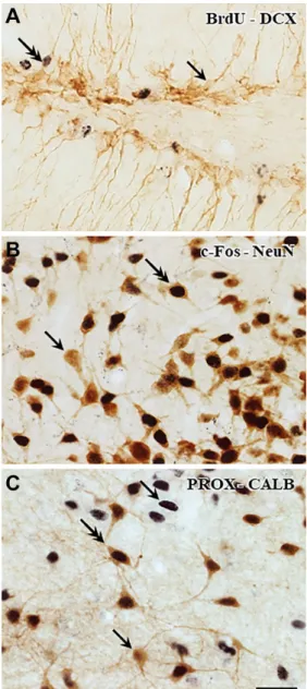
Double Immunostaining for Light Microscopic Observation
Antigens present in different cell compartments of the same cell were stained with the same peroxidase- based technique covering the first stained antigens with SSGI, as shown in Fig. 1. These nuclear antigens (BrdU, Prox-1, and c-Fos) were labeled with the bluish- black Ni-enhanced DAB that was followed by sulfide treatment, silver deposition, and its replacement with gold. The treatment with gold(I) rhodanide strength- ened the color intensity of the nuclei without altering their black appearance during the further incubation steps. The elementary gold successfully blocked the overstaining of the antigen–antibody complexes in the consecutive detection of the second signal.
The presence of the first reaction end product in the nuclei did not interfere with the appearance of the
subsequently stained cytoplasmic antigens (DCX, CALB, and NeuN). The fine processes of neurons were visualized by the brown DAB polymer.
The combinations of antibodies applied in the cur- rent work are listed in Table 1. It is important to note that this double immunolabeling protocol made it pos- sible to apply two kinds of antibodies raised in the same species (e.g., rabbit anti-c-Fos and rabbit anti- NeuN, rabbit anti-Prox-1 and rabbit anti-CALB) in colo- calization without using expensive subtypes of the primary antibodies.
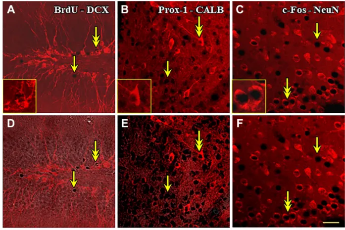
The Combination of SSGI With Immunofluorescence
Images of double-labeled brain tissue captured by con- focal laser scanning microscopy are shown in Fig. 2.
The application of immunofluorescence for DCX, CALB, and NeuN on the gold-toned sections resulted in bright labeling in the cytoplasm (Fig. 2A–C) compa- rable with the immunoperoxidase-stained sections. In fractions of the immunolabeled perikarya, the cyto- plasm enclosed dark roundish areas, which turned to be gold-containing nuclei when viewed in correspond- ing phase-contrast images (not shown). Overlaying of the fluorescent and phase-contrast images confirmed the double labeling of BrdU, Prox-1, or c-Fos-immu- nopositive nuclei and the neuronal perikarya (Fig. 2D–
F). The first immunohistochemical labeling producing the blackish nuclear reaction end product did not inter- fere with the fluorescent visualization of the second cytoplasmic antigen even in those cases when both primary antibodies were produced in the same animal species. Interestingly, when permanent mounting medium Depex was used, instead of aqueous medium like Vectashield, the Cy3-conjugated immunoreagents seemed to be the most effective in terms of longevity and intensity out of the tested fluorochromes in our hands.
It is worth noting that the DAB and SSGI slightly increased the general fluorescent background, but a proper adjustment of the threshold values in laser Table 2. Combinations of Immunohistochemistry and AChE Enzyme Histochemistry Including Animal Pretreatment, Neuronal Markers, Labels (DAB With/Without Ni), Post-DAB Treatments, and Referred Figures.
Versions Pretreatment First Marker First Label Post-DAB Second Marker Second Label Figures
Version 1 DFP ChAT DAB H2O2 AChE DAB-Ni 3A
Version 2 DFP ChAT DAB-Ni H2O2 AChE DAB –
Version 3 None AChE DAB-Ni Na2S + AuSCN CALR DAB-Ni 3B
Version 4 DFP AChE DAB EDTA ChAT DAB-Ni 3C
Abbreviations: AChE, acetylcholinesterase; DAB, diaminobenzidine; CALR, calretinin; DFP, diisopropyl fluorophosphate; ChAT, choline acetyltransferase; HRP, horseradish peroxidase.
Figure 1. Double immunohistochemical staining for light microscopy. Nuclear antigens were stained with primary anti- bodies against BrdU, Prox-1, and c-Fos, which were revealed with DAB-Ni. Immunostaining was followed by SSGI, resulting in black color. Subsequently, perikaryonal staining (DCX, CALB, and NeuN) was carried out applying the same peroxidase-based technique, and it was visualized with brown DAB. Single-head arrows point to single-labeled structures, and the arrows with double heads mark the neurons with immunopositivity both in their nuclei and in cytoplasm. (A) BrdU-DCX. One DCX- immunoreactive granule cell indicates BrdU uptake. (B) c-Fos- NeuN. c-Fos immunoreactivity was enhanced in many branched brown neurons. (C) Prox-1-CALB. The single- and double-labeled cells can be clearly distinguished. Scale bar: 25 µm. Abbreviations:
BrdU, 5-bromo-2´-deoxyuridine; CALB, calbindin; c-Fos, FBJ murine osteosarcoma cellular oncogene; DAB, diaminobenzidine;
DCX, doublecortin; NeuN, neuronal nuclei; Prox-1, Prospero Homeobox 1; SSGI, sulfide-silver-gold intensification.
scanning confocal microscopy greatly eliminated this background fluorescence.
Immunohistochemistry for Neuronal Antigens Before AChE Enzyme Histochemistry
If immunohistochemistry followed by the SSGI method was performed before enzyme histochemistry, the cholinoceptive neuronal elements vanished. In our pilot experiments, the SSGI method was easily adapted as a post-DAB treatment to AChE histochem- istry for the first labeling, but the SSGI with even the mildest power was found to result in unduly intense staining, which means that AChE staining alone usu- ally produced satisfactorily intense labels with high signal-to-noise ratio. The subsequent SSGI was found to thicken the cellular processes artificially and to increase the background slightly, enhancing the sig- nal-to-noise ratio further. Therefore, we sought other colocalization possibilities.
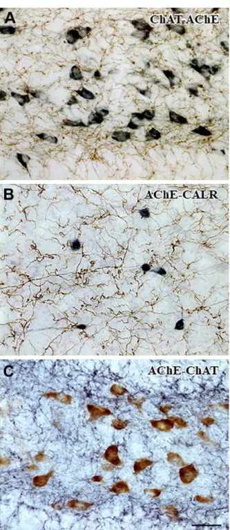
Four sequential versions of immunohistochemistry and AChE enzyme histochemistry were probed to learn about the optimal approach for double staining (see Table 2). The immunohistochemistry for ChAT without SSGI did not interfere with the activity of the AChE (versions 1 and 2). Using hydrogen peroxide treatment was effective to inactivate the HRP, as expected, to prevent further development during the subsequent AChE staining. In version 1, a profuse net- work of cholinergic fibers appeared in brown, and the perikaryonal sites of the de novo synthesis of AChE after DFP pretreatment of the animals (see AChE pharmacohistochemistry25) were labeled with the distinctive bluish-black DAB-Ni precipitate formed by the catalytic end product (i.e., Hatchett’s brown) of the enzyme (Fig. 3A). On the contrary, in version 2, the fine cholinergic fibers, which were originally bluish black, underwent an unintended color conversion dur- ing AChE histochemistry and tended to be brown, sim- ilar to the cholinoceptive neuronal elements, whereas the intense immunoreactive neurons preserved their dark color (not shown). Due to the loss of reliable color contrast between the two kinds of neuronal elements, version 2 is not recommended for use.
Immunohistochemistry for Neuronal Antigens After AChE Enzyme Histochemistry
In our preliminary stainings, the high enzymatic activ- ity of AChE was well preserved in our preparations.
Even the mildest version of the subsequent SSGI pro- duced too intense dark signal. Accordingly, alternative post-DAB treatments were tried to replace the SSGI technique, which, simultaneously, were expected to
permit the use of bluish-black color of DAB-Ni in the subsequent immunohistochemistry for possibly less antigen density.
In version 3, AChE-positive fibers formed dense network among the CALR-immunoreactive neurons in the brain (Fig. 3B). First, AChE staining was applied, resulting in a bluish-black DAB-Ni polymer, which was then converted to a brown nickel sulfide- containing water-insoluble matrix. This compound precipitate was ultimately covered with extremely stable elementary gold. After the gold toning, the immunoreactive neurons labeled with the bluish- black DAB-Ni polymer can be easily distinguished from the first staining.
In version 4, the perikarya showing synthesis of AChE after DFP pretreatment and the cholinergic
axons were simultaneously visualized in the same section (Fig. 3C). Profound enzymatic reaction was detected in many neuronal cell bodies by deposition of the unintensified brown DAB polymer.
AChE histochemistry was terminated with EDTA treatment, because without this treatment, the color of the perikarya would have undergone a brown-to- black conversion during the visualization of the sec- ond neurochemical marker, thereby losing the unambiguous distinction between the sites of the two markers. The application of the EDTA treatment on the sections after the development dissolved the Hatchett’s brown, which would otherwise catalyze the DAB polymerization during the immunohisto- chemical procedure, ended with the bluish-black DAB-Ni precipitation.
Figure 2. Double immunohistochemical staining for combined bright-field/fluorescence microscopy. The nuclear antigens (BrdU, Prox- 1, and c-Fos) were immunostained and enhanced with SSGI similarly as shown in Fig. 1. Thereafter, the perikaryonal markers (DCX, CALB, and NeuN) were visualized with red Cy3-tagged antibodies. Single-head arrows point to immunoreactive nuclei within unlabeled perikarya, whereas the arrows with double heads mark the double-immunoreactive neurons. The same viewfields (A–D, B–E, and C–F) were captured from the sections in two different microscopic modes: laser-scanned confocal (A–C) and combined confocal and phase- contrast (D–F). The marked double-stained cells are magnified in the insets of A to C. Scale bar: 25 µm. Abbreviations: BrdU, 5-bromo- 2′-deoxyuridine; CALB, calbindin; c-Fos, FBJ murine osteosarcoma cellular oncogene; DAB, diaminobenzidine; DCX, doublecortin;
NeuN, neuronal nuclei; Prox-1, Prospero Homeobox 1; SSGI, sulfide-silver-gold intensification.
Discussion
Interference in Colocalization Experiments
The elementary steps of the SSGI method7 were revised and applied in our present colocalization study. The steps were probed either in different cell populations or in different cell compartments of the identical cell. The common efforts in these consecu- tive double labeling methods were to terminate the detection of the first marker to prevent any possible interaction with the detection of the second one and to obtain distinct colors representing the sites of the two markers.
Several factors may cause interference between both the consecutive and simultaneous approaches of double labeling techniques. Certainly, the most severe problem is the relative shortage of the available host species of the primary antibodies. On one hand, the researchers’ choice is usually restricted to monoclonal mouse/rat and rabbit or polyclonal rabbit/guinea pig and goat/sheep primary antibodies. On the other hand, antibodies to two different antigens are often produced from the same animal species, which hin- ders the double labeling experiments. Therefore, we tried to elaborate different ways that may allow con- secutive labeling of two histological items in the same section.
Double Immunostaining for Light Microscopy
Considering the above-mentioned points, not only double but also multiple labeling approaches can be performed, provided that relatively high cellular con- centrations of the markers are present, and the sequence of the detections of the antigens with dis- tinct reaction end product without interference between the immunoreagents and developing condi- tions is elaborated.
Figure 3. Double labeling with AChE enzyme histochemistry and immunohistochemistry for neuronal markers. (A) ChAT- AChE. Cholinergic fibers were visualized with the brown DAB polymer. The residual HRP activity was quenched with hydrogen peroxide treatment before the subsequent AChE enzyme histo- chemistry. The perikaryonal sites of the de novo AChE synthesis of the esterase inhibitor-treated animals were labeled with the bluish-black DAB-Ni polymer. Note that no perikaryonal ChAT immunoreactivity is shown. (B) AChE-CALR. AChE-positive
Figure 3. (continued) fibers were revealed with fine brown gold deposits over the DAB polymer. Quenching of the reac- tion end product of AChE (Cu2[Fe(CN)6]) possessing HRP- like catalytic activity was not necessary before the subsequent immunohistochemistry. CALR-immunoreactive cell bodies were developed with the bluish-black Ni-enhanced DAB polymer. (C) AChE-ChAT. The de novo AChE synthesis in the DFP-pretreated animals were labeled with the brown DAB polymer in the neu- ronal somata. Before immunohistochemistry, the AChE enzyme histochemistry was finalized with EDTA treatment to avoid over- staining and color conversion by the removal of Cu2[Fe(CN)6].
The intense network of ChAT-immunoreactive fibers was labeled with the bluish-black DAB-Ni polymer. Scale bar: 25 µm.
Abbreviations: AChE, acetylcholinesterase; CALR, calretinin;
ChAT, choline acetyltransferase; DAB, diaminobenzidine; DFP, diisopropyl fluorophosphate; HRP, horseradish peroxidase.
(continued)
Although three enzymes (HRP, alkaline phospha- tase, galactosidase) were found to be potentially suit- able for deposition of water-insoluble reaction end products compatible with immunohistochemical proce- dures,26 HRP seems to be the prevailing enzyme out of them due to its various applicable substrates in dif- ferent colors. Obviously, DAB is the most widely used chromogen because of the forming polymer’s versatile properties: the chelating capacity of some metal ions (e.g., Co, Ni) converting the original brown to blackish hues; osmiophilic feature; high insolubility not only in water but also in organic solvents, yielding precise localization of the immunoreactions; and catalytic power of reduction of silver ions in the post-DAB meth- ods also changing the original color.
Using HRP-based double labeling techniques, apparently different approaches are recommended depending on whether the primary antibodies were obtained from the non-cross-reacting animal species (e.g., mouse and goat), allowing simultaneous incuba- tion in the first or even in the second steps of the pro- tocol, or not (e.g., both antibodies are those of mouse).
In the first case, the most critical factor is the sequence of DAB polymers in different colors. Using the same immunoperoxidase-based labeling, it was recom- mended that in the two DAB developments the former one should result in the more intense color including Ni ions, whereas the latter one generates the paler brown DAB.4
If the primary antibodies are derived from phyloge- netically close species, or even if they belong to the same IgG subclass, the incubations in these immu- noreagents must be sequential. To avoid cross- reaction(s) of the detection systems, apparently the researcher may choose between two alternative ways.
The tissue-bound immunoreagents after the first development may be removed by a heat treatment before the subsequent application of the second cycle of antibodies. Elution strategies were elaborated, such as microwave heating or boiling between the two cycles of the visualizations. Microwave treatment was recommended between the applications of the pri- mary antibodies from the same species for colocaliz- ing two different antigens in the same cell of paraffin-embedded tissue.27 Others used traditional boiling for a few minutes to prevent cross-reactions.28 However, these heat-mediated elution techniques were found to be incompatible with the Ni-enhanced DAB polymer by losing its basis color.29 Thus, the first reaction end product was always the DAB polymer, which was followed by elution and subsequent detec- tion of the second antigen by the DAB-Ni polymer. It is worth noting that this approach allows the use of the more intense DAB-Ni polymer as a second signal,
which may be an advantage when the second antigen is experienced to be less detectable, in contrast to the other methods that apply the DAB-Ni polymer as the first signal.
Alternatively, the tissue-bound immunoreagents after the first development may be shielded by atomic silver or gold6,7 before the subsequent cycle of anti- bodies and development. One of the special benefits of this latter way is its compatibility with ultrastructural investigations.30,31 This approach also features the optional intensification of the first signal and thereby a higher contrast between the two signals, which can be well utilized when the first signal is in low intensity (e.g., c-Fos in weakly responsive cells). The very intense staining may be also beneficial in statistical analysis of the immunopositive nuclear antigens (BrdU, Prox-1, or c-Fos) in large areas.
Combined Peroxidase and Fluorescence Immunostaining
Immunofluorescence was combined with immunoper- oxidase technique to detect two cytoplasmic antigens inside the same cell with primary antibodies produced in the same animal.32 These techniques were tried in both orders. It was found that if the immunofluores- cence was used first, it was almost totally quenched by the immunoperoxidase. Thus, the sections after the DAB development required a harsh elution method before the successful application of the fluorescently labeled antibodies, because the intensity of the fluo- rescence was inversely proportional to that of the per- oxidase reaction.
However, in the case of separate intracellular com- partments, two antigens were successfully detected using primary antibodies raised in different animal species by means of combining immunoperoxidase and fluorescence techniques without reported cross-talk.33
Because double immunoperoxidase stainings can be carried out reliably using antibodies from the same spe- cies if the first staining is finalized by silver intensification of the DAB polymer,6 it prompted us to probe whether immunofluorescence can be also effective on the same specimen after SSGI. The small black immunopositive nuclei were easily spotted at low magnification under transmitted bright-field microscopy, which then could be further investigated for colocalization with another anti- gen under incident fluorescence microscopy.
DAB was reported to induce nonspecific fluores- cence in myelinated nerve fibers and in some peri- karya in formaldehyde-fixed nervous tissue34; this background issue can be solved by the appropriate settings of the microscope.
Immunohistochemistry for Neuronal Antigens Before AChE Enzyme Histochemistry
It was reported that the AChE-positive structures were less satisfactorily stained if the immunohistochem- istry was performed before enzyme histochemistry.35 However, we did not experience any hindrance in AChE histochemistry, when it was combined with immunohis- tochemistry, as described here. The SSGI method fol- lowing the immunoperoxidase was found to be incompatible with the subsequent Tago et al.’s16 AChE technique using also DAB development: Some compo- nent (probably silver ion) of the SSGI technique abol- ishes the AChE detectability by its enzyme histochemistry.
Therefore, another post-DAB treatment had to be found for colocalization of AChE and other neuronal markers.
The HRP of the immunohistochemistry was quenched with conventional hydrogen peroxide treat- ment. It is worth mentioning that a modified version of the Karnovsky method using cobalt instead of copper ions was elaborated for a simple double staining proto- col to receive a reaction end product, which lacks the HRP-like activity.35 However, the cationic exchange appears to result in less precise resolution than the Tago’s method.
The approach for the ChAT (revealed by paler DAB polymer) and AChE (revealed by the more intense DAB-Ni polymer) double staining in version 1 was found to be successful, but it did not seem to be opti- mal, as the less intense DAB polymer would suffice for Tago’s AChE histochemistry, including double catalytic reactions first by the enzyme itself and subsequently by Hatchett’s brown. However, in version 2, when the polymers were swapped, the clear color distinction was greatly reduced; thus, version 2 was disapproved.
Immunohistochemistry for Neuronal Antigens After AChE Enzyme Histochemistry
Because the widely used Tago’s AChE staining marks the distribution of this enzyme reproducibly and intensely, the full power of the SSGI was not necessary to mask the enzyme-containing profile. Our primary purpose of avoiding interactions between the enzyme histochemis- try and immunoperoxidase technique was thought to be achieved by means of two different approaches. In one of the protocols, the reaction end product of AChE was shielded by gold plating (see version 3 in section
“Combination of Immunohistochemitry and AChE Enzyme Histochemistry”), which was never found to be overstained by the subsequent DAB treatment, but allowed its clear distinction from the DAB-Ni polymer obtained in the second immunoperoxidase step. In the other protocol (version 4), masking of the DAB polymer
was not performed; instead, Hatchett’s brown with HRP- like activity was removed by the chelating EDTA to abol- ish overstaining of the DAB polymer. In both version 3 and version 4, the more intense DAB-Ni polymer was used as a second signal, which can be advantageous if the immunohistochemical process would yield weaker signal.
In our experiences, the thinner fibers require the more intense staining, over the bigger perikarya.
Accordingly, we recommend the following ranking of versions 1, 3, and 4 for the fibers or lower density anti- gens in general: version 3: DAB + Ni + gold > version 4: DAB + Ni > version 1: DAB.
To conclude, double immunoperoxidase protocols require careful tailoring, because of the potential cross-talks between the two systems, mainly due to the cross-reacting antibodies and the color shift of DAB polymer at the second development. In this study, a revised SSGI as a post-DAB treatment after the first development is recommended for parallel detection of nuclear and perikaryonal antigens to resolve these problems. This technique ensures clear distinction between the sites of the two antigens with high signal- to-noise ratio. Our post-DAB method is shown to be compatible with immunofluorescence.
The versatility of our post-DAB reagents is demon- strated by their combinations with AChE histochemistry with the omission of silver intensification. We compared four versions of combinations of immunohistochemistry and AChE enzyme histochemistry, and we recommend two easy methods for double staining of an antigen and AChE. Our sulfide-gold treatment can be possibly adapted for immunohistochemical purposes as well, if one of the target antigens surpasses the other.
Acknowledgment
We thank Péter Hegyi for kindly providing the confocal microscope.
Competing Interests
The author(s) declared no potential conflicts of interest with respect to the research, authorship, and/or publication of this article.
Author Contributions
ED, IT, EB, and TS designed the study; IT, GS, EP, EB, and TS performed the experiments; IT, GS, and ED wrote the article. All authors approved the final version of the manuscript.
Funding
The author(s) disclosed receipt of the following financial support for the research, authorship, and/or publication of
this article: This study was supported by the Department of Anatomy, Faculty of Medicine, University of Szeged funds.
Literature Cited
1. Graham RC Jr, Karnovsky MJ. The early stages of absorption of injected horseradish peroxidase in the proximal tubules of mouse kidney: ultrastructural cyto- chemistry by a new technique. J Histochem Cytochem.
1966;14(4):291–302.
2. Adams JC. Heavy metal intensification of DAB- based HRP reaction product. J Histochem Cytochem.
1981;29(6):775.
3. Hsu SM, Soban E. Color modification of diaminobenzi- dine (DAB) precipitation by metallic ions and its appli- cation for double immunohistochemistry. J Histochem Cytochem. 1982;30(10):1079–1082.
4. Wouterlood FG, Bol JG, Steinbusch HW. Double-label immunocytochemistry: combination of anterograde neu- roanatomical tracing with Phaseolus vulgaris leucoag- glutinin and enzyme immunocytochemistry of target neurons. J Histochem Cytochem. 1987;35(8):817–823.
5. Gorcs TJ, Leranth C, MacLusky NJ. The use of gold- substituted silver-intensified diaminobenzidine (DAB) and non-intensified DAB for simultaneous electron microscopic immunoperoxidase labeling of tyrosine hydroxylase and glutamic acid decarboxylase immuno- reactivity in the rat medial preoptic area. J Histochem Cytochem. 1986;34(11):1439–1447.
6. Gallyas F. Physicochemical mechanisms of histologi- cal silver staining and their utilization for rendering indi- vidual silver methods selective and reliable. Biotech Histochem. 2008;83(5):221–238.
7. Dobo E, Takacs VT, Gulyas AI, Nyiri G, Mihaly A, Freund TF. New silver-gold intensification method of diamino- benzidine for double-labeling immunoelectron micros- copy. J Histochem Cytochem. 2011;59(3):258–269.
8. Gallyas F, Gorcs T, Merchenthaler I. High-grade inten- sification of the end-product of the diaminobenzidine reaction for peroxidase histochemistry. J Histochem Cytochem. 1982;30(2):183–184.
9. Merchenthaler I, Stankovics J, Gallyas F. A highly sen- sitive one-step method for silver intensification of the nickel-diaminobenzidine endproduct of peroxidase reac- tion. J Histochem Cytochem. 1989;37(10):1563–1565.
10. Hardman CD, Ashwell KWS. Stereotaxic and chemoar- chitectural atlas of the brain of the common marmoset (Callithrix jacchus). Boca Raton, FL: CRC Press; 2012.
11. Zhou J-N, Ni R-J. The tree shrew (Tupaia belangeri chinensis) brain in stereotaxic coordinates. New York:
Springer Berlin Heidelberg; 2016.
12. Palazzi X, Bordier N. The marmoset brain in stereotaxic coordinates. New York: Springer; 2008.
13. Paxinos G, Franklin KBJ. The mouse brain in stereotaxic coordinates. Amsterdam; Boston: Elsevier Academic Press; 2004.
14. Paxinos G, Watson C. The rat brain in stereotaxic coor- dinates. Amsterdam; Boston: Elsevier Academic Press;
2007.
15. Zilles KJ. The cortex of the rat: a stereotaxic atlas.
Berlin; New York: Springer-Verlag; 1985.
16. Tago H, Kimura H, Maeda T. Visualization of detailed acetylcholinesterase fiber and neuron staining in rat brain by a sensitive histochemical procedure. J Histochem Cytochem. 1986;34(11):1431–1438.
17. Reddy DS, Kuruba R. Experimental models of status epilepticus and neuronal injury for evaluation of thera- peutic interventions. Int J Mol Sci. 2013;14(9):18284–
18318.
18. Danscher G, Moller-Madsen B. Silver amplification of mercury sulfide and selenide: a histochemical method for light and electron microscopic localization of mer- cury in tissue. J Histochem Cytochem. 1985;33(3):219–
228.
19. Kornack DR, Rakic P. Continuation of neurogenesis in the hippocampus of the adult macaque monkey. Proc Natl Acad Sci U S A. 1999;96(10):5768–5773.
20. Szakacs R, Weiczner R, Mihaly A, Krisztin-Peva B, Zador Z, Zador E. Non-competitive NMDA recep- tor antagonists moderate seizure-induced c-Fos expression in the rat cerebral cortex. Brain Res Bull.
2003;59(6):485–493.
21. Winawer MR, Makarenko N, McCloskey DP, Hintz TM, Nair N, Palmer AA, Scharfman HE. Acute and chronic responses to the convulsant pilocarpine in DBA/2J and A/J mice. Neuroscience. 2007;149(2):465–475.
22. Knoth R, Singec I, Ditter M, Pantazis G, Capetian P, Meyer RP, Horvat V, Volk B, Kempermann G. Murine features of neurogenesis in the human hippocampus across the lifespan from 0 to 100 years. PLoS ONE.
2010;5(1):e8809.
23. Tang FR, Chia SC, Zhang S, Chen PM, Gao H, Liu CP, Khanna S, Lee WL. Glutamate receptor 1-immunoposi- tive neurons in the gliotic CA1 area of the mouse hip- pocampus after pilocarpine-induced status epilepticus.
Eur J Neurosci. 2005;21(9):2361–2374.
24. Tang FR, Chia SC, Jiang FL, Ma DL, Chen PM, Tang YC. Calcium binding protein containing neurons in the gliotic mouse hippocampus with special reference to their afferents from the medial septum and the entorhi- nal cortex. Neuroscience. 2006;140(4):1467–1479.
25. Di Patre PL, Mathes CW, Butcher LL. Differential visualization of cholinesterasic neuronal somata and fibers by use of modifications of acetylcholinester- ase pharmacohistochemistry. J Histochem Cytochem.
1993;41(1):129–135.
26. Vanderloos CM, Becker AE, Vandenoord JJ. Practical suggestions for successful immunoenzyme dou- ble-staining experiments. Histochemical Journal.
1993;25(1):1–13.
27. Yan J, Catts VS, Chan A, McCombe PA. A simple and reliable immunohistochemical method for colocaliza- tion of 2 antigens in the same cells of paraffin-embed- ded tissues. Appl Immunohistochem Mol Morphol.
2013;21(5):471–477.
28. Cui BP, Gao CQ, Xiong CQ, et al. A novel, convenient and reliable method for immunohistochemical double labeling. Chin Pharmacol Bull. 2015;31(3):436–441.
29. Bauer M, Schilling N, Spanel-Borowski K. Limitation of microwave treatment for double immunolabelling with antibodies of the same species and isotype. Histochem Cell Biol. 2001;116(3):227–232.
30. Takacs VT, Szonyi A, Freund TF, Nyiri G, Gulyas AI.
Quantitative ultrastructural analysis of basket and axo- axonic cell terminals in the mouse hippocampus. Brain Struct Funct. 2015;220(2):919–940.
31. Papp E, Borhegyi Z, Tomioka R, Rockland KS, Mody I, Freund TF. Glutamatergic input from specific sources influences the nucleus accumbens-ventral pallidum information flow. Brain Struct Funct. 2012;217(1):37–48.
32. Lechago J, Sun NC, Weinstein WM. Simultaneous visu- alization of two antigens in the same tissue section by combining immunoperoxidase with immunofluorescence
techniques. J Histochem Cytochem. 1979;27(9):1221–
1225.
33. Young JK, Wu M, Manaye KF, Kc P, Allard JS, Mack SO, Haxhiu MA. Orexin stimulates breathing via medullary and spinal pathways. J Appl Physiol. 2005;98(4):1387–1395.
34. von Bohlen und Halbach O, Kiernan JA. Diaminobenzidine induces fluorescence in nervous tissue and provides intrinsic counterstaining of sections prepared for per- oxidase histochemistry. Biotech Histochem. 1999;74(5):
236–243.
35. Schatz CR, Geula C, Morecraft RJ, Mesulam MM. A one-step cobalt-ferrocyanide method for histochemi- cal demonstration of acetylcholinesterase activity in central nervous system tissue. J Histochem Cytochem.
1992;40(3):431–434.
![degradationofcoatingsisfavourablefrombothclinicalandbiomedicalpointsofview. ofbiomedicalapplications[ , ].Themainapproachisthatthebioceramiccoatingsact toincreasethebiocompatiblecharacteristicsofimplants.Thiscanbebestachievedby theimplants.Generally,thema](data:image/gif;base64,R0lGODlhAQABAIAAAP///wAAACH5BAEAAAAALAAAAAABAAEAAAICRAEAOw==)