Research Article
Antimicrobial Silver-Polyethyleneimine-Polylactic Acid Polymer Composite Film for Coating Methacrylate-Based Denture Surfaces
Zoltán Géczi ,1Péter Hermann,1László Kőhidai,2Orsolya Láng,2Zsófia Kőhidai,3 Tamás Mészáros,4Attila Barócsi,5Sándor Lenk ,5and Tivadar Zelles6
1Department of Prosthodontics, Semmelweis University, Szentkirályi u. 47, 1088 Budapest, Hungary
2Department of Genetics, Cell- and Immunobiology, Semmelweis University, Nagyvárad tér 4, 1089 Budapest, Hungary
3Department of Oral Diagnostics, Semmelweis University, Szentkirályi u. 47, 1088 Budapest, Hungary
4Nanomedicine Research and Education Center, Department of Pathophysiology, Semmelweis University, Nagyvárad tér 4, 1089 Budapest, Hungary
5Department of Atomic Physics, Budapest University of Technology and Economics, Budafoki út 8, 1111 Budapest, Hungary
6Department of Oral Biology, Semmelweis University, Nagyvárad tér 4, 1089 Budapest, Hungary
Correspondence should be addressed to Zoltán Géczi; gaeczi.zoltan@dent.semmelweis-univ.hu Received 4 June 2018; Accepted 5 September 2018; Published 4 November 2018
Academic Editor: Muhamamd A. Malik
Copyright © 2018 Zoltán Géczi et al. This is an open access article distributed under the Creative Commons Attribution License, which permits unrestricted use, distribution, and reproduction in any medium, provided the original work is properly cited.
To prepare an antimicrobial polymer composite composed of silver- (Ag-) polyethyleneimine- (PEI-) polylactic acid (PLA) in chloroform, for coating the mucosal surfaces of methacrylate-based dentures as a prospective therapy for denture stomatitis.
The water-insoluble, tightly bound, hard, micrometre-thin, and colourless film exerts its effects by direct contact with the pathogens and via the active constituents (Ag, PEI, and Ag-PEI) released slowly into the mucosa’s salivary layer. Silver and PEI were blended at 140°C, then bound to PLA. The Ag-PEI complex was characterised by dynamic light scattering and transmission electron microscopy, and the Ag-PEI-PLA composite was examined by atomic force microscopy and micro- computed tomography. The characteristic was measured by atomic force microscopy (AFM) and micro-computed tomography (micro-CT). The quantity of water-soluble Ag-PEI complex released from the compositefilm was measured with gravimetry.
The cellular physiological effects were analysed by impedimetry and computer-based morphometry using human gingival epithelial cells. A real-time cell proliferation assay revealed moderate toxic effects of Ag-PEI on the epithelium. The viscous Ag-PEI-PLA solution produced could be applied as a thin film on methacrylate surfaces. Active antimicrobial components (Ag, PEI, and Ag-PEI) were released from the hard, tightly bound Ag-PEI-PLA coating. This study’s findings verified the applicability of the antimicrobial Ag-PEI-PLA composite for coating the inner surfaces of acrylate dentures. Owing to the well-known antimicrobial effects of silver and PEI and the supplementary effects of chloroform, this composite provides a new therapeutic method for denture stomatitis that can be easily performed by dentists.
1. Introduction
Denture stomatitis (also known as denture-related stomatitis, candida-associated denture stomatitis, denture-associated erythematous stomatitis, prosthetic stomatitis, or a combina- tion of these terms) is a common, multifactorial condition in which mild inflammation and erythema of the oral mucous membrane occur under a removable prosthesis (i.e., a partial
or complete denture or an orthodontic appliance). In patients having removable prostheses, the prevalence of denture stomatitis ranges from 15% to >70%. The most important etiological factors include ill-fitting dentures, poor dental hygiene, high carbohydrate intake, reduced salivaryflow, dia- betes mellitus, and immune deficiency.Candidaspecies are oral fungal commensal organisms that are present in up to 90% of healthy individuals; Candida albicans is the most
Volume 2018, Article ID 1048734, 9 pages https://doi.org/10.1155/2018/1048734
frequent fungal coloniser. Although denture stomatitis is not a particularly severe illness in most cases, it may potentiate additional infections in susceptible patients [1–7].
There is a large body of evidence that demonstrates that Candida albicansis capable of adhering to polymethylmetha- crylate (PMMA) surfaces to form a biofilm. Microbial plaque on the inner surfaces of dentures and the mucosa is com- posed of bacteria and fungi. Changing the local environment favours dominance ofCandidaand causes mucosal inflam- mation; bacteria may support these inflammatory reactions.
Despite these relationships, most forms of therapy focus on anti-Candidatreatment [3, 8–10].
The treatment and management of denture stomatitis can be focused locally and/or systemically. For patients with poor compliance, diabetes, or who are immunocompro- mised, systemic therapy is recommended. In the literature, there are many different forms and local application tech- niques of antimicrobial agents reported, with very diverse results. The most important issues include the development of resistance and recurrent infections. Currently, there is no
“golden rule”for optimal therapy [11–15].
Our goal was to design an antimicrobial, hard polymer composite for coating the inner surface of dentures. This thin, polymer composite film must comply with various requirements: it should be biodegradable, release of the active ingredients from thefilm must be possible, it needs to be sol- uble in a suitable solvent, and it must tightly bond to acrylate.
Another technical requirement is the easy and rapid prepara- tion of a thin layer of the material by a dentist or the dental laboratory. Although many polymers are used for coating, the most common polymer, polylactic acid (PLA) dissolved in chloroform, was chosen because of the fast and complete elimination from the denture surface by evaporation.
We conceptualised that silver-polyethyleneimine (Ag- PEI), with its core-shell micellar structure, excellent colloidal stability, and easy method of hydrothermal synthesis, would be the optimal active antimicrobial ingredient [16–18]. The synergistic antibacterial effect of silver with polyethylenei- mine (PEI) has also been demonstrated [19, 20].
This Ag-PEI nanocomplex is water-soluble; therefore, it is not suitable for acrylate coating on its own. To avoid this dis- advantage, we needed to apply a polymer bulk material to form a stablefilm. We chose PLA, which can easily bind to the Ag-PEI component by electric self-assembly (ESA) and is nearly insoluble in water. The silver-polyethyleneimine- polylactic acid (Ag-PEI-PLA) composite in chloroform is capable of strongly binding to acrylate.
The objective of the present study was to develop an alter- native form of therapy that approaches the problem from a new aspect: to kill the pathogens with the Ag-PEI active ingredient, while a suitable coating material simultaneously solubilises thefirst few superficial micrometres of the acry- late. For this purpose, chloroform promised to be the best sol- vent in which the Ag-PEI and PLA solubilised equally well.
During the coating procedure, thefirst few superficial micro- metres of acrylate will swell and become partially soluble [21], which help kill the pathogens while embedding them.
After the chloroform rapidly evaporates, the composite becomes a thin, hard, and colourless layer. The thickness of
this layer is between 30–40μm, which does not influence the stability of the prosthesis. Considering the novel charac- teristics of this form and application of the Ag-PEI-PLA polymer composite, we propose the name “antimicrobial hard lining” or“antimicrobial hard liner” for this form of therapy and the composite.
Although the cytotoxic natures of Ag and PEI to bacteria are well known, we analysed their biophysical effects on the human gingival epithelium (HGEP) using impedimetry.
Due to the electrical insulator moiety provided by the phospholipid bilayer of the surface membrane, impedime- try allowed us to analyse some basic cellular physiological responses, including viability, cell adhesion, and cell prolif- eration, induced by PEI and Ag-PEI.
The present paper discusses the synthesis and character- isation of the Ag-PEI-PLA composite, including its toxicity and adaptability in human therapy.
2. Materials and Methods
2.1. Materials.High-branched polyethyleneimine (PEI) with a molecular weight of 25,000, silver nitrate (AgNO3), poly- lactic acid (PLA) with a molecular weight of 60,000, and chloroform were purchased from Sigma-Aldrich Chemie, Germany; they were used in this study without further puri- fication. Deionised and ultrapure water were used through- out the study; their resistivity was≥18 MΩcm.
2.2. Preparation of the Ag-PEI-PLA Polymer Composite.Dur- ing thefirst phase, the Ag-PEI complex, the “antimicrobial active component,” was synthesised using a hydrothermal method: 1 g of PEI was diluted in 2 ml of deionised water by stirring in a glass beaker; in another glass beaker, 158 mg of AgNO3(100 mg Ag) was diluted in 1 ml of deionised water by mild shaking the beaker. After the shaking, the solutions were centrifuged for 10 minutes at 20,000g (Micromax RF, Thermo Fisher Scientific, USA). The obtained solutions were slowly unified in the PEI beaker by continuous stirring. The yellowish solution was placed on the plate of a hot plate stirrer (VELP Scientifica, Italy) set at 90°C to evaporate the water. During this process, a continuous darkening of the sample to a deep brown colour and a reduction in its quantity were observed. The temperature was then increased to 140°C;
the substance thickened, and the magnetic stir bar ceased moving. Upon completion of this step, the sample was further heated for 4 h, until it became a deep, dark greyish-brown, thick mass. A dissolution test performed on the obtained Ag-PEI sample proved that the prepared substance was water- and chloroform-soluble [22, 23].
During the second phase, the PLA was conjugated to the Ag-PEI complex by ESA. Two millilitres of 5% (w/v) PLA in chloroform was added to 20 mg of the Ag-PEI sample in a glass tube. At this step, the honey-like, viscous Ag-PEI was first smeared onto the wall of the preweighted tube with a spatula, then the mixture was intensely shaken for 30 min in a laboratory shaker (IKA Works, Wilmington, NC, USA).
The Ag-PEI was very easily solubilised in the 5% PLA solu- tion, which turned yellowish-brown. Subsequently, a 30 min ultrasonic treatment was performed (45 kHz, 100 W; Emmi
12HC, EMAG, Germany). The practically colourless super- natant was gained with a 2 min, 2000 rpm centrifugation (BW41-BR230, Qualitron Inc., Korea), and the aggregated pellet was discarded. The resultant substance was considered suitable for coating acrylic surfaces [24, 25].
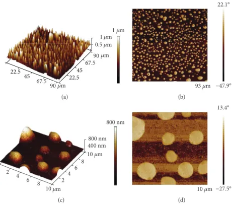
2.3. Atomic Force Microscopy of the Ag-PEI-PLA Composite.
The AFM measurements were performed with an atomic force microscope (Dimension Icon, Bruker, Palaiseau, France) in the tapping mode, under ambient conditions (relative humid- ity,~50%; temperature,~22–24°C). The phase images from the tapping mode operation are a map of how the phase of cantilever oscillation is affected by its interaction with the surface. The physical meaning of this signal is complicated, but in addition to topographic information, the phase can be affected by the relative softness/hardness or chemical nature of the sample [26, 27]. For the tapping mode studies, a cantilever (TESPA-V2, Bruker, Palaiseau, France) with a spring constant of approximately 42 N/m and resonance fre- quencies of approximately 320 kHz was used.
2.4. Micro-Computed Tomography of the Ag-PEI-PLA Composite. The Ag-PEI-PLA polymer composite film was scanned using a micro-computed tomography device (Sky- Scan 1172 microCT, Bruker, Kontich, Belgium). The acquisi- tion parameters were as follows: a 1.91 μm isometric cube voxel with nofilter, at 40 kV, with a 200μA tube current.
The rotation step was 0.5°. From the raw image data set, 3D-rendered images were constructed using CTAn and CTVol software (Bruker, Kontich, Belgium).
2.5. Weight Changes of the Ag-PEI-PLA Composite after 8 Days of Distilled Water (DW) Storage.Fourteen-centimetre- squared portions of PMMA microscopic slides (26×76 mm) were coated with 1–1 ml of the Ag-PEI-PLA composite and PLA in chloroform. After evaporation of the chloroform, the dried slides were placed into DW for 8 days. The weight changes of the air-dried slides were measured on days 1, 2, 3, 4, 5, and 8 with an analytic balance.
2.6. Size Measurement of the Nanoparticles Released from the Polyethyleneimine-Polylactic Acid (PEI-PLA) and Ag-PEI- PLA Membranes. Thin polymer membranes were prepared by casting on a glass surface and pulling out carefully. The compositions of PEI-PLA and Ag-PEI-PLA membranes were identical to those described above. Three cut pieces from both types of membranes (area, 1 cm2; thickness, 0.05 mm) were immersed in 5–5 ml cell culture media free of fetal bovine serum (FBS), representing identical extraction times.
Samples (1.5 ml) containing the released particles were col- lected on day 1, day 5, and day 10 (D1, D5, and D10, respectively). The sizes of the nanoparticles released from the PEI-PLA and Ag-PEI-PLA membranes were measured using a Zetasizer Nano S instrument (Malvern Instruments Ltd., Malvern, UK).
2.7. Impedimetry
2.7.1. Model Cells.Cultures of the HGEP cell line (CELLn- TEC, Bern, Swiss) were maintained in CnT-24.S medium
(CELLnTEC, Bern, Swiss) containing 1% L-glutamine and supplemented with 1% penicillin-streptomycin (Lonza Group Ltd., Switzerland).
The cell cytotoxic and adhesion effects of the PEI-PLA and Ag-PEI-PLA extracts were investigated with a xCELLi- gence RTCA SP (ACEA Biosciences Inc., San Diego, USA) impedance-based system. The real-time assays were imple- mented in E-plate 96 arrays (ACEA Biosciences Inc., San Diego, USA) with a 15 kHz AC system, using a 20 s sampling frequency on noncoated electrodes.
2.7.2. Cytotoxicity Assay.First, growth of a confluent culture of HGEP cells was controlled by impedimetry in the wells of the E-plate 96 arrays. After 24 h, the cultures were treated with the 1- or 5-day PEI-PLA or Ag-PEI-PLA membrane extracts (dilutions: 1000x, 100x, 10x, and 1x) for 168 h.
2.7.3. Cell Adhesion Assay.First, the impedance of the culture medium was recorded as a baseline and absolute control.
Second, the HGEP cells were loaded together with extracts of PEI-PLA or Ag-PEI-PLA (dilutions: 1000x, 100x, 10x, and 1x) prepared by soaking the composite membrane in cell culture medium for 1 or 5 days. The follow-up time was 12 h.
2.8. Apoptosis Measurement.In the background of the cyto- toxic effects, induction of apoptotic processes is frequently revealed. In the present study, our objective was to detect early apoptotic events. The ratio of apoptotic HGEP cells was evaluated after 24 h of treatment with the PEI-PLA and Ag-PEI-PLA composite extracts. For this purpose, two inde- pendent apoptosis assays were performed: (i) computer- based morphometry analysis of HGEP cells treated with the composite extracts and (ii) annexin V staining which is a well-known indicator of the disturbed surface membrane asymmetry, a chief early marker of apoptosis. Computer- based evaluation of the dead cell number on micrograph series taken by light microscopy (obj.: 20x; Axio Observer A1; Carl Zeiss Microscopy GmbH, Jena, Germany) was done by Fiji ImageJ software [28]. The early phase of apoptosis was detected using annexin V labelling (PE Annexin V, BioLe- gend, San Diego, USA) and was measured byflow cytometry (FACSCalibur, BD Biosciences, NJ, USA).
2.9. Statistical Analysis.The delta CI (ΔCI) and slope values were calculated by the integrated software (RTCA 1.2, Roche Applied Science, Indianapolis, IN, USA) of xCELLigence SP System. For the further analysis of the data, the Origin Pro 8.0 (OriginLab Corporation, Northampton, MA, USA) sta- tistical program was used. Data shown in thefigures repre- sent mathematical averages of three parallels and ±SD values. Statistical analysis of data was done by the application of ANOVA of Origin Pro 8.0. The level of significance is shown as follows:x:P< 0 05;y:P< 0 01;z:P< 0 001.
3. Result
3.1. Weight Changes of the Composite after 8 Days of DW Storage. Initially, the average weights of the Ag-PEI-PLA and the control PLA layers prepared on the microscopic glass slides were 57.58 mg and 58.76 mg, respectively. After 8 days,
the weights of both the composite and PLA film had decreased, but the weight lost from the composite was signif- icantly higher than that of the PLA film (33% and 11%, respectively, on day 8; Figure 1).
3.2. Size of the Nanoparticles Released from the PEI-PLA and Ag-PEI-PLA Membranes.Figure 2 presents the size distribu- tion analyses of the particles released from the PEI-PLA and Ag-PEI-PLA membranes and demonstrates that they have characteristic profiles containing populations of particles significantly different from that of the control (medium). A time-course analysis of the peaks shows a decrease with time (Ag-PEI-PLA: D1, 7 nm; D5, 6 nm; D10, 4 nm); nevertheless, no significant difference was detectable in the PEI- vs. Ag- PEI-containing preparations in relation to the duration of the study (data not shown).
3.3. Atomic Force Microscopy of the Ag-PEI-PLA Composite.
The pseudo-3D images of the height data show spherical shapes in the range of 0.5–4.0μm (Figures 3(b) and 3(d)).
In this case, the phase images were primarily determined by the topographic structure on the thinfilm surface. The phase images provided better contrast than the height images (Figures 3(a) and 3(c)).
3.4. Micro-Computed Tomography of the Ag-PEI-PLA Composite. At the maximum enlargement provided by our micro-CT instrument, a granular structure of the composite was evident, indicating the aggregation process (Figure 4).
3.5. Impedimetry
3.5.1. Cytotoxicity.The toxic effects elicited by the nanoparti- cles released from the PEI-PLA and Ag-PEI-PLA were evalu- ated in the day 1 and day 5 samples. The effects of the 1/10 serial dilutions in the range of 1000x–1x were analysed by impedimetry over a 0–168 h period. As depicted in Figure 5, the day 1 samples were capable of eliciting strong and durable cytotoxic effects in the HGEP cells. The cytotoxic sensibility of the HGEP cells proved to be greater to the PEI- PLA D1 extracts (dilution range, 100x–1x) than the Ag-PEI- PLA extracts (dilution range, 10x–1x). For the day 5 extracts (data not shown), the cytotoxic characters of both types of extracts were still detectable; however, the amplitude of toxicity in the PEI-PLA extract was similar to that of the Ag-PEI-PLA extract.
3.5.2. Cell Adhesion. Comparison of the adhesion blocker abilities (Figure 5) demonstrated that both the PEI-PLA and Ag-PEI-PLA day 1 extracts had a significant blocking effect (dilution ranges: 1x–10x); however, the effect of the PEI-PLA extract was more pronounced than that of the Ag-PEI-PLA extract.
3.6. Apoptosis.Our data revealed that both the day 1 and day 5 PEI-PLA and Ag-PEI-PLA extract samples resulted an increased number of dead cells (Table 1) as well as annexin V signals (Figure 6). A comparison of the apoptotic effects elicited by the nanoparticles derived from the PEI-PLA and Ag-PEI-PLA extracts indicated that PEI alone had the
strongest and widest range (100x–10x) effect, while in Ag-PEI, only 10x dilutions were effective.
4. Discussion
Our goal was to extend the possibilities for the management and therapy of denture stomatitis beyond conventional methods. The aim of this study was to produce a thin, hard film on the mucosal surface of acrylate dentures that has anti- microbial effects. Beyond the advantageous on contact killing property of this hard antimicrobial surface, this film has
0 2 4 6 8
55 60 65 70 75 80 85 90 95 100
Percentage of total weight (%)
Days PLA membrane Ag-PEI-PLA membrane
Figure 1: Weight changes of the polymer composite (silver- polyethyleneimine-polylactic acid [Ag-PEI-PLA]) and the polylactic acid (PLA) coating in distilled water (DW) over an 8-day period. Technical parameters of the assay—sample number: 16;
temperature: 25°C; time: 8 days. The error bars represent the standard deviation of measurements for 8–8 samples in 8 days (n= 96).
0 5 10 15 20
0 2 4 6 8 10 12 14 16 18 20
Density of the particles (%)
Size of the particles (nm) PEI-PLA extract
Ag-PEI-PLA extract
P = 0.05
Figure 2: Size distribution of the day 1 polyethyleneimine- polylactic acid (PEI-PLA) and silver-polyethyleneimine-polylactic acid (Ag-PEI-PLA) extracts (medians: PEI-PLA extract, 4.8; Ag- PEI-PLA extract, 5.6). The detected difference indicates that the size of the silver-containing PEI-PLA particles was significantly larger (P= 0 05) than that of the PEI-PLA particles. Technical parameters of the assay—sample number: 3; temperature: 25°C;
time: 60 s.
additional effects owing to the release of active components.
For easy application of the polymer composite on the inner surfaces of prostheses, it is necessary for it to have a suitable viscosity and rapid evaporation of the solvent and be insolu- ble or very slow to solubilise in water.
The presented work is focused on basic research, dealing with in vitro assays; nevertheless, authors complement it with some recommendations for the clinical usage. The application of this hard antimicrobial liner is very similar to the application of the adhesive layer used in the classical relining procedure of dentures by brush. After the evapora- tion, a longer storage in water can induce the release of the active complex.
The silver nanoparticles, which have some unique prop- erties, play a central role in antibacterial and antifungal ther- apies [29]. The effectiveness of these nanoparticles depends on the performance of adequate laboratory procedures dur- ing preparation [29–33]. There are more than one hundred reports regarding the combination of silver with different sta- bilisers, including polymers like polyethyleneimine, while avoiding strong aggregation [17, 18, 19, 34, 35]. All the authors used several alterations, but the basic steps of synthe- sis include the reduction of silver and inhibition of aggrega- tion, followed by functionalization for a given role. The antimicrobial effectiveness primarily depends on the size of the nanoparticles [36, 37].
The method used in the present study uses an easy and common form of synthesis, with mild reduction and aggrega- tion inhibition by PEI. The dense Ag-PEI complex is suitable for long-term storage, and a new sample can be easily pre- pared at chairside for application. According to generally accepted professional opinion, the smaller silver clusters or aggregates provide higher antimicrobial effects [36, 37]. In our composite, the PEI was used for three purposes: (1) the cationic polymer’s antimicrobial effect, (2) the colloidal sta- bility of nanosilver, and (3) the reduction of silver ions in the AgNO3 solution. The antimicrobial effects of Ag and PEI are well known, both individually and in combinations [20, 38–40].
For coating purposes, instead of the denture material (PMMA) which is nonbiodegradable, PLA was used in compliance with the previously stated requirements: it is
22.5 45 22.545
90
90 0.5
1
휇m
1휇m
휇m 휇m 휇m
67.5
67.5
22.5 45 22.545
67 5
67
(a)
93 휇m 22.1°
−47.9° (b)
10 2
2 4 4
6 6
810 휇m 800 nm 800 nm 400 nm
8 휇m
(c)
10 휇m 13.4°
−27.5° (d)
Figure3: Representative atomic force microscopy (AFM) images of the silver-polyethyleneimine-polylactic acid (Ag-PEI-PLA) polymer composite obtained in the tapping mode. (a) A 3D AFM height topography large area scan of the Ag-PEI-PLA complex. (b) A 2D AFM phase large area scan of the Ag-PEI-PLA complex. (c) An enlarged view of the 3D AFM height topography scan of the Ag-PEI-PLA complex. (d) An enlarged view of the 2D AFM phase scan of the Ag-PEI-PLA complex. Relative humidity,~50%; temperature,~22–24°C.
200 휇m
Figure 4: A micro-computed tomography image of the Ag-PEI- PLA composite. Technical parameters of the assay—1.91μm isometric cube voxel with nofilter, at 40 kV, with a 200μA tube current.
biodegradable, soluble in chloroform, and easily binds to the Ag-PEI nanoparticles by ESA. Several methods are available for preparation of the PEI-PLA copolymer, and different polymer combinations are available for drug deliv- ery systems [41–44].
Additionally, the solubilising agent, chloroform, has an antipathogenic effect. It is one of the strongest permeabilising organic solvents with antipathogenic effects onCandida albi- cans [45, 46], and it facilitates intake of active components (e.g., Ag and PEI) into the bacterial and fungal cells. Another
0.00 0.02 0.04 0.06 0.08 0.10
x z
z z
Slope (1/h)
0.00 0.02 0.04 0.06 0.08 0.10
y z
y
z
0 20 40 60 80 100 120 140 160 1.0
1.5 2.0 2.5 3.0 3.5 4.0
Time (h) D1 1x
D1 10x D1 100x
D1 1000x Cont 0 20 40 60 80 100 120 140 160
1.0 1.5 2.0 2.5 3.0 3.5 4.0
ΔCI
Time (h) D1 1x
D1 10x D1 100x
D1 1000x Cont
Cell adhesion slope (1/h)
PEI-PLA extract Ag-PEI-PLA extract
Cytotoxicity
D1 dilutions of Ag-PEI-PLA D1 dilutions of PEI-PLA
D1 1 x D1 10 x D1 100 x D1 1000 x Cont D1 1 x D1 10 x D1 100 x D1 1000 x Cont
ΔCI
Figure5: Effects of the polyethyleneimine-polylactic acid (PEI-PLA) and silver-polyethyleneimine-polylactic acid (Ag-PEI-PLA) extracts (day 1 [D1] extraction: dilution range, 1000x–1x) on the cytotoxicity and cell adhesion of the human gingival epithelium cells using impedimetry (x:P< 0 05;y:P< 0 01;z:P< 0 001). Delta cell index (ΔCI); control (Cont). Technical parameters of the assay—sample number: 5/measuring point; temperature: 37°C; time: 160 h.
Table 1: Computer-assisted morphometry of apoptosis induced by polyethyleneimine-polylactic acid (PEI-PLA) and silver- polyethyleneimine-polylactic acid (Ag-PEI-PLA) extracts. D1: day 1; D5: day 5; and D10: day 10 extractions. Serial dilutions of 1/10, range: 1000x–10x. Control (Cont) in HGEP cells.
Number of apoptotic
cells [±SD] P Apoptosis
Number of apoptotic
cells [±SD] P Apoptosis
PEI-PLA Ag-PEI-PLA
Control 1±0.32 1±0.54
D1—10x 103±3.21 0.001 +++ 20±2.33 0.01 +
D1—100x 37±2.14 0.01 + 1±0.24
D1—1000x 3±0.44 1±0.31
D5—10x 86±3.03 0.001 ++ 24±3.67 0.01 +
D5—100x 48±3.61 0.01 + 2±0.78
D5—1000x 3±0.38 1±0.12
D10—10x 70±4.70 0.01 ++ 45±4.11 0.01 +
D10—100x 35±3.63 0.01 + 4±0.41
D10—1000x 2±0.15 1±0.09
important property of chloroform is that it is a weak hydro- gen donor, which may influence the ESA and the release of components [47, 48].
The Ag+ mild and PEI have medium cytotoxic effects, respectively. All antimicrobial agents have some degree of cytotoxic effect. The degree of cytotoxicity depends on the sensitivity of the microbial species, the characteristics of the environment, and in our study, the modification of agents, notably PEI. The cytotoxicity of PEI depends on the number of reactive amino groups at the free terminals of the chains [49]. Forming complexes with Ag+, the number of free PEI amino groups will decrease. Our comparative evaluation of the extracts revealed that the Ag-PEI-PLA extract had signif- icantly less cytotoxic effects than the derivative PEI-PLA extract containing no silver. The time course evaluation of nanoparticle release (day 1, day 5, and day 10) also demon- strated that over a long period, the Ag-PEI-PLA extract is more advantageous, as it has less apoptosis induction ability.
To summarise, the cell biology analysis data show that the nanosilver-containing novel composite (Ag-PEI-PLA) is better tolerated by the cells of the superficial layers of the oral cavity.
One of the most important clinical requirements of this study was to achieve an antimicrobial effect that extended beyond a week. For practical application of our composite, we need to know when the coating layer partially or completely disappears from the PMMA surface. In a pilot experiment, we measured the weight changes of the compos- ite- and PLA-coatings alone as controls after storage in DW.
Theoretically, the water-soluble Ag-PEI active component should releasefirst. The micellar structure and nanospaces between its individual elements allow the possibility of a slow release of different components via diffusion. This phenome- non may also assist PLA degradation, which is a very slow process. In the literature, there are no data reported regarding the interaction between the individual parts of the composite, or in our case, between PLA and PEI on release.
Over the antibacterial contact killing, the effect of the released components is also notable. As demonstrated in
Figure 1, the weight of our composite decreased over the 1–
8-day period was 33.69%. This result was significantly higher than the PLA release. According to our concept, the PLA, through ESA, decreased the solubility of the PEI or Ag-PEI.
The ratio of the PLA and active components will influence the rate of release. These data indicate that the composite’s weight loss was greater than the entire Ag-PEI content (mean, 16.67%) in the composite sample. It is possible that the released substance was a mix of the components. From another perspective, the weak hydrogen bonding of the sol- vent (chloroform) may influence the physical properties of the polymers and decrease the molecular mobility [47, 48].
The antimicrobial effects of silver and PEI are well known, but the exact, detailed analysis of the Ag-PEI-PLA composite on the different pathogens of oral cavity is neces- sary to be studied. Investigation of the composite is planned to be extended in microbiology and clinical practice.
Further research is required to investigate the viability of the pathogens in the deeper layers of the microcracks covered by the microbialfilm and to determine the potential effects caused by the modification of surface roughness.
5. Conclusions
In this study, a new method was presented for consideration as a novel antimicrobial surface-modifying substance for managing denture stomatitis. The essence of the method was to prepare a nanosilver-PEI-PLA substance, in chloro- form, for coating the mucosal surface of removable dental prostheses. The antimicrobial effects are exerted primarily by Ag and PEI. Additionally, the chloroform had a solubilis- ing effect on the thin, upper acrylate layer of the prosthesis, which may enhance the antimicrobial effects by permeabilis- ing the pathogen membranes. The authors are convinced that this new method, including its multiple combined antimicro- bial effects, provides an opportunity to introduce an effective, novel candidate for managing denture stomatitis. Further, this application procedure could be easily performed chair- side by dentists.
0 1 2 3 4 5
GeoMean intensity
Cont D1 10x D1 100x D1 1000x D5 10x D5 100x D5 1000x D10 10x D10 100x D10 1000x Cont D1 10x D1 100x D1 1000x D5 10x D5 100x D5 1000x D10 10x D10 100x
0 1 2 3 4 5
GeoMean intensity
Dilutions of Ag-PEI-PLA extract Dilutions of PEI-PLA extract
Ag-PEI-PLA extract PEI-PLA extract
Figure6: Apoptotic potencies of the polyethyleneimine-polylactic acid (PEI-PLA) and silver-polyethyleneimine-polylactic acid (Ag-PEI-PLA) extracts. D1: day 1; D5: day 5; and D10: day 10 extractions. Serial dilutions of 1/10, range: 1000x–10x. Control (Cont).
Data Availability
The datasets generated during and/or analysed during the current study are available from the corresponding author on reasonable request.
Conflicts of Interest
The authors declare that there is no conflict of interest regarding the publication of this paper.
Acknowledgments
T. Zelles and Z. Géczi would like to acknowledge Semmelweis Egyetem Grant No. STIA-IN-17.
References
[1] E. A. Field, L. Longman, and W. R. Tyldesley,Tyldesley's Oral Medicine, Oxford University Press, Oxford, UK, 5th edition, 2003.
[2] M. H. Figueiral, A. Azul, E. Pinto, P. A. Fonseca, F. M. Branco, and C. Scully, “Denture-related stomatitis: identification of aetiological and predisposing factors—a large cohort,”Journal of Oral Rehabilitation, vol. 34, no. 6, pp. 448–455, 2007.
[3] L. Gendreau and Z. G. Loewy,“Epidemiology and etiology of denture stomatitis,”Journal of Prosthodontics, vol. 20, no. 4, pp. 251–260, 2011.
[4] M. A. Ghannoum, R. J. Jurevic, P. K. Mukherjee et al.,“Char- acterization of the oral fungal microbiome (mycobiome) in healthy individuals,” PLoS Pathogens, vol. 6, no. 1, article e1000713, 2010.
[5] B. W. Neville, D. D. Damm, C. M. Allen, and J. E. Bouquot, Oral and Maxillofacial Pathology, W.B. Saunders Company, 2nd edition, 2002.
[6] C. Scully,Oral and Maxillofacial Medicine: The Basis of Diag- nosis and Treatment, Churchill Livingstone Elsevier, 2008.
[7] A. Zissis, S. Yannikakis, and A. Harrison, “Comparison of denture stomatitis prevalence in 2 population groups,” The International Journal of Prosthodontics, vol. 19, no. 6, pp. 621–625, 2006.
[8] T. Pereira-Cenci, A. A. Del Bel Cury, W. Crielaard, and J. M. Ten Cate,“Development ofCandida-associated denture stomatitis: new insights,” Journal of Applied Oral Science, vol. 16, no. 2, pp. 86–94, 2008.
[9] D. R. Radford, S. J. Challacombe, and J. D. Walter,“Denture plaque and adherence of Candida albicans to denture-base materials in vivo and in vitro,”Critical Reviews in Oral Biology
& Medicine, vol. 10, no. 1, pp. 99–116, 1999.
[10] J. A. von Fraunhofer and Z. G. Loewy,“Factors involved in microbial colonization of oral prostheses,”General Dentistry, vol. 57, no. 2, pp. 136–143, 2009.
[11] W. M. Amin, M. H. Al-Ali, N. A. Salim, and S. K. Al-Tarawneh,
“A new form of intraoral delivery of antifungal drugs for the treatment of denture-induced oral candidosis,” European Journal of Dentistry, vol. 3, no. 4, pp. 257–266, 2009.
[12] M. G. Bueno, V. M. Urban, G. S. Barbério et al.,“Effect of anti- microbial agents incorporated into resilient denture relines on theCandida albicans biofilm,” Oral Diseases, vol. 21, no. 1, pp. 57–65, 2015.
[13] S. Dalwai, S. J. Rodrigues, S. Baliga et al., “Comparative evaluation of antifungal action of tea tree oil, chlorhexidine gluconate andfluconazole on heat polymerized acrylic denture base resin—an in vitro study,”Gerodontology, vol. 33, no. 3, pp. 402–409, 2016.
[14] K. S. Jang,“Inhibitory effect of antifungal agents incorporated in denture lining materials against candida albicans,”The Jour- nal of Korean Academy of Prosthodontics, vol. 37, no. 5, pp. 293–300, 1999.
[15] S. Sharma and V. Hegde,“Comparative evaluation of antifun- gal activity of melaleuca oil and fluconazole when incorpo- rated in tissue conditioner: an in vitro study,” Journal of Prosthodontics, vol. 23, no. 5, pp. 367–373, 2014.
[16] T. Hasell, J. Yang, W. Wang, P. D. Brown, and S. M. Howdle,“A facile synthetic route to aqueous dispersions of silver nanoparti- cles,”Materials Letters, vol. 61, no. 27, pp. 4906–4910, 2007.
[17] Z. Liu, Y. Wang, Y. Zu et al.,“Synthesis of polyethylenimine (PEI) functionalized silver nanoparticles by a hydrothermal method and their antibacterial activity study,”Materials Sci- ence and Engineering: C, vol. 42, pp. 31–37, 2014.
[18] X. Sun and Y. Luo, “Preparation and size control of silver nanoparticles by a thermal method,”Materials Letters, vol. 59, no. 29-30, pp. 3847–3850, 2005.
[19] J. Tang, L. Dong, W. Su, S. Wang, and L. Xu,“Preparation of colorless polyethyleneimine-silver nanoparticles antibacterial agent and their synergistic antibacterial effect,” Gongneng Cailiao/Journal of Functional Materials, vol. 46, no. 14, pp. 14097–14102, 2015.
[20] D. Xu, Q. Wang, T. Yang et al., “Polyethyleneimine capped silver nanoclusters as efficient antibacterial agents,” Interna- tional Journal of Environmental Research and Public Health, vol. 13, no. 3, p. 334, 2016.
[21] B. A. Miller-Chou and J. L. Koenig, “A review of polymer dissolution,” Progress in Polymer Science, vol. 28, no. 8, pp. 1223–1270, 2003.
[22] A. Sharonova, K. Loza, M. Surmeneva, R. Surmenev, O. Prymak, and M. Epple,“Synthesis of positively and nega- tively charged silver nanoparticles and their deposition on the surface of titanium,”IOP Conference Series: Materials Sci- ence and Engineering, vol. 116, article 012009, 2016.
[23] Y. Zhu, G. Liang, B. Sun, T. Tian, F. Hu, and Z. Xiao,“A novel type of self-assembled nanoparticles as targeted gene carriers:
an application for plasmid DNA and antimicroRNA oligonu- cleotide delivery,” International Journal of Nanomedicine, vol. 11, pp. 399–411, 2016.
[24] H. Kang, S. Jung, S. Jeong, G. Kim, and K. Lee,“Polymer-metal hybrid transparent electrodes forflexible electronics,”Nature Communications, vol. 6, no. 1, p. 6503, 2015.
[25] H. Zhu, J. Ji, M. A. Barbosa, and J. Shen,“Protein electrostatic self-assembly on poly(DL-lactide) scaffold to promote osteo- blast growth,” Journal of Biomedical Materials Research, vol. 71B, no. 1, pp. 159–165, 2004.
[26] R. García and R. Pérez, “Dynamic atomic force microscopy methods,”Surface Science Reports, vol. 47, no. 6–8, pp. 197– 301, 2002.
[27] J. Tamayo and R. García, “Deformation, contact time, and phase contrast in tapping mode scanning force microscopy,” Langmuir, vol. 12, no. 18, pp. 4430–4435, 1996.
[28] J. Schindelin, I. Arganda-Carreras, E. Frise et al., “Fiji: an open-source platform for biological-image analysis,”Nature Methods, vol. 9, no. 7, pp. 676–682, 2012.
[29] Y. A. Krutyakov, A. A. Kudrinskiy, A. Y. Olenin, and G. V.
Lisichkin, “Synthesis and properties of silver nanoparticles:
advances and prospects,”Russian Chemical Reviews, vol. 77, no. 3, pp. 233–257, 2008.
[30] S. Gurunathan, J. W. Han, D. N. Kwon, and J. H. Kim,
“Enhanced antibacterial and anti-biofilm activities of silver nanoparticles against Gram-negative and Gram-positive bac- teria,”Nanoscale Research Letters, vol. 9, no. 1, p. 373, 2014.
[31] C. Marambio-Jones and E. M. V. Hoek,“A review of the anti- bacterial effects of silver nanomaterials and potential implica- tions for human health and the environment,” Journal of Nanoparticle Research, vol. 12, no. 5, pp. 1531–1551, 2010.
[32] M. Rai, A. Yadav, and A. Gade,“Silver nanoparticles as a new generation of antimicrobials,”Biotechnology Advances, vol. 27, no. 1, pp. 76–83, 2009.
[33] V. K. Sharma, R. A. Yngard, and Y. Lin,“Silver nanoparticles:
green synthesis and their antimicrobial activities,”Advances in Colloid and Interface Science, vol. 145, no. 1–2, pp. 83–96, 2009.
[34] H. J. Lee, S. G. Lee, E. J. Oh et al., “Antimicrobial polyethyleneimine-silver nanoparticles in a stable colloidal dispersion,” Colloids and Surfaces B: Biointerfaces, vol. 88, no. 1, pp. 505–511, 2011.
[35] O. Rac-Rumijowska, M. Fiedot, I. Karbownik, P. Suchorska- Woźniak, and H. Teterycz,“Synthesis of silver nanoparticles in NMMO and their in situ doping into cellulosefibers,”Cellu- lose, vol. 24, no. 3, pp. 1355–1370, 2017.
[36] A. Ivask, I. Kurvet, K. Kasemets et al.,“Size-dependent toxicity of silver nanoparticles to bacteria, yeast, algae, crustaceans and mammalian cells in vitro,” PLoS One, vol. 9, no. 7, article e102108, 2014.
[37] Y. Jeong, D. W. Lim, and J. Choi, “Assessment of size- dependent antimicrobial and cytotoxic properties of silver nanoparticles,”Advances in Materials Science and Engineering, vol. 2014, Article ID 763807, 6 pages, 2014.
[38] N. Beyth, Y. Houri-Haddad, A. Domb, W. Khan, and R. Hazan, “Alternative antimicrobial approach: nano- antimicrobial materials,”Evidence-Based Complementary and Alternative Medicine, vol. 2015, Article ID 246012, 16 pages, 2015.
[39] E.-R. Kenawy, S. D. Worley, and R. Broughton,“The chemistry and applications of antimicrobial polymers: a state-of-the-art review,”Biomacromolecules, vol. 8, no. 5, pp. 1359–1384, 2007.
[40] Q. Mu, G. Jiang, L. Chen et al.,“Chemical basis of interactions between engineered nanoparticles and biological systems,” Chemical Reviews, vol. 114, no. 15, pp. 7740–7781, 2014.
[41] H. Zhu, J. Ji, Q. Tan, M. A. Barbosa, and J. Shen, “Surface engineering of poly(DL-lactide) via electrostatic self-assembly of extracellular matrix-like molecules,” Biomacromolecules, vol. 4, no. 2, pp. 378–386, 2003.
[42] Y. M. Park, B. A. Shin, and I. J. Oh, “Poly(L-lactic acid)/
polyethylenimine nanoparticles as plasmid DNA carriers,”
Archives of Pharmacal Research, vol. 31, no. 1, pp. 96–102, 2008.
[43] V. H. Orozco, V. Kozlovskaya, E. Kharlampieva, B. L. López, and V. V. Tsukruk,“Biodegradable self-reporting nanocom- positefilms of poly(lactic acid) nanoparticles engineered by layer-by-layer assembly,”Polymer, vol. 51, no. 18, pp. 4127– 4139, 2010.
[44] J. Zhu, A. Tang, L. P. Law et al., “Amphiphilic core-shell nanoparticles with poly(ethylenimine) shells as potential gene
delivery carriers,” Bioconjugate Chemistry, vol. 16, no. 1, pp. 139–146, 2005.
[45] D. E. Bianchi,“The lipid content of cell walls obtained from juvenile, yeast-like andfilamentous cells ofCandida albicans,” Antonie Van Leeuwenhoek, vol. 33, no. 1, pp. 324–332, 1967.
[46] G. Charlang and N. H. Horowitz, “Membrane permeability and the loss of germination factor fromNeurospora crassaat low water activities,”Journal of Bacteriology, vol. 117, no. 1, pp. 261–264, 1974.
[47] B. G. Wang, T. Yamaguchi, and S. I. Nakao,“Effect of molec- ular association on solubility, diffusion, and permeability in polymeric membranes,” Journal of Polymer Science Part B:
Polymer Physics, vol. 38, no. 1, pp. 171–181, 2000.
[48] B. Wang, Y. Takeo, and S. I. Nakao, “Evidence of hydrogen bonding in chloroform and polyacrylates from NMR measure- ments,”Tsinghua Science and Technology, vol. 7, no. 1, pp. 25– 27, 2002.
[49] A. Zintchenko, A. Philipp, A. Dehshahri, and E. Wagner,
“Simple modifications of branched PEI lead to highly efficient siRNA carriers with low toxicity,” Bioconjugate Chemistry, vol. 19, no. 7, pp. 1448–1455, 2008.
Corrosion
International Journal of
Hindawi
www.hindawi.com Volume 2018
Advances in
Materials Science and Engineering
Hindawi
www.hindawi.com Volume 2018
Hindawi
www.hindawi.com Volume 2018
Journal of
Chemistry
Analytical Chemistry
International Journal of
Hindawi
www.hindawi.com Volume 2018
Scientifica
Hindawi
www.hindawi.com Volume 2018
Polymer Science
International Journal ofHindawi
www.hindawi.com Volume 2018
Hindawi
www.hindawi.com Volume 2018
Advances in
Condensed Matter Physics
Hindawi
www.hindawi.com Volume 2018
International Journal of
Biomaterials
Hindawi www.hindawi.com
Journal of
Engineering
Volume 2018
Applied ChemistryJournal of
Hindawi
www.hindawi.com Volume 2018
Nanotechnology
Hindawi
www.hindawi.com Volume 2018
Journal of
Hindawi
www.hindawi.com Volume 2018
High Energy PhysicsAdvances in
Hindawi Publishing Corporation
http://www.hindawi.com Volume 2013
Hindawi www.hindawi.com
The Scientific World Journal
Volume 2018
Tribology
Advances in
Hindawi
www.hindawi.com Volume 2018
Hindawi
www.hindawi.com Volume 2018
Chemistry
Advances inHindawi
www.hindawi.com Volume 2018
Advances in
Physical Chemistry
Hindawi
www.hindawi.com Volume 2018
BioMed
Research International
Materials
Journal ofHindawi
www.hindawi.com Volume 2018
N a no ma te ria ls
Hindawi
www.hindawi.com Volume 2018
Journal of
![Figure 1: Weight changes of the polymer composite (silver- (silver-polyethyleneimine-polylactic acid [Ag-PEI-PLA]) and the polylactic acid (PLA) coating in distilled water (DW) over an 8-day period](https://thumb-eu.123doks.com/thumbv2/9dokorg/1349304.109624/4.899.530.755.112.318/figure-weight-changes-composite-polyethyleneimine-polylactic-polylactic-distilled.webp)