Inhibitors of Fatty Acid Oxidation and the Pathway of Fatty Acid Biosynthesis from Glucose
I. B. Fritz and M. Halperin
I. Fatty Acid Oxidation 311
A. General 311 B. Inhibitors of Lipolysis 312
C. Inhibitors of Fatty A c y l - C o A Synthetases 315 D . Inhibitors of Carnitine Acyltransferases 317 E. Inhibitors of Enzymes of 0 Oxidation 319
F. Miscellaneous 320 I I . Inhibition of Pathways of Fatty Acid Synthesis 321
A. General Considerations 321 B. Enzymes Involved in Fatty Acid Biosynthesis from Glucose 325
C. The Role of Transport Processes across Mitochondrial Membranes in
the Control of Lipogenesis 333 I I I . Concluding Comments 340
References 342
I. FATTY ACID OXIDATION
A. General
Inhibition of fatty acid oxidation by cells from vertebrate organisms could theoretically be achieved by inhibiting any of the steps leading from the mobilization and hydrolysis of glycerides through each of the reactions required for p oxidation. The regulatory reactions of fatty acid oxidation in vivo are associated with triglyceride hydrolysis and with the initial stages of long-chain fatty acid activation leading to the generation of intramitochondrial fatty acyl-CoA derivatives. In this
311
312 I. B. FRITZ AND M. HALPERIN
Q Acy I — CoA s y nth etase
FIG. 1. T h e fatty acid oxidase spiral. For details, see the review b y Fritz (6).
review, we shall therefore be concerned primarily with inhibitors of these reactions.
The enzymes associated with f$ oxidation of fatty acids in the fatty acid oxidase spiral have been reviewed so competently and extensively elsewhere (1-3), including recent textbooks (4, 5), that we shall not herein attempt to provide detailed considerations. Instead, only a sum
mary of the steps of fatty acid oxidation is depicted in Fig. 1.
B. Inhibitors of Lipolysis
Recent reviews of various reagents that inhibit lipolysis, thereby in
hibiting mobilization of triglyceride, have appeared elsewhere (7-10).
While most agents investigated have been shown to inhibit lipolysis in adipose tissue, one compound (glycodiazine) has been demonstrated to inhibit hepatic lipolysis (11-13).
Since many of the lipolytic hormones [epinephrine, norepinephrine, glucagon, secretin, adrenocorticotropic hormone (ACTH), and others]
appear to act by increasing cyclic AMP levels, presumably by enhancing the activity of adenyl cyclase in adipocytes (14-16), it is appropriate to review briefly the relevant facts about the adenyl cyclase system
in adipose tissue. It has been demonstrated that the hormone-sensitive lipase is a very high molecular weight enzyme (s2 0,w = 34) (17) which may be activated by a protein phosphokinase that is indirectly under the control of cyclic AMP (17, 18). Although it is not proven, investi
gators in this area tend to assume that the controls of hormone-sensitive lipase are likely to be comparable in mechanism and complexity to those influencing phosphorylase activity (16, 19).
The drugs that directly inhibit lipolysis may do so by blocking attach
ment of hormones to their respective receptors, by inhibiting the genera
tion of increased concentrations of cyclic AMP, or by impairing any of the reactions required for the activation of hormone-sensitive lipase by cyclic AMP in the ensuing cascade of events. The generation of high levels of cyclic AMP could be prevented by inhibiting adenyl cyclase activity or by augmenting phosphodiesterase activity. At least one important physiological antagonist of lipolysis, insulin, appears to inhibit triglyceride hydrolysis and subsequent fatty acid oxidation by augmenting phosphodiesterase activity (20). As will be discussed below, drugs have been found which block lipolysis at other sites.
Carlson (9) has classified antilipolytic agents into four major cate
gories, namely, metabolic and hormonal inhibitors of fatty acid mobiliza
tion (e.g., glucose and insulin), drugs that directly inhibit triglyceride hydrolysis in adipose tissue (e.g., nicotinic acid), drugs that indirectly inhibit lipolysis in adipose tissue by virtue of being a- or ^-adrenergic blocking agents (e.g., dibenamine or dichloroisoproterenol), and other drugs that block various levels of neural function, such.as the autonomic nervous system blocking agents (e.g., hexamethonium). Drugs that in
hibit fat mobilization in vivo may or may not do so in vitro, since agents administered parenterally could act deviously by stimulating in
sulin secretion or by blocking various aspects of neural transmission.
We shall not consider here any of those drugs which act in vivo to inhibit lipolysis but which fail to block triglyceride hydrolysis in isolated tissues. Instead, the reader is referred to reviews by others (7-10) for a general consideration of antilipolytic agents. An exhaustive review of factors controlling insulin secretion has also appeared (21). Any of these agents could potentially have antilipolytic activity in vivo.
In general, drugs that have been shown to inhibit lipolysis in vitro have also been observed to lower plasma fatty acid levels in vivo, and this in turn has usually been associated with a decreased rate of fatty acid oxidation resulting from a diminished availability of fatty acids for uptake and metabolism.
Among the many antilipolytic agents investigated, only a few com-
314 I. B. FRITZ AND M. HALPERIN
pounds will be mentioned because of space limitations. One of the earliest such drugs to be identified was nicotinic acid (7, 22). Various nucleoside and nucleotide derivatives have also been reported to inhibit lipolysis (23, 24), perhaps at the same sites at which nicotinic acid acts (7).
The sites of antilipolytic action of salicylates are unknown. Several hormones [insulin, vasopressin, oxytocin, prolactin, and the prosta
glandins (7, 9, 25) ] have been shown to inhibit lipolysis in adipose tissue and to lower fatty acid levels of plasma in vivo. It appears un
likely that all hormones and various chemical agents having antilipolytic properties act by the same mechanisms.
Of the various possible mechanisms previously outlined, only a few observations allow definitive conclusions concerning the sites of action of antilipolytic agents. For example, low doses of /^-adrenergic drugs blocked the activation by epinephrine of the adenyl cyclase system in adipocyte ghosts; they had no effects, however, on the increase in adenyl cyclase activity elicited by ACTH, glucagon, or secretin, all of which are capable of enhancing lipolysis in intact adipocytes, presumably by increasing cyclic AMP (cAMP) levels (26). Similarly, an inactive
ACTH analog inhibited the activation of adipocyte ghost adenyl cyclase by ACTH but not by epinephrine, glucagon, or secretin (26). It therefore appears likely that these hormonal analogs inhibited lipolysis by block
ing the attachment of the natural hormone to its receptor on the plasma membrane. The activation by insulin of a phosphodiesterase having a low K'm for cAMP (20) has been previously mentioned. In this case, the antilipolytic agent acted by lowering cAMP levels by increasing the rate of cAMP removal. An antilipolytic drug, 5-methylisoxazole-3- carboxylic acid ( 5 - M I C A ) , is also reported to act at this site (27).
This drug, as well as 3-methylisoxazole-5-carboxylic acid (3-MICA), prevented the lipolytic effects of caffeine and theophylline but had lesser effects on the lipolytic action of norepinephrine on adipose tissue (27).
A novel site of action has been described by Schreibman et al. (28).
In adipocytes, fat pads, and adipose tissue extracts, cAMP addition activated lipase activity, and this activation was inhibited by a variety of drugs. Propranolol, a /^-adrenergic blocking agent, and phentolamine, an a-adrenergic blocking agent, prevented the activation of the lipase by cAMP under certain conditions (28). In this article, Schreibman et al. (28) review evidence that a variety of other antilipolytic agents (ouabain, dihydroergotamine, insulin, sodium salicylate, iV-ethylmalei- mide, pronethalol, phentolamine, prostaglandin ET, nicotinic acid, etc.) may all influence the same site by inhibiting the activation of lipase by cAMP. This conclusion must be regarded with considerable caution
because of the complexity of cofactors and enzymes involved in the process and the possibility that nonspecific inhibition of protein phospho- kinases or ATP-generating systems is occurring under conditions of the assays.
C. Inhibitors of Fatty Acyl-CoA Synthetases
1. GENERAL
Fatty acids may be activated by either of the following general reactions:
O
R C O O H + A T P + C o A S H ^ > R C — S C o A + A M P + P P (1) O
R C O O H + G T P + C o A S H R C — S C o A + G D P + Pt (2) The GTP acyl-CoA synthetase acts primarily on fatty acids of medium
chain length, but it can also activate long-chain fatty acids (29). It is inhibited by fluoride or by elevated concentrations of inorganic phos
phate (29). The ATP thiokinases (EC 6.2.1.2) form a family of enzymes, some of which are specific for acetate activation (30), others for fatty
acids of medium chain length (31, 32), and still others have a preference for long-chain fatty acids (33-35). Of the latter class, at least three different enzymes have been localized within liver mitochondria (36), but long-chain acyl-CoA synthetases with higher activities have been obtained from the endoplasmic reticulum fraction (33, 37, 38). In mito
chondria, long-chain fatty acyl-CoA synthetases with different properties have been shown to exist on the outer membrane fraction and the inner membrane particles (36, 39). Pande and Mead (40) have reported high levels of this enzyme in plasma membrane fractions of rat liver cells, but other workers have been unable to confirm the observation (37, 41). No specific inhibitors are known, but substrates, pyrophosphate,
and increased concentrations of cofactors have been observed to inhibit the isolated reaction (30, 31). Atractyloside has been shown to inhibit the long-chain fatty acyl-CoA synthetase from liver mitochondria (42).
2. HYPOGLYCIN, DERIVATIVES, AND MODEL COMPOUNDS
The above reactions obviously cannot proceed if any of the substrates is lacking. We will not evaluate compounds that inhibit the thiokinases by lowering ATP or GTP levels, but it is important to consider agents
316 I. B. FRITZ AND M. HALPERIN
that inhibit fatty acid activation, and the subsequent oxidation of the fatty acyl groups, by possibly lowering CoASH levels. Two such drugs are hypoglycin and its analog 4-pentenoic acid. A considerable literature has accumulated on the mechanisms by which "vomiting sickness" is caused by a derivative of a toxin in the fruit of the Jamaican ackee tree (Blighia sapida), namely, hypoglycin (L-a-amino-^-methylenecyclo- propanepropionic acid) (43). Hypoglycin added in vitro does not inhibit fatty acid oxidation, but methylenecyclopropaneacetic acid in vitro is an effective inhibitor, especially of long-chain fatty acids (44)- The analog 4-pentenoic acid also decreases the rate of both short- and long- chain fatty acid oxidation by isolated liver mitochondria (43, 45, 46) and by perfused rat liver (47, 48). In part, this inhibition can be directly associated with decreased levels of CoASH, which are associated with the generation of 4-pentenoyl-CoA and its subsequent /? oxidation to acetyl-CoA and acrylyl-CoA. Acrylyl-CoA cannot be further oxidized in mammalian tissues, but it can be converted to acrylylcarnitine (49).
The CoASH and acetyl-CoA concentrations in perfused rat liver were decreased 80% by addition of 0.1 mM 4-pentenoic acid (47), and the concentrations of free carnitine were also reduced (47, 49). This, how
ever, cannot be the total explanation for the mechanism by which fatty acid oxidation is inhibited, since 2-pentenoic acid, pentenoic acid, and cyclopropanecarboxylic acid also lower CoASH levels appreciably, but they do not inhibit the oxidation of long-chain fatty acids by rat liver mitochondria (50). In addition, 4-pentenoic acid and 4-pentenoyl-
(—)-carnitine strongly inhibit the oxidation of long-chain acylcarnitine derivatives by rat liver mitochondria (51).
The effects of hypoglycin and hypoglycinlike compounds on intermedi
ary metabolism have been recently reviewed by Bressler and colleagues (43) and by Sherratt (52). The former authors emphasized that the in vitro inhibition of fatty acid oxidation by 4-pentenoic acid can be reversed by the addition of both carnitine and CoASH, but not by either alone. From this, they concluded that 4-pentenoic acid probably acts by tying up CoASH and carnitine as derivatives of metabolites of 4-pentenoic acid. Sherratt. however, reviewed data indicating that 4-pentenoic acid and hypoglycin derivatives do not necessarily act at the same sites, and that both compounds have inhibitory effects on in
termediary metabolism that cannot be fully accounted for by a single biochemical effect (52). More recently, observations by Williamson et al. (47) on perfused livers support the conclusion that the mechanisms of inhibition of fatty acid oxidation by 4-pentenoic acid are complex, and that more than one site is involved. Pyruvate dehydrogenase activity
was inhibited greatly by relatively low concentrations of 4-pentenoic acid, presumably associated with the decreased concentrations of intra- mitochondrial CoA (47). The decreased rates of NADH generation asso
ciated with inhibited rates of oxidation of fatty acids and pyruvate in the presence of 4-pentenoic acid probably account in part for the decreased rates of hepatic gluconeogenesis and the subsequent hypo
glycemia that follows administration of this compound (47, 48). An inhibition of isovaleryl-CoA dehydrogenase by hypoglycin and its derivative a-ketomethylenecyclopropylpropionic acid has recently been demonstrated. In contrast, 4-pentenoic acid had no discernible effect on the activity of isovaleryl-CoA dehydrogenase {53). The combined data indicate that 4-pentenoic acid is an interesting inhibitor of many processes, but that studies with this compound do not necessarily provide information on the mode of action of hypoglycin and its derivatives.
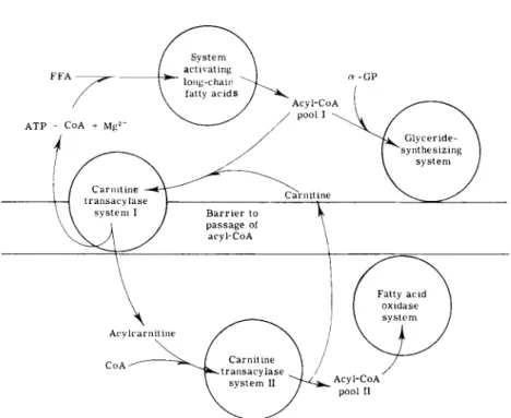
D. Inhibitors of Carnitine Acyltransferases
1. GENERAL
The transfer of fatty acyl groups from extramitochondrial acyl-CoA pools to the fatty acid oxidase complex within the mitochondrial matrix is dependent on operation of the carnitine acyltransferase system
(54-56). The general site of carnitine action on fatty acid oxidation is depicted in Fig. 2, in which the major points summarized include the following, (a) The inner membrane of the mitochondrion provides a barrier to the penetration of CoASH and fatty acyl-CoA compounds;
(b) carnitine acyltransferases, which exist in or on the inner mito
chondrial membrane, transfer acyl groups from CoASH to (—)-carnitine in a freely reversible manner; (c) the acyl groups from fatty acyl- carnitine derivatives are accessible to the enzyme system which generates intramitochondrial acyl-CoA required as substrate for the fatty acid oxidase complex.
Documentation of these conclusions may be found in reviews pre
viously cited (54-56) and in more recent articles (58, 59). Properties of carnitine acetyltransferase (60-62), carnitine octanoyltransferase (63, 64), and carnitine palmitoyltransferases (57, 64-68) have been described.
2 . INHIBITION OF CARNITINE ACYLTRANSFERASES BY ( - f) -ACYLCARNITINE DERIVATIVES AND BROMOACYLCARNITINE DERIVATIVES
Carnitine acetyltransferase (EC 2.3.1.7) is competitively inhibited by (-f-)-acetylcarnitine (61), and membrane-bound carnitinepalmitoyltrans-
318 I. B. FRITZ AND M. HALPERIN
FIG. 2. The actions of carnitine on fatty acyl group translocation across mitochon
drial barriers. For details, see the text and the review b y Fritz (54).
ferase (EC 2.3.1.8) is inhibited by (+)-palmitoylcarnitine or ( + ) - decanoylcarnitine (69, 70). The latter two derivatives do not inhibit the short-chain carnitine acetyltransferase, and (-f-)-acetylcarnitine does not influence the activity of carnitine palmitoyltransferase.
Fatty acid oxidation by mitochondria may be almost completely in
hibited by (-f-)-acylcarnitine derivatives (69), and oleate oxidation by perfused rat livers is also inhibited by ( + ) -decanoylcarnitine (71). The K
f
. in rat heart mitocondria is approximately 10 -5
M for ( + ) - p a l - mitoylcarnitine (69), and the inhibition can be competitively overcome by addition of (—)-carnitine.
More recently, the postulated existence of more than one carnitine palmitoyltransferase (57) has been demonstrated (64, 68). The substrate specificity of the transferases toward different fatty acyl groups reported by Kopec and Fritz (64) differs markedly from that observed by West et al. (68). In the former publication, carnitine palmitoyltransferase I (CPT-I) was reported to have a relatively broad chain length spe
cificity from 10:0 to 18:0, with the highest 7m ax displayed toward the
transfer of myristoyl groups. This solubilized enzyme was not inhibited by ( + )-acylcarnitines (64). In contrast, in the enzyme fractions de
scribed by West et al. (68), the substrate specificity of the enzyme called the "outer enzyme/' or soluble fraction, was quite broad, showing
l^max values toward the transfer of propionyl, butyryl, and hexanoyl
groups as high as that found for the transfer of palmitoyl groups from acylcarnitine to CoASH. The other carnitine palmitoyltransferase, labeled the "inner enzyme" or membrane-bound fraction, showed high
T^max values for the transfer of octanoyl, lauryl, and palmitoyl groups,
with the highest rates for laurylcarnitine conversion to lauroyl-CoA (68).
The basis for the different properties found in the two laboratories re
mains to be resolved. West et al. (68) observed that the "inner enzyme"
fraction was not inhibited by 2-bromopalmitoyl-CoA or (—)-carnitine, whereas the soluble "outer enzyme" fraction was inhibited.
The mechanism of inhibition of carnitine acetyltransferase by (—)-bromoacetylcarnitine has been investigated in detail by Chase and Tubbs (72, 73). A comparable report of the inhibition of the long-chain acyltransferase by bromopalmitoylcarnitine is apparently in preparation
(68). In preliminary communications reviewed by Tubbs and Garland (74), it was reported that 2-bromoacyl-CoA in the presence of carnitine inhibited the oxidation of acyl-CoA but not acylcarnitine by intact rat liver mitochondria, whereas 2-bromoacylcarnitine inhibited the oxidation of both acyl-CoA and acylcarnitine. In a more recent abstract, Tubbs and Chase (75) demonstrated that 2-S-bromomyristoylthiocarnitine in
hibited the oxidation of palmitoyl-CoA or palmitoylcarnitine by intact mitochondria, but it had no discernible effect on the "inner" pool of carnitine palmitoyltransferase (68, 75).
These combined observations demonstrate a complex compartition of long-chain carnitine acyltransferases in mitochondria. Inhibition of the transferases by ( + ) -acylcarnitine derivatives (69, 70) or by the 2-bromoacylcarnitine compounds (72-74) results in inhibition of fatty acid oxidation by mitochondria and perfused rat livers (71). All sub
strates and products inhibit both CPT-I and CPT-II (64).
E. Inhibitors of Enzymes of /? Oxidation
No known inhibitors specifically block the fatty acyl-CoA dehydro
genases (EC 1.3.99.3), the enoyl-CoA hydrolases (EC 4.2.1.17), the 3-hydroxyacyl-CoA dehydrogenases (EC 1.1.1.35), or the thiolases
(acetyl-CoA acetyltransferase, EC 2.3.1.9). Competitive inhibition of
320 I. B. FRITZ AND M. HALPERIN
butyryl-CoA dehydrogenase by crotonoyl-CoA has been reported (76).
Various nonspecific inhibitors, such as sulfhydryl reagents, inhibit the carnitine acyltransferases (61, 66) and the thiolases (77). Fatty acid oxidation in complex systems is blocked by a variety of metals, including Ca
2 + , Zn
2 + , and Cu
2+
(78),-by inhibitors that block any of the reactions of the electron transport assembly, or by sulfhydryl reagents (77-79).
The inhibitory effects of malonate on fatty acid oxidation have been reviewed by Webb (80), who also discussed the possible sites of action.
The inhibitory effects of elevated concentrations of long-chain acyl-CoA derivatives on the carnitine acyltransferases and other enzymes involved in fatty acid metabolism will not be discussed, since these derivatives inhibit a large number of enzymes (81) and the specificity of inhibition is questionable.
F. Miscellaneous
1. DlCHLOROACETIC A d D (DCA)
This compound has the interesting property of lowering blood glucose levels (82) and inhibiting fatty acid oxidation by muscle from diabetic animals (83). Simultaneously, glucose oxidation by muscle from diabetic rats is increased (83), thereby presumably accounting in part for the observed fall in blood glucose levels. Interestingly, this does not occur when DCA is administered to normal, fed rats (82, 83).
The mechanism of inhibition of fatty acid oxidation by DCA is un
known. Administration of DCA is followed by a fall in citrate levels in hearts and diaphragms of diabetic rats, presumably in association with the decreased rate of fatty acid oxidation (84). Decreased citrate concentrations would thereby relieve inhibition of phosphofructokinase (84, 85) and increase rates of glycolysis. It is possible that DCA forms dichloroacetyl-CoA and carnitine derivatives and that these compounds may be inhibitors of various reactions of fatty acid oxidation. Dichloro- acetylcarnitine has been found to inhibit carnitine acetyltransferase but not carnitine palmitoyltransferase activity (85a). Dichloroacetic acid salts have been shown to inhibit gluconeogenesis by isolated liver cells obtained from fasted rats (86). The site of inhibition of gluconeogenesis is associated with an indirect action at the level of triose phosphate dehydrogenase, resulting from inhibition of fatty acid oxidation and concomitant generation of NADH (86). The fall in blood sugar in dia
betic animals following the administration of DCA probably occurs be
cause of the combined effects of an increased rate of oxidation of glucose by muscle (84) and a decreased rate of hepatic gluconeogenesis (86).
2. RELATIONSHIP BETWEEN PATHWAYS OF FATTY ACID OXIDATION AND GLUCONEOGENESIS WITH RESPECT TO INHIBITORS OF FATTY ACID OXIDATION
When fatty acid oxidation is inhibited in the liver, the rate of gluco
neogenesis is also decreased. This has been found to be the case in pigeon liver homogenates (70) and in perfused rat livers (71) treated with
(+)-decanoylcarnitine. Comparable results were obtained when bro- momyristic acid was employed as an inhibitor of fatty acid oxidation
(87). These and other data obtained during investigation on the sites of action of 4-pentenoic acid in perfused livers (47, 48) have been in
terpreted as supporting the concept that the products of fatty acid oxi
dation facilitate rapid rates of gluconeogenesis (55, 71, 88-90). It is of interest that ATP, NADH, and acetyl-CoA, which are generated in larger amounts when fatty acid oxidation rates are enhanced, are each required as substrate or activator for various steps of the multienzyme sequence catalyzing the formation of glucose from lactate. For elabora
tion of the relationships between the pathways of fatty acid oxidation and gluconeogenesis, the reader is referred to various reviews (55, 88-90).
Experimental conditions have been found, however, in which addition of fatty acids results in increased ketogenesis but not in enhanced gluco
neogenesis by perfused rat livers (91). The coordination between these two pathways, which is often correlated in vivo (92), therefore need not be obligatorily coupled (91, 93).
II. INHIBITION OF PATHWAYS OF FATTY ACID SYNTHESIS
A. General Considerations
1. INTRODUCTION
The biosynthetic pathway from glucose to fatty acids is a prominent metabolic route in most organisms. Because there is a limited capacity for polysaccharide storage, carbohydrate ingested in excess of immediate caloric needs and storage capacity is converted to fatty acids and then to triglycerides. Triglycerides are stored primarily in adipose tissue and are quantitatively the major physiological fuel reserve. The biosynthesis of the complex polar lipids (phospholipids) of membranes is also a very important process since they undergo a relatively high rate of metabolic turnover in most cells. These compounds are synthesized by separate metabolic pathways.
3 2 2 I. B. FRITZ AND M. HALPERIN
Fatty acid synthesis as a major biological process occurs mainly in liver, adipose tissue, lactating mammary gland, and intestinal tract.
Rates of lipogenesis are highest in carbohydrate-fed intact animals and are lowest when lipolysis is augmented, as in starvation or insulin de
ficiency. This section will be concerned primarily with fatty acid synthe
sis in adipose tissue and in liver. (For general reviews, see 6, 94-98.) There is still considerable controversy as to the relative importance of liver and adipose tissue as major sites of fatty acid synthesis (99, 100).
In avian species it appears that the liver is the major if not only site of fatty acid synthesis (101), whereas in the rodent there is evidence to support a prominent role for adipose tissue in lipogenesis (99).
There is a complex degree of intracellular compartition during the bio
synthesis of lipids. In the first segment of the pathway, glucose is con
verted to pyruvate in the cytoplasmic compartment. In the next portion, pyruvate enters the mitochondria and is converted to acetyl-CoA and then to citrate. The third segment of the pathway occurs in the cyto
plasmic compartment. This requires citrate exit from mitochondria to the cytoplasm, where it is converted to acetyl-CoA (97), which in turn
is converted to long-chain fatty acids. These fatty acids are now esteri- fied with a-glycerol phosphate to form triglycerides or phospholipids.
2 . PATHWAY FROM GLUCOSE TO PYRUVATE IN LIVER
Glucose enters the hepatocyte from the blood. This entry process is not sensitive to insulin (102). Intracellular glucose is phosphorylated by the enzyme glucokinase to form glucose 6-phosphate (103). This enzyme has two major properties differing from those of hexokinase.
First, it has a high K'm for glucose (10~
2
M), which approximates the normal blood glucose concentration. An increase in blood sugar levels would therefore result in an increased formation of glucose 6-phosphate.
The second unique property of glucokinase is its failure to be inhibited by physiological levels of its product, glucose 6-phosphate, thereby per
mitting high levels of this intermediate to accumulate in hepatocytes when blood sugar levels increase. Glucose 6-phosphate is converted to fructose 6-phosphate by phosphohexosisomerase in a near equilibrium reaction that is not likely to be regulatory significance (104). Fructose 6-phosphate is then converted to fructose 1,6-diphosphate, catalyzed by phosphofructokinase. This reaction is markedly displaced from equilib
rium and is maintained in this state because of inhibition by levels of ATP and citrate existing in the cell (105). However, the inhibition is competitive, and it can be overcome by elevation of fructose 6-phos-
phate concentrations, which may occur secondary to increased blood glucose levels. Fructose diphosphate is converted to pyruvate in a multi- enzyme pathway. The rate of pyruvate formation is therefore indirectly controlled by the blood glucose (substrate) level.
In adipose tissue, the first steps of the glycolytic pathway differ from those in liver in the following respects. The glucose transporting system is insulin sensitive {106, 107). This should result in a low intracellular glucose concentration, which would create a large concentration gradient for glucose across this plasma membrane. Hexokinase, the enzyme that phosphorylates glucose in adipose tissue, has a very low K.'miov glucose
(10 _ 4
-10~
5
M) {103). The rate of glucose utilization by adipocytes is most likely controlled by the rate of glucose entry under physiological conditions rather than by the rate of phosphorylation. Hexokinase is inhibited in a predominantly noncompetitive manner by its product, glucose 6-phosphate {103). The remainder of the glycolytic pathway is similar to that of liver. If phosphofructokinase (PFK) is inhibited in adipose tissue, levels of fructose 6-phosphate and glucose 6-phosphate rise and noncompetitively inhibit hexokinase. This could inhibit further glucose utilization. Therefore, the rate of glucose utilization in adipose tissue is limited by the rate of glucose entry, but it can be secondarily inhibited by PFK inhibition. This contrasts with the regulation of glucose utilization by liver, in which the intracellular glucose concentration and glucokinase activity are the primary determinants.
An additional difference between glycolysis in adipose tissue and liver is the fate of the N A D H2 generated in the cytoplasm in the triose phos
phate dehydrogenase reaction. In liver this reducing power can be readily indirectly transported from the cytoplasm to the mitochondria. In con
trast, mechanisms for transporting reducing equivalents from N A D H2 into mitochondria in adipose tissue are of very low activity {108, 109).
Therefore, lipogenesis in adipose tissue may be limited by the rate of N A D H2 removal from the cytoplasm, since tissue pyruvate concentra
tions are decreased in association with elevated lactate levels {110).
This control is seen when glucose transport is not limiting (i.e., when insulin levels are high).
3. PATHWAY FROM INTRAMITOCHONDRIAL ACETYL-COA TO EXTRAMITOCHONDRIAL ACETYL-CoA IN BOTH LlVER AND ADIPOSE TISSUE
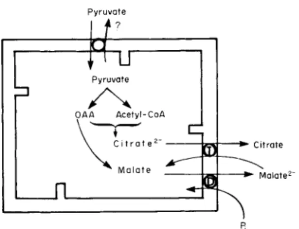
Pyruvate is converted to acetyl-CoA via the pyruvate dehydrogenase complex in the mitochondrion, and oxaloacetate is generated from pyru-
3 2 4 I. B. FRITZ AND M. HALPERIN
P y r u v a t e
Pyruvate
OAA Acetyl-CoA
Fj
FIG. 3. Mitochondrial anion transporters involved in acetyl C o A exit from the mitochondria. Abbreviations: T, tricarboxylate carrier; D , dicarboxylate carrier;
OAA, oxaloacetate; and Pi, inorganic phosphate. For details, see the text.
vate in the pyruvate carboxylase reaction. The importance of the latter reaction has been stressed by Ballard and Hanson (111). Acetyl-CoA in turn combines with oxaloacetate to form citrate. Citrate, so generated, must exit from the mitochondria (97). In a later section, the require
ments for citrate transport in rat liver mitochondrial membranes will be discussed. It is generally accepted that citrate
2-
exits from the mito
chondria in exchange for malate 2-
(112, 113), as illustrated in Fig. 3.
Once citrate has entered the cytoplasmic compartment, it is converted to acetyl-CoA, catalyzed by ATP:citrate lyase.
4. PATHWAY FROM EXTRAMITOCHONDRIAL ACETYL-COA TO FATTY ACIDS
There are two major steps in this pathway. Acetyl-CoA is first con
verted to malonyl-CoA in the reaction catalyzed by acetyl-CoA car
boxylase. This enzyme has two major potential physiological regulators:
citrate, which serves as a positive effector, and long-chain fatty acyl- CoA, which inhibits the reaction. Malonyl-CoA may then be converted to long-chain fatty acyl-CoA in the presence of cytoplasmic NADPH2, catalyzed by the fatty acid synthetase complex. The potential regulatory role of acetyl-CoA carboxylase in this segment of the pathway and the role of acyl carrier protein in fatty acid biosynthesis are discussed in a recent review (114).
5. PATHWAY OF FATTY ACIDS TO TRIGLYCERIDES
This pathway in adipose tissue and liver involves the esterification of 3 moles of fatty acyl-CoA with 1 mole of a-glycerol phosphate {115).
In the intestinal mucosa, however, the monoglyceride pathway is quanti
tatively more important {116). The reader is directed to recent reviews {116-119) for additional information. The monoglyceride pathway ap
pears to be quantitatively significant in hamster adipose tissue {120).
In the section on enzymes to follow, our discussion of the enzymes of the glycolytic pathway will include only phosphofructokinase and pyruvate kinase. The reader is referred to earlier chapters in this series and to various reviews {104, 181) for details concerning the other en
zymes of the pathway.
B. Enzymes Involved in Fatty Acid Biosynthesis from Glucose
1. PHOSPHOFRUCTOKINASE (EC 2 . 7 . 1 . 1 1 )
As pointed out in a previous section, this reaction probably controls the rate of glycolysis in adipose tissue, provided that the rate of glucose transport is not rate limiting. Physiologically, the two primary inhibitors of phosphofructokinase (PFK) are citrate and ATP, both of which are competitive inhibitors even though ATP is also a substrate {105, 122).
The role of cytoplasmic citrate as an inhibitor of PFK, however, could be in conflict with its possible role as an activator of acetyl-CoA car
boxylase in the lipogenic pathway {122).
It is difficult to obtain reliable kinetic values for PFK, but Lowry and Passonneau (105) have determined a K'm for ATP and fructose 6-phosphate ( F 6 P ) of 2 X 1 0 ~
4
and 4 X 1 0 ~ 5
M, respectively. By altering ATP and F 6 P concentrations, the K'{ for citrate was shown to vary
100-fold. A K'i value for citrate of 3 X 1 0 ~ 5
M was measured at pH 8. 0 by these authors (122), while the K[ reported for ATP was 1.4 X 1 0 ~
3 M.
Lowry and Passonneau (105) have interpreted their results to indicate the presence of 7 - 1 2 substrate, inhibitor, and deinhibitor sites on PFK. They postulated that the addition of one inhibitor (ATP) facilitates the addi
tion of the other (citrate) and simultaneously retards the addition of the deinhibitors. In contrast, the addition of any two deinhibitors facilitates the addition of a third but retards the addition of either inhibitor.
Although M g 2+
itself is inhibitory to PFK, the relationship is complex.
Since free ATP is much more inhibitory than MgATP 2
" (105), the addi
tion of M g 2+
in the presence of ATP may decrease inhibition of PFK.
326 I. B. FRITZ AND M. HALPERIN
The effect of pH is also complex. Maximum PFK activity occurs at pH 8.2, and its activity is extremely sensitive to small changes in pH in the physiological range. A decrease in pH decreases the affinity of enzyme for F6P. At pH 5.8, PFK was 90% inactivated, and this activity could not be restored by elevation of pH alone. It required addition of hexose phosphate and adenine nucleotide activators to restore full activity (123). A summary of effectors is provided by Mansour (123).
2. PYRUVATE KINASE (EC 2.7.1.40)
Tanaka and co-workers (124) have reported that there are two types of pyruvate kinase (PK) in liver. Pogson (125) has shown that adipose tissue also has two forms of this enzyme. Pyruvate kinase activity is reported to be of regulatory significance (104). Its concentration varies during different nutritional states, especially in organs that have both a glycolytic and a gluconeogenic pathway. Physiological control of the pyruvate kinase reaction is modulated by levels of the substrate phos- phoenolpyruvate (PEP), and it is inhibited by elevated ATP concentra
tions. In addition, fructose 1,6-diphosphate (FDP) activates the purified liver L type of enzyme at low PEP concentrations, and it reverses the inhibition of ATP of this enzyme form. Fructose 1,6-diphosphate does not influence the M form of this enzyme.The enzyme PKA is converted to PKB by FDP, and P KA is also activated by FDP. Kinetic constants are summarized in Table I. Bailey et al. (126) state that there are two L forms of this enzyme distinct from the M form reported by Tanaka (124). They also indicate that Cu
2+
inhibits the L form with a K[ of 1.0 X lO"
5 M.
Weber et al. (127) reported that acetyl-CoA inhibited PK, with a K[
of 3.0 X 10~
5
M, under conditions cited. However, the K\ increased two orders of magnitude when a preincubation period was absent.
3. PYRUVATE DEHYDROGENASE (EC 1.2.4.1)
Pyruvate formed from glucose in the cytoplasm must enter the mito
chondria to be converted to acetyl-CoA by the pyruvate dehydrogenase (PD) complex (128). There seems to be a mitochondrial transport system for pyruvate, according to Papa et al. (129), but no inhibitors of this process have yet been found. The pyruvate dehydrogenase com
plex isolated from liver and adipose tissue mitochondria is a multienzyme unit, and the activity of the complex is regulated by phosphorylation
T A B L E 1
PROPERTIES OF PYRUVATE K I N A S E "
Liver enzyme
Property M T y p e L T y p e P KB P K A
K'm P E P (M) 7.5 X 10"
5
8.4 X 10~
4
6.7 X 10~
5
6.0 X 10~
4
K'm A D P (M) 2.7 X 10~
4
1.0 x 1 0 -
4
6.7 X 10~
4
3.3 X 10"
4
K\ATP ( M ) 3.5 X 10"
3
1.0 X 1 0 -
4
1.5 X 1 0 -
3
1.5 X 10"
3
K\ p - C M B
6
(M) 2.5 X 1 0 -
5
7.42 X 10"
7
Conditions for Regeneration
a
N o r m a l or car
increased of liver bohydrate-
activity tumor fed, insulin
F D P activation —
+
—c
Kinetics Michaelis- Substrate Michaelis- Substrate
+
Menton activation Menton activation
a
Data from 124, 125.
b
p-Chloromercuribenzoate.
c
The P KA form is converted to P KB b y F D P and reversed ty E D T A , A T P , or citrate.
and dephosphorylation reactions (128, 130-132). Phosphorylation and concomitant inactivation are catalyzed by an ATP-specific protein kinase (PD kinase), resulting in phosphorylation of the pyruvate de
hydrogenase fraction. Dephosphorylation and concomitant reactivation are catalyzed by a phosphatase that requires M g
2+
for its activity. This activation can be seen in adipose tissue with insulin in vitro (132-134).
The P D kinase is inhibited by ADP. This is more evident in enzyme preparations from kidney than from liver. Adenosine monophosphate and 3',5'-cyclic A M P were without effect. The apparent K'm for ATP was 20-90 JJLM and the apparent K'{ for A D P was approximately 100 \xM.
Pyruvate and a-ketobutyrate, which is a weaker substrate for PD, pre
vented in part the inactivation of PD by ATP in the presence of P D kinase. Very high pyruvate concentrations (10 times greater than the K'm of 50 y.M) were required for almost complete protection. The P D kinase is active at low M g
2+
concentrations, whereas the phosphatase requires a concentration of 10 m l Mg
2
+ for optimal activity (131).
Garland and Randle (135) have reported another form of control of PD. They showed that the PD complex from pig hearts was inhibited by the products of pyruvate oxidation (acetyl-CoA and NADH) and
328 I. B. FRITZ AND M. HALPERIN
that these inhibitions were reversed by CoA and NAD, respectively.
The acetyl-CoA to CoASH mole ratio for 50% inhibition was approxi
mately 0.75 in rat liver (136), whereas the actual ratio in livers from fed rats was 0.5 and that from livers of fasted rats with 1.5 (137). Wieland et al. (136) calculated a K'. for acetyl-CoA of 29 /xM.
4. PYRUVATE CARBOXYLASE (EC 6.4.1.1)
Pyruvate carboxylase (PC) catalyzes the synthesis of oxaloacetate from pyruvate. This enzyme contains biotin, as does acetyl-CoA car
boxylase. The reaction occurs in the mitochondrial compartment and has been recently reviewed (138).
Under physiological circumstances, a short-chain acyl-CoA derivative is required as an activator of this enzyme from tissues of vertebrate organisms. The most potent activators include acetyl-CoA, propionyl- CoA, crotonyl-CoA and isobutyryl-CoA. The pH optimum is at pH 7.8.
The K'm for ATP is 5.8 X 10~
5
M, that for bicarbonate is 1.0 X 10"
3 M, and that for pyruvate is 4.4 X 10~
4
M . The K'a for acetyl-CoA is 3.3 X 1 0
-5
M, but it can be as low as 4 X 10~
6
M at a different pH. Propionyl- CoA is less active, having a K'a of 1.1 X 10~
4
M. If the pyruvate concen
tration is increased above 1 mM, it shows apparent activation of pyruvate carboxylase (139).
Three main classes of reversible inhibitors of pyruvate carboxylase have been described: (a) nucleotides that are competitive inhibitors with respect to ATP, (6) analogs of the activator acyl-CoA derivatives which are competitive inhibitors with respect to acetyl-CoA, and (c) pyruvate analogs, many dicarboxylic acids, and their derivatives which are specific inhibitors of the transcarboxylation step of the pyruvate carboxylase reaction, inhibiting the enzyme as a result of interaction with the bound manganese. The inhibitor constants obtained for repre
sentatives of each of these classes of inhibitors are summarized in Table II (139). Pyruvate carboxylase purified from yeast is inhibited by L-aspartate, but pyruvate carboxylase prepared from chicken liver is not inhibited by L-aspartate (0.2-20 mM) at either saturating or non- saturating concentrations of acetyl-CoA (H0).
Pyruvate carboxylase is inactivated irreversibly by incubation with avidin, due to binding of the biotin residues in the enzyme (HI). Other less specific inhibitors of PC include sulfhydryl reagents, such as p-chloromercuribenzoate and 5,5'-dithiobis(2-nitrobenzoic acid), and various protein-denaturing agents, such as guanidine salts and sodium dodecylsulfate.
T A B L E I I
SOME INHIBITORS OF PYRUVATE C A R B O X Y L A S E
0
Inhibitor (M)
Nucleotides
C T P 1.6 X 1 0 "
5
U T P 4.5 X 1 0 "
4
T T P 1.3 X 10~
3
C D P 1.6 X 1 0 "
3
5 ' - C M P 1.2 X 10~
2
A c y l - C o A analogs
M a l o n y l - C o A 8.3 X 1 0 "
6
Methylmalonyl-CoA 1.0 X 10~
4
Acetylpantetheine 2.8 X 10~
4
Pyruvate analogs and dicarboxylic acids
Fluoropyruvate 1.7 X 10~
4
Phenylpyruvate 4.8 X 10~
4
Oxalate 1.2 X 10~
5
Oxamate 1.6 X 1 0- 3
Malonate 2.2 X 10~
2
Mesoxalate 2.1 X 1 0- 3
L-Malate 6.5 X 1 0- 3
Glyoxal 5.6 X 1 0- 3
° From 139.
5. CITRATE SYNTHASE (EC 4.1.3.7)
The condensation of acetyl-CoA with oxaloacetate to form citrate is catalyzed by the mitochondrial enzyme citrate synthase. This enzyme has been well characterized from many sources. The pH optimum for this enzyme is approximately 9.0 but becomes lower in the presence of adenine nucleotides. The K
r
values for acetyl-CoA and oxaloacetate are of the order of 10~
5
M (for review, see 142).
Three major groups of physiological inhibitors have been discovered for this enzyme. Atkinson (143) and Garland (144) have reported that citrate synthase is inhibited by adenine nucleotides. Concentrations re
quired for 50% inhibition (shown in parentheses) are most marked for ATP (0.42 mitf), less extensive for ADP (0.6 milf), and least of all for AMP (2.0 m l ) . The inhibition is competitive with acetyl-CoA in that, in the presence of 4 m l ATP, the K'm for acetyl-CoA is raised
from 14 to 160 /zM. A similar phenomenon occurs with respect to oxalo
acetate, although the potential release of ATP inhibition by elevated
3 3 0 I. B. FRITZ AND M. HALPERIN oxaloacetate concentrations is masked by substrate inhibition. When added in vitro, M g
2+
tends to overcome the inhibition of citrate synthase by adenine nucleotides, and considerable doubt remains concerning the physiological implications of the control of citrate synthase activity by these cofactors (145).
Another group of inhibitors of citrate synthase includes the long-chain fatty acyl-CoA compounds. Wieland and Weiss {146) and Tubbs (147) reported that low concentrations (5 X 10~
6
M ) of palmitoyl-CoA inhibit this enzyme. Studies by Srere (148) have shown that this interaction is dependent not on the concentration of palmitoyl-CoA but rather on the molar ratio of this compound to citrate synthase. Srere (148) and Fritz (149) have shown that a number of substances (palmitoylcarnitine, albumin) can protect citrate synthase from inhibition by long-chain acyl-CoA derivates and that inhibition occurs only when large amounts of palmitoyl-CoA are enzyme bound.
Williamson (150) has studied a third group of inhibitors of citrate synthase. He showed that succinyl-CoA is a potent competitive inhibitor for acetyl-CoA (K'{ = 1 4 0 nM). Propionyl-CoA has a similar effect (K'{ = 5 0 fiM) when studied with the beef heart enzyme. Coenzyme A had a low K\ ( 6 7 /xM) for this enzyme, but showed a mixed form of inhibition. Citrate itself was a weak (K[ = 1.6 X 1 0
-3
M ) inhibitor of citrate synthase, competitive with oxaloacetate. Fluoroacetyl-CoA, which may function as a substrate for this enzyme, has been shown to be a competitive inhibitor of citrate synthase with respect to acetyl-CoA.
Desulfo-CoA is also a competitive inhibitor for citrate synthase with respect to acetyl-CoA (142).
6. ATP .'CITRATE OXALOACETATE LYASE (EC 4.1.3.8)
Once citrate has exited from the mitochondrion, it is converted to acetyl-CoA, catalyzed by the enzyme ATP:citrate oxaloacetate lyase.
The properties of this enzyme have been described by Srere (151).
Watson et al. (152) have restudied the K'm for citrate at low (85 meq/
liter) and high ( 5 0 0 meq/liter) chloride concentrations. With low chloride levels, there were two apparent K'm values for citrate ( 0 . 1 6 and 5 mM), whereas at high chloride levels only one K
f
m of 0 . 1 4 mM was seen but at a l^max of 5 0 % of that seen with low chloride levels. One of the reaction products, ADP, inhibits this reaction competitively with respect to ATP.
The K'i value for ADP at pH 8.4 is 0.172 mM. These authors (152) have described two additional inhibitors of this enzyme. The stereoisomer of ( —)-hydroxycitrate obtained from Garcinia cambozia inhibits this enzyme
competitively with citrate when the citrate concentration is less than 1 m l . The K' values for homocitrate are 0 . 1 5 and 0.57 \xM at low and high chloride concentrations. Watson and Lowenstein {153) demonstrated in a cell-free hepatic system that this agent strongly inhibited fatty acid synthesis from [
1 4
C ] alanine. The concentration required in this system (1 m l citrate as substrate) for 5 0 % inhibition was 2 0 juM. The au
thors state that ( — )-hydroxycitrate does not inhibit citrate exit from mitochondria.
The other agent to inhibit ATP:citrate oxaloacetate lyase in vitro is tricarballylate (1,2,3-tricarboxypropane). Its mechanism of action is complex. This enzyme can convert tricarballylate to its CoA derivative.
When citrate is present, its cleavage is inhibited by tricarballylate with a K'. of approximately 1 mM. However, this inhibition is dependent on the time of preexposure of the enzyme, inhibitor, ATP, and CoASH
(158).
7. ACETYL-COA CARBOXYLASE (EC 6.4.1.2)
This enzyme is located in the cytoplasmic compartment in liver and adipose tissue. It catalyzes the first reaction in the synthesis of fatty acids which is unique to this biosynthetic pathway. It has been ex
tensively reviewed elsewhere (114, 154-156). This enzyme has four non- identical subunits and contains covalently bound biotin as a prosthetic group. It therefore can be inhibited by avidin, a specific biotin-binding protein (141)-
In 1952, Brady and Gurin (157) reported that the synthesis of long- chain fatty acids was greatly stimulated by citrate in a pigeon liver cell-free extract. This effect was later localized to acetyl-CoA car
boxylase. Activation by anions such as citrate is associated with poly
merization of inactive protomers to form active polymeric filaments (158). This affects the Fm ax rather than the K' for substrates. Acetyl-
m
CoA potentiates this action of citrate. The extent of the activation by citrate depends on both the citrate concentration and the time of activa
tion in an interdependent manner. Tricarballylate can replace citrate as an activator, but its presence inhibits fatty acid synthesis, possibly by inhibiting citrate lyase (152). The interdependence between citrate concentration and the time of incubation, as well as the temperature dependence, makes it difficult to assign a value for the citrate concentra
tion required to give half-maximal activation in fatty acid synthesis (159). A concentration of 10 mM ATP is required to inhibit the citrate- induced activation of acetyl-CoA carboxylase by about 5 0 % . The ATP
3 3 2 I. B. FRITZ AND M. HALPERIN
inhibition of acetyl-CoA carboxylase is strong even in the presence of magnesium ions (159).
Acetyl-CoA carboxylase is inhibited by long-chain fatty acyl-CoA derivatives (114), competitively with respect to citrate and noncompeti- tively with respect to substrates. The K'm is approximately 0.8 pM. As stated previously, the specificity of the inhibitory effects of acyl-CoA derivatives has been questioned because these agents are potent de
tergents and inhibit many enzymes in vitro (81). Elevation of protein concentrations prevents the inhibitory effects of palmitoyl-CoA on many enzymes (81, 160), and palmitoylcarnitine is also capable of reversing this inhibition (159,161).
Malonyl-CoA has been reported to be a potent inhibitor of acetyl-CoA carboxylase, competitively with respect to acetyl-CoA. The K'. for malonyl-CoA for both the rat liver and adipose tissue enzymes is ap
proximately 1 0 i*M (156). Inhibition ( 5 0 % ) of this enzyme was seen at concentrations of 10~
3
M salicylate or 6 X 10~
3
M nicotinate (162).
Maragoudakis (163, 164) has shown that a number of hypolipidemic agents, namely, 2-methyl-2-[p-(l,2,3,4-tetrahydro-l-naphthyl)phenoxy]
propionate, ethyl 2-(p-chlorophenyl)-2-methylpropionate, and 2-methyl- 2 - (p-chlorophenylphenoxy) propionate, inhibit acetyl-CoA carboxylase.
This inhibition is competitive for both the substrate acetyl-CoA and the activator isocitrate and is noncompetitive for ATP and HCO~. These drugs do not inhibit fatty acid synthetase.
8. FATTY ACID SYNTHETASE
This major enzyme in the lipogenic pathway has been extensively reviewed elsewhere (114) - The reader is referred to this and other reviews for details required to supplement general considerations referred to in the previous sections. In summary, this enzyme complex is stimulated by FDP or other phosphorylated sugars (165) and inhibited by one of its substrates, malonyl-CoA (166), and the product, long-chain fatty acyl-CoA (167).
In bacterial systems, the key dehydrase that initiates their unique pathway to long-chain unsaturated fatty acids is inhibited by 3-decanoyl-Af-acetylcysteamine (168). The other known mechanism for fatty acid desaturation in higher living forms is inhibited in vivo and in vitro by the cyclopropene fatty acid, sterculic acid (169). Methyl- malonyl-CoA has been shown by Cardinale et al. (170) to inhibit fatty acid synthesis in vitro. It may also lead to the formation of unusual fatty acids, probably by substituting for malonyl-CoA.
9. INHIBITION OF THE PATHWAYS OF GLYCERIDE BIOSYNTHESIS
Phosphatidic acid phosphohydrolase (L-a-phosphatidate phospho
hydrolase, EC 3.1.3.4) is the best-characterized enzyme specific to the esterification pathway. This enzyme can be inhibited in vitro by 5 0 mM fluoride, detergents such as long-chain fatty acyl-CoA and Tween 20, and SH reagents such as A^-ethylmaleimide (119). Rosenthal and Ching-Hsien Han (171) have shown that certain synthetic phosphatidic analogs (2-hexadecoxy-3-octadecoxypropylphosphonate and 2-octade- cyleicosylphosphonate) can also act with some specificity as inhibitors of this enzyme. These agents are less effective on the particulate than on the soluble enzymes. This latter point illustrates the importance of physical factors in enzyme-catalyzed reactions of phospholipids.
The diglyceride acyltransferase can be inhibited by fluoride (172), whereas the monoglyceride one cannot (173), and the converse is true with SH-binding agents. Both acyltransferases are inhibited by long- chain fatty acyl-CoA (174).
C. The Role of Transport Processes across Mitochondrial Membranes in the Control of Lipogenesis
1. GENERAL CONSIDERATIONS
In this section, we shall discuss in detail the pathways of carbon and reducing-power transport across the mitochondrial membranes and indicate, where known, the inhibitors of these pathways. As indicated earlier, there is a complex coordination of various portions of the lipo- genic pathway within cells of such organs as liver and adipose tissue, and the interactions between mitochondrial and cytoplasmic compart
ments play a paramount role in supplying and possibly regulating the flow of carbon and reducing power required for fatty acid synthesis in the cytoplasm.
The systems used for the transport of substrates into and out of mito
chondria have been discussed in a recent review (175). The outer mito
chondrial membrane does not appear to hinder the passage of small molecules. If a substrate is to react with an enzyme located within mitochondria, it must first penetrate the inner mitochondrial membrane.
This membrane does not permit free diffusion of most molecules and ions in either direction. Consequently, the entry of important metabolic anions such as malate and citrate into mitochondria and into the cyto
plasm requires specific transporting systems.
334 I. B. FRITZ AND M. HALPERIN
2 - O x o g l u t a r a t e - M a l o n a t e
m
L- M a l a t eXL m
m • P i
S u c c i n a t e M a l o n a t e
C i t r a t e I s o c i t r a t e
^ / s - A c o n i t a t e
FIG. 4. Mitochondrial anion transport systems for intermediates of the tricarbo
xylic acid cycle. Abbreviations: 0 , oxoglutarate carrier; D , dicarboxylate carrier; T, tricarboxylate carrier; and Pi, inorganic phosphate.
Robinson and Chappell first showed that 2-n-butyl malonate inhibited dicarboxylate oxidation by intact mitochondria to a far greater extent than it influenced their oxidation by mitochondria fragmented by ultra- sonication. They later accounted for these results by demonstrating that 2-n-butyl malonate inhibited the passage of L-malate across the inner mitochondrial membrane (176). More recent work has shown that there are at least three separate mitochondrial carriers for L-malate, the specificity of which is determined by the nature of the countertransport- ing anion (see 175). These are illustrated in Fig. 4. The initial indication that three separte carriers existed for L-malate was provided by ob
servation of the different properties in various tissues of oxoglutarate and isocitrate oxidation in the presence and absence of L-malate. Thus, malate greatly stimulated oxoglutarate oxidation in heart mitochondria, whereas there was a much lesser effect on isocitrate oxidation. In ad
dition, malonate was observed to activate the oxidation of oxoglutarate but not of citrate, whereas the converse was obtained with 2-hydroxy- malonate. More recent studies with various other inhibitors have con
firmed and extended this hypothesis (113,177-179).
2. INHIBITORS OF ANION TRANSPORT ACROSS MITOCHONDRIAL MEMBRANES
Since, as mentioned above, the dicarboxylate and tricarboxylate inter
mediates of the Krebs cycle do not diffuse freely across the inner mito-
chondrial membrane, these metabolites must exit by specific carriers.
Two of these carriers are primarily involved in the fatty acid synthesis pathway. The tricarboxylate carrier allows citrate
2-
to exit from the mitochondrion in exchange for malate
2-
{112, 113), while the dicar- boxylate carrier permits malate-phosphate exchange {175). The di- carboxylate carrier transports malate from the mitochondrion to the cytoplasmic compartment, where it serves as a counteranion to permit citrate exit from the mitochondrion (see Fig. 3). Citrate exit into the cytoplasmic fraction is required for fatty acid synthesis from glucose (97).
a. Inhibitors of the Tricarboxylate Transporting System. Several in
hibitors of this transport system have recently been reported (113, 177-179). Three compounds have been found which inhibit only the tri
carboxylate carrier, namely 2-ethyl citrate (177), 2-propyl citrate (177), and 1,2,3-tricarboxy benzene (113, 178). The latter compound is the most potent inhibitor (K
r
. = 0.3 mM) and weakest countertransporter of the three. Substitutions at the 5 position of 1,2,3-tricarboxybenzene of either a carboxyl, a nitro, an amino, or an acetamido substituent significantly decreases the inhibitor potential of this compound (179).
p-Iodobenzyl malonate is also a very potent inhibitor of the tricar
boxylate carrier (178), but it equally well inhibits the dicarboxylate and oxoglutarate carriers. Another group of inhibitors of the tri
carboxylate carrier is comprised of the iodobenzoates (179). These com
pounds, in contrast to those mentioned above, are noncompetitive in
hibitors. The kinetic constants of inhibitory compounds described are summarized in Table III.
b. Inhibitors of the Dicarboxylate Transport System. A number of dicarboxylic acids has been shown to inhibit the dicarboxylate transport
ing system of rat liver mitochondria. This list includes 2-butyl malonate (176), 2-pentyl malonate (113), p-iodobenzyl malonate (180), and 2-phenyl succinate (181). Other compounds that have recently been shown to be inhibitors of this dicarboxylate transport system are those compounds with adjacent cts-carboxyls such as 2,3-pyridine dicarboxyl
ate, m-l,2-cyclohexane dicarboxylate, phthalate, and 3-nitro-l,8-naph- thalate (179). The latter compound consistently showed the highest activity of this group. This finding was rationalized by the fact that the intercarboxyl distance in this compound (0.25 nm) more closely resembles that of malate and malonate (0.24 nm) than does that of the less active inhibitors such as phthalate (0.28 nm) (179). A detailed description of these inhibitors may be found in a publication of Robinson
336 I. B. FRITZ AND M. HALPERIN T A B L E I I I
COMPOUNDS INFLUENCING TRICARBOXYLATE TRANSPORT IN R A T L I V E R MITOCHONDRIA"
Substrate
K'm for transport
( m M ) Inhibitor
Inhibition of citrate exchange
( % )
Citrate 0.25 1,2,3-Benzene tricarboxylate 92 Isocitrate 0 . 2 2 5-Nitro-l,2,3-benzene tricarboxylate 77
cis-Aconitate 0 . 2 2 2-Ethyl citrate 58
L-Malate 0.35 2-Propyl citrate 50
p-Iodobenzyl malonate 89
m- or p-iodobenzoate 72
m-Bromobenzoate 84
p-Iodophenyl acetate 60
a
All inhibitors were present at 10 m M final concentration, and all incubations were carried out at 10°C. For details, see Robinson et al. (179).
et al. {179) and is summarized in Table IV. All of the above studies were performed with rat liver mitochondria. In white adipose tissue of the rat, the only compound reported to inhibit the dicarboxylate trans
porting system has been butyl malonate {182). Other inhibitors remain to be tested in mitochondria from organs other than liver.
T A B L E I V
COMPOUNDS INFLUENCING THE DICARBOXYLATE TRANSPORTER IN R A T L I V E R M I T O C H O N D R I A
0
Substrate
K'm for transport
( m M ) Inhibitor
Inhibition of malate- phosphate exchange
( % )
L-Malate 2-n-Pentyl malonate 116
Malonate 2-(5
/
-Bromoamyl) malonate 115 Succinate 0.83 2-p-Iodobenzyl malonate 108
2-n-Butyl malonate 101
2-Benzyl malonate 96
2-Cinnamylidine malonate 88
2-Phenyl malonate 84
3-Nitr o-1,8-naphthalate 75 2,3-Pyridine dicarboxylate 43
° All inhibitors were present at a concentration of 10 m M . All incubations were carried out at 10°C. For details, see Robinson et al. (179).
Inhibition of mitochondrial anion transport should theoretically block fatty acid synthesis by inhibiting carbon flow to the cytoplasm. An ideal inhibitor should possess the following properties. It must be able to cross the plasma membrane of the cell and achieve the required cyto
plasmic concentration. The hypothetical compound must be a strong and specific inhibitor of this carrier, and it should have no significant effects on other steps in this or other metabolic pathways. The hypotheti
cal inhibitor should not act as a substrate for the carrier, as this property itself would allow exit of the tricarboxylic and dicarboxylic cycle anions.
In practice, it is difficult to meet these criteria. Consider the problem related to citrate analogs employed to inhibit the tricarboxylate carrier.
Since citrate is a very potent inhibitor of phosphofructokinase, citrate analogs may also inhibit this step and therefore reduce glucose conversion to pyruvate, thereby blocking fatty acid synthesis at an earlier site than transport of anions across mitochondrial membranes. This result was obtained when 2-ethyl citrate was tested (177). A further complica
tion can occur if citrate analogs share citrate's ability to activate acetyl- CoA carboxylase. At present, the "ideal inhibitor" having properties enumerated above remains elusive, and we know of no published reports concerning cellular systems in which inhibition of lipogenesis has been obtained by exclusively inhibiting citrate transport.
3. REDTJCING-POWER REQUIREMENTS FOR FATTY ACID SYNTHESIS
In the pathway of glucose to fatty acids, N A D H2 is formed in the cytoplasmic compartment. If it were to accumulate there, the pyruvate formed in the glycolytic pathway would be converted to lactate and thereby diminish fatty acid synthesis because less substrate would be available for acetyl-CoA formation. There are two primary pathways that permit reoxidation of the cytoplasmic NADH2, namely, N A D H2 transport into mitochondria or N A D H2 transfer to cytoplasmic NADP via the malate cycle (183). These pathways will now be discussed.
a. Transport of Reducing Equivalents across the Mitochondrial Mem
branes. It is generally accepted that NADH cannot cross the inner mito
chondrial membrane (184). In order for the reducing power of N A D H2 to enter mitochondria a "shuttle system" is required. Potentially, two pathways for this transport exist: the L-glycerol 3-phosphate pathway and the "Borst shuttle." The Borst shuttle is a malate-oxaloacetate cycle modified to include transamination on each side of the mito
chondrial membranes (185). The Borst cycle is illustrated in Fig. 5.
338 I. B. FRITZ AND M. HALPERIN
N A D H2
N A D
FIG. 5. The role of transporting systems in the oxidation of cytoplasmic N A D H2
b y mitochondria. Abbreviations: 0 , oxoglutarate carrier; G, glutamic carrier; A, glutamate-activated aspartate carrier; O A A , oxaloacetate; l,NAD-malate dehydro
genase; 2, aspartate aminotransferase.
The liver has an active "Borst shuttle" system (175). The steps in this pathway include the cytoplasmic and mitochondrial malate de
hydrogenases and aspartate aminotransferases as well as the oxoglutarate and aspartate-glutamate mitochondrial anion transport systems. The only reported inhibitors of the oxoglutarate carriers are p-iodobenzyl malonate (179), which also inhibits the dicarboxylate and tricarboxylate carriers, and L-aspartate, which inhibits only oxoglutarate entry into rat liver mitochondria (186). Rognstad and Katz (186a) have used aminooxyacetate to inhibit specifically the aspartate aminotransferase.
This compound does inhibit this step but it also combines with a-keto acids (such as pyruvate), thereby limiting its usefulness in studies on fatty acid synthesis. There are no known specific inhibitors of the gluta
mate-aspartate exchange, but the glutamate transporter is inhibited by 4-hydroxyglutamate, 2-aminoadipate or ^reo-hydroxyaspartate (see 175), and avenaciolide (187). The reader is referred to Barman (188) for a discussion of additional inhibitors of malate dehydrogenase and aspartate aminotransferase.
Although the "Borst shuttle" is active in liver, its activity is very limited in adipose tissue (108), which contains low amounts of intra- mitochondrial aspartate aminotransferase activity. When lipogenesis is proceeding rapidly in adipose tissue, the restricted flow of reducing power from N A D H2 into mitochondria limits the rate of fatty acid synthesis, because tissue pyruvate concentrations are decreased (110).