Review
Multidrug Resistance (MDR) and Collateral
Sensitivity in Bacteria, with Special Attention to Genetic and Evolutionary Aspects and to the
Perspectives of Antimicrobial Peptides—A Review
András Fodor1,* , Birhan Addisie Abate2, Péter Deák1,3 , LászlóFodor4, Ervin Gyenge5,6, Michael G. Klein7, Zsuzsanna Koncz8, Josephat Muvevi9, LászlóÖtvös10,11,12,
Gyöngyi Székely5,6,13 , Dávid Vozik14and LászlóMakrai4,*
1 Department of Genetics, University of Szeged, H-6726 Szeged, Hungary; deakp@brc.hu
2 Ethiopian Biotechnology Institute, Agricultural Biotechnology Directorate, Addis Ababa 5954, Ethiopia;
birhanaddisie@gmail.com
3 Institute of Biochemistry, Biological Research Centre, H-6726 Szeged, Hungary
4 Department of Microbiology and Infectious Diseases, University of Veterinary Medicine, P.O. Box 22, H-1581 Budapest, Hungary; Fodor.Laszlo@univet.hu
5 Hungarian Department of Biology and Ecology, Faculty of Biology and Geology, Babes,-Bolyai University, 5-7 Clinicilor St., 400006 Cluj-Napoca, Romania; gyenge_ervin@yahoo.com (E.G.);
gyongyi.szekely@ubbcluj.ro (G.S.)
6 Institute for Research-Development-Innovation in Applied Natural Sciences, Babes,-Bolyai University, 30 Fântânele St., 400294 Cluj-Napoca, Romania
7 Department of Entomology, The Ohio State University, 1680 Madison Ave., Wooster, OH 44691, USA;
klein.10@osu.edu
8 Max-Planck Institut für Pflanzenzüchtungsforschung, Carl-von-Linné-Weg 10, D-50829 Köln, Germany;
zskoncz@mpipz.mpg.de
9 National Cereals and Produce Board, Mombasa 80100, Kenya; jmuvevi@gmail.com
10 OLPE, LLC, Audubon, PA 19403-1965, USA; lotvos@comcast.net
11 Institute of Medical Microbiology, Semmelweis University, H-1085 Budapest, Hungary
12 Arrevus, Inc., Raleigh, NC 27612, USA
13 Centre for Systems Biology, Biodiversity and Bioresources, Babes,-Bolyai University, 5-7 Clinicilor St., 400006 Cluj-Napoca, Romania
14 Research Institute on Bioengineering, Membrane Technology and Energetics, Faculty of Engineering, University of Veszprem, H-8200 Veszprém, Hungary; vozik.david@drv.hu or
vozik.david@mk.uni-pannon.hu or vozikd@gmail.com
* Correspondence: fodora@expbio.bio.u-szeged.hu or fodorandras@yahoo.com (A.F.);
Makrai.Laszlo@univet.hu (L.M.); Tel.:+36-(30)-490-9294 (A.F.);+36-(30)-271-2513 (L.M.)
Received: 23 March 2020; Accepted: 23 June 2020; Published: 29 June 2020 Abstract: Antibiotic poly-resistance (multidrug-, extreme-, and pan-drug resistance) is controlled by adaptive evolution. Darwinian and Lamarckian interpretations of resistance evolution are discussed. Arguments for, and against, pessimistic forecasts on a fatal “post-antibiotic era” are evaluated. In commensal niches, the appearance of a new antibiotic resistance often reduces fitness, but compensatory mutations may counteract this tendency. The appearance of new antibiotic resistance is frequently accompanied by a collateral sensitivity to other resistances. Organisms with an expanding open pan-genome, such asAcinetobacter baumannii, Pseudomonas aeruginosa, andKlebsiella pneumoniae, can withstand an increased number of resistances by exploiting their evolutionary plasticity and disseminating clonally or poly-clonally. Multidrug-resistant pathogen clones can become predominant under antibiotic stress conditions but, under the influence of negative frequency-dependent selection, are prevented from rising to dominance in a population in a commensal niche. Antimicrobial peptides have a great potential to combat multidrug resistance, since antibiotic-resistant bacteria have shown a
Pathogens2020,9, 522; doi:10.3390/pathogens9070522 www.mdpi.com/journal/pathogens
high frequency of collateral sensitivity to antimicrobial peptides. In addition, the mobility patterns of antibiotic resistance, and antimicrobial peptide resistance, genes are completely different. The integron trade in commensal niches is fortunately limited by the species-specificity of resistance genes. Hence, we theorize that the suggested post-antibiotic era has not yet come, and indeed might never come.
Keywords: MDR; intrinsic/acquired resistance; collateral sensitivity; negative frequency-dependent selection; experimental evolution; pangenome; global dissemination; mobility patterns of resistance genes; adaptive evolution
1. Introduction
The rapid and ongoing spread of antibiotic resistance poses a serious threat to global public health [1]. Considering the complexity of the sophisticated resistance mechanisms [2,3], recruiting cooperating scientists with different backgrounds seems reasonable and necessary. This review is addressed to committed scientists, biologists, geneticists, and evolutionary biologists who may be attracted by fundamental, rather than applied, research perspectives. We focused on selected aspects.
One is the danger posed by globally spreading monoclonal pathogens (Acinetobacter baumannii[4–6], Pseudomonas aeruginosa[7–9]), with an expanding open pangenome, being aware that they are not the only ones displaying a threat.
Antibiotic resistance was discovered in 1940 [10]. The selective pressure of antimicrobials is favorable for resistant clones to survive and spread [11]. The emergence of antibiotic multiresistance (MDR) in pathogenic bacteria has become alarming in the last decades [12]. MDR appeared not only in human clinical pathogens [13–16] but also in zoonic bacteria [17,18], veterinary pathogens [19–26], and in plant pathogens [27–32]. Although application technologies are improving for plant pathogens [33], the trend has been that the use of antibiotics as plant medicines has gradually been restricted [34, 35]. A precondition for elaborating appropriate therapies is having a better understanding of antibiotic resistance mechanisms [36] in Gram-negative [13,16,37] and Gram-positive [38–41] pathogens.
New “bugs” are continuously appearing, and new antibiotics are needed. Conly and Johnson asked in 2005 “Where are the new antibiotics?” [42]. The tantalizing answer came 11 years later: Antibiotics are
“right under our nose!” [43]. Unfortunately, they still have not made it to market [44], at least not in the required number [45,46].
2. Multidrug Resistance: Updated Terms and Definitions
The genomes of different bacterium species harbor silent genes that code for resistance, but in the absence of antibiotics it is not manifested. Consequently, a large pool of potential of antibiotic resistance genes is hidden in various niches. The term antibiotic resistome covers the collection of all antibiotic resistance genes, including those associated with both pathogenic and non-pathogenic bacteria, but not ones which produce antibiotics [47]. Pál and his associates provided a comprehensive characterization of resistance genes, mobile genetic elements (MGEs), and bacterial taxonomic compositions for 864 metagenomes from humans (n=350), animals (n=145), and external environments (n=369), all deeply sequenced using Illumina technology. They concluded that antibiotic-polluted environments are probably under-estimated transmission routes, and indeed reservoirs for antibiotic resistance [48].
2.1. Antibiotic Resistance as a Phenotype
The resistance genotype [49–51] determines the resistance phenotype. Take the evolutionary history ofMycobacterium tuberculosisresistance to second-line anti-tuberculous as a scholarly medical genetic example to illustrate the simple Mendelian genotype–phenotype relations [52–55].
Genetic suppression of the cellular autolytic system of pneumococci causes simultaneous resistance to penicillin, D-cycloserine, and phosphonomycin [56], providing an example of the iso-allelic
inheritance phenotype [57,58]. Physiological suppression is similar [56], providing an example of the old genetic term, phenocopy [59,60]. The clarification of genotypic/phenotypic relations is a prelude to the application of clinical metagenomics [61].
2.2. The Location and Harboring of the Resistance Genes
The resistance gene is a peptide-coding open reading frame, genetically regulated as structure gene of an operon. The higher level of organization is the antibiotics resistance cassette [62–64]. The respective resistance gene(s) could be localized either on the chromosome [51], or in a plasmid [65–69], either as non-conjugative [62], conjugative [70,71], or in an episome [72].
2.3. Insertion and Excision
The resistance gene as an integron component [71,73] can be harbored by a mobile genetic element capable of inserting into, and excising from, another DNA molecule like a plasmid. This has been reviewed by several authors [74–79].
2.4. Homolog Recombination
Some mobile elements are capable of recombining with the bacterial chromosome, allowing transfer of large segments, as shown with vancomycin and penicillin resistance transfer, at least in Gram-positive (Enterococcus) pathogens [80,81]. Horizontal gene transfer (HGT) could happen even between the different bacteria taxa [82,83].
2.5. Intrinsic Resistance (IR)
IR is the phenotypic expression of a resistance gene originally present, resulting in structural and functional changes of the original gene product [84,85]. This can be either a decomposing enzyme [86–88], or the target site of the drug [89,90]. The most efficient resistance mechanisms are the multidrug efflux pumps [91–94].
When resistance to antimicrobial compound is a phenotypic expression of a resistance-encoding gene coming from outside via horizontal gene transfer HGT [95–100], it is called “acquired”
resistance [85,101]. The gene from outside had been harbored by a plasmid and was taken up from the environment, such as from soil [102,103] or the gastrointestinal [83,99] microbiota community.
Expansion of KPC-producingKlebsiella pneumoniaewith various mgrB mutations giving rise to colistin resistance [104], and the bla (VIM-1) metallo-beta-lactamase producing. E. coliwas published to spreading over in a university hospital in Greece [105]. These are examples of concerted activities of mobile genetic elements, including insertion sequences, transposons, and gene cassettes/integrons, and has been summarized by Partridge et al. [79]. Horizontal gene transfer (HGT) could even happen between different bacteria taxa [82,83].
The novel phenomenon called distributive conjugal gene transfer (DCT) was recently discovered in mycobacteria. DCT involves the transfer of chromosomal DNA between mycobacteria and, most significantly, generates trans-conjugants with mosaic genomes of the parental strains [106].
2.6. Definitions of Antibiotic Polyresistant Strains
Antibiotic resistances [2,3] are classified as follows: multidrug resistant (MDR) is not susceptible to at least one representative from each of three categories of selected antimicrobial compound families [3].
Extreme drug-resistant (XDR) is not susceptible to at least a single representative of all but very few categories of antimicrobial compound families. Pan-drug resistant (PDR) is not susceptible to any of the tested representatives of all known antimicrobial compound families [3].
2.7. The Updated List of the “ESKAPE” Polyresistant Pathogenic Bacterium Species
In 2006, the Antimicrobial Availability Task Force (AATF) of the Infectious Diseases Society of America (IDSA) prepared a review that highlighted frequently resistant pathogens to licensed antimicrobials [36], characterized by high antibiotic resistance, and extremely versatile MDR phenotypes which are responsible for many nosocomial infections [36]. As for their clinical significance [107], the updated the list of six ESKAPE Pathogen Bacterium Species appeared in 2008 [18]. The six letters are the initials of the genera of these bacteria: Enterococcus faecium, Staphylococcus aureus, Klebsiella pneumoniae, Acinetobacter baumannii, Pseudomonas aeruginosa,andEnterobacterspp. [108–112].
There is “NO DRUG” against them and “NO ESKAPE” from them [109]. They include causative pathogens of nosocomial diseases, such as ventilator-associated pneumonia [113,114], infections in burn wounds [115], and pathogens which “escape” from antimicrobial agents [109,116,117].
Immune-compromised patients are most exposed to ESKAPE pathogens causing bacteremia [118].
The ESKAPE list has recently been updated by the World Health Organization (WHO) [119].
It includes the carbapenem-resistant Klebsiella pneumoniae, Acinetobacter baumannii, Pseudomonas aeruginosa, carbapenem-resistant and third-generation cephalosporin-resistant Enterobacteriaceae, clarithromycin-resistantHelicobacter pylori, fluoroquinolone resistant Campylobacterspp., Neisseria gonorrhea, andSalmonella typhias community-acquired infection-causing pathogens. The highest-ranked Gram-positive members are vancomycin-resistantEnterococcus fecalisandEnt. faecium, as well as the methicillin-resistant (MRSA)S. aureus[119]. The resistance mechanisms used by the ESCAPE pathogens were recently reviewed [120].
Some other dangerous Gram-negatives such asEscherichia coli, Francisella tularensis, and Gram- positivesMycoplasma bovis and Bacillus anthracismay be considered as putative “ESKAPE Club-Members”
in the future. We discuss this option in Supplementary Material 1.
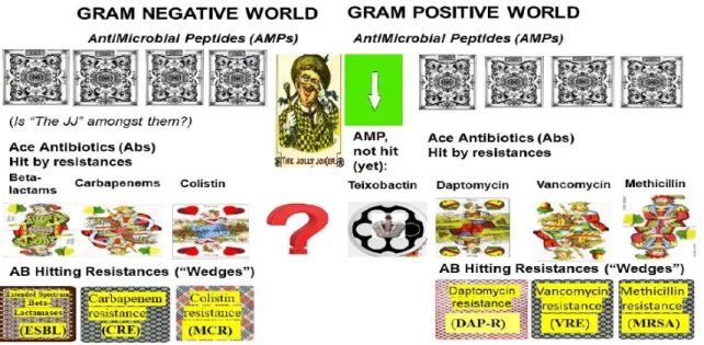
3. Resistance Problems Related to Gram-Negative Pathogens
The logical chain of events leading to multidrug resistance in Gram-negative pathogens is illustrated in Figure1.(LEFT).
3.1. B-Lactams vs. ESBL Resistance in Enterobacteriaceae and Klebsiella pneumoniae
Theβ-lactams were introduced to overcome penicillin resistance by preventing re-assembly of peptidoglycan bonds, which eventually leads to cell lysis [121,122]. Extended-Spectrum B-Lactamase (ESBL) producing Enterobacteriaceae andK. pneumoniaepathogens are resistant toβ-lactam-based antibiotics. The emergence of, and challenges by, ESBL-producing Enterobacteriaceae has been reviewed over the last three decades [123–126]. Pitout suggested that in vitro resistance to ceftazidime, and/or aztreonam, should be used as phenotypic markers of ESBL [127]. In Gram-negative bacteria, theβ-lactamase production is frequently associated with reduced permeability of the outer membrane efflux [128].
Biochemistry has increased the number of β-lactamase enzymes with enlarged substrate specificities [129]. The substrate range includes cephalosporins (cefotaxime, ceftriaxone [130–132]), monobactam aztreonam [133–135], amino-penicillin combinations [136], lactamase inhibitor ampicillin- sulbactam [137–140], ureido-penicillins [141,142] including piperacillin/tazobactam [143–146], temocillin [147], piperacillin/tazobactam [148], and ceftolozane-tazobactam [149]. Furthermore, the appearance of new enzymes is not associated with the loss of ability to hydrolyze the earlier lactams, such as ampicillin [36]. In addition, the prevalence of ESBL production amongE. coliand Klebsiellaspecies is variable [13]. ESBL can be considered as first strike back from the Gram-negative pathogens or as the first “Wedge” from Nature.
3.2. Carbapenems: A Strong Antibiotic (“High Card”) to Beat the “Wedge” ESBL
To overcome ESBL problems, a new beta-lactam class of antibiotics, the carbapenems [150,151], was developed [152], including blood and respiratory isolates ofP. aeruginosa[153]. As Breilh and his associates summarized, they act either as “slow substrate”β-lactamase inhibitors or by binding to penicillin-binding proteins [154]. This “value-added feature” of inhibitingβ-lactamases serves as a major rationale for the expansion of this class ofβ-lactams [155]. Carbapenem antibiotics, including imipenem, are Gram-negative cell-wall synthesis interrupting molecules, which bind to penicillin-binding proteins [156]. Cilastatin is a human enzyme, dehydropeptidase, in the kidney that inhibits imipenem degradation and meropenem [156], as well as ertapenem [157]. Each one alone, or in combination, had been a putative last line of defense against multidrug-resistant Gram-negative organisms.
3.3. Carbapenem Resistance: The Second Strike Back from the Gram-Negative Pathogens (“Wedge” from Nature)
Since 2006, the number of carbapenem-resistant Enterobacteriaceae (CRE) has significantly increased [158–160]. A novel, epidemic, serine class-A type enzyme (KPC) is behind carbapenem resistance. It is encoded by the Bla (Oxa) gene family [161–165]. KPC exhibits powerful activity against all types of Beta-lactam molecules [166–168].
3.4. NDM1
A novel type of plasmid-encoding carbapenem-resistant metallo-β-lactamasecarbapenemase NDM 1 [169] was identified in 2008 in two Enterobacteriaceae isolates, both recovered from a Swedish patient transferred from India [170]. The emergence of NDM-expressing enterobacteria [171]
andKlebsiella[172,173] was then reported from all continents, but it recently reappeared in Italy (Toscana, Z. Koncz, personal communication). There are publications on NDM 1 expressing Acinetobacter [174], Pseudomonas [175], and only one E. coli publication [105]. It has never been found in Gram-positive bacteria. Klebsiella pneumoniae, Acinetobacter,andPseudomonasare spread by clonal dissemination [176], whileE. coliand other Enterobacteriaceae spread through polyclonal dissemination [177]. Many carbapenem-resistance genes localized on mobile genetic elements have been previously reviewed [178–184].
3.5. “Dropped and Rediscovered” Colistin, as a Large Spectral Antibiotic (a “Trump Card”)
The cationic lipopeptides polymyxin B, E, and colistin [185] disrupt outer membranes (OMs) and have been used since 1959 for treating infections caused by Gram-negative MDR pathogens. However, nephrotoxic side effects were discovered [186]. Since the appearance of several new infections caused by MDR Gram-negative organisms [187,188], and intensive searches of old antibiotic options, colistin was rediscovered and considered to be a potential trump card [189–194].
3.6. Colistin Resistance: The Third Unexpected Attack (“Wedge”) from Nature
The first colistin-resistant mutant was reported in 1981 [195]. The appearance of colistin resistance, especially plasmid-mediated transferable ones [196–201], questioned whether the polymyxins should be considered as the last “life-savers” [202]. Colistin resistance is a consequence of post-translational modification, or loss, of the lipopolysaccharide (LPS) molecules [203]. The first colistin-resistant mutant was reported in 1981. The appearance of colistin resistance, especially the plasmid-mediated transferable ones, questioned whether the polymyxins were still be considered as the last “life-savers”.
The mechanisms of acquired and intrinsic resistance of polymyxin in different bacteria have recently been reviewed by many authors [204–209]. The genetic factors behind colistin resistance aremcr genes [198,210,211]. Colistin resistance has been evolving under clinical conditions [212,213] in A. baumannii [214–216], P. aeruginosa [217], K. pneumoniae [218,219], and Enterobacter cloacae[220].
The evolution of colistin resistance was more than a one step process, requiring mutation in at least five independent loci synergistically, creating the resistant phenotype [211,212,221,222].
3.7. Efforts to Overcome MDR Problems in Gram-Negative Pathogens in the Absence of Omnipotent (“Jolly Joker”) Antibiotics
The potentiation ofβ-lactam antibiotics andβ-lactam withβ-lactamase inhibitor combinations was effective when used against MDR and XDR P. aeruginosa, using non-ribosomal tobramycin–
cyclam conjugates [223,224]. A detailed evaluation of publications reviewing arguments for and against combining colistin and carbapenems controlling infections caused by carbapenem-resistant Enterobacteriaceae (CRE), K. pneumoniae carbapenemase (KPC)-producing bacteria, and carbapenem-resistantA. baumannii(CRAB), based on randomized controlled trials in which treatment with colistin was in combination with meropenem or rifampin, showed it did not work as expected. This demonstrates that the use of some polymyxin has always been necessary when either CRAB, CRE, or CRPA harboring metallo-beta-lactamases are at stake [225]. The combination strategy is especially useful when the applictiin design is syncronized with the results by obtained the recently published cassette assay [226] (aiming efficiently quantifying the outer membrane (OM) permeability of multipleβ-lactams in carbapenem and colistin-resistantKlebsiella. pneumoniae) and enabling to rationally optimize the use of the dose of synergisticβ-lactam antibiotics, [226].
4. Resistance Problems Related to Gram-Positive Pathogens
The logical chain of events leading to multidrug resistance in Gram-positive pathogens is illustrated in Figure1.(RIGHT).
StreptococcusandEnterococcusspecies are intrinsically resistant to beta-lactams [227]. They use beta-lactamases and/or just [228] penicillin-binding polypeptides [229]. This has been reviewed by Funda and his associates [230].
4.1. Methicillin-Resistant Staphylococcus aureus (MRSA)
Since the early sixties [231], multiresistant pathogen strains [232,233], including those resistant to vancomycin, have appeared [234]. MRSA is the causative agent in many diseases worldwide [235].
This includes nosocomial infections like pneumonia [236], spondylodiscitis [237], colonization of burns and wounds [238,239], endocarditis [240], and renal problems [241], all of which have greatly increased hospital death tolls [242]. Methicillin-resistantS. aureus(MRSA), andS. epidermisisolates, from companion animals have been reviewed [243,244]. Previously, the clinical use of vancomycin, synergistically combined with antibiotics, was suggested against poly-resistant clinical isolates [245].
However, vancomycin and daptomycin resistance have developed in the same patient within hours [246].
4.1.1. The Molecular Basis of MRSA
Extreme methicillin resistance is polygenicly inherited [247]. The responsible chromosomal DNA segment of ~50 kb, named mec, consists of the open-reading frame mecA coding for the penicillin-binding protein 2a (PBP 2a), linked to the regulatory genes mecI and mecR1, which control mecA expression. A variable number of resistance determinants are also included [248]. Strains lacking mecA may show low-level methicillin resistance, due to modifications in native PBPs, or the expression of beta-lactamase [249]. However, there is only one publication on methicillin decomposing an enzyme [250], which has not been confirmed.
4.1.2. The Accelerated Evolution of MRSA
MRSA evolution started when the mecA was acquired, probably when the penicillinase-resistant oxazolidines were introduced [251]. The genetic profile, antibiotic resistance and bacteriophage profiles,
and the pulse-field patterns of contemporary epidemic MRSA clones were very similar to those of the early methicillin-sensitive (MSSA) isolates [252].
The evolutionary history of MRSA was reviewed by Antignac and Tomasz [253]. The donorS.sciuri strain has a mecA-homolog gene, capbpD, coding for the penicillin-binding protein PBP 4. The mecA gene is harbored by a SCCmec element and was horizontally transferred to an originally sensitive (MSSA) strain ofS. aureus[253]. Since this discovery, an international working group (IWG-SCC) has been concentrating on the classification of “Staphylococcal Cassette Chromosome Elements” [254,255].
Another team (Cepheid Healthcare-Associated Infection (HAI) Consortium) has continuously been addressing the changing epidemiology of MRSA isolates, with the recognition of both “empty cassette”
strains, where mecA is lost from the SCCmec cassette, and the emergence of SCCmec variants [256].
The first MRSA isolates, followed by applied Bayesian phylogenetic reconstruction, provided an option for reconstructing further details of the evolutionary history of the archetypal MRSA [257]. Harkins and his associates assumed the approximate date at which the earliest MRSA lineage harboring the SCCmec appeared was about the mid-1940s, the era of methicillin [257]. The research field of staphylococcal cell wall structure has gradually gained in its significance [258].Streptococcus pneumoniaehas a complex cell wall that plays a key role in contributing to pneumococcal resistance to lysozymes [259].
4.2. Enterococci: The Gram-Positive “Vanguards” of the “MDR Movement”
Enterococcusspecies(Ent. faecium, Ent. faecalis, Ent. gallinarum, andEnt. cecorum; for taxonomy, see Wikipedia) are facultative anaerobes that exist as commensals in the gastrointestinal tract of a variety of organisms, including humans [260]. Six penicillin-binding proteins (PBPs) were identified in the first clinical isolates [261]. From samples studied,Ent. faeciumwas found to be the most resistant to, and showed the lowest affinities for, penicillin, whileEnt. boviswas the most penicillin-sensitive and showed the highest affinity [261–263]. When streptomycin was discovered [262], it was applied together with penicillin [262], and the synergistic effect seemed to work [264]. However, this combination provided a selective condition for MDR enterococci [263].
MDR enterococci strains are adapted to the gastrointestinal tract and can become the dominant flora [265,266]. The first epidemic MDREnt. faeciumstrain emerged from animal and commensal strains [267,268]. Enterococci have been, and remained, a prominent Gram-positive pathogen in the SENTRY (Antimicrobial Surveillance) Program from 1997 until now [269]. The most common Enterococcuspathogen species areEnt. faecalis(64.7%) andEnt. faecium(29.0%) [269]. Enterococcus faecumbecame the most important nosocomial pathogen, posing a growing clinical challenge because of its rapidly evolving antibiotic resistance repertoire, practically against all clinically used antimicrobials.
Enterococci use many spectacular genetic strategies [270–272].
The MDR enterococci have individual combinations of genuine antibiotic resistance mechanisms [38–41,273–278]. The resistance mechanisms include modification of drug targets [38], inactivation of therapeutic agents [272,279,280], and overexpression of efflux pumps [38,281,282].
For more details, see Supplementary Material 2.
Their dynamic cell envelopes serve as their first line of defense against antimicrobials [264,283].
Peptidognt, ycan (PG), is a well-established target for antibiotics [284–287]. Components, like teichoic acids, capsular polysaccharides (CPS), surface proteins, and phospholipids, can undergo modifications and reduce the susceptibility to antibiotics [288–290].
4.2.1. Comparative Genomics of Enterococci: Species, Strains, Clades Resistance Groups
The recent way to identify ofEnterococcusspecies has been based on a simple assay of the groEL gene [291]. Comparative genomics showed that the MDREnt. faeciumgenetic clade A1 [291–295] had separated evolutionarily from the animal-adaptedEnt. faecium[295–297] at about the same time as penicillin and streptomycin were jointly introduced into clinical use [297].
4.2.2. Clinically Adapted and Non-ClinicalEnterococcusstrains
Clinically adaptedEnterococcusstrains are still evolving from commensal strains, such as CC17 fromEnt. foecium,but not fromEnt. faecalis[266,267,298,299]. Clinical and non-clinical strains of Ent. faeciumhave distinct structural and functional genomic features [300,301].
4.2.3. MDR Potential of Enterococci
For the genetics and genomics of ampicillin resistance inEnt. faecium, see the comprehensive review by Zhang et al. [302]. Evolutionarily separated clinical strains intensively collect mobile genetic elements, resulting in alterations in hyper-mutability that lendsEnt. faeciumits remarkable genome plasticity [303]. Enterococci serve as donors of antibiotic resistance gene clusters, including vancomycin resistance [304–309] and daptomycin resistance [310], to other Gram-positive pathogenic micro-organisms including MRSA [310].
4.2.4. Vancomycin-resistantEnterococcus faeciumVRE [298] as a Leading Cause of MDR Hospital Infections
In theEnterococcus genus, the vancomycin resistance (VRE) gene clusters are classified into nine types according to their gene sequences and the organization (reviewed by Chen and Xu) [311].
The latest review on the genomics ofEnt. faeciumVRE summarized the new insights into VRE evolution, drug resistance, and hospital adaptation based on whole-genome sequencing [312,313].
Two genetically definableEnt. faeciumpopulations are the hospital-adapted MDR (including vancomycin-resistant) isolates (Clade A) [268,293,312,313] and vancomycin-susceptible commensal strains (Clade B). The majority of five VRE isolates collected from different sites belong to VanA [269,314].
However, the isolate VanN [315], which had previously been found in chickens in Japan [316], and then was also found in a hospitalized patient in Canada [317], emerged recently from a commensal (CladB) isolate [268].
4.2.5.Enterococcus cecorum: An Example for the Recent Speedy Development of Antibiotic Multiresistance in GenusEnterococcus
A normal commensal intestinal inhabitant is increasingly responsible for outbreaks of arthritis and osteomyelitis in chickens. Since 2002,Ent. cecorumhas been recognized as the causative pathogen of enterococcal spondylitis (ES) [260,318–328].Enterococcus cecorumwas known as a harmless commensal of the gastrointestinal tract of chickens. However, in the last one-and-a-half decades, new pathogenic isolates ofEnt. cecorumhave been an increasingly significant cause of morbidity and mortality in broiler chickens, and frequent outbreaks are reported, although an environmental reservoir for pathogenic Ent. cecorumhas not been found. Classical genetic analyses ofEnt. cecorumdemonstrated that strains with increased pathogenicity are genetically related, and share several virulence genes and antibiotic resistance genes, when compared to commensal strains. These pathogenic strains can be recovered from retail meat, and they may serve as a reservoir for further spread of antimicrobial resistance among otherEnterococcusspp. [329]. For more details, see Supplementary Material 3.
4.2.6. Vancomycin, Vancomycin Resistance; Daptomycin, Daptomycin Resistance
Vancomycin is a non-ribosomal (NRP)-templated glycopeptide fromAmycolatopsis orientalis[330,331].
It was used against Gram-positive targets, both intravenously and per os [332,333]. When taken by mouth it is poorly absorbed. Vancomycin acts by inhibiting proper cell wall synthesis in Gram-positive bacteria [38,334].
Vancomycin resistance: After the discovery and introduction of the omnipotent antibiotic vancomycin, it was considered the last resort, or “card” in the “card game” of pharmaceutical science, for Gram-positive pathogens [38]. Since the appearance of vancomycin-resistant enterococci strains in the late 1980s, the number of resistances has been steadily rising, both in Enterococci [14,271,335],
and Staphylococci [336,337]. Many resistance isolates appeared, often with life-threatening consequences [337–340]. As an alternative to the generation of completely new substances, novel approaches have focused on structural modifications of vancomycin to overcome these resistances and to restore its efficacy against vancomycin-resistant enterococci [334,340]. The most recent knowledge about the mechanism of vancomycin resistance has been summarized [341].
4.2.7. Daptomycin (DAP)
Daptomycin [342–352] is a cyclic anionic lipo-(dipepsi) lipopeptide, which includes three D-amino acid residues (D-asparagine, D-alanine, and D-serine) linked to a hydrocarbon tail, ten carbons in length, derived from decanoic acid [347]. It is biosynthesized byS. roseosporusfrom a soil sample gathered from Mount Ararat, Armenia [348]. DAP was introduced as a new drug againstS. aureus (MRSA) and vancomycin-resistant enterococci (VRE), vancomycin-intermediateS. aureus (VISA), and penicillin-resistantS. pneumoniae[344,349–351].
A spectacular genetic analysis, including transposon mutagenesis [352] and molecular cloning [353], allowed discovery of the biosynthetic pathway of DAP. There are perspectives of improved production of DAP by exploiting the potential of genetic manipulation of secondary metabolite biosynthesis [354]. The total chemical synthesis has also been done [354].
4.2.8. Daptomycin Resistance DAP (R)
The discovery of the mode of action of daptomycin [355] helps to handle daptomycin resistance problems. Intensive studies on daptomycin-resistant (DAP-R) mutants have also been discovered in enterococci [349] [356,357]. This has resulted in revealing the mechanisms of DAP-R in different Gram-positive bacteria [38–41,273], includingB. subtilis[358],Enterococci[359–362],Ent. faecalis[361], Ent. faecium[362–364], andS. aureus[365–368]. Mutations of DNA “mismatch repair” genes in a DAP-R pleiotropic phenotype were discovered in a clinical isolate ofEnt. faecium[369].
5. The Efforts to Discover Omnipotent (“Jolly Joker”) Antibiotics to Overcome MDR Problems
5.1. Teixobactin: The First Omnipotent (“Jolly Joker”) Antibiotic Active Against Gram-Positive Targets The first antibiotic without detectable resistance was discovered in Kim Lewis’ laboratory.
Teixobactin inhibits cell wall synthesis by binding to a highly conserved motif of precursor peptidoglycan lipid II and precursor of cell wall teichoic acid-lipid III (a) [370]. NoS. aureusnor Mycobacterium tuberculosismutants have been found to be resistant to teixobactin so far [371]. However, resistance mediated by D-stereospecific peptidases cannot be ruled out in the future [372]. Teixobactin is a non-ribosomal peptide (NRP) antibiotic, which contains D-amino acid. The total synthesis and structure–activity relationships of teixobactin have been conducted [373], and lactam and ring-expanded analogs are also available [374]. Teixobactin provides a new perspective for using antibiotics in the treatment of mycobacterial infections [375].
5.2. Narrow/Spectral Pluripotent Antimicrobial Peptides
We consider antimicrobial peptides (AMPs) as any polyamide (or even biopolymer with ester, thioester, or otherwise modified backbone) that can be made on a contemporary chemical peptide synthesizer, and that exerts antimicrobial activity [376]. The antibiotic-resistant bacteria show widespread collateral sensitivity to antimicrobial peptides [377], as discussed later in detail.
AMPs are either of natural origin, such as those isolated from insects [378], or designed [379]
either by computer [380], following QASR-studies on the designer molecule [381] or after synthetic (experimental) precursor manipulation [382]. The natural AMPs are part of the inherent immune system in all known taxa but Archaea, [380,383], and many of them were found to be effective against multiresistant pathogens, usually, with high selectivity [383] Encodings and models for antimicrobial peptide classification for multi-resistant pathogens have then been developed, [383].
Based on conclusions drawn from QSAR and in vitro bioassays the putative promising chemical derivatives will then be structurally optimized [384]. Synthetic candidate derivatives including those substituted with unusual amino acids [385] share unique features such as serum stability, lack of side effects, and owing intracellular target specificity. The discovery of a new natural AMP molecule is usually followed by quantitative structure/activity (QSAR) studies [381], and then the design and optimization of analog molecules [379–385] then the best ones finally will chemically be synthesized.
The most promising molecular family is the proline-arginine rich peptide (PrAMP) [386]. including pyrrochorrycins [386,387], apeadicins [388,389], and oncocins [390]. Some of them are in a preclinical stage [391,392]. All of these are narrow spectral AMPs. Some were more efficient in vivo than in vitro, supposedly due to cooperation with the innate immune system of the host to be protected [376,386–392].
5.3. Large Spectral Pluripotent (“Jolly Joker” Candidate) Non-Ribosomal Encoded (NRP) Biosynthetic Antimicrobial Peptides
Omadacycline aminomethylcycline is a semisynthetic derivative of tetracycline, and it is active against many Gram-negative bacteria and Gram-positives, including MRSA,S. aureus,S. pneumoniae, β-hemolytic streptococci, Enterococcus VRE, and Enterobacteriaceae in vitro [393] and in vivo in clinical practice [394]. Similar activity has also been found in the thermostable antimicrobial peptide discovered in Hungary inXenorhabdus budapestensisandX. szentirmaii[31,395–399] and later identified as fabclavine [400–405].
5.4. Efflux Pump Inhibitors (EPIs)
Efflux pump inhibitors (EPIs) had great potential, but the putative drug candidates were toxic [406].
Two new carbapenem–β-Lactamase inhibitor combinations were introduced [151]. In the last years, several hundred thousand people developed multidrug-resistant tuberculosis (MDR-TB), requiring a new effective treatment. It was found that efflux pumps play an important role in the evolution of drug resistance. New strategies are required to mitigate the consequences of the activity of efflux pumps [130].
6. Adaptive Evolution: Trends and Mechanisms in Relation to MDR-Posed Danger
Antibiotic multiresistance has an obviously increasing worldwide tendency an evolutionary trend.
The real question is whether the evolution of MDR is an irreversible one-way street.
6.1. Resistance vs. Persistence: Darwinian and Lamarckian Approaches
The proper approach for trying to slow down the rapid evolution of resistant bacteria requires a full understanding of the mechanisms of adaptive evolution [68,407–409]. The problem with the similarities between resistant and tolerant phenotypes is that tolerance can be associated with the failure of antibiotic treatments [410]. The correct and precise definitions of the terms of tolerance, persistence, persistent, and resistance have been provided in “opinion papers” [410] and confirmed by the Consensus Statement found in Balaban and her associates [411].
The phenotypic similarities between resistance and persistence led to Darwinian- and Neo-Lamarckian-inspired research approaches. Resistance is a phenotypic expression of a resistance allele and is specific for an antibiotic with a unique structure. This allows the microorganism to grow in the constant presence of the antibiotic within a concentration range [412].
This can be quantitatively characterized by a Minimal Inhibiting Concentration (MIC). On the other hand, tolerance (the synonym of persistence) [413] provides a general, not antibiotic-specific protection. Inheritable persistence is a pleiotropic phenotype with loss-of-function alleles from many genes with completely different functions [414], called toleromes by Brauner et al. [410].
Non-inherited persistence is inducible [415–417], fitness-decreasing [418], and in a dormant stage [413]. This is the phenotype of the epigenetic subpopulation [419–421] that remains in the culture after the actively respiring bacteria have been vanquished by antibiotics [422,423].
The Minimum Duration for killing (MDK99) is the quantitative parameter designed to evaluate the tolerance level of the population treated with antibiotics [410]. The epigenetic persisting subpopulation, with a non-inherited persistent phenotype, has a clear adaptive value [424–427]. It serves as a sheltered evolutionary reservoir [428] from which antibiotic-resistant mutants may emerge [426,427]. Potentially,
“persistence invites resistance” [429], but it appears that persistence may, but not must, precede resistance [429–431]. Persistence-based antibiotic resistance research efforts are of key importance from the aspect of elaborating anti-persistence strategies [432–441], but this is out of the scope of this review.
6.2. An Introduction to Experimental Evolution
The 60-year history of genomic research, which helped reconstruct mobile genetic elements harboring genes responsible for multidrug resistance of pathogens, has recently been reviewed [407,442].
The spread of many multidrug-resistant (MDR) bacteria is predominantly clonal. Interestingly, international clones/sequence types (STs) of most pathogens emerged and disseminated during the last three decades [443]. This kind of evolutionary processes can be monitored or experimentally recapitulated. Bottlenecks reduce the size of the gene pool within populations of all, with implications for their subsequent survival. By reducing genetic diversity, bottlenecks may alter individual or population-wide adaptive potential [444]. Here we discuss some other aspects directed to antibiotic resistance.
Resistance Evolution and Mobile Genetic Elements in Enterococci
Gilmore and his associates wanted to determine how the enormous accretion of mobile elements affect the competitive growth of enterococci in the gastrointestinal tract consortium [303,445].
They observed that the prototype clinical isolate strain (V583) was actively killed by the gastrointestinal tract flora, while commensal enterococci flourished. They found that the death of V583 resulted from lethal crosstalk between accumulated mobile elements, and that this crosstalk was induced by a heptapeptide pheromone produced by nativeEnt. faecalispresent in the fecal consortium [303].
They concluded the accumulation of mobile elements in hospital isolates of enterococci can include those that are incompatible with native flora. That is, in the absence of antibiotics, wild type (commensal) bacteria can win the competition with MDR pathogens.
6.3. Resistance Is Not a Positive Selection Marker in a Commensal Niche—The Lesson Learned from Population Genetics and Monitoring Evolutionary Processes
The virulent, globally disseminated, multidrug-resistant lineage ST131 ofE. colican cause urinary tract infections and bacteremia [443,444]. To differentiate ST131 from the largerE. colipopulation associated with a disease, Kallonen and his colleagues isolated samples from various parts of England for genomic analysis under the framework of a systematic 11-year hospital-based survey [446].
They concluded that antibiotic resistance must not have been the predominant reason for the prevalence ofE. colilineages in this population, but the frequency ofE. colilineages in an invasive disease was driven by negative frequency-dependent selection, acting in the commensal niche outside of the hospitals. This research team later showed that the predominant multidrug resistantE. coliclones, under the influence of negative frequency-dependent selection, are prevented from rising to dominance in a population [447].
In a long-term (2001–2014), worldwide analysis of enterococci and vancomycin-resistant enterococci (VRE) causing invasive infections, Mendes and his team evaluated the prevalence and in vitro susceptibility of enterococci and VRE in bloodstream infections in European and US hospitals [448]. Through a SENTRY Surveillance Program, Mendes and his associates found that the VRE rates amongEnt. faeciumincreased both in Europe and the USA. The multidrug-resistant (MDR) phenotype was a positive selective trait of antibiotic stress under hospital conditions.
6.4. The Loading Capacity of the Bacterial Genome Is Limited—The Lesson Learned from Experimental Evolution
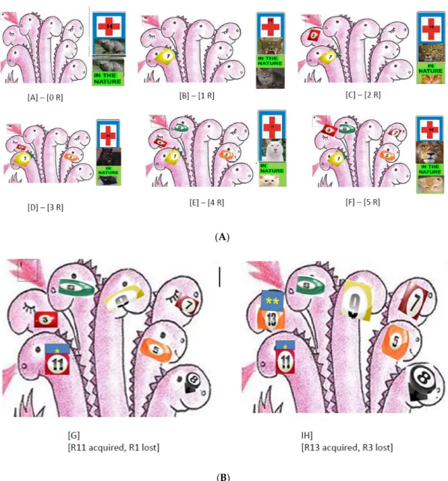
Escherichia coliis an ideal organism for experimental evolution. Most changes to its metabolic network that survived over the past 100 million years, which are adapted genetically to the environment, are due to horizontal gene transfer, with a very low contribution from horizontal gene transference [407,442]. Experimental microbial evolution studies use controlled laboratory populations to evaluate the mechanisms of evolution and are based on molecular, genomic, and maybe transcriptomic and proteogenomic analysis over several generations of evolving populations in controlled, pre-established conditions in a morbidostat [449–451] or in a DiVERGE [452]. The srecently developed technolog, enables researchers to carry out testing on empirical prediction ns of a given evolutionary theory [453,454]. The data from the literature gave an impression that a new antbitoc resistance is a positive selection marker in antibiotic stress conditions but is a negative selection marker in commensal milieu (as demonstrated on Figure2A).
6.4.1. Evolutionary Trends in The Presence of Antibiotics
Toprak and his associates experimentally recapitulated the evolution of antibiotic resistance developed in E. coli against several different antibiotics, and they found a gradual increase in resistance levels of each antibiotic tested during the experimental period [455]. The whole-genome sequencing of the evolved strains revealed mutations including resistant ones to the given drug or resistant ones to more than one drug. Resistance could be developed either in a stepwise manner in replicates (trimethoprim) or via diverse combinations of mutations in different genes (chloramphenicol and doxycycline). For interactions between the presence of an antibiotic and resistance evolution, the evolutionary dynamics were monitored by whole-genome deep sequencing every 3 to 4 days in another Gram-negative pathogen organism,P. aeruginosa. During the experimental period, the resistance level to colistin increased by one order of magnitude in 10 days and two orders of magnitude in 20 days, supposedly due to mutations in themutS mutator gene [456].
Resistance, Fitness, and Compensatory Mutations
The antimicrobial pressure may drive the evolution of its resistance, which may be associated with reduced bacterial fitness. There is evidence that the probable “evolutionary price” of a new antibiotic resistance is lowering fitness, and it must be determined genetically [457–460].
The phenotypic consequences are reflected in the geometry adaptations [461,462]. However, compensatory mutations [461] may change the pattern and might be an explanation of the dissemination of colistin-resistantA. baumanniiisolates [63,463]. This might be an explanation for the dissemination of colistin-resistantA. baumanniiisolates [203]. Using precise, high-throughput fitness measurements for genome-wideE. coligene deletion strains, with eight antibiotics, the width of the distribution of fitness effects (so-called DFE) mutations, which occur spontaneously from one antibiotic to another, is lower than in the absence of the antibiotic stress [462]. Unlike the DFE mutations, the magnitude of the changes in tolerated drug concentration, resulting from genome-wide mutations, are similar for most drugs but are exceptionally small [464].
Collateral Sensitivity
In a large-scale laboratory evolutionary experiment, Lázár and her associates discovered a novel trend in the evolution of antibiotic hypersensitivity inE. colicalled collateral sensitivity [377,465–468].
Some populations which became adapted to amino-glycosides have especially low fitness in the presence of several other antibiotics. The whole-genome sequences of 63 independently evolved amino-glycoside-resistant strains demonstrated multiple mechanisms based on reduced proton-motive force (PMF) through the inner membrane. The comparison of captured determinants of the antibiotic cross-resistance interaction network demonstrated that convergent molecular evolution was predominant across antibiotic treatments, and the resistance encoding mutations simultaneously
enhanced sensitivity to many other drugs [465]. This phenomenon is called collateral sensitivity [and illustrated on Figure2B. As mentioned before, antibiotic-resistant bacteria show widespread collateral sensitivity to antimicrobial peptides [377].
Antibiotic and Antimicrobial Resistance Genes are of Different Mobility Patterns
The antibiotic-resistant bacteria not only showed an unexpectedly high frequency of collateral sensitivity to antimicrobial peptides, while cross-resistances between the strains were rather rare [390], but clinically relevant multidrug-resistance mutations also increased susceptibility to antimicrobial peptides. As for mechanisms, collateral sensitivity in multidrug-resistant bacteria arise partly through regulatory changes shaping the lipopolysaccharide composition of the bacterial outer membrane.
This information allows the identification of antimicrobial peptide and antibiotic combinations that enhance antibiotic activity against multidrug-resistant bacteria and slow down de novo evolution of resistance [377,468]. Furthermore, while the antimicrobial peptide (AMP) resistance genes are widespread in the gut microbiome, their rate of horizontal transfer is lower than that of antibiotic resistance genes [469]. Gut microbiota culturing and functional metagenomics have revealed that AMP resistance genes originating from phylogenetically distant bacteria have only a limited potential to confer resistance inE. coli, an intrinsically susceptible species [469].
6.4.2. Evolutionary Trends of Antibiotic Resistance in the Absence of Antibiotic Exposure
Antibiotic use is the main driver in the emergence of antibiotic resistance. In a set of experiments withE. coli, it was demonstrated that in the absence of an antibiotic and resistance evolution, drug-resistance frequently declined within 480 generations during exposure to an antibiotic-free environment.
The extent of resistance declination seemed antibiotic-specific and driven by mutations influencing both the resistance level and fitness probably pleiotropically [469]. It was concluded that a phenotypic reversion to the antibiotic-sensitive state may be mediated by the acquisition of additional mutations while maintaining the original resistance mutations [469]. This result might be an important argument against the selective virtue of antibiotic resistance in general.
However, in a set of experiments on another Gram-negative pathogen,P. aeruginosa, an opposite trend was demonstrated. It is difficult to interpret this contradiction. It was demonstrated that selection in the absence of antibiotics did co-select for decreased susceptibility to several antibiotics.
This was considered as experimental evidence confirming the selective virtue of antibiotic resistance in a commensal niche [468]. The interpretation of these results by the authors is that resistance evolved coincidentally in response to other selective pressures. Thus, genetic adaptation of bacteria to natural environments may drive resistance evolution by generating a pool of resistance mutations, where selection could act to enrich resistant mutants when antibiotic exposure occurs [470].
6.4.3. Phylogenetic Limitations: Species Specificity of the Antibiotic Resistance Genes
Recent studies on transferring resistance-conferring mutations and full resistance genes intoE. coli, from and to closely related species, have not been very successful. Resistance mutations originated from one bacterial species and transferred to another, and they rarely invoke a resistance phenotype.
More frequently, the yield was drug hypersensitivity in close relatives, since the new gene could not fit the epistatic system of the new host, as was demonstrated in the case of the transferred aminoglycoside resistance (trkH) gene [471].
6.5. The CRISPR/Cas Bacterial Immune System: A Molecular Tool to Get Rid of Unwanted Antibiotic Resistance
Bacteria and Archaea have developed a system based on clustered, regularly interspaced, short palindromic repeats (CRISPR) from peculiar genetic loci. These repeats provide acquired immunity against viruses and plasmids by targeting the nucleic acid in a sequence-specific manner [472].
These hypervariable loci take up genetic material from invasive elements and build up inheritable DNA-encoded immunity over time. It was demonstrated that theS. thermophilusCRISPR1/Cas system can also naturally acquire spacers from a self-replicating plasmid containing an antibiotic-resistance gene, leading to plasmid loss. The most recent evolutionary classification of CRISPR-Cas systems and cas genes includes two classes, 6 types, and 33 subtypes. The discovery of numerous derived CRISPR-Cas variants is often associated with mobile genetic elements that lack the nucleases required for interference [473].
7. Clonally Evolving Pan-Genomic ESKAPE Pathogens
7.1. Pseudomonas aeruginosa: A Pan-Genomic Hotbed of Multidrug Resistance
The latest review on antibiotic resistance mechanisms inP. aeruginosaand alternative therapeutic strategies came out last year [474]. Our latest knowledge about the lifestyle ofP. aeruginosa (for taxonomy: see Wikipedia) is that of a ubiquitous [475], invasive [476], opportunistic [477–480], facultative, pathogenic bacterium species. It can cause diseases in plant and animal species, as well as in humans [481], which has recently been summarized [482]. As a consequence of its evolutionary plasticity [483], based on open genome expandability [484–487], it is armored with a full arsenal of antibiotic multiresistance [3,488,489]. The majority of MDRP. aeruginosadisplays a wide repertoire of antibiotic resistance mechanisms, including posttranslational modification of drug targets [490–492], enzymatic inactivation of antibiotic molecules [493], encoding genes of carbapenem-resistance preferably those coding for metallo-β-lactamase; overexpressing efflux pumps [494–496], and showing strong ability to form a biofilm [497–499].
Pseudomonas aeruginosais one of the two pangenomic [499,500], Gram-negative pathogenic species, which has permanently acquired MDR encoding genes. It is a threatening source of resistance genes, which are transmittable via horizontal gene transfer [339,501].
7.1.1. A Shortlist of Intrinsic and Acquired Antibiotic Resistance inP. aeruginosa
The most recent overviews [487,489] provides “an ocular perspective” of the different mechanisms of antibiotic resistance inP. aeruginosa. Most strains are intrinsically resistant to third-generation cephalosporins due to chromosomal-encoded C beta-lactamase and AmpC [488,501]. The original intrinsic MDR-arsenal includes the production of beta-lactamases, loss of outer membrane proteins [500], and up-regulation of efflux pumps [502–507]. This species also acquired resistance to aminoglycosides and fluoroquinolones [487]. One of the acquired enzymes taken-up byP. aeruginosa,Pseudomonas extended resistance (PER) [508–511], a class of extended-spectrum beta-lactamase (ESBL), occurs less frequently but still is of clinical importance [512,513].
Pseudomonas aeruginosauses different mechanisms that can jointly contribute to its multiresistant phenotype [512] and multidrug efflux systems [492,495,496,514,515]. All of this makesP. aeruginosa extremely invasive. The rapidly increasing number of newP. aeruginosaisolates of MDR, XDR, and PDR phenotypes severely reduces the antibiotic therapy options available [3]. Valuation of the economic cost of antimicrobial resistance (AMR) is important for decision making and should be estimated accurately, [516]. Colistin, which acts on outer membranes of bacteria resulting in its permeability and cell-death, was suggested five years ago as a salvage therapy in the treatment of life-threatening infections due to MDRP. aeruginosablood-stream infections (BSI) [517]. Articles and reviews on resistance problems appeared one year later [518] and have been appearing since [205,214–217].
7.1.2. Pseudomonas Genetics and Genomics
Genetics: Most genes of intrinsic resistance are on the chromosome, including multidrug efflux pumps and enzymes responsible for resistance to beta-lactam and aminoglycoside antibiotics [519].
Genomics: A challenging option, provided by the next-generation whole-genome sequencing, as an approach to better understand and combat antibiotic resistomes [520], has been beneficially used
inPseudomonasresearch, as exemplified by Cao et al. [521]. An international consortium has been continuously providing comparative genomic information [522,523] and the prognosis [500] for clinical use. In addition. an antimicrobial resistance prediction concluded from the comparative genomics data of comparative sequence analysis of all available drug-resistantP. aeruginosagenomes could be realized [486,501,516,522,523].
Ramanathan and his associates studied single-nucleotide polymorphisms (SNPs) in ten multiantibiotic resistant P. aeruginosa clinical isolates [519]. Non-synonymous single-nucleotide polymorphisms (nsSNPs) were found in more clinical isolates compared to the reference genome (PAO1). The nsSNPs identified in the multidrug-resistant clinical isolates were found to alter a single amino acid in several antibiotic-resistant genes. They also found mutations in genes encoding efflux pump systems, cell wall, DNA replication, and genes involved in repair mechanisms. Furthermore, nucleotide deletions in the genome and mutations leading to a generation of stop codons were also observed in the antibiotic-resistant clinical isolates, and in specific mutations within antibiotic-resistant genes, compared to the susceptible strain of the same bacterial species [519].
7.1.3. Transcriptomics
So far, we have detailed knowledge of the behavior ofP. aeruginosaunder standard laboratory conditions, but we only have a superficial understanding of bacterial functions and behaviors during human infection. However, a recent study on transcriptomes in human infection revealed that multiple genes known to confer antibiotic resistance had substantially higher expression in human infection than under laboratory conditions [524].
7.1.4. Resistance Phenotypes
The first comprehensive review on this subject was published by Kempf and Rolain [525].
Mutations in pmrB confer cross-resistance between the LptD inhibitor POL7080 and colistin in P. aeruginosa[217]. Integrated whole-genome screening revealed thatP. aeruginosavirulence genes use multiple disease models, and the pathogenicity is host-specific [8]. Biofilm formation is an important part of the pathogen strategy of this bacterium [526]. A list (incomplete) ofPseudomonas-caused human diseases is discussed in Supplementary Material 5.
7.2. Acinetobacter baumannii
7.2.1. Species with a Gradually Expanding (Open) Pangenome
The bacteriumMicrococcus calcoaceticuswas isolated from soil by enrichment in a calcium-acetate- containing minimal medium [527]. It was finally moved to Moraxella [528] as a single representative of genusAcinetobacterin Bergey’s Manual [529] under the name ofA. calcoaceticus(type strain ATCC 23055) [529]. Acinetobacter baumanniiwas designated taxonomically after appearing as a human pathogen [530–533]. Acinetobacter baumanniiis also a veterinary pathogen [534–538], but, unlike P. aeruginosa, it has not appeared as a plant pathogen. Acinetobacter baumanniiis a very successful pathogen [539]. WHO declared thatA. baumanniiis one of the two most serious pangenomic ESKAPE organisms [540]. Since then, the rapid global evolution of MDR inA. baumanniihas been carefully monitored, and clonal lineages of old and new isolates have been revealed [541]. Two long-term international global surveys have recently been carried out onA. baumanniias a potential health risk factor, as reported by Gales et al. and Flamm et al. [540,541].
7.2.2. Resistance Mechanisms and Diseases
The mechanisms of disease caused by A. baumanniistrains have recently been reviewed by Morris et al. [8]. The latest review on the resistance mechanisms ofA. baumannii[542] focuses on XDR resistance.
As for its resistance mechanisms, tigecycline efflux was described as a mechanism for non- susceptibility inA. baumannii. In addition, a deletion of TnAbaR23 resulted in significant antibiogram changes in a multidrug-resistantA. baumanniistrain [543]. For more information see Supplementary Material 6.
7.2.3. Genetic Dissection of Colistin Resistance inA. baumannii: Trebosc et al., 2019
The transcriptional regulator PmrA was considered as the only potential drug target to restore colistin efficacy inA. baumannii[510], but the deletion ofpmrA, the gene responsible for overexpression of the phosphoethanolamine, PetN, only restored susceptibility in a few strains. A detailed genetic analysis revealed a new colistin resistance mechanism mediated by genomic integration of the ISAbaI insertion element upstream of the PmrC homolog (eptA), leading to its overexpression. The gene eptA is present in each international clone 2 clinical strain, and a duplicated ISAbaI-eptA cassette was present in at least one clinical isolate, thus indicating this colistin resistance determinant may be embedded in a mobile genetic element [216].
7.2.4. Genetics Toolkits to Becoming Multiresistant
Acinetobacter baumannii, like other non-glucose-fermenting Gram-negative species such as P. aeruginosa, has increasingly been acquiring carbapenem resistance [544,545]. An argument against the worries [546] thatA. baumanniiincreases the risk of carbapenemase spread in general, since horizontal resistance gene the transfer can occur between Gram-negative species, regardless of their ability to ferment glucose [545], is that experimentally transferred resistance mutations from other species to E. colihave been mostly silent, and frequently yield drug hypersensitivity [469].
7.2.5. Sword of Damocles: Clonal Evolution, Global Spread, and Epidemic Potential ofA. baumannii The different antibiotic resistance genes are organized into resistance islands in the genome of A. baumannii. The impression of the reviewers is that the MDR, XDR, and PDR must not be considered as simple collections of different antibiotic resistances, but they may represent a coordinated system based on sophisticated genetic background and genomic organizations. Based on the evolutionary analysis of six housekeeping genes,A. baumanniiis of monophyletic origin [531,546]. Originally three predominant pathogen clones, called ‘international clonal lineages’ (ICLs), were known as being responsible for hospital outbreaks worldwide [547]. The monophyletic status of ICLs 1 and 2 have also been shown [38,548]. As reviewed first by Dijkshoorn et al. [546], the genome of a representative ICL1 (AYE) [549–551] strains include 52 genes associated with resistance to anti-microbial drugs, and 45 are localized in an 86 kb resistance island called AbaR5 [552]. ABAR1 is also present in otherA. baumannii strains, but at a much smaller size. The presence of an extraordinary 22 gene-cassette coding for transposases and insertion sequences may be responsible for the acquisition of resistance genes into the AbaR1 island of the AYE ICL1-type strain [543]. Almost half are orthologous to coding sequences ofPseudomonas[546].
7.2.6. Genome Plasticity
The Acinetobacter baumannii calcoaceticus complex is considered as a species with an open pan-genome and is well-defined on Wikipedia. The complete genome [553–555] includes the core genome (genes present in all isolates) with extreme antibiotic resistance traits [554]. In addition, the accessory genome [555], including the genes absent from one or more isolates, or unique to a given isolate, also contains hosts antibiotic resistance genes but in different arrangements [555].
Antibiotic resistance genes are located both in the core and the accessory genomes [556]. In the latter, they were found in alien islands or flanked by integrases, transposases, or insertion sequences [556].
They must be acquired via horizontal gene transfer from otherAcinetobacterstrains to colonize the same environment [556]. The first detailed genomic analyses revealed that the existingA. baumanniiclinical population consists of low-grade pathogens, whose pathogenicity relies mainly on an ability to persist
in the hospital setting and survive antibiotic treatment [4]. This analysis led to the conclusion that A. baumanniihas a high capacity to acquire new genetic determinants and displays an open pan-genome.
This feature may have played a crucial role in the evolution of this human opportunistic pathogen towards clinical success [4]. The whole pangenome ofA. baumanniiconsists of>8800 orthologous coding sequences, and it has exponentially been increasing as new genomes become available (an open pan-genome), mainly due to unique accessory genomes of different isolates enriched with acquired genes of transport and transcription regulation functions [4]. Since then, at least 15 complete, and 180 draft, chromosomalA. baumanniigenomes, 31 plasmids, and six bacteriophage sequences have been available on the NCBI database, together with those of other species of the genus, and antibiotic treatment induces important genes [557]. The open pan-genome ofA. baumanniialso includes lots of plasmids, transposons, integrons, and genomic islands, which may contribute to the evolutionary success of this clinical pathogen. This is particularly true in the acquisition of multidrug resistance determinants [5], a tool for redefining this, and other, pathogenic bacterium species [162,547,557,558].
7.2.7. Will the Epidemic Threat Be Materialized?
The population structure ofA. baumanniicomprises of a set of expanding multiresistant clones raised from an ancestral susceptible genetic pool [559–561], confirming thatA. baumannii used to have low phylogenetic diversity, providing a narrow evolutionary bottleneck through which a micro-evolutionary tree with many branches has emerged [546]. Making this metaphor complete, each branch of that tree carries “dangerous fruit” called antibiotic-resistance (ABR) genes. All the other knownAcinetobacterspecies are harmless soil-inhabiting ones [546].
Until recent years,A. baumanniihas been almost exclusively isolated from locations of antibiotic stress conditions, that is, in hospital environments, (especially from intensive care units of hospitals) and veterinary clinics [562–564]. However, in 2017,A. baumanniiDSM30011 was obtained from the resinous desert shrub guayule and may be considered as a “copy” of the first isolate, and whole-genome sequencing and phylogenetic analysis based on core genes confirmed DSM30011 affiliation to A. baumannii [565]. No antimicrobial resistance islands were identified in DSM30011, agreeing with a general antimicrobial susceptibility phenotype. The marginal ampicillin resistance of DSM30011 most likely derived from chromosomal ADC-typeampC and blaOXA-51-type genes [566]. The environmental Acinetobacter baumanniiisolate DSM30011 reveals clues into the preantibiotic era genome diversity, virulence potential, and niche range of a predominant nosocomial pathogen [567] armed with efficient multidrug efflux pumps [567].
The “branches” of the original “tall” “micro evolutionary tree” are rather “long.” ln the frame of a SENTRY study [543],Acinetobacterclinical isolates from 10 South Asian and Pacific countries were tested for being non-susceptible to imipenem or meropenem and positive for OXA-23-, OXA-24/40-, OXA-58-, and MBL-encoding genes [540,541]. The sword of Damocles (a globalAcinetobacterepidemic) is still possible, but has not yet been dropped down.
8. Concluding Remarks
We must apologize for omitting so many excellent publications from this field because of space limits.
8.1. Forcasts Based On Evolutionary Data
8.1.1. The Main Question
Whether the post-antibiotic era is at the gate, or there is a chance to prevent its coming, is the question of questions. The literature convinced us that the trend of the global expansion of clonally spreading MDR and XDR pathogens is not irreversible. The post-antibiotic era is not inevitable. Let us discuss the arguments for and against these statements.
8.1.2. The “Combat Scenario”
Nature and science have long been battling. Science produces antibiotics, nature presents pathogens with resistances. Nature has gradually become more sophisticated and better organized.
The MDR or XDR phenotype cannot be considered as a brute summary of the expression of ad hoc collected (intrinsic and acquired) antibiotic resistance alleles. MDR (or XDR) represents a concerted phenotypic expression of structurally and functionally organized (operons, cassettes, integrons), and coordinated (resistomes) resistance alleles, which had been selected by the evolution. Both the intrinsic occurrence and the uptake of resistance alleles are random.
8.1.3. Adaptive Evolution
The selective drives acting in the commensal niche and those acting in a milieu under selective (antibiotic) pressure are different. A neutral mutant allele, including those encoding resistance, in a commensal niche may be fixed in the population on the condition that it does not display a heavy genetic load, that is it does not express a negative fitness affecting (DFE) phenotype]. That is, it does not have either a positive or a negative selective value. Therefore, new antibiotic resistances are expected to appear in the future as in the past.
Neutral mutant alleles, including resistance-encoding ones, in a commensal niche may be fixed in the population on the condition that it does not display a heavy genetic load, or if it does not express negative fitness affecting (DFE) phenotype. That is, it does not have either a positive or negative a selective value. Therefore, new antibiotic resistances are expected to appear in the future, as in the past.
A mutant allele with a strong antibiotic resistance phenotype will expectedly be lost, or at least prevented from domination, or selectively lost in commensal conditions if it represents a heavy genetic load. This phenomenon is demonstrated in the monitoring studies on Gram-negative and Gram-positive MDR pathogens. However, the same allele will be enriched in the population up to 100% under selective (antibiotic) stress conditions, either in a hospital environment or in the laboratory.
8.1.4. The “Dialectics” of Resistance and Sensitivity
There are exceptional situations when the same gene can act as a resistance or a sensitivity gene, in the same species, depending upon genetic background (Supplementary Material 4).
8.1.5. Collateral Sensitivity: A Biochemically Proven Limiting Factor in MDR Evolution
For evolutionary success (winning the “permanent war”), the pathogen needs to carry strong and heavy “weapons” (MDR combinations), which also increases the genetic load. The rediscovery, genomic, and evolutionary interpretation of the collateral sensitivity is the most important experimental proof that the resistance capacity of a bacterium is limited. It means that “trees cannot grow up to the sky”.
The credibility of this discovery is also supported by biochemical evidence]. The molecular technique for getting rid of an unwanted resistance is probably the CRISPR/Cas Bacterial Immune System.
This conception is illustrated in an allegoric way on Figure2. The higher resistance level makes the pathogen stronger in selective but weaker in non-selective (commensal) conditions. (Figure2A–H).
This explains collateral sensitivity as well. A limited number of resistances can be harbored, but not more. If the pathogen gains a new one (#11 in Figure2G, and #13 in Figure2H) it must get rid of another one (#1 Figure2G and #3 on Figure2H). The data in the last report of the HAI Consortium on changes in antibiotic resistance profiles of MRSA clones in the USA indirectly confirm that collateral sensitivity is not a laboratory artifact [256].
![maltophilia ;nevertheless,otherlessfrequentlyisolatedgenerashouldalsobetakeninto functions[ , ].NFGNBincludesomecommonlyisolatedgenera,suchas Pseudomonas , inanoxidativefashion)rods.Inaddition,theyareallsimilarinthattheyareunableto Burkholderiamallei , an](data:image/gif;base64,R0lGODlhAQABAIAAAP///wAAACH5BAEAAAAALAAAAAABAAEAAAICRAEAOw==)