Clinical microbiologic examination of Stenotrophomonas maltophilia strains
Ph.D. thesis
Dr. Juhász Emese Réka Semmelweis University Doctoral School of Clinical Medicine
Supervisor: Kristóf Katalin, MD, Ph.D.
Official reviewers:
Deák Judit, MD, Ph.D.
Kenesei Éva, PharmD, Ph.D.
Head of the Complex Examination Committee:
Benyó Zoltán, MD, DSc.
Members of the Complex Examination Committee:
Tulassay Tivadar, MD, DSc.
Vásárhelyi Barna, MD, DSc.
Folyovich András, MD, Ph.D.
Budapest
2018
1. Introduction
Stenotrophomonas maltophilia has emerged as an important opportunistic and nosocomial pathogen in the past two decades worldwide. Behind Pseudomonas aeruginosa and Acinetobacter baumannii, S. maltophilia is the third most common non-fermenting Gram-negative bacillus responsible for healthcare-associated infection. The World Health Organisation listed S. maltophilia as one of the leading antibiotic-resistant pathogens in hospitals. Community acquired infections have also been reported. Pneumonia and bacteraemia are the most frequent infections. Due to enzyme production (L1- and L2-β-lactamases, different aminoglycoside transferases, etc), efflux pumps (SmeABC – SmeYZ, MacABC, etc) and low outer membrane permeability, it has inherent extended antibiotic resistance. Therapeutic options are strongly limited. Currently only trimethoprim-sulfamethoxazole (SXT) is recommended for therapy, but some circumstances (hypersensitivity of the patient, resistance of the bacterium) can limit the use of this drug. In such cases, susceptibility of S. maltophilia isolates for other antimicrobials, for example fluoroquinolones or tetracycline derivates must be tested, even if clinical evidences for their efficacy are lacking yet. Furthermore, the recommendation by the European Committee on Antimicrobial Susceptibility Testing (EUCAST) includes species specific clinical breakpoint only for SXT. Antibiotic combination therapy can be required in severely immunocompromised patient, in cystic fibrosis patients, in patients with SXT intolerance or in case of SXT-resistant (defined as MDR) S. maltophilia infection.
Growing number of patients at risk (immunocompromised patients, cancer patients, patients under long-term intensive care, etc) and the necessity of the usage of broad- spectrum antibiotics such as carbapenems (which cause selective pressure for the inherently carbapenem-resistant S. maltophilia) have the consequence of increasing number of S. maltophilia infections. This fact together with the natural features of this bacterium (biofilm forming ability, ubiquiter, colonizing moist hospital environments) and its inherent and acquired antibiotic resistance mean that S. maltophilia is a continuous threat we have to face. The high attributable mortality rates (14-69%) in S.
maltophilia infection highlight the clinical significance of this bacterium. The aim of this work is to describe the clinical microbiology of this well-known, but still news
hiding, globally challenging bacterium, especially focusing on antibiotic treatment options.
2. Objectives
The aim of our studies was to investigate S. maltophilia isolates collected in Diagnostic Laboratory of Clinical Microbiology, Institute of Laboratory Medicine, Semmelweis University in order to find answers to the following questions:
1. How susceptible were clinical S. maltophilia isolates to the first-line antibiotic agent SXT and to the second-line drugs?
2. What was the clinical background of S. maltophilia strains?
a. Were they infective or just colonized patients?
b. What was the outcome of S. maltophilia infections?
c. What kind of risk factors influenced the outcome of infections?
3. How frequent was the S. maltophilia co-colonization or co-infection?
4. How susceptible were SXT-resistant S. maltophilia isolates to other antibiotic agents and what was the genetic background of their SXT resistance?
5. Was there any in vitro effective antibiotic combination against extremely drug resistant S. maltophilia isolates?
6. Was colistin+SXT combination in vitro effective against S. maltophilia, MDR P. aeruginosa and MDR A baumannii strains isolated together from lower respiratory tract co-infections?
3. Methods
3.1. Identifications and collection of strains
A total of 160 consecutive non-duplicate S. maltophilia isolates from a three-year collection period (2009-2011) were investigated in the first part of the study. If more strains were collected from the same patient, the first clinically relevant one (presumed on the type of the sample) was included in the study. Thirty consecutive non-duplicate S. maltophilia isolates collected in a five-year period (2010 – 2014) in Diagnostic Laboratory of Clinical Microbiology, Institute of Laboratory
Medicine were resistant to SXT. These ones were tested in the second part of the study. The identification of the isolates was done initially by the classic methods (OF probe, oxidase test, catalase test) and VITEK 2 Gram-negative identification cards (bioMérieux), later by matrix assisted laser desorption ionization time of flight mass spectrometry (MALDI-TOF MS) (Bruker Daltonics). Strains were stored at -20°C in brain-heart infusion broth containing 30% glycerine until investigations. Strains identified by biochemical methods were re-tested by MALDI-TOF MS technique. The MALDI-TOF MS identification was performed by direct overlay method (isolates were smeared on target plate and overlayed by 1 µl matrix solution). Bruker Biotyper 2.0 softver was used for analysis. Matrix solution contained 5 mg α-cyano-4-OH- cinnamic acid dissolved in one milliliter of water, acetonitril and trifluoroacetic acid 4,75:5:0,25 mixture. Parameters of mass spectrometer device (ion source 1 20kV, ion source 2 18,5 kV, lenses 8,5 kV, detector 2,65 V, without gating, positive linear mode, spectra detected in 2000-20000 Da range, maximal laser frequency) were not changed.
Biotyper scores ≥2.0 were accepted as valid species level identification. Identification results as „Stenotrophomonas maltophilia”, „Stenotrophomonas maltophilia (Pseudomonas beteli)”, „Stenotrophomonas maltophilia (Pseudomonas hibiscicola)”
and „Stenotrophomonas maltophilia (Pseudomonas geniculata)” were all accepted, as these species were belonging to Stenotrophomonas rRNA lineage.
Clinical data and laboratory findings (white blood cell number (WBC), C- reactive protein (CRP), procalcitonin (PCT)) of patients at the time of S. maltophilia infections were collected retrospectively from clinical final reports. Based on clinical data, isolates were divided into two groups: isolates of patients who were infected by S. maltophilia and of those who were colonized only. Infection or colonization was distinguished according to clinical diagnoses given in final reports. The definitive diagnosis of infection was clinically established. Colonization was defined as the presence of on skin, mucous membranes, in wounds, or in excretions or secretions without causing adverse clinical signs or symptoms.
In our centre 58% of S. maltophilia isolated from LRT specimens was cultured as co-pathogen or co-colonizer in 2013-2014. Pseudomonas aeruginosa was found to be the most frequent co-pathogen, but A. baumannii was also a significant co-habitant. Those lower repiratory tract samples (tracheal aspirate or bronchoalveolar lavage sample) were included in our study, from which MDR P. aeruginosa, MDR A.
baumannii and S. maltophilia were isolated at the same time. In the study period 20 bacterial triplets were collected from different patients.
3.2. Antibiotic susceptibility testing
The minimal inhibitory concentrations (MIC) of antibiotics were determined by the broth microdilution method in cation-adjusted Mueller-Hinton broth (BectonDickinson) on 96-well plates (TOMTEC), according to CLSI testing conditions. The antibiotics tested included SXT (Sigma-Aldrich) in 0,25-128 mg/l, ciprofloxacin (Fresenius Kabi) in 0,5-256 mg/l, moxifloxacin (Bayer Pharma) in 0,064-32 mg/l, levofloxacin (TEVA) in 0,064-32 mg/l, colistin (colistin-sulphate, Sigma-Aldrich) in 0,25-256 mg/l, doxycycline (Pfizer) in 0,064-32 mg/l and tigecycline (Wyeth) in 0,064-32 mg/l concentrations. The MIC values of invasive isolates were tested by agar dilution method in case of SXT and fluoroquinolones and by gradient diffusion tests (bioMérieux Etest® and Liofilchem) in case of SXT, fluoroquinolones, tigecycline and colistin, too.
Isolates resistant to SXT were tested for ceftazidime (Fresenius Kabi) in range 1-512 mg/l and chloramphenicol (Sigma-Aldrich) in range 0,5-256 mg/l, too.
The SXT and colistin ranges were modified to 0,5-256 mg/l and 1-512 mg/l, respectively. Colistin was tested at 0,06-32 mg/l and SXT at 2-1024 mg/l in case of MDR P. aeruginosa strains isolated from co-infections. Colistin was tested at 0,06-32 mg/l and SXT at 0,5-256 mg/l in case of MDR A. baumannii strains isolated from co- infections.
To interpret MIC results, EUCAST breakpoints were applied. The S.
maltophilia specific breakpoint for SXT, and the non-species related breakpoints for fluoroquinolons and tigecycline were applied. For doxycyline – lacking either specific or non-species related breakpoints – the epidemiological cut-off (ECOFF) value of S. maltophilia (8 mg/l) was applied. For colistin – lacking non-species related breakpoints or determined ECOFF - Pseudomonas sp. specific breakpoint (4 mg/l) was used (according to EUCAST clinical breakpoints version 4.1.). For chloramphenicol the S. maltophilia specific, Clinical Laboratory Standards Institute (CLSI) declared breakpoints were used. Escherichia coli ATCC 25922, Pseudomonas aeruginosa ATCC 27853 and Staphylococcus aureus ATCC 29213 were used as
quality control strains, which correspond with the quality control recommendation of CLSI.
3.3. Testing of antibiotic combinations
The extremely resistant S. maltophilia strains (n=4) were tested for antibiotic combinations. The twenty different combinations tested were followings: a) ceftazidime plus ciprofloxacin, moxifloxacin, levofloxacin, doxycyline, tigecycline, colistin, SXT; b) colistin plus ciprofloxacin, moxifloxacin, levofloxacin, doxycyline, SXT; c) tigecycline plus ciprofloxacin, moxifloxacin, levofloxacin, colsitin, SXT; d) SXT plus ciprofloxacin, moxifloxacin, levofloxacin. Antibiotic combinations were analysed by checkerboard (CB) technique on 96-well plates. Microbroth plates were inoculated with bacteria to yield 5x10⁵ CFU/mL in 100 µL final volume of Mueller- Hinton broth completed with antibiotic agents. Plates were incubated at 35°C for 18- 22 h. When synergy was detected, results were confirmed by E-test combination method. Double disk agar diffusion method was tried to use for screening, placing disks in 1 cm distance from each other.
Fractional inhibitory concentration indices (ΣFICI) were calculated following the formula: FIC(A)+FIC(B)= ΣFICI, where FIC(A)= MIC of antibiotic agent A in combination with antibiotic agent B/MIC of antibiotic agent A alone and FIC(B)= MIC of antibiotic agent B in combination with antibiotic agent A/MIC of antibiotic agent B alone. The ΣFICI of two antibiotics tested defines the effects of antimicrobial combinations as antagonistic (ΣFICI > 4), indifferent (0,5 = ΣFICI ≤ 4) or synergic (ΣFICI < 0,5). The 0,5 = ƩFICI ≤ 1 values can be defined as additive or partial synergic.
Susceptible breakpoint indices (SBPI) were also used to interpret the results. SBPI was calculated as follows: SBPI = (susceptible breakpoint of antibiotic agent A / MIC of antibiotic agent A in combination with antibiotic agent B) + (susceptible breakpoint of antibiotic agent B / MIC of antibiotic agent B in combination with antibiotic agent A). An SBPI of 2 indicates that the MIC values of the two antibiotics tested in combination are either equivalent to their respective susceptible breakpoints or the MIC of one of the antimicrobials in a combination is less than its susceptible breakpoint. Greater SBPI values indicate more effective combinations
Antibiotic combination of colistin-plus-SXT on S. maltophilia, P.
aeruginosa and A. baumannii co-isolates was analysed initially by CB technique.
Stenotrophomonas maltophilia isolates were tested in 7 doubling dilutions of colistin and 11 doubling dilutions of SXT, whereas P. aeruginosa and A. baumannii strains were tested in 7 doubling dilutions of SXT and 11 doubling dilutions of colistin.
When synergy was detected by CB, a time-kill assay (TKA) was performed at 1xMIC.
When MIC values were above the therapeutic level, SXT was used at 8 mg/l and colistin at 4 mg/l, which fits the peak serum levels of these agents. Twenty ml of SXT, colistin and SXT-plus-colistin containing Mueller-Hinton broth were inoculated with bacteria to yield a density of 106 CFU/ml in the final volume. Tubes were incubated at 37°C with constant agitation. After 1, 2, 4, 6 and 24 h incubation aliquots were removed, serially diluted (10-1 – 10-6) in 0,9% sodium chloride solution and plated on sheep blood agar plates (bioMérieux). Colony-forming units were counted on agar plates after 24 h incubation at 37°C. The lower limit of detection by this method was 20 CFU/ml. Synergy was defined as a ≥ 2 log10 decrease in CFU/ml at 24 h for the antibiotic combination compared with CFU/ml decrease of S. maltophilia in presence of SXT, and CFU/ml decrease of P. aeruginosa and A. baumannii in presence of colistin.
3.4. Genetic testing
Enterobacterial Repetitive Intergenic Consensus PCR (ERIC-PCR) was used for molecular typing of isolates. Bacteria were suspended in 100 µl of PCR- grade water and heated at 100 °C for 15 minutes. After centrifugation at 12000 rpm for 2 minutes, supernatant was removed. One µl of the supernatant was used as DNA for PCR. Primers of ERIC1 5’-ATGTAAGCTCCTGGGGATTCAC–3’ and ERIC2 5’-AAGTAAGTGACTGGGGTGAGCG-3’ (Bio Basic Inc.) and REDTaq Ready Mix PCR reaction mix (Sigma-Aldrich) were used for DNA amplification, in 50 µl final PCR reaction volume. PCR conditions were the following: initial denaturation at 95°C for 2 minutes, 30 cycles at 90°C for 30 seconds, 52°C for 1 minute, 65°C for 8 minutes. Electrophoresis was performed in 1.5% agarose gel (Sigma Aldrich), in Tris- boronicacid-EDTA buffer (Sigma Aldrich), stained with 0.01% GelRed (Biotium).
Isolates from the same wards were tested in the same PCR and gelelectrophoreses run.
Band patterns were visually evaluated in the absence of appropriate software. Isolates with two or more different bands were interpreted as unrelated.
The presence of sul1 gene was tested by F 5’- ATGGTGACGGTGTTCGGCATTCTGA-3’ and R 5’-CTAGGCATGATCTAAC CCTCGGTCT-3’ primers, the sul2 gene by F 5’-GAATAAATCGCTCATCATT TTCGG-3’ and R 5’-CGAATTCTTGCGGTTTCTTTCAGC-3’ primers. The parameters of PCR were the followings: 1 x 95°C 10 minutes, 30 x 94°C 45 seconds, 64°C 45 seconds, 72°C 2 minutes, 1 x 72°C 10 minutes. For detection of class 1 integron, primers F 5’-CAACACATGCGTGTAAAT-3’ and R 5’-CTTGCTGC TTGGATGCC-3’ were used. The annealing temperature was 55°C, the other parameters of PCR were the same as described above.
3.5. Statistical analysis
Patients’ characteristics were tested for their association with overall mortality. First, univariate analysis using χ2 test or Fisher test was performed. A p value of <0,05 was considered as significant. Variables with significant association with mortality in the univariate analysis were entered in a multivariate forward stepwise logistic regression model to identify independent risk factors for death. The odds ratio (OR) with the corresponding 95% confidence interval (95% CI) for each variables were calculated. A p value of <0,05 was considered as indicative of statistical significance. Stata 12 software (StataCop LP) was used for the analysis.
4. Results
Biotyper 2.0 software of Bruker MALDI-TOF MS classified 74% of the isolates as S. maltophilia, 16% as S. maltophilia (P. hibiscicola), 7% as S. maltophilia (P. beteli), 3% as S. maltophilia (P. geniculata). As P. hibiscicola, P. beteli and P.
geniculata belong to Stenotrophomonas rRNS lineage, they were reclassified to Stenotrophomonas genus. Now they are synonims of S. maltophilia.
Isolates of infected patients (n=100) were cultured from blood (n=25), bronchoalveolar aspirate (n=30), tracheal aspirate (n=31), sputum (n=7), central venous catheter (n=4), peritoneal fluid (n=3). Isolates of colonized patients (n=60) were cultured from rectal swab (n=11), urine (n=8), ear swab (n=6), throat - (n=3),
nose - (n=4), eye sample (n=7), catheter (n=1), sputum (n=7), tracheal aspirate (n=7) and wound sample (n=6).
Isolates resistant to SXT were cultured from blood (n=2), bronchoalveolar lavage sample (n=8), tracheal aspirate (n=3), sputum (n=3), endotracheal tube (n=3), rectal swab (n=7), urine (n=1), ear swab (n=1) and throat swab (n=2). The most of these isolates (n=23) were considered clinically to be colonizer or contaminant bacteria. Only 7 isolates were infective; they caused lower respiratory tract infections or bacteriaemia. Thirteen strains were isolated from neonates cared in neonatal intensive care unit (ICU), six strains from adult patients treated in ICUs.
4.1. Results of ERIC-PCR
ERIC-PCR resulted in highly diverse patterns, however seven identical patterns among the colonizer (4 timer 2 isolates and 3 timers 3 isolates with the same band patterns) and twelve among the infective isolates (6 timers 2 isolates, 4 times 3 isolates, once 5 isolates and one 6 isolates with tha same band patterns) were found. In order to prevent the distortion of values by clonal isolates, 10 colonizer and 23 infective isolates were excluded from the final evaluation of antibiotic susceptibility testing results.
Four identical patterns were found among SXT resistant isolates (2 times 2 isolates, once 3 isolates and once 9 isolates with the same band patterns). Despite the detected clonality, isolates with the same band pattern showed different MIC values.
All isolates were included in the final evaluation of antibiotic susceptibility testing results.
4.2. Results of antibiotic susceptibility testing
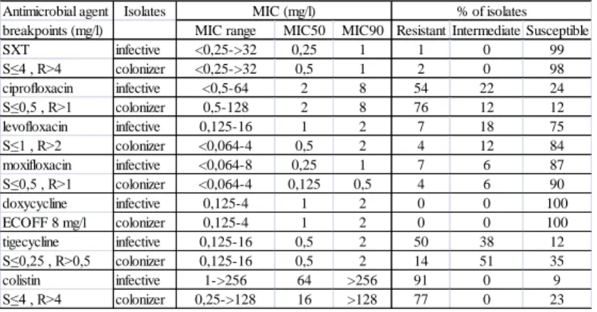
The results obtained by microdilution, gradient diffusion and agar dilution methods were in concordance. The MIC50 and MIC90 values, MIC ranges and susceptibility in % of selected isolates with different ERIC-PCR patterns (77 infective and 50 colonizer) were summarized in Table 1. However, MIC values did not differ significantly if the total of 100 infective and 60 colonizer isolates were evaluated.
Susceptibility for doxycycline was investigated instead of the often tested minocycline, since the latter is not distributed in Hungary. Applying S. maltophilia specific ECOFF, all the isolates seemed to be sensitive to doxycyline. The
susceptibility rates of of infective and colonizer isolates were compared. Both groups high rate of susceptibility to SXT. Apart from ciprofloxacin, the infective isolates had higher rates of non-susceptibility than the colonizers. Non-susceptibility rates for colistin and tigecycline were 1,2-1,3 times higher in the infective groups.
Table 1. Summary of MIC values and interpretations of 127 S. maltophilia strains (77 infective, 50 colonizer) with different ERIC-PCR patterns.
Antimicrobial agent Isolates
breakpoints (mg/l) MIC range MIC50 MIC90 Resistant Intermediate Susceptible
SXT infective <0,25->32 0,25 1 1 0 99
S≤4 , R>4 colonizer <0,25->32 0,5 1 2 0 98
ciprofloxacin infective <0,5-64 2 8 54 22 24
S≤0,5 , R>1 colonizer 0,5-128 2 8 76 12 12
levofloxacin infective 0,125-16 1 2 7 18 75
S≤1 , R>2 colonizer <0,064-4 0,5 2 4 12 84
moxifloxacin infective <0,064-8 0,25 1 7 6 87
S≤0,5 , R>1 colonizer <0,064-4 0,125 0,5 4 6 90
doxycycline infective 0,125-4 1 2 0 0 100
ECOFF 8 mg/l colonizer 0,125-4 1 2 0 0 100
tigecycline infective 0,125-16 0,5 2 50 38 12
S≤0,25 , R>0,5 colonizer 0,125-16 0,5 2 14 51 35
colistin infective 1->256 64 >256 91 0 9
S≤4 , R>4 colonizer 0,25->128 16 >128 77 0 23
% of isolates MIC (mg/l)
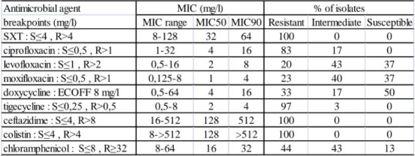
The antibiotic susceptibility of SXT resistant isolates is summarized in Table 2. The MIC values of SXT ranged 8 - 128 mg/l. None of the isolates was susceptible to ciprofloxacin, tigecycline, ceftazidime or colistin. Only 37% of them were susceptible to levofloxacin or moxifloxacin. Interpreting isolates with MIC value under ECOFF as susceptible, 50% of the strains were susceptible to doxycycline.
Thirteen % of the isolates were susceptible to chloramphenicol. Two isolates resistant to all tested antibiotic agents and two others susceptible only to doxycycline were detected. These four isolates were further investigated; susceptibilities for combinations of 20 different antibiotics were tested on them.
Table 2. Summary of MIC values and interpretations of 30 SXT-resistant S.
maltophilia strains (n=30).
Antimicrobial agent
breakpoints (mg/l) MIC range MIC50 MIC90 Resistant Intermediate Susceptible
SXT : S≤4 , R>4 8-128 32 64 100 0 0
ciprofloxacin : S≤0,5 , R>1 1-32 4 16 83 17 0
levofloxacin : S≤1 , R>2 0,5-16 2 8 20 43 37
moxifloxacin : S≤0,5 , R>1 0,125-8 1 4 23 40 37
doxycycline : ECOFF 8 mg/l 0,5-64 4 16 33 17 50
tigecycline : S≤0,25 , R>0,5 0,5-8 2 4 97 3 0
ceftazidime : S≤4, R>8 16-512 128 512 100 0 0
colistin : S≤4 , R>4 8->512 128 >512 100 0 0
chloramphenicol : S≤8 , R≥32 8-64 16 32 44 43 13
MIC (mg/l) % of isolates
The rate of SXT-resistant S. maltophilia isolates didn’t change significantly in 2009-2017. Annually 4-12 patients were detected with MDR S. maltophilia positive samples. In the whole nine-year period 81 consecutive, non-duplicate SXT-resistant isolates were cultured. The overall resistance rate was 1,9%. The stagnating app. 2%
resistance rate indicates that neither an MDR clone nor a mobile genetic element spread effectively. Furthermore, there isn’t any effective pressure selecting SXT resistance.
All P. aeruginosa and A. baumannii strains were susceptible to colistin, with MIC50 1 mg/l and MIC90 2 mg/l. Fifteen percent of A. baumannii strains were susceptible to SXT, MIC50 32 mg/l and MIC90 128 mg/l was found. Pseudomonas aeruginosa strains showed a high level of intrinsic resistance to SXT, with MIC50 256 mg/l and MIC90 512 mg/l. All S. maltophilia strains were sensitive to SXT, with MIC50 0,25 mg/l and MIC90 1 mg/l, and resistant to colistin, with MIC50 256 mg/l and MIC90 >512 mg/l.
4.3. Results of antibiotic combination testing
Most of the combinations, tested by CB, had indifferent effect on extremely resistant S. maltophilia strains (n=4), based on the calculated ƩFICI values from the lowest MIC values. Antagonistic combination was not found. Synergic effect at least in one isolate was found in combinations of ceftazidime+ciprofloxacin, ceftazidime+moxifloxacin, ceftazidime+levofloxacin, ceftazidime+colistin,
colistin+ciprofloxacin, colistin+moxifloxacin, colistin+levofloxacin, colistin+doxycycline, tigecycline+colistin, SXT+moxifloxacin, SXT+levofloxacin.
Ceftazidime+moxifloxacin and ceftazidime+colistin were the only combinations that showed synergic or at least partial synergic effect on each tested isolate. Taking into consideration the peak serum levels during antibiotic treatment (ceftazidime: 60 mg/l, ciprofloxacin: 1,8-4,6 mg/l, levofloxacin: 5,7-8,6 mg/l, moxifloxacin: 4,5 mg/l, colistin: 5-7,5 mg/l, SXT: 1-2/40-60 – 9/105 mg/l, tigecycline: 0,63 mg/l, doxycyline:
1,5-2,1 mg/l, chloramphenicol: 11-18 mg/l), only the combinations of ceftazidime+fluoroquinolons and colistin+fluoroquinolons can be accepted as synergic, in certain cases. Furthermore, if EUCAST susceptibility breakpoints are also taken into consideration, just the MIC values of fluoroquinolons (in combination with ceftazidime: SBPICAZ+MXF = 2.125, SBPICAZ+LEV = 4.125) and colistin (in combination with fluoroquinolons: SBPICOL+MXF = 2.125) decreased into the susceptible range, and only in one-one isolate out of the tested four ones.
Synergic and indifferent effects of colistin+SXT were found by CB in cases of co-isolated S. maltophilia, MDR P. aeruginosa and MDR A. baumannii strains. Synergy was detected in 50%, 35% and 45% of S. maltophilia, P. aeruginosa and A. baumannii strains, respectively. Antagonistic effect was not found.
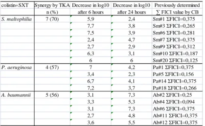
Results gained by TKA are summarized in Table 3. The tested S.
maltophilia, P. aeruginosa and A. baumannii strains showed synergy in 70% (7 strains out of 10), 57% (4 strains out of 7) and 56% (5 strains out of 9) of cases, respectively.
Considering that for most combinations with colistin against Gram-negative species initial killing is usually dramatic, but is followed by significant regrowth, the rates of log10 decrease after 6 and 24 h are summarized in Table 3. Synergic activity of the combination was already detected after 6 h incubation in 86% of S. maltophilia isolates and 50% of P. aeruginosa strains. In the case of A. baumannii, synergy was detected just after 24 h incubation. The results gained by CB and TKA methods correlated in 61% of cases, but the ΣFIC values did not correlate with the rate of log10 decrease in TKA. Colistin-plus-SXT combination had synergic effect on seven S.
maltophilia (35%), four P. aeruginosa (20%) and five A. baumannii (25%) strains by both methods. The combination had synergic effect on all co-isolates from two patients.
Table 3. Summary of the synergic results gained by TKA. Colistin+SXT combination was tested on strains which were previously tested by CB method and ƩFICI values were <0,5. Decrease of colony count was compared with that in presence of SXT in case of S. maltophilia, and in presence of colistin in case of P. aeruginosa and A.baumanni strains.
colistin+SXT Synergy by TKA Decrease in log10 Decrease in log10 Previously determined n (%) after 6 hours after 24 hours ∑ FICI value by CB
S. maltophilia 7 (70) 5,9 2,4 Sm#1 ΣFICI=0,375
7,7 3,8 Sm#3 ΣFICI=0,265
7,5 3,9 Sm#6 ΣFICI=0,281
2,4 4,7 Sm#7 ΣFICI=0,375
2,7 2,9 Sm#9 ΣFICI=0,312
6,3 3,1 Sm#10 ΣFICI=0,187
6 6 Sm#20 ΣFICI=0,125
P. aeruginosa 4 (57) 7 4,2 Pa#1 ΣFICI=0,375
3,4 2,3 Pa#5 ΣFICI=0,156
6,7 4,1 Pa#14 ΣFICI=0,375
7,2 3,7 Pa#18 ΣFICI=0,266
A. baumannii 5 (56) 3,1 7,3 Ab#2 ΣFICI=0,25
3,3 5,3 Ab#4 ΣFICI=0,094
3,1 7,3 Ab#6 ΣFICI=0,375
2,7 4,8 Ab#11 ΣFICI=0,375
3,6 5,5 Ab#12 ΣFICI=0,375
4.4. Results of resistance genes testing
Genetic testing could be performed only from 27 strains out of the 30 (three strains were unculturable after storage). Except of two isolates carrying sul2, all the others were positive for sul1. None of the strains contained both genes. Our study found that 7 strains (23%) carried a class 1 integron when integrase-specific primers were used.
4.5. Results of clinical data analysis
Analysis of clinical background of 100 infective isolates revealed that 70
% of the isolates were cultured from patients admitted to intensive care units. Sixty- two isolates were obtained from patients with pneumonia, 12 of them developing respiratory failure. Fourty-six patients had sepsis (23 of them developing severe sepsis, septic shock or multiorgan failure), in 19 cases S. maltophilia was considered
as the etiological agent. The co-morbidity was chronic obstructive pulmonary disease (COPD) in 19 cases and malignancy in 19 cases, respectively. Nine patients were immunsuppressed (3 of them had lung transplantation).
The all-cause mortality was 45%. In 25% of fatal cases S. maltophilia was regarded to have direct role in death. Clinical data and their correlation with mortality are shown in Table 4. Twenty patients did not receive specific therapy for S.
maltophilia (13 of them died); however, patients received antimicrobial agents against the co-infective bacteria. In 11 cases colistin was applied, 9 of them were fatal.
Twenty-nine patients were treated with SXT, 7 of them died. Six patients were treated with ciprofloxacin (5 died), 17 with moxifloxacin (3 died), 16 with levofloxacin (7 died), one with tigecycline (died).
The mortality was significantly associated with the following variables:
ICU admission, need for mechanical ventilation, vasopressor therapy, presence of multiorgan failure and central venous catheter. The association with mortality remained significant after their adjustment for age and gender. Multivariate analysis with forward stepwise logistic regression identified central venous catheter (OR: 0,15, 95% CI: 0,03-0,59, p=0,007) and central venous catheter with vasopressor therapy (OR: 0,23, 95% CI: 0,08-0,65, p=0,006) as independent determinants of mortality.
The count of WBC of infected patients ranged 0,05-37,7 G/l, median value was 11,2 G/l. Values of CRP and PCT ranged 0,4-423 mg/l and 0,15-100 µg/l, median values were 86 mg/l and 1,6 µg/l, respectively. Laboratory results were determined from blood samples collected at the same time when specimens for microbiologic testing were collected. When WBC, CRP and PCT values were not tested on the day of specimen collection for microbiologic culturing, those determined at the nearest date (within two days) were accepted.
Table 4. Univariate analysis of overall mortality of 100 patients infected by S.
maltophilia, performed by χ2 test or Fisher test. p <0,05 values are highlighted.
Died (n=45) Survived (n=55) p vaue
n (%) n (%)
Age in years, median (range) 67 (0-88) 62 (0-88) -
Gender, male 23 (51,1) 35 (63,6) 0,29
Hematological malignancy 3 (6,6) 3 (5,4) 0,31
Advanced cancer 10 (22,2) 8 (14,5) 0,54
Diabetes mellitus 16 (35,5) 16 (29,1) 0,22
Corticosteroid use 4 (8,8) 8 (14,5) 0,31
Chemotherapy 4 (8,8) 11 (20) 0,2
Neutropenia (<0,5 G/l) 5 (11,1) 2 (3,6) 0,24
Post-transplantation stage 0 5 (9,1) -
Chronic heart disease 15 (33,3) 18 (32,7) 0,88
Chronic kidney disease, hemodialysis 10 (22,2) 5 (9,1) 0,12
Chronic lung disease 13 (28,8) 14 (25,4) 0,87
Chronic liver disease 5 (11,1) 7 (12,7) 0,95
Hypertension 30 (66,6) 25 (45,4) 0,05
Admission to intensive care unit 43 (95,5) 32 (58,2) 0,00005
Need for vasopressors 26 (57,7) 8 (14,5) 0,00001
Central venous catheter 42 (93,3) 29 (52,7) 0,00001
Need for mechanical ventilation 41 (91,1) 28 (50,9) 0,00004 Severe sepsis, septic shock, multiorgan failure 23 (51,1) 5 (9,1) 0,00001 Non-S. maltophilia bloodstream infection 15 (33,3) 9 (16,3) 0,08 S. maltophilia bloodstream infection 12 (26,6) 13 (23,6) 0,9
Recent surgery 18 (40) 12 (21,8) 0,08
Polymicrobial infection 35 (77,7) 33 (60) 0,09
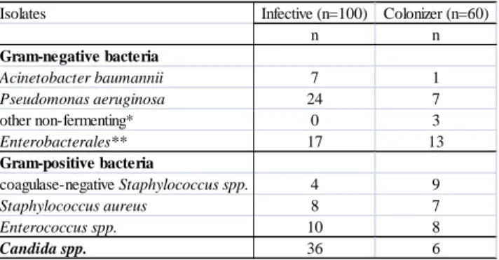
Other microorganisms were isolated together with S. maltophilia from 68%
of specimens. Numbers of these isolates are shown in Table 5.
Table 5. Other microorganisms isolated together with 160 S. maltophilia isolates.
* Alcaligenes faecalis, Achromobacter xylosoxidans, Pseudomonas fluorescens.
** Proteus mirabilis, Serratia marcescens, Escherichia coli, Klebsiella pneumoniae, Klebsiella oxytoca, Enterobacter aerogenes, Enterobacter cloacae
Isolates Infective (n=100) Colonizer (n=60)
n n
Gram-negative bacteria
Acinetobacter baumannii 7 1
Pseudomonas aeruginosa 24 7
other non-fermenting* 0 3
Enterobacterales** 17 13
Gram-positive bacteria
coagulase-negative Staphylococcus spp. 4 9
Staphylococcus aureus 8 7
Enterococcus spp. 10 8
Candida spp. 36 6
5. Conclusions
1. Our results show that SXT is still the most potent antibiotic drug against S. maltophilia; the susceptibility rate was 98-99%. Due to the low frequency of moxifloxacin and levofloxacin resistance, these drugs can be used either in high dose monotherapy or rather in combination with other antibiotics. Doxycycline was proved to have excellent in vitro activity against S. maltophilia. Based on this results and previous reports, minocycline would have similar effect. Considering SXT resistant isolates, severe infections of patients with chronic respiratory illnesses and the guideline given by CLSI, the distribution of minocycline in Hungary would be suggested. Colistin was found to have weak in vitro activity against S. maltophilia, therefore, it cannot be used in monotherapy in S. maltophilia infections. The establishment of S. maltophilia specific clinical breakpoints for agents other than SXT by EUCAST is strongly required in the near future. Furthermore, the most reliable antibiotic susceptibility testing method for alternative antibiotics should be declared.
2. Most infections caused by S. maltophilia were associated with severe co- morbidity and long-term, extensive ICU treatment. The high all-cause in hospital
mortality rate (45%) we observed can be connected to the serious underlying illnesses rather to S. maltophilia itself. However, the strict attributable mortality rate (11%) was also high. Especially patients admitted to ICU have the risk of S. maltophilia infections, therefore screening them for S. maltophilia colonization should be considered. Patients with central venous catheter must be taken special attention, because central venous catheter is associated with mortality in S. maltophilia infections.
3. Physicians have to consider that S. maltophilia as a co-pathogen or co- colonizer in polymicrobial infections can have negative impact on the success rate of antibiotic treatment and clinical outcome. The carbapenem, cephalosporine and aminoglycoside treatment of P. aeruginosa, which was found to be the most frequent co-pathogen, can be negatively effected by co-colonizer S. maltophilia. As S.
maltophilia can influence the virulence of surrounding bacteria, its eradication can be reasonable even if it just colonize a patient.
4. The occurance of SXT-resistant, MDR S. maltophilia is rare yet. While sul genes are associated with mobile genetic elements, ongoing resistance surveillance is required. The higher antibiotic resistance rates of MDR strains have significant clinical consequences: levofloxacin and moxifloxacin mean alternative therapeutic options yet, but in case of resistance to them just few antibiotic combinations or tetracycline derivates can be used. However, the clinical evidence of their efficacy is lacking yet.
5. Combinations including fluoroquinolons – especially moxifloxacin – can be effective, even if the isolate was resistant to them. Ceftazidime improves in vitro effect of other antibiotic agents in combination, despite the high level resistance of the strains to itself. An antibiotic agent in aerosolised form in combination with an intravenous one can be effective, even if the ΣFICI and SBPI do not show synergism.
It has to be kept in mind that results of synergy testing are isolate specific.
6. According to our in vitro results colistin-plus-SXT combined therapy can be used efficiently in clinical practice as no antagonistic effect was detected.
Moreover, in 20-35% it was found to have synergic effect against co-isolated MDR P.
aeruginosa, A. baumannii and S. maltophilia strains. Regrowth of MDR A. baumannii after 24 hour in the presence of colistin can be prevented by colistin-plus-SXT combination.
List of publications
Publications related to present thesis:
Juhász E, Krizsán G, Lengyel G, Grósz G, Pongrácz J, Kristóf K. (2014) Infection and colonization by Stenotrophomonas maltophilia: antimicrobial susceptibility and clinical background of strains isolated at a tertiary care centre in Hungary. Ann Clin Microbiol Antimicrob, 13: 333.
Juhász E, Pongrácz J, Iván M, Kristóf Katalin. (2015) Antibiotic susceptibility testing of sulfamethoxazole-trimethoprim resistant Stenotrophomonas maltophilia strains isolated at a teriary care centre in Hungary. Acta Microbiol Immunol Hung, 62: 295- 305.
Juhász E, Kovács A, Iván M, Pongrácz J, Kristóf K. (2017) In vitro activity of colistin and trimethoprim/sulfamethoxazole against consortia of multidrug resistant non- fermenting Gram-negative bacilli isolated from lower respiratory tract. Jund J Microbiol, 10: e14034.
Juhász E, Iván M, Pongrácz J, Kristóf K. (2018) Ritkábban előforduló, alsó légúti fertőzést okozó Gram-negatív nem fermentáló pálcák. Orv Hetil, 159: 23-30.
Other publications:
Sárvári P, Sóki J, Kristóf K, Juhász E, Miszti C, Melegh Sz, Latkóczy K, Urbán E.
(2018) A multicentre survey of the antibiotic susceptibility of clinical Bacteroides species from Hungary. Infect Dis, 50: 372-380.
Sárvári P, Sóki J, Kristóf K, Juhász E, Miszti C, Latkóczy K, Melegh Sz, Urbán E.
(2017) Molecular characterization of multidrug resistant Bacteroides isolates from Hungarian clinical samples. J Glob Antimicrob Resist, 13: 65-69.
Juhász E, Pintér E, Iván M, Pongrácz J, Kristóf K. (2017) Colistin resistance among blood culture isolates at a tertiary care centre in Hungary. J Glob Antimicrob Resist, 11: 167-170.
Adegathe J, Juhász E, Pongrácz J, Rimanóczy E, Kristóf K. (2016) Does Staphylococcus saprophyticus cause acute cystitis only in young females, or is there more to the story? A one-year comprehensive study done in Budapest, Hungary. Acta Microbiol Immunol Hung, 63: 57-67.
Pongrácz J, Benedek K, Juhász E, Iván M, Kristóf K. (2016) In vitro biofilm production of Candida bloodstream isolates: Any association with clinical characteristics? J Med Microbiol, 65: 272-277.
Pongrácz J, Juhász E, Iván M, Kristóf K. (2015) Significance of yeasts in bloodstream infection: epidemiology and predisposing factors of candidaemia in adult patients at a university hospital (2010-2014). Acta Microbiol Immunol Hung, 62: 317-329.
Nagy E, Ábrók M, Bartha N, Bereczki L, Juhász E, Kardos G, Kristóf K, Miszti C, Urbán E. (2014) Mátrix-asszisztált lézer deszorpciós, ionizaciós, repülési idő mérésen alapuló tömegspektrometria speciális alkalmazása a klinikai mikrobiológiai diagnosztika területen. Orv Hetil, 155: 1495-1503.
Juhász E, Jánvári L, Tóth A, Damjanova I, Nobilis A, Kristóf K. (2012) Emergence of VIM-4- and SHV-12-producing Enterobacter cloacae in a neonatal intensive care unit.
Int J Med Microbiol, 302: 257-260.
Juhász E, Béres J, Kanizsai S, Nagy K. (2012) The Consequence of a Founder Effect:
CCR5-Delta 32, CCR2-64I and SDF1-3 ' A Polymorphism in Vlach Gypsy Population in Hungary. Pathol Oncol Res, 18: 177-182.
Guba Z, Hadadi E, Major A, Furka T, Juhász E, Koos J, Nagy K, Zeke T. (2011) HVS-I polymorphism screening of ancient human mitochondrial DNA provides evidence for N9a discontinuity and East Asian haplogroups in the Neolithic Hungary.
J Hum Genet, 56: 784-796.
Juhász E, Nagy K. Comparison of thirteen methods for aDNA (1731-1841) extractions. In: Gyulai G (szerk.), Plant Archaeogenetics (Botanical Research and Practices). Nova Science Publishers, New York, 2011:129-134.
Juhász E, Ostorházi E, Pónyai K, Silló P, Párducz L, Rozgonyi F. (2011) Ureaplasmas: from commensal flora to serious infections. Rev Med Microbiol, 22: 73- 83.
Dobay O, Juhász E, Ungvári Á, Jeney C, Amyes S, Nagy K. (2009) Taguchi optimisation of a multiplex pneumococcal serotyping PCR and description of 11 novel serotyping primers. Acta Microbiol Immunol Hung, 56: 327-338.
Talha E, Juhász E, Kanizsai S, Nagy K. (2009) Molecular detection of T. pallidum by PCR in seronegative cases. Acta Microbiol Immunol Hung, 56: 181-189.
Juhász E, Ghidán A, Kemény B, Nagy K. (2008) Emergence of antiretroviral drug resistance in therapy-naive HIV infected patients in Hungary. Acta Microbiol Immunol Hung, 55: 383-394.