Examination of colistin-resistance in Gram-negative bacteria
PhD thesis
Dr. Béla Kádár
Semmelweis University
Doctoral School of Pathological Sciences
Supervisor: Dr. Dóra Szabó, D.Sc.
Official reviewers: Dr. József Kónya, Ph.D.
Dr. Szabolcs Sipeki, Ph.D.
Head of the Final Examination Committee: Dr. Károly Cseh, D.Sc.
Members of the Final Examination Committee: Dr. Gábor Pálos, Ph.D.
Dr. János Sinkó, Ph.D.
Budapest
2017
INTRODUCTION
Antibiotic resistant pathogens are an increasing healthcare problem worldwide.
WHO predicts that by 2050 antibiotic-resistant microbes will have caused the death of ten million people every year, of which 390,000 will be Europeans. Infections caused by multidrug-resistant and extensively drug-resistant Gram-negative are typically nosocomial infections – catheter-associated urinary tract infections, bloodstream infections related to use of intravenous devices, lower respiratory tract infections caused by prolonged lying or mechanical ventilation. Primary pathogens are non-fermenting Gram-negative bacteria (Acinetobacter spp., Pseudomonas aeruginosa, Stenotrophonomonas maltophilia), members of Enterobacteriaceae family (Escherichia coli, Klebsiella pneumoniae, Serratia marcescens, as well as Proteus, Enterobacter, Citrobacter spp.), and Gram-positives e.g. methicillin-resistant Staphylococcus aureus (MRSA) and vancomycin-resistant Enterococcus strains. For decades these bacteria have been developing resistance to different antibiotic families.
Treatment options for infections caused by multiresistant Gram-negative bacterial strains are limited. Long-existing but previously unheeded antibiotics like polymyxins (polymyxin B and colistin), nitrofurantoin and fosfomycin are living their renaissance, but besides these agents there are newer antimicrobials at disposal, as well, e.g.
tigecycline, ceftazidime/avibactam and ceftolozane/tazobactam.
Polymyxins have been subjects of intensive studying for years, because their selective antimicrobial activity against Gram-negatives is making them „last resort”
antibiotics against multiresistant bacteria. Acquired resistance to them was relatively rare in the past due to scarce usage. However, the more frequent employment has led to the appearance of colistin-resistant strains (since colistin is more widely used in clinical practice than polymyxin B, the term „colistin-resistance” became general). These strains have shown increased tolerance to antimicrobial peptides, as well. The molecular basis of resistance is the enzymatic modification of lipopolysaccharide which is an integral part of Gram-negative’s outer membrane. These modifications enable the outer membrane to electrostatically repel polymyxin molecules. Alterations are attained by addition of either phosphoethanolamine or 4-amino-4-deoxy-L-arabinose to the free
regulatory mechanism: (i) direct regulation is performed by PmrA-PmrB system – it activates the expression of genes responsible for phosphoethanolamine addition (pmrC, cptA), and of arn operon carrying out 4-amino-4-deoxy-L-arabinose synthesis and transfer, (ii) PhoP-PhoQ system enhances activity of PmrA-PmrB through PmrD connector protein, (iii) MgrB membrane protein stimulates PhoP-PhoQ system. All the aforementioned proteins are products of chromosomal genes, but in 2015 the first representative of a plasmid-encoded, mobile colistin-resistance gene family (mcr gene family) coding phosphoethanolamine-transferases was identified.
One possible option for treating infections caused by colistin-resistant strains is antibiotic combinations. In vitro synergism of polymyxins and rifampin against colistin- resistant Gram-negative bacteria was proved in numerous studies. Colistin–imipenem and colistin–meropenem were proved to be efficient against carbapenemase-producing K. pneumoniae strains. The combinaton of colistin and tetracycline were in vivo succesfully used for treatment of bloodstream infection caused by panresistant K.
pneumoniae.
In our studies we examined the susceptibility of acquired colistin-resistant bacterial strains isolated in Hungary to antibiotic combinations, the genetic factors behind their resistance, their tolerance to cationic antimicrobial peptides, and the changes in the composition of their outer membrane proteins.
OBJECTIVES
• Defining the antibiotic susceptibility of available bacterial strains.
• Finding antibiotic combinations of synergistic interactions that are in vitro effective against colistin-resistant bacterial strains.
• Examining the susceptibility of colistin-resistant bacterial strains to lactoferrin, lysozyme, and protamine.
• Identifying the presence of genes responsible for developing colistin-resistance with PCR (phoP, phoQ, pmrA, pmrD, mgrB, mcr-1), then measuring expression of present genes with reverse transcription PCR.
• Analyzing the outer membrane protein composition of colistin-susceptible and colistin-resistant bacterial strains.
MATERIALS AND METHODS
Bacterial strains
Klebsiella pneumoniae and Enterobacter asburiae strains isolated from clinical samples were investigated in our studies. The K. pneumoniae ATCC 700603 strain was used as control in the antibiotic susceptibility tests.
All eight K. pneumoniae strains belonged to international clone ST258. They were multiresistant, KPC-2 enzyme producers isolated and identified in 2008–2009, during the first colistin-resistant K. pneumoniae epidemic in Hungary. The two examined E.
asburiae strains originated from sporadic cases.
Determination of antibiotic susceptibility
Antibiotic susceptibility of the strains was checked by microdilution method and E-test. The following agents were tested by microdilution: ceftazidime, cefotaxime, ceftriaxone, imipenem, ertapenem, amikacin, tobramycin, ciprofloxacin, levofloxacin, moxifloxacin, rifampin, polymyxin B and colistin. E-test was used for separating colistin-heteroresistant subpopulations within colistin-susceptible strains. After overnight incubation the valid recommendations of the European Committee on Antimicrobial Susceptibility Testing (EUCAST) were followed for minimal inhibitory concentration (MIC) value interpretation.
Checkerboard analysis
Efficiency of antibiotic combinations was tested with checkerboard method.
Using the lowest individual MIC values FIC indices (fractional inhibitory concentration index) were calculated as follows: ΣFICI = FICIA + FICIB, where FICIA = MICA(c) / MICA(a), and FICIB = MICB(c) / MICB(a). Based on ΣFICI values synergistic (ΣFICI ≤ 0,5), partially synergistic (0,5 < ΣFICI < 1), additive (ΣFICI = 1), indifferent (1 < ΣFICI
≤ 4) and antagonistic (ΣFICI > 4) relations were established.
Susceptibility to lactoferrin, protamine and lysozyme
Bacteria were cultured in Luria-Bertani (LB) broth, then were centrifugalized in the exponential phase of cell division. Bacterial solutions of 2,1 x 10⁵ CFU/ml (CFU =
colony-forming unit) were prepared, from which 10–10 µls were added to 50–50 mg/ml protamine, lysozyme and lactoferrin, respectively, making 200 µl final volumes. One hundred µl of each solution was inoculated onto LB agar plates after incubation, and after a 18-hour incubation the number of colonies were counted by naked eye (one colony considered as one CFU). Finally, percental changes of germ numbers were calculated.
Examination of colistin-resistance genes with PCR
Two or three colonies of the bacterial strains were taken from Mueller–Hinton (MH) agar, then were solved in distilled water, and the solutions were centrifugalized after a 100°C water bath. Oligonucleotides for amplification of phoP, phoQ, pmrA, pmrB and pmrD genes were designed with MWG Eurofins Primer Design programme, while the primers of mcr-1 and mgrB genes were produced based on previous publications. PCR amplicons were run in 1,5% agarose gel, then were painted in special gel dye, and finally were detected under UV light. Identification of pmrB and mgrB amplicons’ nucleotide sequence was carried out by BIOMI Ltd. (Gödöllő, Hungary).
Results were analyzed using NCBI GenBank database.
Examination of gene expression with RT-qPCR
The complete RNA content of bacterial cells was extracted with RNeasy Mini Kit (QIAGEN, Hilden, Germany), then was processed with RNase-free DNase (QIAGEN).
Reverse transcription PCR was performed with LightCycler RNA Master SYBR Green I. kit (Roche Applied Science, Penzberg, Germany). Oligonucleotides for amplification of phoP, pmrD and arn genes were designed with MWG Eurofins Primer Design programme, while the primers of housekeeping genes rpoB and rrsE were produced based on previous publications.
Isolation of outer membrane proteins
Bacterial strains were cultured in 500 ml MH broth, then were centrifugalized, and at last the sediment was resolved in normal saline solution. After resolving the sediment in Tris-HCl icy cooling was implemented, then ultrasonic disruption was performed for 2x2 minutes with 500 W (MSE Soniprep 150 Ultrasonic Disintegrator,
MSE Ltd., London, UK). After centrifugation the supernatant was ultracentrifugalized, and the resulting sediment was solved in sodium lauryl sarcosine solution (Sigma- Aldrich Ltd., Budapest, Hungary), then was incubated. Following another ultracentrifugation the sarcosine-insoluble outer membrane proteins (OMP) were isolated from the sediment.
One dimensional gel electrophoresis of outer membrane proteins (1-DE)
The outer membrane protein sample was mixed with Lämmli solution [1 M Tris (pH 6,8), 50% glycerol, 10% sodium dodecyl sulfate (SDS), β-mercaptoethanol, bromophenol blue, distilled water (Bio-Rad Magyarország Kft., Budapest, Hungary)], then the mixture was warmed. After cooling electrophoresis was carried out in Bio-Rad Mini Protean 3 system. Gels were incubated in dying solution, then were steeped in differentiating solution.
Analysis of outer membrane proteins with Microchip
Outer membrane proteins were extracted, marked with fluorescent dye, centrifugalized, then separated electrophoretically in Agilent 2100 Bioanalyzer System Microchip (Agilent Technologies, Santa Clara, CA, USA).
Two dimensional gel electrophoresis of outer membrane proteins (2-DE)
Outer membrane proteins were solved in 2-DE sample buffer [8 M urea, 2%
CHAPS (3-[(3-cholamidopropyl)-dimethylammonio]-1-propanesulfonate), 50 mM dithiothreitol, 0,2% Bio-Lyte® 3/10 Ampholyte, bromophenol blue (Bio-Rad)], were applied onto immobilized pH gradient (IPG) strips, the were separated according to their charges by isoelectric focusing. After this the strips were washed in equilibration buffer, then the proteins were separated according to their masses by polyacrylamide gel electrophoresis. Protein bands were made visible with dying solution, and the gel areas of interest were cut out for further examination.
In-gel digestion
Gel parts containing protein bands were cut out and sliced into smaller pieces. The dye was removed, gel pieces were dehydrated, disulfide bridges were reduced and
sulfhydryl groups were alkylated, then side-chain-protected trypsin (Promega, Madison, WI, USA) was used for in-gel proteolysis. Digested peptides were extracted from the gel with formic acid solution (Sigma-Aldrich).
MALDI-TOF/MS mass spectrometry
Mass spectrometry examination was carried out with Autoflex II MALDI- TOF/MS method (Bruker Daltonics, Bremen, Germany). Digested peptides were solved in a mixture of α-cyano-4-hydroxycinnamic acid (Bruker Daltonics), acetonitrile and trifluoroacetic acid (Scharlau Chemie, Barcelona, Spain). Peptides were identified by 1000 shots, data processing was executed with flexAnalysis software package version 3.1 (Bruker Daltonics), and Sequence Editor software (Bruker Daltonics) was used for analysis. The MASCOT algorithm (http://www.matrixscience.com) and the Swiss-Prot database (Swiss Institute of Bioinformatics, Genf, Svájc) were utilized for protein identification.
RESULTS
Antibiotic susceptibility
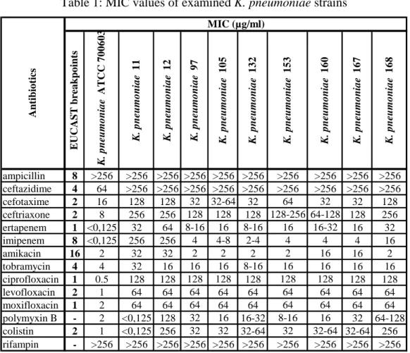
All K. pneumoniae strains belonging to ST258 clone were resistant to 3rd generation cephalosporins, ertapenem, tobramycin, fluoroquinolones and rifampin. All strains were resistant to polymyxins except the No. 11 (Table 1).
Enterobacter asburiae strains isolated from sporadic cases were susceptible to 3rd generation cephalosporins, carbapenems, fluoroquinolones and amikacin. Within the colistin-susceptible strain No. 0821 a colistin-heteroresistant subpopulation was identified with E-test (Table 2).
Table 1: MIC values of examined K. pneumoniae strains
EUCAST breakpoints K. pneumoniae ATCC 700603 K. pneumoniae 11 K. pneumoniae 12 K. pneumoniae 97 K. pneumoniae 105 K. pneumoniae 132 K. pneumoniae 153 K. pneumoniae 160 K. pneumoniae 167 K. pneumoniae 168
ampicillin 8 >256 >256 >256 >256 >256 >256 >256 >256 >256 >256 ceftazidime 4 64 >256 >256 >256 >256 >256 >256 >256 >256 >256
cefotaxime 2 16 128 128 32 32-64 32 64 32 32 128
ceftriaxone 2 8 256 256 128 128 128 128-256 64-128 128 256
ertapenem 1 <0,125 32 64 8-16 16 8-16 16 16-32 16 32
imipenem 8 <0,125 256 256 4 4-8 2-4 4 4 4 16
amikacin 16 2 32 32 2 2 2 2 16 16 2
tobramycin 4 4 32 16 16 16 8-16 16 16 16 16
ciprofloxacin 1 0.5 128 128 128 128 128 128 128 128 128
levofloxacin 2 1 64 64 64 64 64 64 64 64 64
moxifloxacin 1 2 64 64 64 64 64 64 64 64 64
polymyxin B - 2 <0,125 128 32 16 16-32 8-16 16 32 64-128
colistin 2 1 <0,125 256 32 32 32-64 32 32-64 32-64 256
rifampin - >256 >256 >256 >256 >256 >256 >256 >256 >256 >256
Antibiotics
MIC (µg/ml)
Table 2: MIC values of examined E. asburiae strains
EUCAST breakpoints
K. pneumoniae ATCC 700603
E. asburiae 0821
E. asburiae 0821/H
E. asburiae 148
ampicillin 8 >256 256 256 32
ceftazidime 4 64 0,25 1 <0,125
cefotaxime 2 16 0,5 1 <0,125
ceftriaxone 2 8 0,5 1 <0,125
ertapenem 1 <0,125 <0,125 <0,125 <0,125
imipenem 8 <0,125 0,5 0,5-1 0,25-0,5
amikacin 16 2 0,5 0,5 0,5-1
tobramycin 4 4 0,25 <0,125 0,5
ciprofloxacin 1 0,5 <0,125 <0,125 <0,125
levofloxacin 2 1 <0,125 <0,125 <0,125
moxifloxacin 1 2 <0,125 <0,125 <0,125
polymyxin B - 2 0,125 >256 64-128
colistin 2 1 0,125 >256 256
rifampin - >256 >256 >256 >256
Antibiotics
MIC (µg/ml)
Results of checkerboard analysis
Antibiotic interactions observed in K. pneumoniae strains No. 11 and 12 were different from several aspects (Table 3–4). In case of No. 11 pure synergism was detected only between imipenem and tobramycin, but partial synergism was noted in combinations comprised of rifampin, ciprofloxacin, imipenem and ceftazidime.
Imipenem combined with rifampin, tobramycin or ciprofloxacin was synergistic against the colistin-resistant strain No. 12. Rifampin combined with colistin or polymyxin B was also synergistic against it.
Table 3:
MIC values and FIC indices of antibiotics tested on K. pneumoniae strain 11
1. ABalone 2. ABalone 1. ABcombination 2. ABcombination
colistin - ceftazidime 0,125 256 0,25 1 2,004
colistin - ciprofloxacin 0,125 128 0,25 1 2,008
colistin - imipenem 0,125 8 0,25 1 2,125
colistin - rifampin 0,125 256 0,25 1 2,004
polymyxin B - ceftazidime 0,125 256 0,25 1 2,004
polymyxin B - ciprofloxacin 0,125 128 0,25 1 2,008
polymyxin B - imipenem 0,125 8 0,25 1 2,125
polymyxin B - rifampin 0,125 256 0,25 1 2,004
rifampin - ciprofloxacin 256 128 0,25 64 0,501
rifampin - imipenem 256 8 4 4 0,516
imipenem - ciprofloxacin 8 128 4 1 0,508
imipenem - tobramycin 8 32 1 4 0,250
ceftazidime - ciprofloxacin 256 128 0,25 64 0,501
ceftazidime - tobramycin 256 32 1 16 0,504
tobramycin - ciprofloxacin 32 128 32 1 1,008
K. pneumoniae 11
Antibiotic combinations MIC (µg/ml)
FICI
Table 4:
MIC values and FIC indices of antibiotics tested on K. pneumoniae strain 12
1. ABalone 2. ABalone 1. ABcombination 2. ABcombination
colistin - ceftazidime 256 256 64 2 0,258
colistin - ciprofloxacin 256 128 2 16 0,133
colistin - imipenem 256 8 32 8 1,125
colistin - rifampin 256 256 0,25 1 0,005
polymyxin B - ceftazidime 128 256 64 1 0,504
polymyxin B - ciprofloxacin 128 128 1 16 0,133
polymyxin B - imipenem 128 8 2 4 0,516
polymyxin B - rifampin 128 256 0,25 1 0,006
rifampin - ciprofloxacin 256 128 64 64 0,750
rifampin - imipenem 256 8 1 4 0,504
imipenem - ciprofloxacin 8 128 1 2 0,141
imipenem - tobramycin 8 32 1 2 0,188
ceftazidime - ciprofloxacin 256 128 256 64 1,500
ceftazidime - tobramycin 256 32 0,25 16 0,501
tobramycin - ciprofloxacin 32 128 16 1 0,508
K. pneumoniae 12
Antibiotic combinations MIC (µg/ml) FICI
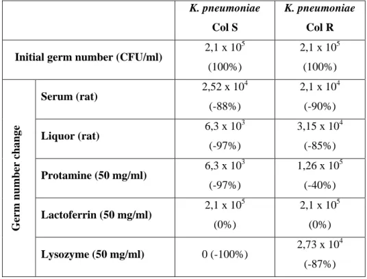
Susceptibility to lactoferrin, protamine and lysozyme
The K. pneumoniae strains were proved to be resistant to lactoferrin – we did not observe any decrease of colony forming units after exposure. Protamine caused 97%
colony forming unit fall in the colistin-susceptible K. pneumoniae strain, however, CFU number only decresed with 40% in the colistin-resistant strain. Lysozyme exhibited 100% bactericidal effect on the colistin-susceptible strain, and the effect was also notable albeit of lesser extent in case of the colistin-resistant one (Table 5).
Protamine did not cause any significant change in CFU numbers of the E.
asburiae strains. On the other hand, the colistin-resistant strains showed high tolerance to lactoferrin and lysozyme (Table 6).
Table 5: CFU changes in K. pneumoniae strains after exposition to serum, liquor, lactoferrin, lysozyme and protamine (Col S = colistin-susceptible; Col R = colistin-resistan)
K. pneumoniae Col S
K. pneumoniae
Col R Initial germ number (CFU/ml) 2,1 x 10⁵
(100%)
2,1 x 10⁵ (100%)
Germ number change
Serum (rat) 2,52 x 10⁴
(-88%)
2,1 x 10⁴ (-90%)
Liquor (rat) 6,3 x 10³
(-97%)
3,15 x 10⁴ (-85%)
Protamine (50 mg/ml) 6,3 x 10³ (-97%)
1,26 x 10⁵ (-40%)
Lactoferrin (50 mg/ml) 2,1 x 10⁵ (0%)
2,1 x 10⁵ (0%)
Lysozyme (50 mg/ml) 0 (-100%) 2,73 x 10⁴ (-87%)
Table 6: CFU changes in E. asburiae strains after exposition to lactoferrin, lysozyme and protamine (Col S = colistin-susceptible; Col R = colistin-resistan)
E. asb 0821 Col S
E. asb 0821/H Col R
E. asb 148 Col R
Initial germ number (CFU/ml) 2,1 x 10⁵ (100%)
2,1 x 10⁵ (100%)
2,1 x 10⁵ (100%)
Germ number change Protamine (50 mg/ml) 1,995 x 105 (-5%)
2,1 x 10⁵ (0%)
2,1 x 10⁵ (0%)
Lactoferrin (50 mg/ml) 2,1 x 10⁵ (0%)
2,919 x 105 (+39%)
2,373 x 105 (+13%)
Lysozyme (50 mg/ml) 2,184 x 105 (+4%)
2,646 x 105 (+26%)
2,163 x 105 (+3%)
Examination of colistin-resistance genes with PCR
Presence of phoP, phoQ, pmrA, pmrB and pmrD genes were verified in the colistin-susceptible and colistin-resistant K. pneumoniae strains, as well. Neither the E.
asburiae nor the K. pneumoniae strains carried the mcr-1 gene.
The mgrB gene was detected in all K. pneumoniae strains. A 954 basepair-long amplicon was detected in the colistin-susceptible strain. Sequence analysis showed that the amplicon contained a new, previously undescribed mgrB variant, and a type 5 insertion sequence (IS5). Uniformly 540 basepair-long amplicons lacking IS5 were detected in the colistin-resistant strains, and these identical mgrB genes also coded a new variant of the protein (Figure 1).
Sequence of PmrB demonstrated wild-type protein in the colistin-susceptible and colistin-resistant strains, as well.
Figure 1:
Length of mgrB genes isolated from K. pneumoniae strains (upper amplicon: colistin- susceptible K. pneumoniae strain; lower amplicon: colistin-resistant K. pneumoniae strain)
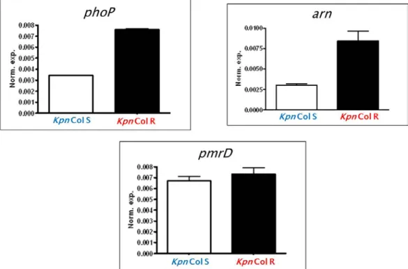
Examination of gene expression with RT-qPCR
The PhoP-PmrD-arn regulatory system’s role in colistin-resistance was confirmed in the colistin-resistant K. pneumoniae strains: increased expression of phoP and arn genes was detected (Figure 2).
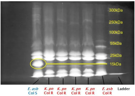
One dimensional gel electrophoresis of outer membrane proteins (1-DE)
Absence of a ca. 15–16 kDa protein fraction was detected in the colistin-resistant K. pneumoniae and colistin-resistant E. asburiae strains (Figures 3–4).
Figure 2: Relative gene expression of K. pneumoniae colistin-resistance genes
(Kpn Col S = colistin-susceptible K. pneumoniae; Kpn Col R = colistin-resistant K. pneumoniae)
Figure 3: 1D gel electrophoresis of K. pneumoniae outer membrane proteins
(K. pn Col S = colistin-susceptible K. pneumoniae; K. pn Col R = colistin-resistant K. pneumoniae)
Figure 4: 1D gel electrophoresis of E. asburiae outer membrane proteins
(K. pn Col R = colistin-resistant K. pneumoniae;
E. asb Col S = colistin-susceptible E. asburiae; E. asb Col R = colistin-resistant E. asburiae)
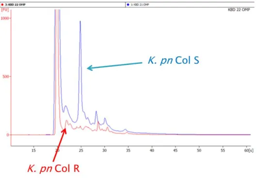
Analysis of outer membrane proteins with Microchip
Running of outer membrane proteins marked with fluorescent dye in Agilent 2100 Bioanalyzer System microchip demonstrated absence of a protein fraction in the colistin-resistant K. pneumoniae and colistin-resistant E. asburiae strains, respectively, 20–25 seconds into the run (Figures 5–6).
Analysis of outer membrane proteins with MALDI-TOF/MS mass spectrometry Substantial amount of DNA starvation/stationary phase protection proteins (Dps) and LysM domain/BON superfamily proteins was found in the colistin-susceptible K.
pneumoniae strain, while the colistin-resistant strain lacked these.
Outer membrane proteins OmpC and OmpW were present in the colistin- susceptible E. asburiae strain, but were absent in the colistin-resistant ones. OmpA and OmpX were only detected in the colistin-resistant strains.
Figure 5: Analysis of K. pneumoniae outer membrane proteins by Microchip
(K. pn Col S = colistin-susceptible K. pneumoniae; K. pn Col R = colistin-resistant K. pneumoniae)
Figure 6: Analysis of E. asburiae outer membrane proteins by Microchip
(E. asb Col S = colistin-susceptible E. asburiae; E. asb Col R = colistin-resistant E. asburiae)
DISCUSSION
Our experiments on colistin-resistant, KPC-2 producing K. pneumoniae strains confirm previous reports about importance of antibiotic combination therapy versus monotherapy. Synergistic interaction was detected between the following antibiotic combinations: rifampin–colistin, rifampin–polymyxin B, imipenem–rifampin, imipenem–tobramycin and imipenem–ciprofloxacin.
Our findings on the presence of a colistin-heteroresistant subpopulation within a colistin-susceptible E. asburiae strain correlate with international reports of recent years. Colistin-heteroresistance concomitant with lysozyme-tolerance was previously observed in Enterobacter cloacae. Cationic antimicrobial peptides bind to the PhoP- PhoQ proteins in the outer membrane of Gram-negative bacteria similarly to polymyxins. This regulatory protein pair has an important role in the development of colistin-resistance. Our K. pneumoniae and E. asburiae strains developed colistin- resistance without previous polymyxin exposure, which suggests the possibility of cross-tolerance/-resistance. Tolerance to human antimicrobial peptides (e.g. lactoferrin, lysozyme) may contribute to development of colistin-resistance. This phenomenon might applies backwards, too – bacteria may become tolerant to host antimicrobial peptides due to colistin-resistance.
Presence of several chromosomal genes responsible for colistin-resistance (phoP, phoQ, pmrA, pmrB, pmrD and mgrB) was successfully verified with PCR in K.
pneumoniae strains. However, the mcr-1 plasmid-encoded resistance gene first described in November 2015 was not detectable in any examined strains.
Changes in amino-acid sequence of PmrB are knowingly contribute to development of colistin-resistance, but PmrB proteins of our colistin-resistant K.
pneumoniae strains were all wild-type. On the other hand, MgrB proteins of the colistin- susceptible and colistin-resistant K. pneumoniae strains were equally different from wild-type, and there was an IS5 nucleotide portion adjoining mgrB gene in the susceptible strain which was absent in colistin-resistant ones. This is remarkable because insertion sequences usually lead to colistin-resistance by inserting themselves into mgrB, deactivating it.
Role of PhoPQ and arn systems in developing colistin-resistance in our strains was verified with RT-qPCR. Although expression of pmrD gene did not demonstrate
significant difference between colistin-susceptible and colistin-resistant K. pneumoniae strains, substantial overexpression of arn and phoP was detected in the colistin-resistant strain.
During gel electrophoresis of colistin-resistant K. pneumoniae and E. asburiae strains’ outer membrane proteins absence of a ca. 15-16 kDa protein fraction was detected. Absent proteins in case of K. pneumoniae were DNA starvation/stationary phase protection proteins (Dps) and LysM domain/BON superfamily proteins. Colistin- resistant E. asburiae strains lacked OmpC and OmpW, but OmpA and OmpX were present.
Main function of Dps and their homologues is the protection of bacterial cells during stationary phase of cell division. The bind to bacterial chromosome in a non- specific way, creating a stable, protected Dps-DNA complex. They bind and oxidize intracellular Fe2+ ions, decreasing the amount of reactive oxygen species.
LysM and BON domains are conserved protein sections. These are mainly structural proteins and enzymes responsible for maintaining cell membrane integrity. As colistin-resistance is based on molecular changes in the cell wall, the alterations in the expression of these proteins is logical, although its background is not clear, yet.
OmpC and its homologues are porin-type transport proteins found in the outer membrane of Enterobacteriaceae. They are responsible for transporting several types of molecules into the cells, including antibiotics. Their loss or decreased expression leads to antibiotic resistance and diminished susceptibility to serum antimicrobial activity in E. coli and Enterobacter spp.
OmpA is a multifunctional membrane protein: in addition to maintaining integrity of the outer membrane, it is responsible for serum resistance in E. coli and antimicrobial peptide resistance in K. pneumoniae.
OmpX is a protein structurally similar to OmpA. Its overproduction was observed in multiresistant Enterobacter aerogenes strains with the simultaneous underproduction of OmpF and Omp36 porins, as well as structural changes of LPS. Upregulation of ompX and downregulation of omp36 together cause the decrease of outer membrane permeability.
CONCLUSIONS
We have reached the following conclusions and results in our studies:
• Klebsiella pneumoniae strains
– ST258 clone is prone to developing colistin-resistance
– combinations of polymyxins and rifampin, as well as imipenem–
rifampin, imipenem–tobramycin and imipenem–ciprofloxacin have in vitro antibacterial activity against colistin-resistant strains
– colistin-resistant strains develop tolerance to antimicrobial peptides – colistin-resistant strains are overexpressing phoP, pmrD and arn genes – new MgrB variants were identified in ST258 clone
– outer membrane protein assortment changes in colistin-resistant strains (loss of LysM/BON superfamily proteins and DNA starvation proteins)
• Enterobacter asburiae strains
– certain E. asburiae strains are able to develop colistin-resistance while retaining their susceptibility to β-lactams, aminoglycosides and fluoroquinolones
– colistin-resistant strains develop tolerance to antimicrobial peptides – outer membrane protein assortment changes in colistin-resistant strains
(absence of OmpC and OmpW, and presence of OmpA and OmpX)
LIST OF OWN PUBLICATIONS
Articles published in the subject of dissertation
Kádár B, Kocsis B, Tóth Á, Damjanova I, Szász M, Kristóf K, Nagy K, Szabó D.
(2013) Synergistic antibiotic combinations for colistin-resistant Klebsiella pneumoniae.
Acta Microbiol Immunol Hung, 60(2): 201-209., IF: 0,780
Kádár B, Kocsis B, Kristóf K, Tóth Á, Szabó D. (2015) Effect of antimicrobial peptides on colistin-susceptible and colistin-resistant strains of Klebsiella pneumoniae and Enterobacter asburiae. Acta Microbiol Immunol Hung, 62(4): 501-508., IF: 0,568
Kocsis B, Kádár B, Tóth Á, Fullár A, Szabó D. (2017) MgrB variants in colistin- susceptible and colistin-resistant Klebsiella pneumoniae ST258. J Microbiol Immunol Infect, 50(5): 735-736., IF: 2,973
Kádár B, Kocsis B, Tóth Á, Kristóf K, Felső P, Kocsis B, Böddi K, Szabó D. (2017) Colistin resistance associated with outer membrane protein change in Klebsiella pneumoniae and Enterobacter asburiae. Acta Microbiol Immunol Hung, 64(2): 217- 227., IF: 0,921*
Articles published in other subjects than the dissertation
Kádár B, Szász M, Kristóf K, Pesti N, Krizsán G, Szentandrássy J, Rókusz L, Nagy K, Szabó D: In vitro activity of clarithromycin in combination with other antimicrobial agents against biofilm-forming Pseudomonas aeruginosa strains. ACTA MICROBIOLOGICA ET IMMUNOLOGICA HUNGARICA 57:(3) pp. 235-245. (2010), IF: 0,625 (Kádár B és Szász M megosztott első szerzők)
Kádár B, Kocsis B, Nagy K, Szabó D: The renaissance of polymyxins. CURRENT MEDICINAL CHEMISTRY 20:(30) pp. 3759-3773. (2013), IF: 3,715
Szabó Bálint Gergely, Lénárt Katalin Szidónia, Kádár Béla, Gombos Andrea, Dezsényi Balázs, Szanka Judit, Bobek Ilona, Prinz Gyula: A Streptococcus pneumoniae (pneumococcus) -infekciók ezer arca. ORVOSI HETILAP 156:(44) pp. 1769-1777.
(2015), IF: 0,291
Szabó BG, Kádár B, Lénárt KS, Dezsényi B, Kunovszki P, Fried K, Kamotsay K, Nikolova R, Prinz G: Use of intravenous tigecycline in patients with severe Clostridium difficile infection: a retrospective observational cohort study. CLINICAL MICROBIOLOGY AND INFECTION 22:(12) pp. 990-995. (2016), IF: 5,292