DOKTORI (PhD) ÉRTEKEZÉS
Christopher Fenila
Soproni Egyetem
Sopron
2017
Doktori (PhD) értekezés Soproni Egyetem
Kitaibel Pál Környezettudományi Doktori Iskola
Doktori program: K1. Biokörnyezettudományi program Programvezető: Prof. Dr. Albert Levente
TARGETING HISTAMINE H4 RECEPTORS IN ALLERGIES
CAUSED BY AIR POLLUTANTS
HISZTAMIN H4 RECEPTOROK VIZSGÁLATA LÉGSZENNYEZ Ő ANYAGOK ÁLTAL KIVÁLTOTT
ALLERGIÁS ESETEKBEN
Készítette: Christopher Fenila
Témavezető: Dr. Fekete Gusztáv Társ-témavezető: Prof. Dr. Tóth Péter
Sopron
2017
ALLERGIES
CAUSED BY AIR POLLUTANTS
című értekezés doktori (PhD) fokozat elnyerése érdekében készült a Soproni Egyetem Kitaibel Pál Környezettudományi Doktori Iskolája
K1. Biokörnyezettudományi program keretében.
Írta: Christopher Fenila
Témavezetők:
Dr. Fekete Gusztáv,
Elfogadásra javaslom (igen / nem)
(aláírás)
Prof. Dr. Tóth Péter,
Elfogadásra javaslom (igen / nem)
(aláírás) A jelölt a doktori szigorlaton ...%-ot ért el.
Szombathely, ...
a Szigorlati Bizottság elnöke
Az értekezést bírálóként elfogadásra javaslom (igen / nem) Első bíráló (Dr. ...) igen / nem
(aláírás)
Második bíráló (Dr. ...) igen / nem
(aláírás) A jelölt az értekezés nyilvános vitáján ...%-ot ért el.
Szombathely, ...
a Bírálóbizottság elnöke
A doktori (PhD) oklevél minősítése ...
Az EDT elnöke
Alulírott Christopher Fenila jelen nyilatkozat aláírásával kijelentem, hogy a Targeting histamine h4 receptors in allergies caused by air pollutants című PhD értekezésem önálló munkám, az értekezés készítése során betartottam a szerzői jogról szóló 1999. évi LXXVI.
törvény szabályait, valamint a Kitaibel Pál Környezettudományi Doktori Iskola által előírt, a doktori értekezés készítésére vonatkozó szabályokat, különösen a hivatkozások és idézések tekintetében.1
Kijelentem továbbá, hogy az értekezés készítése során az önálló kutatómunka kitétel tekintetében témavezetőimet, illetve a programvezetőt nem tévesztettem meg.
Jelen nyilatkozat aláírásával tudomásul veszem, hogy amennyiben bizonyítható, hogy az értekezést nem magam készítettem, vagy az értekezéssel kapcsolatban szerzői jogsértés ténye merül fel, a Soproni Egyetem megtagadja az értekezés befogadását.
Az értekezés befogadásának megtagadása nem érinti a szerzői jogsértés miatti egyéb (polgári jogi, szabálysértési jogi, büntetőjogi) jogkövetkezményeket.
Sopron, 2017.………….
………..
doktorjelölt
1 1999. évi LXXVI. tv. 34. § (1) A mű részletét – az átvevő mű jellege és célja által indokolt terjedelemben és az eredetihez híven – a forrás, valamint az ott megjelölt szerző megnevezésével bárki idézheti.
36. § (1) Nyilvánosan tartott előadások és más hasonló művek részletei, valamint politikai beszédek tájékoztatás céljára – a cél által indokolt terjedelemben – szabadon felhasználhatók. Ilyen felhasználás esetén a forrást – a szerző nevével együtt – fel kell tüntetni, hacsak ez lehetetlennek nem bizonyul.
It does not; therefore depend on man's desire or effort, but on God's mercy Romans 9:16
Abstract
Inhalation of airborne pollutants can result in allergic sensitization.
Responses may occur in the upper respiratory tract, the lower respiratory tract or systemically, for example, a febrile response. The mechanisms underlying these responses are not always clear but include production of reaginic antibody, activation of T-lymphocyte subsets, and release of inflammatory mediators. A variety of agents have been associated with elicitation of these reactions including chemical vapors, dusts and particulates, and microbial organisms. As a result of the widespread occurrence of allergens in both indoor and outdoor environments, development of allergy and its treatment management has received close attention.
Six common pollutants listed in the EPA website have been used to check for currently available background literature showcasing the role of those pollutants in allergy. However, no studies have been performed to demonstrate the relation of Histamine receptor with the pollutants, except for mRNA expression studies.
Evidence-based information about the major pollutants responsible for causing or exacerbating allergic diseases is lacking. Hence in this study we have prioritised the major pollutants and its involvement in allergy.
Histamine has long been known to be the mediator that orchestrates inflammatory and allergic responses acting through H1, H2, H3 and H4 receptors.
Recent reports suggest the involvement of H4R in the control of immune cell trafficking and pro-inflammatory responses. This was derived from H4R-mediated histamine-induced activation of eosinophils, increased expression of adhesion molecules and rearrangement of the actin cytoskeleton leading to immune cell migration from bloodstream to sites of inflammation. Consequently, H4 receptors are currently an attractive target for the pharmacological modulation of histamine transferred signals in inflammatory conditions and for the development of therapeutic strategies for allergic conditions. The 3D structure of H4R has not been experimentally elucidated till date. Hence in this study, 3D structure of the Histamine H4 receptor was generated using iTASSER with human β2-adrenergic GPCR as a top template. This is the first Histamine H4 receptor structure model developed where human template is being used. From the five models generated by iTASSER only one model was chosen for further analysis after various validation tests.
Subsequently, the active site of the receptor model was identified using Discovery studio 2 and previous literature as evidence. Among the 11 binding sites predicted by the ligand fit module of Discovery studio, site 2 was found to possess most of the key residues that were identified in previous works. Three databases containing similar structures of the known ligand JNJ7777120, Thioperamide and
Vuf6002 were constructed from PubChem database. The molecular docking of the three separate databases onto the binding site of the modelled receptor revealed that 148 of 150 JNJ7777120 analogues, 42 of 49 Thioperamide analogues, 193 in a total of 198 Vuf 6002 analogues successfully docked onto the binding site. Out of these, six compounds with high docking scores were identified (Compound I, J, A, E, F, K). The ADMET properties as analyzed by the module in Discovery studio show that except 2 compounds (Compounds E, F) others have good ADMET properties.
Similarly the molecular dynamic simulations which determine the conformational variations of the H4R-ligand complexes clearly show that the compounds are well stabilized at their respective active sites.
Structure based virtual screening has resulted in scaffolds from which new compounds could be developed. The identified structures have to be further analyzed and optimized for interesting chemical structures. The receptor model developed in this study also could serve as a basis for future investigations since it is modelled based on the human GPCR. To conclude, this study has given insights for the development of new antagonists.
Acknowledgement
I would like to thank all the people who contributed in some way to the work described in this thesis.
First and foremost, I am grateful to the God for giving me the opportunity to do Ph. D and being with me throughout this period.
I whole heartedly thank my academic advisor, Dr. Gusztáv Fekete, for accepting me as his student. I would like to express my sincere gratitude to my advisor for the continuous support of my Ph. D study, for his patience and motivation. I could not have imagined having a better advisor and mentor for my Ph.D study.
Besides my advisor, I would like to thank the rest of my thesis committee:
Prof. Tóth Péter, Prof. Péter Molnár and Prof. Dr. Bayoumi Hamuda Hosam for their insightful comments and encouragement, and also for their questions which incented me to widen my research from various perspectives.
I wish to express my sincere gratitude to the University of West Hungary, my special thanks to all the staff members of the Faculty of Natural and Technical Sciences, Savaria Institute of Technology.
My sincere thanks also go to Berla-mam and Xavier-sir, for their technical advices I also take this opportunity to thank my friend Dr. Saranya Nallusamy for her suggestions and technical advice.
I take this opportunity to express gratitude to all of the my friends who were there to help and support during my stay in Hungary, Evelien Impens, Mátyás Andó, Kitti Rábi, Timkó Györg, Zsuzsanna Timkó and Miklós Andó. They all made me feel at home with their hospitality.
I also take pleasure in thanking Rev. Stephen Murray and Pleuntje Jellema Murray for their moral and prayer support. Also my sincere thanks go to the home prayer group and members of St Johns Anglican Church. I extend my thanks to my friends Saranya Shanmugasundaram, Vijay Anand, Bliss Ramya Joan, Feng Lan and Leydi Carolina for their encouragement.
My sincere gratitude goes to my parents S. Christopher and S. Laila for their support in all my pursuits and their honest love and prayers. I also thank my sister C. Anila Gifty who has also been there as a naughty and loving sister. Words cannot express how grateful I am to them. I also offer my special thanks to my family-in-law. I am always grateful to my father-in-law (J. Sukumaran), mother-in
law (R. Prema Sukumaran), brother in law (Russel Sugumaran), co-sister (C. S Dyana) for their unyielding support and prayers and specially my cheerful nephew (Ryan Jeremy Russel). I also would like to thank my extended family members who have always been my strength. Last but not the least; I would like to thank my beloved husband Jacob Sukumaran. Thank you for supporting me in everything, and especially I can’t thank you enough for encouraging me throughout this experience. I appreciate for your love and faithful support during the stages of this Ph.D. Finally to add, thanks to my little son Ragnar Joey who will be able to witness my Ph.D unexpectedly. His smiles makes me happy and endearing
Contents
LIST OF FIGURES ... 3
LIST OF TABLES ... 5
ABBREVIATIONS ... 6
CHAPTER 1 INTRODUCTION ... 8
1.1 General Introduction... 9
1.2 Objectives of the research ... 14
CHAPTER 2 REVIEW OF LITERATURE ... 15
2.1 Definition of Environment ... 16
2.2 Air pollution affecting Environment ... 17
2.3 Air Pollution and health ... 18
2.4 Interaction of the pollutant with the immune system ... 20
2.4.1 Acute phase allergy ... 21
2.4.2 Late phase of allergic reaction: ... 24
2.5 Role of Histamine receptors in Allergic diseases ... 25
2.5.1 Histamine H4 receptor (H4R) ... 26
2.6 Therapeutic potential of Histamine receptors for allergy ... 29
2.6.1 Antihistamines: ... 29
.6.2 H4R receptor antagonists ... 32
2.7 Structure based virtual screening ... 34
2.7.1 SBVS and histamine receptors ... 35
CHAPTER 3 MATERIALS AND METHODS ... 36
3.1 Description of tools and Software ... 37
3.1.1 Basic Local Alignment Search Tool (BLAST) ... 37
3.1.2 Transmembrane helix predictors ... 37
3.1.3 Q-site finder ... 39
3.1.4 I-TASSER ... 39
3.1.5 ERRAT ... 39
3.1.6 PROCHECK ... 40
3.1.7 PubChem ... 40
3.1.8 ChemSketch ... 41
3.1.9 Discovery Studio ... 41
3.2 Methodology ... 42
3.2.1 Evidence based information ... 42
3.2.2 Sequence analysis ... 43
3.2.3 Transmembrane helix Predictions... 45
CHAPTER 4 RESULTS AND DISCUSSIONS ... 55
4.1 Evidence based information... 56
4.1.1 Ozone:... 56
4.1.2 Particulate matter: ... 57
4.2 3D structure development ... 62
4.2.1 Sequence analysis ... 62
4.2.2 Automated generation of the structures by fold recognition ... 64
4.2.3 Choosing the best model ... 76
4.2.4 Transmembrane topology of Histamine H4 receptor ... 76
4.2.5 Analysis of the binding site ... 78
4.2.6 Structure based virtual screening ... 80
4.3 Lead compounds ... 89
4.4 ADMET prediction ... 90
4.5 Molecular dynamics ... 91
CHAPTER 5 SCIENTIFIC FINDINGS ... 94
CHAPTER 6 CONCLUSIONS AND FUTURE RESEARCH ... 96
6.1CONCLUSIONS ... 97
6.2FUTURE RESEARCH ... 98
CHAPTER 7 SUMMARY ... 99
CHAPTER8 PUBLICATIONS ... 102
PAPERS WITH IMPACT FACTOR ... 103
CONFERENCE PROCEEDINGS ... 103
CHAPTER 9 REFERENCES ... 105
List of figures
Figure 1 Number of deaths by indoor and outdoor pollution ... 10
Figure 2 Changes in the immune system in response to pollutants (Bahadar et al., 2015) ... 20
Figure 3 Processing of the antigen (http://www.uic.edu/) ... 22
Figure 4 Allergy mechanisms (Derendorf et al., 2008) ... 24
Figure 5 Signalling mechanism of GPCR (Ghosh et al., 2015) ... 26
Figure 6 Chemical structure of JNJ7777120 ... 32
Figure 7 Chemical structure of Thioperamide ... 33
Figure 8 Chemical structure of Vuf 6002 ... 34
Figure 9 SBVS flowchart (Lionta et al., 2014) ... 34
Figure 10 FASTA format of hH4R ... 44
Figure 11 HMMTOP ... 46
Figure 12 TM HMM ... 47
Figure 13 TMPred ... 47
Figure 14 SOSUI ... 48
Figure 15 BLAST of hH4R ... 63
Figure 16 BLAST of 2R4R A against hH4R ... 64
Figure 17 BLAST of 2RH1 A against hH4R ... 64
Figure 18 3D Model 1 of hH4R predicted by I-TASSER ... 65
Figure 19 3D Model 2 of hH4R predicted by I-TASSER ... 66
Figure 20 3D Model 3 of hH4R predicted by I-TASSER ... 66
Figure 21 Model 4 of hH4R predicted by I-TASSER ... 67
Figure 22 Model 5 of hH4R predicted by I-TASSER ... 67
Figure 23 Ramachandran plot of Model 1 ... 71
Figure 24 Ramachandran plot of Model 2 ... 72
Figure 25 Ramachandran plot of Model 3 ... 73
Figure 26 Ramachandran plot of Model 4 ... 74
Figure 27 Ramachandran plot of Model 5 ... 75
Figure 28 TM of hH4R ... 78
Figure 29 Ligand binding site of hH4R model ... 80
Figure 30 Binding mode of Compound A with the receptor ... 82
Figure 31 Chemical structure of Compound 12 ... 83
Figure 32 Binding mode of Compound E with the receptor ... 84
Figure 33 Chemical structure of Immepip ... 85
Figure 34 Chemical structure of Impentamine ... 85
Figure 35 Chemical structure of Methimepip ... 85
Figure 36 Binding mode of Compound F with the receptor ... 86
Figure 37 Binding mode of Compound I with the receptor ... 87
Figure 38 Binding mode of Compound J with the receptor ... 88
Figure 39 Binding mode of Compound K with the receptor ... 88
Figure 40 ADMET Plot of polar surface area versus AlogP. ... 91
Figure 41 Compound I ... 92
Figure 42 Compound L ... 92
Figure 43 Compound A ... 92
Figure 44 Compound E ... 93
Figure 45 Compound F ... 93
Figure 46 Compound K ... 93
List of tables
Table 1 Source of air pollutants ... 10
Table 2 Structural classification of H1 antihistamines ... 30
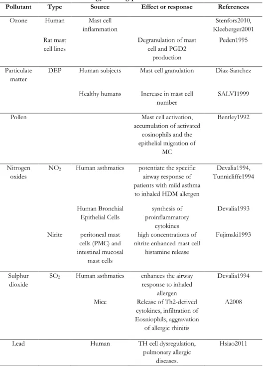
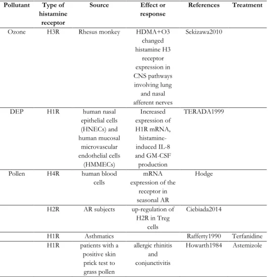
Table 3 Allergy causing pollutants and its effect ... 60
Table 4 Pollutant and Histamine receptors ... 61
Table 5 C-score of the models ... 68
Table 6 Main geometric parameters of the model prediction and validation ... 70
Table 7 ERRAT ... 76
Table 8 Prediction of transmembrane regions using various web servers... 77
Table 9 Top 4 compounds from JNJ777120 database ... 82
Table 10 Top 4 compounds from Thioperamide database ... 83
Table 11 Top 4 compounds from Vuf 6002 database ... 87
Table 12 Top six compounds of high docking score and their interactions with Asp94 ... 89
Abbreviations
ADMET Absorption Distribution Metabolism, Excretion and Toxcity
AIP4 Atrophin-1-interacting protein 4 APC Antigen presenting cell
AR Allergic rhinitis
Arah1 Arachis hypogaea
Betv1 Betula verrucosa
BLAST BLAST for Basic Local Alignment Search Tool BLOSSUM BLOcks SUbstitution Matrix
cAMP Cyclic adenosine monophosphate CD4+ Cluster of differentiation 4
CHARMM Chemistry at HARvard Molecular Mechanics
CO Carbon monoxide
CO2 Carbon dioxide
COPD Chronic obstructive pulmonary disease CXCL12 Chemokine (C-X-C motif) ligand 12 CXCR4 Chemokine (C-X-C motif) receptor 4
DC Dendritic cell
DNA Deoxy nucleic acid
EPA Environmental Protection Agency FcεRI Fc region of immunoglobulin E FEV1 Forced expiratory volume in 1 second
GM-CSF Granulocyte-macrophage colony-stimulating factor GPCR G-protein-coupled receptors
H1R Histamine 1 receptor
H2R Histamine 2 receptor
H3R Histamine 3 receptor
H4R Histamine 4 receptor
hH4R human Histamine 4 receptor
HMMTOP Hidden Markov Model for TOpology prediction
IgE immunoglobulin E
IL Interleukin
IP3 Inositol trisphosphate
I-TASSER Iterative Threading ASSEmbly Refinement JNK c-Jun N-terminal kinases
LC-1 Liver-cytosol type 1
MAPKs Mitogen-activated protein kinases
MC Mast cells
MHC Major Histocompatibility complex MoDC Monocyte derived dendritic cells mRNA messenger Ribonucleic acid
NCBI National Center for Biotechnology Information NMR Nuclear Magnetic resonance
NO Nitric oxide
O3 Ozone
PDB Protein data bank
Phlp1 Pollen allergens pKd dissociation constant PM10 Particulate matter
PSA Polar surface area
SBVS Structure based virtual screening LVS Ligand-based virtual screening
SO2 Sulphur dioxide
Th2cells T helper cells
TM HMM Trans membrane hidden Markov model TNF-α Tumour necrosis factor
TRAP Traffic associated air pollution VOC5 Volatile organic chemicals WHO World Health Organization
Chapter 1
Introduction
1.1 General Introduction
Environment can be defined as “the complex of physical, chemical, and biotic factors (such as climate, soil, and living things) that act upon an organism or an ecological community and ultimately determine its form and survival”. It therefore includes everything that may directly affect the behaviour of a living organism or species, including light, air, soil, water and other living organisms. For human well-being, we interact with the environment for our benefits. As the population increased, humans started to exploit the environment intensely. This ultimately had led to detrimental effects on the environment which directly affects humans. Pollution is one of the major adverse outcomes in response to the exploitation of the environment. Environmental pollution is popularly grouped into air, water, light, land, noise and thermal pollution. Air is important for sustaining life. Approximately we breathe over 3,000 gallons of air each day. Therefore, clean air is vital for healthy living. Air pollution can damage trees, crops, other plants, lakes, and animals. In addition to damaging the natural environment, air pollution also damages buildings, monuments, and statues. Hence air pollution is a major threat to human health and his ecosystem.
Air pollution has long been recorded from the prehistoric period where soot deposition in the caves were identified by Spengler (Spengler et al., 1983), but air pollution by itself has gained a major concern for health hazards only from the last century. World Health Organisation (WHO) defines air pollution as
“contamination of the indoor or outdoor environment by any chemical, physical or biological agent that modifies the natural characteristics of the atmosphere.
Household combustion devices, motor vehicles, industrial facilities and forest fires are common sources of air pollution. Pollutants of major public health concern include particulate matter, carbon monoxide, ozone, nitrogen dioxide and sulphur dioxide. Outdoor and indoor air pollution cause respiratory and other diseases, which can be fatal. WHO has estimated a total of 7 million deaths in 2012 as a consequence of air pollution (http://www.who.int/ mediacentre/news/releases/
2014/air-pollution/en/). This accounts for 12.5 % of the total global human deaths.
This data is twofold larger than the previous estimates and confirms that air pollution is now the world’s largest single environmental health risk. Some unconfirmed reports say that in the last 20 years the mortality rate due to air pollution is far high compared to the mortality rate from the infectious diseases.
The number of deaths caused by both indoor and outdoor pollution has been depicted in Figure 1. In 2010, WHO estimated that more than 6 million people die prematurely every year because of air pollution (Wong, 2013).
The Western Pacific region (WPr) and South East Asian regions (Sear) bear most of the environmental hazard burden with 2.8 and 2.3 million deaths,
respectively. Nearly 680,000 deaths occur in Africa, about 400,000 in the Eastern Mediterranean region, 287,000 in Europe and 131,000 in the Americas. Comparing to the low income countries (LMI), high income countries (HI) such as Europe (295,000), Americas (96,000), Western Pacific (68,000) and Eastern Mediterranean region (EMR) (14,000) have lesser number of deaths.
Figure 1 Number of deaths by indoor and outdoor pollution
Based on the chemical structure, air pollutants can be classified into primary and secondary pollutants as categorized in Table 1. Substances that are directly released from the source are primary pollutants while these primary pollutants react to form the precursor of the secondary pollutants (Stern et al., 1973)
Table 1 Source of air pollutants
Source Pollutants
Primary pollutants Carbon dioxide, Carbon monoxide, Sulphur dioxide, Nitric oxide Secondary pollutants Dioxins
Based on their physical state, air pollutants are further classified as: 1) gaseous pollutants 2) particulate organic pollutants 3) heavy metals 4) particulate matter (Kampa et al., 2008).
With the technological advances, we as a society have disturbed the environment which has ultimately affected the human health. Among the various consequences, allergy is the most common disorder influenced by air pollution.
Allergic diseases reduce the quality of life and negatively impact the socio-economic welfare of the society. These air pollutants act allergens to cause several allergic disorders in humans. The prevalence of the allergic diseases has increased in the
past three decades with the increased exposure to allergens (Holgate, 2004). Since this increase in prevalence has occurred in this short duration, genetic changes cannot only be the major reason. External environment factors can be attributed as a major contributor to the increase in allergic diseases (Bartra et al., 2007).
Any foreign substance when contacted by human body can induce allergy, thereby referred here as allergen and this can be a product of environmental pollution. Though our immune system defends from the alien substance, some of them react differently leading to allergic reactions. When the allergen enters the human body through inhalation, the body produces a certain type of antibody called Immunoglobulin E (IgE). This IgE is very specific in nature which means each pollutant induces specific IgE. A person can be allergic to one pollutant but not necessarily to the other. When a susceptible person encounters an allergen, large amounts of IgE will be produced. Subsequent exposure to the allergen cause allergic reaction which depends on the type and amount of the allergen encountered. A wealth of evidence suggest that atmospheric concentrations of pollutants such as ozone (O3), nitric oxides (NO), respirable particulate (PM10) and volatile organic chemicals (VOC5), which result from increased use of liquid petroleum gas or kerosene, may be linked to the increased prevalence of allergic diseases which develop more frequently in urban areas of developed countries (Brauer et al., 2007;
D'Amato et al., 2000). It is estimated that over 20 percent of world population suffers from IgE mediated allergic diseases i.e. allergic asthma, allergic rhinitis, allergic conjunctivitis, atopic eczema/atopic dermatitis and anaphylaxis. Though stringent pollution control measures may reduce the release of pollutants, in industrialised society it is inevitable to find a solution to existing establishment (automobiles, industries) which will continue emitting allergens to the environment.
Hence treatment and prevention of the allergic disease has been a major concern.
Though these allergic diseases already have established treatment pathways and medications, it gets more challenging with the addition of new type of pollutants to the existing spectrum. This has urged us to target the modus in developing a new drug lead against the allergens from the air pollution. Before going into the therapeutics a clear insight into the molecular mechanism of the development of the allergy is necessary.
Allergies occur when our immune system becomes hypersensitive to particular substances. Various cell molecules and mechanisms are involved in this process of mediating allergy in our body. The IgE produced during the allergic reaction is primarily produced by the plasma cells. This IgE binds to its receptor on mast cells which leads to the production of histamine. Histamine is considered the first allergic mediator implicated in the process of allergy because the levels of histamine is elevated in plasma and tissue when an allergen encounters the human body. The pleiotropic effects of histamine are mediated by different histamine membrane receptors. So far four different sub types of G protein-coupled histamine
receptor, designated as H1, H2, H3 and H4 have been identified and are found to be expressed on various immune cells (Hill et al., 1997; Hough, 2001).
The H4 receptor is more widely distributed, especially in organs associated with the immune system. It is preferentially expressed in intestinal tissue, spleen, thymus, medullary cells, bone marrow and peripheral hematopoietic cells, including eosinophils, basophils, mast cells, T lymphocytes, leukocytes and dendritic cells.
These cell types are primarily involved with the development and continuation of allergic responses. Recent evidences of the in vivo and in vitro studies using animal models and human biological samples elucidate the role of H4 receptor in histamine-induced chemotaxis of mast cells, eosinophils and other immune cells which are hallmark characters of allergic diseases (Hanuskova et al., 2013). Thus these biological functions and the expression pattern indicate a crucial role of H4R in allergy caused by the pollutants. These Histamine H4 receptors have become an attractive target for anti-allergic therapy.
Conventionally, the prevention and management of allergic disorders is fundamental to avoid allergen exposure. Apart from this, several pharmacotherapies like anti-histamines, cortisone, dexamethasone, hydrocortisone, theophylline, cromolyn sodium etc. are prescribed to block the action of allergic mediators. With the increased prospects of H4R, antagonism of histamine's action at H4R has been the key for an immense market for pharmacological treatment. The pathophysiological significance of H4R in inflammatory conditions, such as asthma and allergic disorders, as well as its contribution in acute and chronic inflammation was initially suggested by using the dual H3R/H4R antagonist thioperamide (Hofstra et al., 2003) and subsequently by the use of the selective H4R antagonist JNJ-7777120 (Thurmond et al., 2004). With the success of the H4R antagonist, more researches are focused on the antagonism of the receptor to identify a competitive lead for the development of drug, thereby providing a remedy to the allergic responses caused by the environmental pollution.
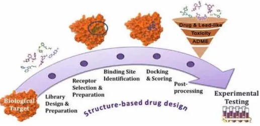
In our attempt to identify a potential antagonist we followed structure based virtual screening. This structure-based method confide on the 3D structure of the target receptor or protein which can be obtained either experimentally or by homology modelling to develop a new drug. The identification of new ligands for a given receptor is necessary. A ligand is a substance or a chemical that binds to the receptor and invokes biological response. Ligand can either be agonist, antagonist or inverse agonist. An agonist binds to a receptor and activates the receptor to produce a biological response. Whereas an agonist causes an action, an antagonist blocks the action of the agonist and an inverse agonist causes an action opposite to that of the agonist. In this study, we aimed to identify a potential antagonist that can block the function of H4R by searching from large databases of 3D structures of small molecules. The structures are tried to fit into the binding pocket of the
receptor using fast approximate docking programs. The promising ligand best fits onto the binding site. This method is known as virtual screening. The physical properties of these compounds can be tested using in silico assays. These in silico hits or lead structures can serve as a suitable starting point for the development of novel, potent and selective antagonist. Thus the identification of these compounds will open new avenues for the development of a new drug for allergy.
1.2 Objectives of the research
The main objectives of this research are to develop a potential lead candidate drug for allergy caused by the air pollutants. As mentioned above a structure based drug designing was followed to achieve this. Some of the main objectives of this research are
1. Though it is known that pollution is an important factor in contributing allergy, there is not much evidence based information on it. There are many pollutants which are hazardous to health but we aim to discuss and identify the environmental pollutants that trigger allergic response. Secondly, it is also intended to determine the involvement of H4R in eliciting this immune response. In order to counteract the effect of histamine released with the intake of allergen, an antagonist against the immune response elicited has to be developed. To serve this purpose, a 3D structure model of the receptor H4, in which the histamine binds, is necessary.
The main aim of this section is to generate a high quality structure of the H4R.
Since the experimental 3D structure is not available, the structure has to be modelled and the binding site has to be determined.
2. Next a database with all the known small structure has to be built which allows to choose the best drug lead from a wide range of structures. To achieve this 3 already commercially available ligands were used as a template to filter the structures from PubChem and construct 3 separate databases.
3. Our aim is virtual screening the best lead structures from the 3 sets of databases. The dock score and hydrogen bond formation are the parameters used to identify the best hit or lead. Once the lead structure has been identified to be potential to bind the target molecule, they are made to go through few in silico assays to determine its efficacy.
Chapter 2
Review of Literature
2.1 Definition of Environment
The word “Environment” is originated from French meaning “encircle”, therefore in simple terms it means one's own encircling surrounding. Environment can be defined in many ways, some of the commonly used definitions are
1. It is in totality of all social, physical, biological and chemical individually as well as collectively that composes man made surroundings.
2. It refers to sum total conditions which surround man at a given point of space and time.
3. It is the representativeness of the physical component of the earth wherein man is the important factor influencing his environment.
4. Environment is the holistic view of the world as its functions at any time with multitude of special elemental and socio-economic distinguished by quality and attributes of space and mode of behaviour of biotic and abiotic forms (Prof. R. K. Agarwal, 2010).
5. The non-genetic conditions and circumstances that affect person's conduct and health (Sharma et al., 2005).
Thus environment is a very broad concept and it involves everything that affects an organism in its lifetime and includes both abiotic (non-living) and biotic (living) substances. Some components of the environment serve as a resource such as soil, water etc., while others act as a regulatory factor such as temperature, light etc. Different components of the environment are interdependent and interlinked.
In general, the environment can be described as the physical surrounding and conditions affecting the lives of people and animals (Chauhan, 2008). Human health, well-being and survival ultimately depend on the health and integrity of the whole environment (Nadakavukaren, 2011). The environment accounts for almost 20% of all deaths in the WHO European Region.
From the beginning humans have been dependent on the world for air, water, food and solar energy which drives the whole complex system of living organisms. This relationship between living things to their environment is called Ecology. In the book ''A perspective of environmental pollution'' the author terms this study as the study of the first world of man. According to him, the second world of man is his own handicraft with the use of technology. With the start from forest clearance for expansion of pasture and extensive cultivation, human have extensively changed the productivity of the world. As a by-product of the increase in the technology, the waste materials disposed into the environment is substantial increased. This waste can be harmful to the ecological system and humans and are called pollution (Holdgate, 1979).
The word pollution is derived from a Latin word ''polluere'' which means ''to soil'' or ''to defile''. It has threatened the well-being of human and other living organisms. Just as weed is a plant out of place, pollutant is a chemical out of place.
Oil enclosed in a tanker is not a pollutant but if it is spilled it is a pollutant (Hill, 2010; Hill et al., 1997). Pollution could also be defined as when a substance occurs in a location or organism at higher levels than normal (Rieuwerts, 2015). Pollution could be an end result of any forms of human activity. It can vary from a simple form of human smoking to a breakdown of a huge nuclear power plant.
Environmental pollution is commonly classified into air, water, light, land, noise and nuclear pollution. All these pollution types ultimately affect the major life supporting system which includes air, water and soil. Some of the pollution is localized such as cigarette smoking or smoke from a wood fire, but air and water pollution can be transboundary (Rana, 2011). Willerroider (Willerroider, 2003) reported that polar bears which travel long distance for food tend to accumulate high levels of industrial pollutants such as polychlorinated bi phenyls in their body.
Therefore major concern has been the air and water pollution. In this study our focus will be on air pollution.
Though air pollution has become a hot topic in the recent times, its prevalence was documented in the 2nd century. In 79 AD Pluinus described the death of his uncle due to volcanic fumes. Beginning from that, various air pollutants have been reported, differing in their chemical composition, reaction properties, emission and persistence in the environment. Air pollution can cause concern in two ways 1) by affecting individual health 2) by disturbing the environmental set up.
2.2 Air pollution affecting Environment
In this section, we discuss on the global effects of the air pollution. Air pollution has been a major player in altering the environmental patterns. The natural greenhouse gases such as CO2 and methane help to prevent the heat entering into the earth atmosphere, thereby providing a good environment for the living beings.
However, the increased production of CO2 from the industries has started to slowly reverse the protection provided by these greenhouse gases. These gases have started to increase the temperature of the earth atmosphere thereby leading to global warming (Green, 2011). It has been reported that the global surface temperature has increased by 0.5 °C since 1975 (Hansen et al., 1999; Jones et al., 1999). The global warming of 0.1 - 0.2 °C every decade has resulted in increasing loss of snow cover and Arctic sea ice, frequent occurrence of heavy precipitation, rising sea level, and shifts in the natural ranges of plants and animals. It is estimated that the global average temperature is already approximately 0.8 °C above its preindustrial level, and present atmospheric levels of greenhouse gases will contribute to further warming of 0.5 - 1 °C (MC, 2008). Additionally, the increase in CO2 content also affects the pollination of plants. CO2 is an important component for the
photosynthesis; thus increased CO2 exposure in turn increases the pollen production and biomass (Ziska et al., 2008; Ziska et al., 2011). The increase in pollen indirectly affects the population with allergy and asthma. In Ambrosia, high CO2 increased pollen production by 32% and 55% (Rogers et al., 2006; Wayne et al., 2002).
Another adverse effect of air pollution is acidification of lakes and rivers which affects the soil and vegetation of the land. Acidification is mainly due to the emission of pollutants such as sulphur di oxide and nitrous oxide. These compounds mix with the water vapour and travel thousands of kilometres and result in acid rain (Kaitala et al., 1992; Green, 2011). The acid rain can damage trees, crops, wildlife, lakes and other bodies of water. Those pollutants can also harm fish and other aquatic life.
2.3 Air Pollution and health
Air pollution gained global concern when intense smog with 2000µg of SO2 surrounded London and killed 4000 people in 1952 (Bartra et al., 2007). It was reported by Ministry of Health that the people suffering from cardiorespiratory diseases were amenable to the smog. Thus it was concluded that this extravasation of the cardiorespiratory diseases is particularly due to the air pollution. Pollution caused by traffic is a major risk for respiratory diseases. The gaseous and particulate emissions caused by automobiles remains the major cause for the urban pollution. It has been reported that for every 100 km, a car emits a mean value of about 1 kg of pollutants into the atmosphere. Traffic associated air pollution (TRAP) leads to increased incidence of childhood asthma and allergy. Studies by Bowatte et al in birth cohort studies have highlighted that increased longitudinal childhood exposure to PM 2.5 and black carbon was associated with increased risk of subsequent asthma in childhood. This early childhood exposure to TRAP was associated with development of asthma across childhood up to 12 years of age (Bowatte et al., 2015).
Population-based, cross-sectional studies of metropolitan areas in the United States have also found associations between particulate air pollution and annual mortality rates. In India and China, the increase in PM10 of 10 µg/m3 is associated with an increase in mortality of 0.6% in daily all natural cause mortality (Committee. 2010). Air pollution was positively associated with death from lung cancer and cardiopulmonary diseases but not with death from other causes considered together (Dockery et al., 1993). Epidemiological studies have revealed that increased air pollution has been a reason for lower lung function and increased susceptibility to Chronic obstructive pulmonary disease (COPD) (Sunyer, 2001).
Thus it has been a common thought that air pollution increases the risk of respiratory and other diseases. On the contrary, Bouhys et al in 1978 have alleged
that higher air pollution has no considerable effects on chronic lung diseases (Bouhuys et al., 1978). Pollutants also affect the function of lungs, increase in total lung resistance by an average of 67% and decrease in FEV1 (Forced expiratory volume) by 23 % was evident with inhalation of 1.0 ppm of SO2 for 10 minutes (Koenig et al., 1982; Koenig et al., 1983). It was later found that exposure of SO2 for 2.5min can lead to bronchoconstriction in asthmatic patients. This action of SO2 can be reverted with the use of albuterol, cromolyn, theophylline, and corticosteroids (Koenig et al., 1991).
A study based on daily counts of hospital admissions for 1999-2002 obtained from billing claims of Medicare enrollees clearly showed that the risk of cardiovascular and respiratory hospital admission with the short term exposure of air pollutants (Dominici et al., 2006). Air pollution has been thought to be a major reason for aggravation of asthma leading to increased hospital admission and drugs.
In controlled experiments, asthmatic patients are found to be more sensitive to SO2. Nielsen and colleagues recently reported an 18% increase in lung cancer incidence for each 5 μg/m3 increase in PM2·5 concentration in a longitudinal cohort study (Raaschou-Nielsen et al., 2013).
Another health hazard from increased air pollution is allergy. The incidence of allergy and bronchial asthma is elevated in the last decade in the industrial countries. It has been reported that the prevalence of Allergic rhinitis is high in the developed countries affecting 40 % population worldwide (Bousquet et al., 1990;
Long et al., 2002), 23%-30% in Europe (Bachert et al., 2006; Bauchau et al., 2004) and 12%- 30% in US (Nathan et al., 2008). In Hungary 52.5% of patients suffered from seasonal AR and 35.1% from perennial AR (Szilasi et al., 2012). In a cross sectional study on Taiwanese children by Hwang et al, it was concluded that exposure to pollutants such as SO2, NO2 and Nox increase the risk of allergic rhinitis (Hwang et al., 2006). In a longitudinal study performed by Martimoer et al, it was predicted that in children who have asthma or had prenatal and early exposure to air pollutants like CO2, SO2, CO, O3 had high sensitization to allergens (Mortimer et al., 2008).
Airborne allergens such as ragweed pollen, mold spores, cat dander and dust mites affect the respiratory system, producing classic hay fever-type symptoms of sneezing, runny nose and congestion. Patients with untreated allergies can then often experience sinusitis. If untreated, bacterial or fungal sinus and ear infection may arise in people with immune system weakened by frequent allergic reaction.
Untreated allergies make asthma patients sick more often and subject to severe, life- threatening asthma attacks. Hence proper diagnosis and treatment of allergies is important.
2.4 Interaction of the pollutant with the immune system
Our immune system encounters the environment in an outstanding manner; however the environmental pollutants interfere with the immune response thereby leading to immunological disorders. Bahadar et al in his review have briefed that environmental toxicants can affect the immune system ranging from organ to cellular components. They have also detailed the changes in the immune response to the pollutants based on the composition, structure and function. These changes are illustrated in Figure 2 (Bahadar et al., 2015).
Figure 2 Changes in the immune system in response to pollutants (Bahadar et al., 2015)
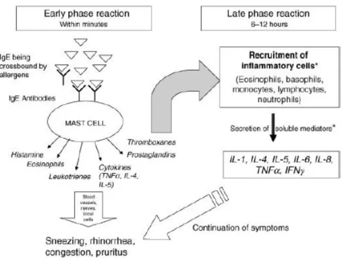
Immune responses may occur in the upper respiratory tract (rhinitis), the lower respiratory tract (wheeze, bronchospasm) or systemically, for example, a febrile response. The underlying mechanism is still ambiguous. A variety of pollutants have been associated with elicitation of these reactions. As a result of the widespread occurrence of allergy caused by environmental pollution, mechanism in the development of allergy has received close attention. With regard to the above Figure 2, increase in the production of the Immunoglobulin which changes the release of the mediators is the clinical feature of Allergy. In immunological aspect, any particles that elicit the production of IgE antibody are termed allergen which in this study relates to the pollutant. The clinical manifestation of IgE dependent immunological reaction is Allergy whereas Atopy is the genetic tendency to generate IgE response (Holgate et al., 2011). The allergic reaction in the nose involves a complex interaction between allergen and multiple effector cells. Allergic reactions consists of two phases 1) Acute phase that typically subsides within 60 minutes 2) Late phase which is developed in 3 to 12 hours by the development of an intense
inflammatory reaction (Lemanske, RobertF. et al., 1988). Acute phase is mast cell mediated whereas the late phase is believed to be dependent on the local accumulation and activation of leukocytes, including neutrophils, eosinophils, and basophils (Salvi, Sundeep and Blomberg, Anders and Rudell, Bertil And Kelly, Frank And Sandstram, Thomas And Holgate, Stephen‚t. And Frew, 1999; Gleich, 1982; Dolovich et al., 1973). The delayed or late response can be attributed to the chronic allergic diseases (Gleich, 1982).
The steps involved in the process of both acute and late phase allergy caused by pollutants are discussed in the following sections. The overall process of allergy is depicted in Figure 4.
2.4.1 Acute phase allergy
Acute phase is the immediate reaction, taking effect within minutes of allergen (pollutant) provocation, results in the release of mediators that lead to symptoms characteristic of the target organ (Adelman et al., 2012). Following sequence of events happens in the process.
2.4.1.1 Presentation of antigen/pollutant to the immune system
Pollutants can make entry into the respiratory tract as volatile gas (ozone, benzene), liquid droplets (sulphuric acid, nitrogen dioxide), or particulate matter (diesel exhaust, aromatic hydrocarbons). Initial entry of the pollutant into the respiratory system is mediated by coupling of the pollutant with protein or conjugates (Albright et al., 1996). Other allergens such as pollen, dust mites and animal dander are protein in nature with molecular weight of 10 to 20 kDa (Adelman et al., 2012). For example, the pollen particles contain pollenic allergens, high environmental humidity conditions can result in osmotic shock of this pollen particle. This leads to the release of microparticles or paucimicronic particles that contain allergenic proteins. Allergens are mostly lipid binding proteins (e.g. Bet v 1 and homologues, house dust mite group 2 allergens, lipocalins of pets, plant lipid transfer proteins), and some are glycoproteins (e.g. peanut Ara h 1 and grass pollen Phl p 1). After the entry of the allergen, it leads to the reduced epithelium and facilitate the contact of the inhalatory allergens with the network of antigen presenting cells (APC). Cells that act as APCs in the airway include mucosal macrophages, dendritic cells, pulmonary alveolar macrophages, and B cells themselves. These lipid ligands and conjugated glycans have been shown to interact with pathogen recognition receptors such as Toll-like receptors (TLR) and C-type lectins (CTL) on antigen-presenting cells. All of them take up the antigen by the process of endocytosis. Then the antigen is degraded or processed and the linear peptides are presented to the T cells (Figure 3).
Figure 3 Processing of the antigen (http://www.uic.edu/)
2.4.1.2 Activation of T cells
The processed antigen is co-presented along with Major histocompatibility complex (MHC) class 2 molecules to CD4+T cells (Th2 lymphocytes or Th2 cells), which are the major regulators (Young, 1998). The APC exert their effects through production of interleukins such as IL-4, IL-5, and IL-13. IL-4 that is critical to the development of Th2 cells (Woodfolk, 2007). Next step is the T cell activation for the induction of Th2 cell response which is an important step in the process of allergy (Cezmi A, 2007). There are two known mechanisms for the activation of the Th2 response.
1. Dendritic cells (DC) are APC which present the antigen but cannot themselves activate the T cells. It requires additional factors like ligands for TLR to migrate and then activate T cells by increased production of CD40, CD80 and CD86.
2. In addition, there is accumulating evidence that mast cells also modulate DC into Th2 immune response. Caron et al have suggested a mechanism for the maintenance of Th2 based responses in allergic disorders. From their study they concluded that histamine released by mast cells of allergic subjects upon contact with the sensitizing allergen, polarizes maturing DC into DC2 through both H1 and H2 receptors. By this polarization of DC2, histamine favours the induction of Th2-biased responses and sensitization to diverse encountered allergens, as observed in atopics (Caron et al., 2001).
Once the T cells are activated by the above mechanism, clonal expansions of the cells occur and begin cognate interaction with the B cells.
2.4.1.2 IgE production
IgE antibodies play a key role in instigating immediate hypersensitivity reactions and contribute to the pathophysiology of a wide range of allergic diseases.
As mentioned before, activation of T cells lead to the production of interleukins which in-turn stimulate B cells to produce Ig E. IgE production is carried out in B cells. B cells are developed in the bone marrow and leave as mature B cells, expressing IgM and IgD on their surfaces. These IgM or IgD B cell receptors recognize the antigen specifically and thereby activate the B cell. However, if the mature B cell binds to the processed antigen presented on a T helper (Th) cell, the B cell gets activated which is followed by Ig class switch recombination (Luger et al., 2010). The B cells entail class-switch recombination at the immunoglobulin heavy chain locus into the IgE heavy chain (Cε). Furthermore, CD4+ Th2 cells that produces IL-4 also orchestrate this class switching of IgE mediated in B cells.
2.4.1.3 Sensitization and degranulation of mast cells
IgE sensitizes mast cells by binding to their high-affinity Fc receptor (FcεRI) on tissue mast cells or blood basophils. An allergic reaction is initiated when an antigen crosslinks immunoglobulin E (IgE) antibody bound to the Fc receptor (Sutton et al., 1993). Subsequently, mast cells degranulate, releasing vaso active amines (mainly histamine), lipid mediators (prostaglandins and cysteinyl leukotrienes), chemokines and other cytokines (Larche et al., 2006). TNF- α, GM- CSF, macrophage inflammatory protein-1α, and a number of “T helper (Th) 2-cell type” cytokines, such as interleukins IL-3, IL-4, IL-5, IL-6, IL-9, IL-10, and IL-1 are also released upon mast cell activation (Gould et al., 2003). The activities of allergen- activated mast cells are said to “orchestrate the allergic response”.
Figure 4 Allergy mechanisms (Derendorf et al., 2008) 2.4.2 Late phase of allergic reaction:
Late phase reactions are sequel to the early phase reactions because in some individuals this initial response evolves slowly into extensive inflammation (Lemanske, Robert. et al., 1988). It lasts longer than the initial phase reaction. It usually peaks between 4th and 8th hour after allergen challenge and subsides after 12h to 24 h. IgE binds to the FcεRI at the surface of dendritic cells (DCs) and monocytes, as well as to the low-affinity receptor for IgE, FcεRII (also known as CD23) present at the surface of B cells. This process increases the uptake of allergen by these antigen presenting cells (APCs) and the subsequent presentation of allergen-derived peptides to specific CD4+ T cells, which drive the late phase of the allergic reaction. A late-phase response associated with the influx of T cells, monocytes, and eosinophils may ensue some hours later (Gould et al., 2003). Mast cell derived mediators and cytokines are responsible for cellular recruitment and development of the late-phase response. The late phase reaction was first noticed by Blakley et al around 100 years ago where he found the association of allergen inhalation and asthma after several hours later.
2.5 Role of Histamine receptors in allergic diseases
Histamine was initially thought to be obtained from non-human source.
Only in the late 1920s it was found to be a natural constituent of human. Riley and West initially determined those mast cells are the storage cabin of histamine.
However in their later studies they proved that basophils, platelets and other cells can also accommodate histamine (Riley, 1953). In mammals the histamine amounts from 1 to 100 g-1 in tissues. Early researchers found that it had a stimulant effect on smooth muscle of the gut and respiratory tract, caused vasodepression, stimulated cardiac contractility and induced a shock-like syndrome when introduced into animals. These and other effects are mediated by histamine specific receptors expressed on the respective target cells. Until now four histamine receptors have been characterized and cloned in humans and mice, referred to as histamine H1R, H2R, H3R, and H4R, (Hill, 1990; Liu et al., 2001; Nguyen et al., 2001; Oda et al., 2000). These receptor subtypes display homologies of about 20% within a species and of about 70–95% of a given receptor subtype between humans and mice (Liu et al., 2001; Thurmond et al., 2008; Hill, 1990). All of the histamine receptors belong to the family of G protein coupled receptors (GPCR). GPCR are heptahelical transmembrane molecules that transduce the extracellular signal by using G- proteins and intracellular second messenger systems. The molecular mechanism of GPCR is depicted in the Figure 5. Binding of an agonist (activating ligand) induces a conformational change in the G protein-coupled receptor (GPCR) to activate it.
Activated receptors couple to heterotrimeric G proteins composed of Gα, Gβ and Gγ subunits. Subsequently, the heterotrimeric G proteins dissociate and G protein signaling mediates the generation of second messengers such as cyclic AMP, inositol triphosphate (IP3) and Ca2+. Activated receptors are phosphorylated, primarily in the carboxyl terminus, by GPCR kinases. Phosphorylated receptors recruit β- arrestins, which are multifunctional adaptor proteins that block further G protein–
GPCR coupling, potentially through a steric hindrance mechanism (referred to as desensitization). β-arrestins also mediate clathrin-dependent endocytosis of activated GPCRs as well as independent signalling pathways downstream of GPCRs. β-arrestins scaffold mitogen-activated protein kinases (MAPKs; such as extracellular signal-regulated kinase (ERK) and c-Jun N-terminal kinase (JNK)), tyrosine kinases and E3 ubiquitin ligases (such as atrophin-1-interacting protein 4 (AIP4)). The arrows next to cAMP indicate that cAMP levels can go up or down in response to GPCR activation.
Figure 5 Signalling mechanism of GPCR (Ghosh et al., 2015)
The four Histamine receptor subtypes are distinct in terms of their pharmacology and molecular biology and have been implicated in diverse biological effects. The affinity of histamine binding to different Histamine receptors varies significantly, with Ki values ranging from 5-10 nM for the H3 and H4 receptors to 2-10 mM for the H1 and H2 receptors (Thurmond et al., 2004; Endo, 1982). This difference in affinities of receptors determines the biological effects of histamine upon activation.
H1 receptors are expressed on multiple cell types including endothelial cells and smooth muscle cells, where they mediate vasodilation and bronchoconstriction.
Because of its varied role, antihistamine specific to this receptor were developed in 1930s and widely used. Many effects of histamine were not blocked by these antihistamine and this made Ash and Shild propose that a second type of histamine receptor might exist in heart and stomach tissues (Ash et al., 1966). This was later experimentally identified by Black et al and was named H2 receptor (Black et al., 1972). In 1980s Schwartz et al identified a third class of histamine receptors (H3 receptor) which mainly influences the histamine synthesis and release in CNS neurons.
The discovery of this fourth histamine receptor, and the evidence that it is expressed on many cell types involved in allergic responses, suggested that the H4R play an important role in mediating the histamine effects in asthma and allergic diseases. Hence in this study H4R is focused, thus a detailed description of its role and mechanism in contributing the allergic process is given.
2.5.1 Histamine H4 receptor (H4R)
The H4 receptor which is the latest discovered was not identified using the traditional pharmacological means. It was cloned by several groups independently in 2000 and 2001 (Liu et al., 2001; Oda et al., 2000; Nguyen et al., 2001). The H4R
protein shows high level of homology towards H3R. Both H3R and H4R have splice variants and also exhibit species to species specificity. For example, histamine display 25 times higher activity in human H4R than in rat H4R (Timmerman et al., 2009). H4R displayed 40% structural homology and 58% transmembrane homology with the H3R.
2.5.1.1 Expression and activation of H4R
Histamine H4 receptor is a pertussis-toxin-sensitive GPCR predominantly expressed on cells of the immune system, including MCs, monocytes, eosinophils, dendritic cells (DCs), T cells and natural killer cells; in peripheral tissues such as spleen, thymus, colon, blood leukocytes and bone marrow, its expression being induced or altered in response to inflammatory stimuli.
APC
APC which includes monocytes and DC are primary immune cells which aid in the uptake and presentation of the antigen into the system and initiate allergic inflammation. The expression of H4R protein was confirmed in human monocyte derived dendritic cell (MoDC). Furthermore they also showed that H4R mediated histamine induced chemotaxis and calcium mobilization in MoDC (Damaj et al., 2007; Gutzmer et al., 2005), which directly suggest the chemotactic and immunomodulatory effect of Histamine via H4R. IL-12p70 is an interleukin produced by APCs which is important for the elucidation of a Th1-type immune response, whereas the absence of IL-12p70 indicates Th2-type immune responses.
This indicates the role of H4R in allergic diseases which are characterized by Th2 mediated responses. Gutzmer et al have elucidated that the suppression of IL-12p70 is mediated by H4R. The downstream signal transduction of IL-12p70 follows MAPK and cAMP pathway where cAMP signalling is thought to be induced by H2R. Experiments show that, preincubation of MoDC with U0126, an inhibitor of MEK 1/2 that blocks the phosphorylation of ERK1/2, rescued IL-12p70 suppression via the H4R-mediated signalling but not via the H2R-mediated response (Gutzmer et al., 2005). Taken together we can conclude that H4R plays a vital role in promoting Th2 immune responses, chemotaxis and intermodulation effect thereby mediating allergy.
T cells
In CD4 (+) T cells, H4R expression is present both in mRNA and protein levels. This upregulation is favoured in Th2 environment than in Th1 or naive T cells. Treatment of the cells with H4R specific agonist exhibited the mRNA induction of AP1 and IL-33 (Gutzmer et al., 2009).
Mast cells and basophils
Mast cells are important effector cells in allergic diseases. Mast cells bind IgE with IgE receptor, and subsequent contact with antigens triggers IgE receptor cross-linking and the release of preformed mediators, such as serotonin and histamine, and de novo produced mediators, such as prostaglandins and leukotrienes. The release of mediators dictates the signs and symptoms of allergic diseases (Galli et al., 2012). In mouse cells, H4R was found to mediate mast cell migration in response to histamine. This effect can be attributed to the accumulation of mast cell in allergic tissue (Hofstra et al., 2003). In addition, histamine H4 receptor has been reported to mediate mast cell migration toward CXCL12, a constitutive chemokine (ligand of CXCR4 and CXCR7) that is expressed in the skin and airway epithelium and plays a significant role in allergic airway diseases (Godot et al., 2007). Human mast skin cells and tissue mast cells were found to express H4R in 2004 (Lippert et al., 2003). However only recently Jemima et al in their study have characterized the functional expression of h4R in human mast cells that leads to the stimulation of Th2 cytokines(IL-5, IL-4 and IL- 13) (Jemima et al., 2014).
Basophils, unlike mast cells which reside in the tissue, circulate in the blood and migrate to sites of inflammation. Human basophil expressed H4R (Hofstra et al., 2003). In humans, basophils are the prominent sources of the biologically active Th2-type cytokines IL-4 and IL-13, which cause IgE class switching in B cells (Yanagihara et al., 1997). Recently the interplay between mast cell, basophil and H4R have been elucidated which stresses the importance of the receptor in allergic diseases (Shiraishi et al., 2012).
Eosinophils
Eosinophils are bone marrow-derived granulocytic leukocytes, which reside in tissues, especially in the respiratory and intestinal systems and in the uterus.
Eosinophil numbers in the blood stream are relatively low, and the control of eosinophil migration towards the tissues has been attributed to adhesion molecules and chemokines (Tachimoto et al., 2002; Lukacs, 2001). Eosinophils are important effector cells in the late phase allergic response, and they have been implicated in the pathogenesis of allergic disease (Bousquet et al., 1990). Ling et al have demonstrated a new mechanism of eosinophil recruitment driven by mast cells via the release of histamine. They conclude that histamine released from mast cells mediates eosinophil chemotaxis, cell shape change and upregulation of adhesion molecules via H4R (Ling et al., 2004; O'Reilly et al., 2002).
Neutrophils
It was also demonstrated that H4R antagonists cause a significant inhibition of polymorphonuclear cell influx into the peritoneum or pleural cavity in zymosan-induced neutrophilic inflammation models (Thurmond et al., 2004;
Takeshita et al., 2003). H4R blocked adhesion dependent degranulation of neutrophils in response to mast cells (Dib et al., 2014).
Taken all these together, H4R are functionally present in diverse immune cells and mediate biological activities leading to allergy and inflammation. Therefore H4R makes a promising target of drug design for allergic diseases.
2.6 Therapeutic potential of Histamine receptors for allergy
2.6.1 Antihistamines:The effect of histamine had urged the researchers to find a way to thwart the response. This was first started in Pasteur institute by Boven where he had access to Fourneau’s bank of compounds (Bovet et al., 1937). The first compound reported as an antihistamine by Ungar, Parrot and Bovet was the adrenolytic benzodioxan, piperoxan (933F) in 1937, which blocked the effect of histamine on the guinea-pig ileum. This was followed by the search of more antihistamines. After 1945, these antihistamines became widely used in the treatment of various allergic disorders such as hay fever, allergic rhinitis and urticaria (Parsons et al., 2006).
The prevalence rates of allergic diseases such as allergic rhinitis and asthma can be attributed to the increased level of pollutants in the environment and is increasing in many countries. As mentioned earlier, although several mediators are involved in the pathophysiology of allergic diseases, histamine plays a fundamental role, particularly in allergic rhinitis and urticaria. Antihistamines combine with and stabilize the inactive conformation of H1-receptors and thus interfere with the actions of histamine. The antihistamines down regulate the antigen presentation, expression of pro-inflammatory cytokines and cell adhesion molecules and chemotaxis (Simon et al., 2008). Antihistamines provide symptomatic relief of allergic symptoms caused by histamine release. Antihistamines have remained at the forefront of treatment for allergic diseases for many years and are among the most commonly prescribed medicines (van Schoor, 2008). Antihistamines are chemically and functionally classified into 6 types namely; Alkylamines, Piperazines, Piperidines, Ethanolamines, Ethylenediamines, Phenothiazines (Table 2) (van Schoor, 2008; Simon et al., 2008). The antihistamines can be broadly classified into 1) first generation antihistamine 2) second generation and 3) third generation.