Analysis of safety and efficacy of minimal invasive pacemaker electrode repositioning
PhD Thesis
István Osztheimer Semmelweis University Doctoral School of Basic Medicine
Supervisor:
Dr. László Gellér, med. habil, PhD, associate professor Official reviewers:
Dr. László Sághy PhD, senior lecturer Dr. Viktor Horváth PhD, assistant lecturer Head of the Final Examination Committee:
Dr. Lívia Jánoskúti, med. habil, PhD, associate professor Members of the Final Examination Committee:
Dr. Zoltán Járai, med. habil, PhD, professor Dr. Krisztina Madách, PhD, associate professor
Budapest 2017
1
I. Introduction
Since the appearance of implantable pacemaker systems between 1958 and 1963 a lot of development and expansion of indication range was observed, including CRT in the years after 2000. With increasing numbers of CIED (cardiac implantable electronic device) numbers of complications is not decreasing, moreover a proportional increase can be observed.
Infection involving the leads and device is of high clinical importance, causing endocarditis and necessitating explantation with all of its complications. Complications (especially infection) are more common after repeated procedures. Repeated procedures are necessitated primary by electrode dysfunctions, electrode dislocations.
Right atrial and right ventricular electrode stabilization is achieved either by active or passive mechanism. Electrode dislocation is less using active fixation. In this case the lead tip is stabilized by a screw in helix in the myocardium. Passive fixation electrode has tines to hold the lead tip in position between myocardial trabeculae. Latest shows less risk for myocardial perforation, but shows higher dislocation rate.
Left ventricular electrodes are advanced into the coronary sinus side branches. Active fixation is rarely used with this electrodes. Lead position is supported by the shape and loop of the lead. Our institution widely utilizes coronary stentimplantation for left ventricular lead stabilization.
A high number of complex CIED implantations and management of complications is performed at the Semmelweis University.
2
II. Objective
Electrode repositioning without opening of the pacemaker pocket would be of enormous clinical importance, since complications after reoperation are characteristically associated with opening of the pocket, haematoma, infection and pain is the result.
We sought to investigate electrode repositioning procedures without opening of the pacemaker pocket, efficacy and safety was evaluated. We also studied methods not recommended for left ventricular lead stabilization.
Particular objectives:
1. To find and develop right atrial, right ventricular and left ventricular pacemaker lead repositioning procedures which can be performed without opening of the pacemaker pocket (further on: minimal invasive procedures).
2. To analyze safety and efficacy of minimal invasive electrode repositioning procedures performed with individual lead types.
3. To compare minimal invasive lead repositioning procedures performed with individual lead types with procedures with opening of the pacemaker pocket in the same time period.
4. To compare our patient group with lead repositioning procedures performed with opening of the pacemaker pocket with data from the literature showing increased risk for complication of reoperation. With the help of this data we would like to reproduce the data from the literature and validate our own data.
5. To identify not recommended and dangerous procedures for left ventricular pacemaker lead stabilization.
3
III. Methods
III/a Right ventricular and atrial pacemaker lead minimal invasive lead repositioning (MILR)
Without opening of the pacemaker pocket just the distal end of the electrode can be moved with intravascular tools. Not all electrode types are suitable obviously for minimal invasive repositioning. The helix of active fixation electrodes can just be moved from the proximal part of the electrode which is connected to the pacemaker device. Contrary to this, in case of passive fixation electrodes there is no need for pocket opening and theoretically, there is no need for manipulation of the proximal part of the electrode.
Based on the above, we performed a large number of right atrial and right ventricular MILR procedures on passive fixation electrodes (36 atrial, 9 ventricular) in our institution between September 2006 and December 2012. (1. figure). Femoral vein access was used for the procedure. After introducer insertion into the vein a deflectable ablation catheter was introduced into the right side of the heart. We repositioned the dislodged electrodes with the help of the ablation catheter. We used lumen less, non- irrigated catheters in every case. The pacemaker electrode and the ablation catheter can slide or roll on each other, so the we considered the used tool atraumatic and not dangerous for lead integrity. We never used a snare or other catching tool for our MILR procedures.
Complications after femoral vein puncture are rare and we use ablation catheters routinely in our institution. In case of failure of MILR procedure a reposition with opening of the pacemaker pocket can be performed in every case.
4
1. figure: Right ventricular and atrial MILR
A1: Passive right ventricular electrode dislocated into the right ventricular outflow tract
A2: Lead tip is pulled back with an ablation catheter
A3: The electrode is advanced to the intraventricular septum by the aid of the suction force generated in the right ventricle A4: The passive fixation electrode is stabilized in the myocardium in the low intraventricular septum
B1: Passive right atrial lead is dislocated into the tricuspid valve B2: The lead tip is liberated with the aid of the ablation catheter.
With the torque form the ablation catheter, the atrial led tip is moved anteriorly.
B3: The ablation electrode is straightened, so the atrial electrode springs back to its original J shape.
B4: Final atrial electrode position in an anterior, most probably near appendage position.
III/b. Left ventricular or coronary sinus electrode minimal invasive lead repositioning (MILR)
5
Our institution published the minimal invasive repositioning process of coronary sinus leads to avoid phrenic nerve stimulation earlier. The first procedure was performed in December 2005. We used this published MILR technique and its simplified version (without stent implantation) routinely beginning from the first patient in every case it was performable. 42 left ventricular MILR cases were performed between December 2005 and December 2012 (2. figure).
Left ventricular MILR can only be performed in case of distal electrode position in the coronary sinus side branch, when the electrode causes PNS or has a high threshold, or if there is non- capture in this position.
2. figure: left ventricular MILR procedure ABL: ablation catheter, AMPL: amplatz guide, STENT: stent
A: Initial left ventricular lead position. Left ventricular lead is hooked around by an ablation catheter in the chamber of the right atrium.
B: By pulling back the ablation catheter, the left ventricular lead tip is pulled proximally in the coronary sinus side branch.
C: The electrode position is stabilized with a stent implantation.
6
III/c Right atrial, right ventricular and left ventricular lead repositioning with opening of the pocket (LROP)
LROP can be performed in every clinical case: electrode fracture, electrode dislocation, electrode perforation, and with active and passive electrodes. This is the standard and universally applied method by every implanting center. Advantage is that the electrode can be exchanged in case of electrical issues or in order to optimize implantation and lead patency.
III/d Retrospective data collection on MILR and LROP procedures performed in the same time window, ethical aspects
Right atrial and ventricular MILR is performed since September 2006. Left ventricular MILR is performed since December 2005. Both procedures were often performed in the first years afterwards due to the high rate of passive fixation right atrial and right ventricular lead as well as unipolar left ventricular leads used. Rate of MILR procedures decreased in the years later.
Studies showing elevated risk in case of repeated pacemaker pocket opening were published mostly after the year 2011
We decided to conduct retrospective data collection from the time point of the first MILR procedures until December 2012. In order to compare our results all LROP procedures in the same time period were collected as well for analysis.
Ethics approval was waived by the institutional review board due to the retrospective data analysis (No.94/2016) All patients gave written informed consent before the procedures.
7
III/e Data collection about left ventricular lead stabilization procedures causing severe complication
One severe complication was seen and treated by our hospital, the implantation was primarily not performed at our institution. Detailed describing and publication of this complication was conducted.
IV. Results
IV/a Efficacy and complication rate of right ventricular and atrial pacemaker lead MILR procedures and comparison with right ventricular and atrial LROP procedures performed in the same time window.
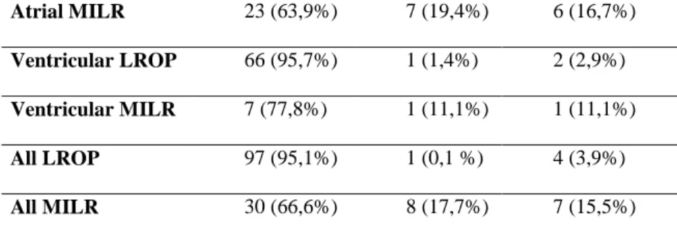
37 MILR procedures were successful from the 45. In eight patients appropriate mechanical and electrical properties could not be achieved. In seven patients repeated dislocation was noted during follow up. Only one LROP procedure was acutely unsuccessful and one repeated dislocation happened during follow up. (1 table).
1. table Success after right atrial and ventricular lead repositioning (mean follow up time 29 Months)
Long term success
Acutely unsuccessful
Re-dislocation during follow up Atrial LROP 31 (93,9%) 0 (0,0%) 2 (6,1%)
8
Atrial MILR 23 (63,9%) 7 (19,4%) 6 (16,7%) Ventricular LROP 66 (95,7%) 1 (1,4%) 2 (2,9%) Ventricular MILR 7 (77,8%) 1 (11,1%) 1 (11,1%)
All LROP 97 (95,1%) 1 (0,1 %) 4 (3,9%)
All MILR 30 (66,6%) 8 (17,7%) 7 (15,5%)
MILR procedures show a completely different complication profile compared to LROP procedures. We observed one minor complication during MILR: left ventricular lead micro- dislodgement during atrial lead repositioning. It is important to note, that no infection was observed after MILR procedures (2.
table). Large number of bleeding and infection complications were noted after LROP procedures.
2. table Complications during and after right ventricular and right atrial lead repositioning
Procedure Complications Incidence
Atrial MILR Minor complication:
o LV electrode micron dislodgement 1/8 Ventricular MILR
Atrial LROP Minor complication:
o elevated troponin
o RV electrode dislodgement o wound erythema and fever
1/33 1/30 1/33 Ventricular LROP Minor complication:
9
o atrial fibrillation induction o haematoma
o fever
o deterioration in renal function o presyncope and hypotension o atrial electrode dislocation Major complication:
o pericardial effusion, PM pocket decubitus, full system explantation due to infection(active electrode)
o pneumothorax
o subclavian artery puncture and haematoma
o bleeding from the wound o anaemia and haematoma on the thoracic wall
1/69 1/69 1/69 1/69 1/69 1/36
1/69
1/69 1/69
1/69 1/69 1/69
IV/b Success and complication rates of left ventricular MILR procedures and their comparison with LROP procedures performed in the same time window.
We performed successful left ventricular MIRL in 29 patients from the 42 clinical cases (69%) (3. figure). Left ventricular LROP procedures were successful in 39 clinical cases form the 48 (81%) (4. figure).
10
3. figure: Success of left ventricular MILR
4. figure: Success of left ventricular LROP
Complications show a different pattern also between left ventricular LROP and MILR procedures (3. table).
29; 69%
7; 17%
6; 14% Successful all during
follow up
Repeated dislocation during follow up Acutely unsuccessful
39; 81%
6; 13%3; 6% Successful all during follow up
Repeated dislocation during follow up Acutely unsuccessful
11
3. table: complications after left ventricular MILR and LROP
Procedure Complication Incidence
Left ventricular MILR
Minor complication:
o LV electrode integrity problem (tear)
1/42
Left ventricular LROP
Minor complication:
o atrial fibrillation 1/48
o fever 1/48
o haematoma 1/48
o RV electrode dislocation while LV repositioning
1/48
Major complication:
o pericardial effusion 1/48 o pocket infection 15 Months
after reposition
1/48
IV/c Methods not recommended for left ventricular lead stabilization
We observed one major complication caused by a left ventricular lead stabilization mechanism. Due to the intraoperative dislodgement of the unipolar left ventricular lead, the stylet was retained in order to stabilize the lead and it was connected with
12
the retained stylet to a CRT-P device. Two years later pectoral muscle stimulation was observed due to fracture of the lead close to the pacemaker device. Again two years later the patient was examined due to chest pain. Electrode fracture at the right atrial level and penetration of the fractured stylet to the right inferior pulmonary lobe was diagnosed. Percutaneous electrode extraction was performed (5. figure)
5. figure: Fracture of the left ventricular lead stabilized with retained stylet and penetration of the fractured stylet through the lateral wall of the atrium into the right inferior pulmonary lobe..
A: before beginning of the procedure: left ventricular lead fracture, and the stylet (arrow) penetrates into the right inferior pulmonary lobe B: Removal of the proximal stylet part with a snare C: Removal of the distal stylet which penetrated into the lung
13
V. Conclusion
1. It was possible to find an appropriate method for right atrial and right ventricular minimal invasive lead repositioning after consideration of the theoretical restrictions. Only in case of passive fixation can this method be applied. We also found an alternative reposition method used to treat left ventricular lead problems, which was used and published at our institution. We also used a modified version of this without stent implantation.
2. 67% of our patients has good electrode function on long term after right atrial and right ventricular MILR. 69% of our patients has a good functioning left ventricular lead after left ventricular MILR on long term.
3. While MILR procedures showed no complications connected to the pacemaker pocket, after LROP procedures a large number of infection, bleeding and other complications were observed.
4. During discussion we showed, that our LROP patient group showed a similar infection and overall complication rate expected from previously published international data.
5. Left ventricular lead stabilization with the retained stylet technique is not recommended and can be dangerous. Late electrode and stylet fracture, as well as the penetration of the fractured stylet into neighboring organs can result.
14
New scientific statements reviewed by the dissertation:
Pacemaker electrode repositioning can be performed without opening of the pacemaker pocket on right ventricular, right atrial, and left ventricular leads in case of appropriate patient population.
We performed our minimal invasive lead repositioning procedures routinely in clinical practice, so we cold investigate safety and efficacy for an alternative lead repositioning method on a large patient population for the first time in scientific literature. Minimal invasive electrode reposition is safe and effective in a large number of cases.
The use of retained stylet to stabilize the left ventricular lead is dangerous, due to fracture and penetration of the fractured parts into neighboring organs.
15 Publication record:
Related to the dissertation
1. Osztheimer I, Szilagyi S, Pongor Z, Zima E, Molnar L, Tahin T, Merkely B, Geller L. (2017) Minimal Invasive Left Ventricular Lead Repositioning is Safe and Effective in Distal Left Ventricular Lead Positions. Pacing Clin Electrophysiol.
DOI:10.1111/pace.13068 IF:1,44
2. Osztheimer I, Szilagyi S, Pongor Z, Zima E, Molnar L, Tahin T, Ozcan EE, Szeplaki G, Merkely B, Geller L. (2017) Minimal invasive right ventricular and atrial pacemaker lead repositioning as a first alternative is superior in avoiding pocket complications with passive fixation leads. J Interv Card Electrophysiol. DOI:10.1007/s10840-017-0242-x IF: 1,676
3. Osztheimer I, Duray G, Huttl K, Merkely B. (2016) Fracture and Lung Penetration of a Left Ventricular Lead Stabilized by Retained Stylet. Can J Cardiol. 32(12):1576 e19- e20.
IF: 3,112
Not related to the dissertation
3. Lux A, Osztheimer I, Merkely B. (2017) Left-to-right shunt induced by an active fixation electrode. Europace. 19(2):302.
4. Neuhoff I, Szilagyi S, Molnar L, Osztheimer I, Zima E, Dan GA, Merkely B, Geller L. (2016) Transseptal Leftventricular Endocardial Pacing is an Alternative Technique in Cardiac Resynchronization Therapy. One Year Experience in a High Volume Center. Rom J Intern Med. 54(2):121-8.
16
5. Ozel E, Osztheimer I, Ozturk A, Geller L, Ozcan EE. (2016) Percutaneous right atrial pacemaker lead repositioning using a regular deflectable ablation catheter. Postepy Kardiol
Interwencyjnej. 12(2):183-4.
IF:0,358
6. Szeplaki G, Geller L, Ozcan EE, Tahin T, Kovacs OM, Parazs N, Karady J, Maurovich-Horvat P, Szilagyi S, Osztheimer I, Toth A, Merkely B. (2016) Respiratory gating algorithm helps to reconstruct more accurate electroanatomical maps during atrial fibrillation ablation performed under spontaneous respiration. J Interv Card Electrophysiol. 46(2):153-9.
IF: 1,676
7. Szeplaki G, Boros AM, Szilagyi S, Osztheimer I, Jenei Z, Kosztin A, Nagy KV, Karady J, Molnar L, Tahin T, Zima E, Geller L, Prohaszka Z, Merkely B. (2016) Complement C3a predicts outcome in cardiac resynchronization therapy of heart
failure. Inflamm Res. 65(12):933-40.
IF: 2,557
8. Ozcan EE, Szilagyi S, Sallo Z, Molnar L, Zima E, Szeplaki G, Osztheimer I, Ozturk A, Merkely B, Geller L. (2015) Comparison of the Effects of Epicardial and Endocardial Cardiac Resynchronization Therapy on Transmural Dispersion of Repolarization. Pacing Clin Electrophysiol.
38(9):1099-105.
IF:1,44
9. Szegedi N, Széplaki G, Kovács A, Nagy KV, Németh T, Kutyifa V, Molnár L, Osztheimer I, Zima E, Szilágyi Sz, Özcan EE, Gellér L, Merkely B. (2015) Reszinkronizációs
17
Terápia – Primer implantáció és upgrade. Cardiologia Hungarica 45:(1) pp. 5-11
10. Ozcan EE, Szeplaki G, Tahin T, Osztheimer I, Szilagyi S, Apor A, Horvath PM, Vago H, Merkely B, Geller L. (2014) Impact of respiration gating on image integration guided atrial fibrillation ablation. Clin Res Cardiol. 103(9):727-31.
IF: 4,56
11. Szeplaki G, Ozcan EE, Osztheimer I, Tahin T, Merkely B, Geller L. (2014) Ablation of the epicardial substrate in the right ventricular outflow tract in a patient with Brugada syndrome refusing implantable cardioverter defibrillator therapy. Can J Cardiol. 30(10):1249 e9- e11.
IF:3,711
12. Ozcan EE, Szeplaki G, Tahin T, Osztheimer I, Szilagyi S, Merkely B, Geller L. (2013) When to go epicardially during ventricular tachycardia ablation? Role of surface electrocardiogram. Interv Med Appl Sci. 5(4):182-5.
13. Ozcan EE, Szeplaki G, Osztheimer I, Tahin T, Geller L.
(2013) Catheter ablation of electrical storm triggered by monomorphic ventricular ectopic beats after myocardial infarction. Anadolu Kardiyol Derg. 13(6):595-7.
IF: 0,755
14. Ozcan EE, Osztheimer I, Szeplaki G, Merkely B, Geller L.
(2013) Successful ablation of atrioventricular nodal re-entrant tachycardia in a patient with interruption of inferior vena cava and azygos continuation. Can J Cardiol.29(12):1741 e9-11.
IF:3,94
18
15. Janosi A, Osztheimer I, Merkely B. (2012) [QT prolongation and ventricular fibrillation caused by acute necrotising pancreatitis in a young female patient]. Orv Hetil.
153(48):1918-20.
16. Kutyifa V, Merkely B, Szilagyi S, Zima E, Roka A, Kiraly A, Osztheimer I, Molnar L, Szeplaki G, Geller L. (2012) Usefulness of electroanatomical mapping during transseptal endocardial left ventricular lead implantation. Europace.
14(4):599-604.
IF:2,765
17. Bárány T, Muk B, Osztheimer I, Szilágyi Sz, Molnár L, Kutyifa V, Becker D, Gellér L, Merkely B, Zima E. (2011) Pacemaker-implantált betegetk telekardiológiai utánkövetése- Hazai tapasztalatok a Home Monitoring rendszerrel.
Cardiologia Hungarica 41:(4) pp. 231-238.
18. Geller L, Szilagyi S, Zima E, Molnar L, Szeplaki G, Vegh EM, Osztheimer I, Merkely B. (2011) Long-term experience with coronary sinus side branch stenting to stabilize left ventricular electrode position. Heart Rhythm. 8(6):845-50.
IF: 4,102
19. Szilágyi Sz, Merkely B, Molnár L, Zima E, Osztheimer I, Végh EM, Gellér L (2011) CRT Implantation: Lead stabilization using coronary sinus side branch stenting Interventional Medicine and Applied Science 3:(3)pp. 142- 145.
20. Vegh EM, Szeplaki G, Szilagyi S, Osztheimer I, Tahin T, Merkely B, Geller L. (2011) [Electroanatomical mapping and
19
radiofrequency ablation of tachycardia originating in pulmonary vein in an adult patient]. Orv Hetil. 152(34):1374- 8.
21. Osztheimer I, Széplaki G, Szilágyi Sz, Végh E, Solymossy K, Tahin T, Srej M, Bettenbuch T, Merkely B, Gellér L (2010) EnSite/Velocity rendszer hatékonysága a pitvarfibrilláció ablációja során klinikánkon. Cardiologia Hungarica 40:(4) pp.
279-285.
22. Széplaki G, Tahin T, Szilágyi Sz, Osztheimer I, Bettenbuch T, Srej M, Merkely B, Gellér L (2010) Ablation of premature ventricular complexes originating from the left ventricular outflow tract using a novel automated pace-mapping software Interventional Medicine and Applied Science 2:(4) pp. 181- 183
23. Rosianu S, Paprika D, Osztheimer I, Temesvari A, Szili- Torok T. (2009) Echocardiographic evaluation of patients with undocumented arrhythmias occurring in adults late after repair of tetralogy of Fallot. Eur J Echocardiogr. 10(1):139-43.
IF: 1,476
24. Orosz M, Agh-Biro Z, Osztheimer I, Panczel P. (2007) [Clinical symptoms and treatment of cervicofacial actinomycosis. Literature survey and case report]. Fogorv Sz.
100(4):135-40.