NON-PHARMACOLOGICAL TREATMENT OF CHRONIC SYSTOLIC HEART FAILURE
OPTIMIZATION OF CARDIAC RESYNCHRONIZATION THERAPY FOR THE TREATMENT OF CHRONIC HEART FAILURE: RESPONSE OF
PATIENTS AND NEW INDICATIONS
PhD thesis
Annamária Kosztin MD
Semmelweis University
Doctoral School of Basic Medical Sciences
Supervisors: Béla Merkely MD, DSc Gábor Földes MD, PhD Reviewers: Lívia Jánoskúti MD, PhD
Róbert Pap MD, PhD
Chairman of the Commitee: Péter Sótonyi MD, DSc Members of the Commitee : István Lőrincz MD, PhD
Gergely Szabó MD, PhD
Budapest 2017
TABLE OF CONTENTS
1 ABBREVIATIONS ... 3
2 INTRODUCTION ... 5
2.1 Prevalence and incidence of chronic systolic heart failure ... 5
2.2 Diagnosis of heart failure ... 5
1.1.1 Signs and symptoms of heart failure ... 5
2.2.2 Gold standard clinical tools to diagnose heart failure: Echocardiography and NT- proBNP ... 5
2.3 Treatment of chronic systolic heart failure ... 6
2.3.1 Pharmacological treatment ... 6
2.3.2 Non-pharmacological treatment: Implantable Cardioverter Defibrillator, Cardiac Resynchronization Therapy ... 7
2.4 Efficacy of cardiac resynchronization therapy ... 8
2.4.1 Mechanism of action ... 8
2.4.2 Current indications ... 9
2.4.3 Investigation of response: definition of responder patients ... 9
2.4.4 New indications: ... 16
3 OBJECTIVES ... 18
4 METHODS ... 19
4.1 Patient population ... 19
4.1.1 Inclusion and exclusion criteria of patients in Part 1. ... 19
4.1.2 Patient population and randomization in BUDAPEST CRT upgrade study ... 20
4.2 Follow up and investigations ... 22
4.2.1 Follow up ... 22
4.2.2 ECG ... 23
4.2.3 Echocardiography ... 24
4.2.4 Serum biomarker measurements ... 26
4.3 Device implantation and programming ... 26
4.3.1 Device implantation procedure in Part 1... 26
4.3.2 Upgrade procedure in BUDAPEST CRT UPGRADE study ... 26
4.3.3 RV-LV AD measurement at implantations ... 28
4.3.4 Device programming during BUDAPEST CRT UPGRADE study ... 29
4.4 Endpoints ... 29
4.4.1 Endpoints of Part 1... 29
4.4.2 Endpoints of Part 2... 31
4.5 Statistics and methods for analyses ... 31
4.5.1 Statistical analysis ... 31
4.5.2 Study selection for systematic review and meta-analyses ... 32
4.5.3 Sample size calculation and statistical methods in the BUDAPEST CRT UPGRADE study... 35
5 RESULTS ... 36
5.1 Part 1 – Optimization of patient selection and intraoperative techniques in order to achieve a more beneficial clinical response ... 36
5.1.1 Optimal patient selection by measuring NT-proBNP and a novel biomarker, serum CT-apelin ... 36
5.1.2 The role of an intraoperative parameter, the RV-LV AD measuring during CRT implantation ... 45
5.2 Part 2 - The question of CRT upgrade ... 60
5.2.1 A systematic review and meta-analyses from the literature about the outcome of patients after CRT upgrade vs. de novo CRT implantation ... 60
5.2.2 Current status and preliminary results of the BUDAPEST CRT upgrade study ... 79
6 DISCUSSION ... 84
6.1 Optimization of patient selection and intraoperative techniques in order to achieve a beneficial clinical response after CRT implantation ... 84
6.2 Part 2 - The question of CRT upgrade ... 88
6.3 Limitations ... 97
7 CONCLUSIONS ... 99
8 SUMMARY ... 102
9 ÖSSZEFOGLALÁS ... 103
10 REFERENCES ... 104
11 PUBLICATIONS ... 124
12 AKNOWLEDGEMENT ... 127
1 ABBREVIATIONS
ACE: Angiotensin Converting Enzyme AE: Adverse Event
ARB: Angiotensin Receptor Blocker AV: Atrio-Ventricular
AUC: Area Under the Curve BNP: Brain Natriuretic Peptide BUN: Blood Urea Nitrogen
CABG: Coronary Artery Bypass Graft CI: Confidence Interval
COPD: Chronic Obstructive Pulmonary Disease CRT: Cardiac Resynchronization Therapy
CRT-D: Cardiac Resynchronization Therapy with defibrillator CRT-P: Cardiac Resynchronization Therapy - Pacemaker CT-apelin: C-Terminus Apelin
EDV: End-diastolic Volume EF: Ejection Fraction
ELISA: Enzyme-linked Immunosorbent Assay ESV: End-systolic Volume
EDV: End-diastolic Volume HF: Heart Failure
HR: Heart Rate
HTX: Heart Transplantation
ICD: Implantable Cardioverter Defibrillator IQR: Interquartile Range
IVCD: Intraventricular Conduction Disorder LBBB: Left Bundle Branch Block
LVEF: Left Ventricular Ejection Fraction LVESV: Left Ventricular End-systolic Volume NYHA: New York Heart Association
NT-proBNP: N-terminal prohormone Brain Natriuretic Peptide PCI: Percutaneous Coronary Intervention
PM: Pacemaker
PI: Principal Investigator PTX: Pneumothorax
ROC: Receiver Operating Characteristic Curve RBBB: Right Bundle Branch Block
RVAP: Right Ventricular Apical Pacing
RV-LV AD: Right to Left Ventricular Activation Delay SAE: Serious Adverse Event,
TDI: Tissue Doppler Imaging
UADE: Unanticipated Adverse Device Effect
USADE: Unanticipated Serious Adverse Device Effect VT: Ventricular Tachycardia
VF: Ventricular Fibrillation VV: Ventriculo-Ventricular 6MWT: 6-minute Walk Test
77-aa apelin peptide: 77-aminoacid apelin peptide
2 INTRODUCTION
2.1 Prevalence and incidence of chronic systolic heart failure
During the past half century, cardiovascular disease has become the largest cause of mortality worldwide (1), the prevalence is approximately 1–2% of the adult population in developed countries, rising to ≥10% among people over 70 years of age (2). Heart failure (HF) is still a major and rising healthcare problem, due to the successful acute coronary syndrome-treatment and ageing, the previously fatal condition turned to a prolonged chronic disease with subsequent hospital admissions (1). Based on data about causes of cardiovascular hospitalization suggests, the ratio of HF hospitalization is decreasing, but primarily in the population with reduced, not with preserved ejection fraction (2). The European Society of Cardiology Heart Failure Registry, where approximately 70% of patients had reduced ejection fraction (<45%), showed that 12-month all-cause mortality rates for hospitalized and ambulatory patients were 17% and 7%, respectively (3), while the 12-month hospitalization rates were 44% and 32%, respectively (3).
2.2 Diagnosis of heart failure
1.1.1 Signs and symptoms of heart failure
Heart failure is a complex clinical syndrome with non-specific signs and symptoms of fluid retention and increased sympathetic activity (2), therefor the most accurate diagnostic tools of HF are supposed to provide objective evidences of a structural or functional cardiac abnormality (2).
2.2.2 Gold standard clinical tools to diagnose heart failure: Echocardiography and NT-proBNP
Echocardiography is the most useful, widely available and easily reproducible test to confirm the diagnosis of HF. It provides immediate information on ejection fraction, systolic and diastolic function, chamber volumes and dimensions and valve function, which are essential for the diagnosis and treatment of HF (2).
The other gold standard diagnostic tool is the B-type natriuretic peptide (BNP) and N-
terminal prohormone BNP (NT-proBNP), which is broken down by an enzyme called neprilysin. The negative predictive values of these peptides are very similar and high (0.94–0.98) during the chronic and acute HF events, however the positive predictive values are lower in chronic (0.44–0.57) and in acute settings (0.66–0.67) as well (4).
Therefor the evaluation of natriuretic peptides is primarily recommended for excluding, not for confirming the diagnosis of HF.
2.3 Treatment of chronic systolic heart failure 2.3.1 Pharmacological treatment
In patients with chronic systolic HF the aim of the treatment includes the improvement of their symptoms and quality of life, decrease the number and duration of hospital admissions and reduce mortality. By evidences of high-volume, randomized trials, the basic pharmacological regime has been confirmed as the most effective therapy which improves these endpoints.
Groups of neurohormonal antagonists reduce all-cause mortality, thus present as IA evidence level in the current guidelines: these are the beta blockers, ACE inhibitors and Mineralocorticoid Receptor Antagonists (2). However a new compound, CLCZ (combination of ARB and the neprilysin inhibitor, sacubitril) has been already shown to be superior to an ACE inhibitor, enalapril (2). Until further evidences are coming, it is recommended as a IB-drug for replacement of an ACE-inhibitor in ambulatory patients with systolic HF who remain symptomatic despite optimal medical treatment.
Angiotensin Receptor Blockers are alternative therapies when ACE inhibitors are contraindicated or not tolerated. Any other pharmacological treatments such as diuretics, ivabradine, direct vasodilators and digoxin can be added for selected patient populations with symptoms (NYHA II-IV)(2).
2.3.2 Non-pharmacological treatment: Implantable Cardioverter Defibrillator (ICD), Cardiac Resynchronization Therapy (CRT)
2.3.2.1 Implantable Cardioverter Defibrillator
A high proportion of deaths among patients with severe systolic HF occur suddenly and unexpectedly due to electrical disturbances, including ventricular arrhythmias, bradycardia or asystole (2). We can account approximately 20% of incidence of sudden cardiac death in patients with lower than 30% of left ventricular ejection fraction (5). To prevent sudden cardiac death and to terminate potentially lethal ventricular arrhythmias, the most effective therapy is the implantable cardioverter defibrillator compared to antiarrhythmic agents.
As a choice of secondary prevention, ICD was investigated in AVID(6), CIDS(7), and CASH(8) trials, where ischemic and non-ischemic patients were enrolled after ventricular tachycardia (VT) or ventricular fibrillation (VF) with syncope and low ejection fraction (except for CASH trial, where the mean ejection fraction was 46 ± 18%). In these trials ICD was compared to amiodarone, sotalol, metoprolol or propafenon. Each trials confirmed that ICD group experienced 20-31% relative risk reduction in all-cause mortality in the first three years compared to groups treated with antiarrhytmic agents.
For primary prevention MADIT I (9), MADIT II (10), MUSTT (11) and SCAD-HeFT (12) trials confirmed the effectivity of ICD compared to conventional therapy. The first, MADIT I trial investigated ischemic HF patients with mild to moderate symptoms (NYHA I-III) with inducible or asymptomatic VT and low ejection fraction. The ICD group experienced a 31% risk reduction in all-cause mortality, while on conventional treatment arm 22% risk reduction was observed during the mean follow up time of 27 months. In MADIT II 1232 patients were enrolled who experienced a myocardial infarction less than 30 days prior to enrolment and had low EF (≤30%) and they were randomized to ICD or non-ICD arm in a 3:2 manner. The study ended with similarly favourable results as MADIT I.
SCD-HeFT (12) was the latest of those studies demonstrating the benefits of ICDs. In the trial 2521 mild to moderate HF patients (NYHA II-III) with low EF (≤35%) were investigated. The results concluded the evidences of studies mentioned above, that ICDs significantly reduce the risk of all-cause mortality (from 31 to 55%) in groups suffering or in potentially risk of sudden cardiac death caused by malignant ventricular
arrhythmias.
Thus by the current guidelines (2) patients have to be implanted an ICD after 3 months of optimal medical treatment with ≤35% ejection fraction and symptomatic HF (NYHA II-IV functional class) with narrow QRS <120ms in primary prevention. In secondary prevention survivors of sudden cardiac death or patients who have experienced sustained symptomatic ventricular arrhythmias should be implanted an ICD. In the decision of performing the implantation we should take into consideration the candidate’s co- morbidities, etiology, quality of life, the left ventricular ejection fraction and the expected survival over the following year.(2)
2.3.2.2 Cardiac Resynchronization Therapy
While the ICD reduces only the risk of sudden cardiac death, CRT has been shown to improve cardiac function, HF symptoms, and to reduce hospitalization and all-cause mortality in patients with mild to severe HF and a prolonged QRS (13-15). By implanting an additional left ventricular lead into a side branch of the coronary sinus or by surgical or transseptal technique to the left ventricle directly, it is possible to pace both ventricles simultaneously resolving the intra- and interventricular electromechanical delay.
2.4 Efficacy of cardiac resynchronization therapy 2.4.1 Mechanism of action
Due to the progression of the disease, conduction delay - manifested as prolonged QRS - is frequent in HF patients and associated with increased prevalence of mechanical dyssynchrony. Primarily by pacing along the latest activated part of the left ventricle simultaneously with the right ventricle, results in a better activation pattern. The impact of decreasing the interventicular dyssynchony might be less valuable, by the diminishing of intraventricular dyssynchrony of the left ventricle could CRT mostly exert its beneficial effect.
Secondarily CRT devices with atrial electrodes also allow the optimization of the atrioventricular interval for patients with sinus rhythm, which produces a better filling time.
The acute haemodynamic and electromechanical effect can be observed in the increasing
stroke volume, decreasing mitral regurgitation, pulmonary wedge pressure and narrowing QRS. These actions turn to reverse remodeling which is expressed from cellular to morphological levels, and it is reflected in the beneficial long-term outcome such as reduced mortality and HF hospitalization.
2.4.2 Current indications
There is a conclusive evidence of short- and long-term effect of CRT on symptoms, exercise capacity, left ventricular function and reduced HF hospitalization and all-cause mortality in symptomatic patients (NYHA II-IV functional class) with sinus rhythm and typical LBBB morphology and wide QRS (>150ms). In this patient population, CRT implantation is recommended with IA evidence level. In those patients, whom QRS is between 120-150 ms with Left Bundle Branch Block (LBBB) morphology, the evidence level is B (16).
The benefit in those, whose non-typical LBBB is less pronounced, thus those with wide QRS >150ms, IIa class B level, while in those patients who has QRS of 120-150 ms, CRT is less recommended as IIb class B evidence level. CRT is not recommended in patients with narrow QRS, Class III. (16).
2.4.3 Investigation of response: definition of responder patients
The definition of responder patients is primarily based on echocardiographic parameters, since improvement of left ventricular ejection fraction and left ventricular dimensions are strongly correlated with the clinical outcome and proved to be surrogate endpoints of respond (17). However there has been a mild heterogeneity in defining response to CRT, based on the most frequently used end-systolic volume (ESV) reduction, patients can be classified as super responders (≥30% ESV decrease), responders (30-15% ESV decrease), non-responders (<15% ESV decrease) and negative responders (ESV increase)(18).
Defining responder criteria also involve functional parameters in some studies such as NYHA class, 6 minute walk test or quality of life questionnaires, which show less comprehensive results in detecting the positive response to CRT (19). Besides there have been some additional parameters such as detection of the decrease of functional mitral regurgitation or septal dyskinesis (20,21) which might also reflect the beneficial response
(2) to the therapy.
Based on the definitions mentioned above, approximately 22% of patients are super- responders, further 35% are responders, while 43% response less favorably to CRT (non- responders or negative responders)(18). Mainly patients with non-ischemic etiology, women and patients with typical LBBB morphology seem to be the most optimal candidates (2).
2.4.3.1 Before CRT implantation - optimal patient selection
2.4.3.1.1 QRS width and morphology
The prognostic implications of QRS width and morphology are between the main predictors of long-term outcome after CRT implantation although partly still debated.
However either the early haemodynamic, echocardiographic investigations or randomized trials confirmed the poor response to CRT in patients with QRS<150ms, the first recommendations of ESC guidelines were derived from the inclusion criteria of two initial high-volume randomized studies, the COMPANION(22) and CARE-HF(23) studies, which used QRS>120ms. Although 130 or 150 ms cut off values also appeared in some trials (MUSTIC(24) or MIRACLE(25)), the initial 120 ms was accepted continuously year after year. The findings of MADIT CRT (21) were incorporated in the guidelines as the next milestones, confirming patients with mild symptoms also benefit from CRT over 150 ms QRS duration. While most of the clinical trials and meta-analyses suggest a moderate clinical improvement to CRT between 120-150 ms QRS regardless of symptoms, there are limited and controversial data for echocardiographic dyssynchrony parameters, which may additionally help to appoint responder patients in this grey zone (26).
Beside the QRS duration, the morphology is also a crucial parameter. The sub-study of MADIT-CRT(25) showed that the presence of LBBB morphology was associated with 53% reduction in the risk of all-cause mortality and HF events, while patients with non- LBBB morphology did not show any clinical benefit to CRT. These findings were also confirmed by recent meta-analyses, which showed 36% and 24% risk reduction in all- cause mortality in patients with LBBB, whereas no clinical benefit could be observed in non-LBBB respectively(27). However regarding to a recent meta-analysis from Cleland
et al., the impact of QRS morphology is still questionable, while only QRS duration predicted the magnitude of the effect of CRT on outcomes.(28)
The 2013 ESC guideline provides IA evidence level for NYHA II-IVa patients with QRS
>150ms and IB with QRS 120-150 ms and LBBB morphology, while III B for narrow QRS (<120ms).
2.4.3.1.2 Ejection fraction
Left ventricular ejection fraction (LVEF) is one of the basic parameters that determine the selection of patients for resynchronization, while the baseline value and its improvement strongly correlate with the outcome, thus regards as a surrogate endpoint in chronic systolic HF (29).
The first large randomized trials – COMPANION (22) and CARE HF(23) included patients with LVEF≤ 35% and NYHA III-IV functional class. Their findings were conclusive in this severely symptomatic patient population, less than 35% patients had a clear benefit from resynchronization. Further studies with higher inclusion criteria for EF and mild symptoms were also designed. In the REVERSE(30) trial patients with ≤40%
of EF were included. Based on the core lab measurements, approximately 30% of the patients had >30% EF, which population also showed a significant improvement in echocardiographic parameters and composite clinical endpoint of HF events and all-cause mortality.
In the MADIT-CRT trial(31) despite the inclusion criteria of ≤30% LVEF, patients with higher ejection fraction were also enrolled assessed by the core lab. Kutyifa et al. found the beneficial effect of CRT could be detected regardless of ejection fraction, moreover patients with higher than 30% of LVEF showed the largest echocardiographic reverse remodeling (31). Based on the results of previous trials, the current guidelines recommend CRT for patients with LVEF ≤ 35% and NYHA II-IVa.
However the role of right ventricular function improvement after CRT is less evaluated and described in the literature, there have been evidences about a more favourable clinical outcome and long-term results in those patients who has a better baseline right ventricular function assessed by sophisticated parameters such as longitudinal and global strain (32).
2.4.3.1.3 Symptoms
There is a clear evidence for device implantation in patients with mild to severe symptoms
(NYHA II-IVa). The first randomized trials included patients with severe symptoms (NYHA III-IV), thereafter MADIT-CRT(21), REVERSE(33) ad RAFT(33) trials supported the benefits of CRT in mildly symptomatic patients. Based on MADIT- CRT(21) and REVERSE(33), where 18% and 15% of included patients were asymptomatic - NYHA I respectively, the trials confirmed that CRT did not reduce all- cause mortality or HF events in this patient population. In NYHA II, MADIT-CRT long term follow up results showed 35% risk reduction in patients in NYHA II functional class with ischemic etiology, while 43% risk reduction could be observed in the composite primary endpoint in non-ischemic patients compared to ICD alone patients (34). In a recent meta-analysis Al Majed et al. found that CRT reduces the risk of all-cause mortality and HF hospitalization in patients with NYHA I-II 29% and 17% respectively, which is comparable to patients with severe symptoms, NYHA III-IVa as well(35).
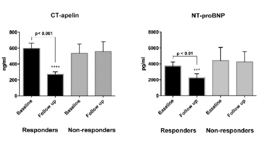
2.4.3.1.4 Predictors of response – biomarkers, CT-apelin
An optimal biomarker in chronic HF should be specific enough to detect the disease, provide an estimation of the prognosis and guide the treatment. The gold standard HF biomarker is the NT-proBNP. However, in patients who underwent CRT implantation, the cross-sectional values are suitable for describing the current status of the patient but prior studies failed to confirm its role as an independent predictor of response to CRT (36,37). Thus novel biomarkers are being investigated, in which inflammatory factors can take a part. Due to the low cardiac output and relating hypoperfusion all over the body, a systematic inflammation can occur during chronic HF. By activating the complement system, its components such as C3a might have an important role and has a predictive value for the response to CRT (38): elevated C3a levels increase the risk of mortality independent of the NT- proBNP levels, while CRT has an anti-inflammatoric effect by reducing the complement activation, thus measuring of the alteration of C3a might be a potential biomarker in the future. There are some other routinely measured laboratory parameters, which might help tailoring the therapy and predict the outcome after CRT implantation. Based on the above mentioned immuno-pathophysiology, the ratio of neutrophil leukocytes to the lymphocytes (39) or due to the congestion, the red blood cell distribution width might be novel prognostic markers in chronic HF (40). From the state-of-art HF biomarkers such as galectin-3, copeptin, NGAL, adrenomedullin or apelin (41,42), the latter has been emerged as a promising biomarker and investigated
comprehensively. Pre-pro-Apelin is expressed as a pro-hormone from several tissues. The apelin and its G-coupled receptor are expressed early during the embryonic development of the heart and affect the angiogenesis and maturation of cardiovascular cells (43). Its expression is also detected in adults where apelin has a paracrine effect as one of the most potent stimulators of cardiac contractility (44), moreover acts as a mediator of blood pressure via nitric-oxide dependent pathways (45). However, the role of apelin in HF is still unclear as changes of plasma levels are controversial in humans during the progression of HF (46-48). In addition, no data was available on its value in predicting or evaluating the response to CRT until now.
2.4.3.2 During the implantation
2.4.3.2.1 The role of intra- and interventricular delay
During the progression of HF, prolonged atrio-ventricular (AV) and ventriculo- ventricular (VV) delay can be observed. Interventricular dyssynchrony refers to prolonged activation between the ventricles, while intraventricular dyssynchrony develops by the late activation of mostly the postero-lateral / lateral region of the left ventricle (49). Several studies tested imaging (transthoracal echocardiography and MRI) and electrophysiological techniques to assess the localization and the role of intra- and interventricular dyssynchrony in CRT response.
The echocardiographic evaluation of interventricular dyssynchrony is based on the delay between the beginning of aortic and pulmonary velocity curves and the QRS, over 40ms delay the dyssynchrony can be confirmed. Intraventricular dyssynchrony can be evaluated by TDI or speckle tracking methods by segments, the latest activated part should contract with at least 50ms delay (49). These methods reflect primarily the mechanical dyssynchrony, thus there might have been differences between dyssynchrony assessed by echocardiogaphic or electrophysiological techniques.
The electroanatomical mapping is a more precise technique, imaging the electrical activation pattern directly (50). In this regard the most often investigated phenomenon is LBBB. Auricchio et al. (50) found that an U-shaped pattern of activation can be observed, where the line of block generally paralleled the septum.
Regardless of the method, the assessment of the latest activated part in the left ventricle
could be essential in order to perform a guided left ventricular lead implantation, thus achieve a better clinical response to CRT.
2.4.3.2.2 Targeting of the LV lead implantation
It has been proposed that optimal LV lead placement is an important determinant of response to CRT. The location of the left and right ventricular leads affects clinical outcome, and the incidence of ventricular tachyarrythmias (51). There have been positive results for echocardiography-guided left ventricular lead implantation. Those who were randomized to planned lead implantation by evaluating the latest site of peak contraction by strain analyses, yielded 15% higher amount of echocardiographic responder patients (52).
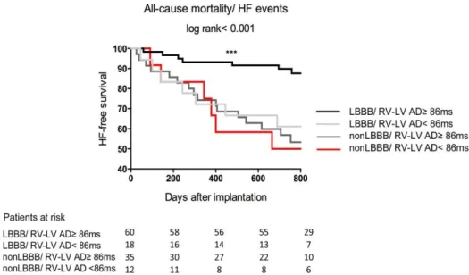
Furthermore, few smaller studies have indicated that the electrical delay between the signals sensed by the LV lead and the beginning of QRS duration (Q-LV), or the distance between the electrical signals of the left and right ventricular leads (RV-LV AD) predicted echocardiographic improvement and clinical outcome (53-55).
By measuring LV lead activation time from the beginning of the QRS (Q-LV), Gold et al. showed significant increase in functional and echocardiographic improvement in those patients who had greater Q-LV time.
RV-LV activation delay may also reflect the distance of RV and LV leads, moreover shows the electrical dyssynchrony and prolonged activation pattern derived from the slow conduction due to e.g. a scar tissue. Those studies, which used RV-LV activation delay (55,56), also showed significant improvement in echocardiographic response and in clinical outcome in patients with longer measured activation delay. However, none of these studies looked specifically at sub-groups of LBBB and non-LBBB patients.
Based on these prior studies the assessment the RV-LV delay during the implantation seems essential in the terms of the further response.
2.4.3.2.3 Multipolar pacing
However in novel therapeutic attempts multiple right and left ventricular stimulations are performed by multiple leads, in the recent thesis we are focusing on the comparison of bipolar and quadripolar left ventricular pacing.
During the implantation procedure, it can happen that only suboptimal target vein can be found for LV lead implantation or difficult to avoid phrenic nerve stimulation. By using multipolar pacing, a better clinical response and lower number of phrenic nerve stimulation (57) can be observed. Several investigations (58-60) confirmed a more pronounced improvement by quadripolar lead implantation and optimization of the pacing site compared to bipolar stimulation. This effect was reflected either in haemodynamic (58,60) or echocardiographic response (59).
2.4.3.3 Follow up - patient management and device optimization
2.4.3.3.1 AV and VV delay
The proper programming of the device such as AV and VV delay seems slightly controversial, while prior studies (61,62) found that it has an impact on better response to CRT. However large randomized trials (63,64) could not confirm these data, therefor in the current guidelines, it is not recommended routinely, but supported to use in non- responder patients (65).
The optimization is classified into two groups: echocardiography-based and device- specific measurements and settings. Regarding the AV delay, a suboptimal AV programming can result in a 10-15% decrease in cardiac output. The optimal setting was investigated by a large randomized trial (63), where no difference was found in the echocardiographic response in cases of fix 120ms AV delay, echocardiographic optimization or a device-specific optimization (SMART AV function).
The VV delay optimization can also be controlled by echocardiography-based higher stroke volume, ECG-based QRS narrowing or device-specific programs. However, these methods are not corroborated by tough evidences.
2.4.3.3.2 Remote monitoring
Remote monitoring is presented as IIa A evidence in the current ESC guidelines (65). By home monitoring it is considered to detect the arrhythmias or technical issues earlier, moreover HF hospitalizations or malignant arrhythmias can be prevented (66,67).
2.4.4 New indications:
2.4.4.1 Non-Left Bundle Branch Block morphology
Recent studies have suggested that patients with LBBB derive a significant benefit from CRT implantation, while in patients with a non-LBBB (as Right Bundle Branch Block - RBBB or intraventricular conduction delay - IVCD) the benefit is less if at all discernible (68,69). In non-LBBB patients with a prolonged PR interval, resynchronization with ICD treatment (CRT-D) was also associated with a 73% reduction in the risk of heart failure/death and 81% decrease in the risk of all-cause mortality compared with implantable cardioverter defibrillator therapy without CRT (70). In non-LBBB patients with normal PR, CRT-D therapy was associated with a trend toward an increased risk of heart failure/death (HR 1.45; 95%, CI 0.96-2.19; P=0.078; P<0.001) and a more than 2- fold higher mortality (HR 2.14; 95%, CI 1.12-4.09; P=0.022; P<0.001) compared with implantable cardioverter defibrillator therapy without CRT(70).
2.4.4.2 Upgrading to Cardiac Resynchronization Therapy
2.4.4.2.1 Lack of evidences
Since chronic right ventricular pacing is thought to be deleterious by increasing the risk of atrial fibrillation, HF and all-cause mortality (71,72), patients already carrying conventional pacemaker (PM) or ICD systems are often considered for upgrading to CRT.
Recent studies have suggested that only patients with typical LBBB ECG morphology derive a significant benefit from CRT(68,69). Although right ventricular pacing could reveal ventricular dyssynchrony similar to LBBB, data are scarce regarding the benefit of upgrade CRT in patients with previously implanted cardiac pacemaker or ICD systems.
The latest ESC guidelines on cardiac pacing and resynchronization therapy recommended CRT upgrade as a class I indication (level B) for symptomatic patients (NYHA III-IV) with low ejection fraction (LVEF≤35%)(65), while the most recent European heart failure guidelines restrict this indication as a class IIb (level B)(2). The ACC guidelines focused on the percentage of pacing rather than symptoms.
2.4.4.2.2 The BUDAPEST CRT upgrade study
About 28% of CRT implantations in Europe are upgrade procedures after previously implanted cardiac devices(73). To date, there are no conclusive results on the outcome of patients who underwent CRT-D upgrade from having previously implanted pacemaker or ICD devices, symptomatic HF, reduced ejection fraction and relatively high percentage (>20%) ventricular pacing. Furthermore, recent data indicate that upgrade procedures to biventricular pacing are associated with a relatively high complication rate (74), suggesting that a large, multicenter, randomized trial is required. We have designed an investigator initiated, prospective, randomized, multicenter trial, the BUDAPEST CRT upgrade study to clarify the question with 21 European and Israeli sites’ participation.
The study is conducted in accordance with the Helsinki Declaration, the Good Clinical Practice and the applicable regulatory requirements (75).
3 OBJECTIVES
Our aim was to determine novel parameters that might improve the clinical outcome after CRT implantation in regard of optimal patient selection and special methods during the implantation or early detection of response.
In order to optimize the patient selection and early assessment of the response to CRT, the serum levels of a novel biomarker, CT-apelin were measured. Its predictive value for the echocardiographic response was investigated and compared to the gold standard NT- proBNP levels at baseline and 6 months after resynchronization.
Moreover we examined the impact of an easily measured parameter during the implantation, the RV-LV activation delay. Its predictive role in the functional, echocardiographic and clinical outcome such as heart failure, all-cause mortality or laboratory parameters including NT-proBNP and renal function was assessed by the baseline QRS morphology of patients who underwent CRT implantation.
We would also focus on those questions, which are not entirely covered by the current ESC guidelines: patients who have an already implanted conventional pacemaker or ICD and referred to CRT upgrade. By concluding the available evidences of the literature, we analysed the clinical outcome, adverse events and long-term survival after upgrading compared to de novo implantation. However conclusive data will be provided by the BUDAPEST-CRT Upgrade Study, which investigates the all-cause mortality, heart failure events and echocardiographic response as primary endpoint besides functional response and safety after 12 months. In the current thesis the actual status, rationale and design of this investigator initiated trial is discussed in details.
4 METHODS
For a better interpretation, those studies in which optimal patients selection and intraoperative parameters were evaluated, are shown separately (Part 1) in the Methods and Results sections. In Part 2 the questions of CRT upgrade are shown by concluding the results of the currently available data in the literature in a meta-analysis and the rationale and status of the BUDAPEST CRT upgrade study.
4.1 Patient population
4.1.1 Inclusion and exclusion criteria of patients in Part 1.
Between September 2009 and December 2010 a prospective, observational, cohort study was designed to investigate patients undergoing successful CRT implantation at the Heart and Vascular Center, Semmelweis University, Budapest, Hungary. Patients with both ischemic and non-ischemic etiology were enrolled. Inclusion criteria were low left ventricular ejection fraction (EF≤35%), a prolonged baseline QRS interval (≥120 ms) and symptoms of HF (NYHA II-IVa functional class) despite optimal medical treatment.
Before the enrolment all patients underwent diagnostic coronarography or recoronarography in order to tailor the implantation by images of coronary sinus, moreover atherosclerosis as a secondary cause of HF could be verified. In those cases, where a percutaneous coronary intervention (PCI) was performed, patients were enrolled after 3 months of the procedure.
Exclusion criteria were patients with genetic HF, known malignant or inflammatory disease or severely reduced life expectancy, less than 1 year. We did not include those patients who were geographically unstable, or unwilling to attend regular follow ups or did not consent to the study.
In the investigation of optimal patient selection by serum biomarker measurements, from the total included patient cohort those who died before 6 month-follow up or unable or unwilling to give serum samples for biomarker assessments were censored due to the lack of ability to classify them according to response criteria and further biomarker measurements. Therefor it can be viewed as a substudy in the current thesis.
The study was approved by the Institutional Scientific Ethics Committee. All patients provided written informed consents and all data were anonymized prior to utilization.
4.1.2 Patient population and randomization in BUDAPEST CRT upgrade study
This prospective, multicenter, randomized trial was prepared and designed in 2013 by the principal investigator (PI), Professor Bela Merkely, co-PIs as Dr. Valentina Kutyifa and Professor Ilan Goldenberg and members of Steering Committee. High-volume, experienced centers were contacted in Europe and Israel, each sites which would participate got the opportunity to enroll patients after contracting and initiation.
From November 2014 patients are enrolled to the study regardless of the HF etiology with reduced LVEF (≤ 35%), symptoms (NYHA functional class II-IVa) despite optimal medical treatment with single or dual chamber pacemakers or ICD devices implanted at least 6 months before the inclusion (with ≥20% RV pacing over 90 days prior to enrolment and wide paced QRS duration ≥150 ms) with sinus rhythm, atrial fibrillation/flutter or atrial tachycardia as per protocol. The rate or frequency or rhythm control management is based on the physician’s discretion. Patients are excluded with typical LBBB intrinsic QRS morphology, severe right ventricular dilatation (>50mm), severe renal disease (serum creatinine >200umol/l) or other co-morbidities, which might influence the outcome of the patient. Those who had acute events (e.g. PCI, myocarditis, Coronary Artery Bypass Graft - CABG) or in those cases where heart transplantation (HTX) is planned. Detailed inclusion and exclusion criteria are listed in Table 1.
In the Semmelweis University after physicians pre-screened their patients and those who are thought to be eligible for the study, are referred to Annamaria Kosztin for screening and consent.
Those subjects, who proved to be eligible for the study, could be randomized in a 3:2 manner (CRT-D:ICD). Altogether a total of 360 patients are planned to be enrolled.
Table 1. Inclusion and exclusion criteria of the BUDAPEST CRT upgrade study
Inclusion criteria Exclusion criteria
1. Age: over 18 years
2. Cardiomyopathy with LVEF ≤35%, ischemic or non-ischemic
3. Single or dual chamber PM or ICD implanted ≥6 months prior to enrolment (battery depletion or another indication for upgrade is not required)
4. RV pacing ≥20% in the prior ≥90 days (use of algorithms to avoid ventricular pacing is recommended, per discretion of the clinician) 5. Paced QRS duration ≥150 ms 6. Symptomatic heart failure with
NYHA functional class II-IVa ≥3 months prior to enrolment, despite optimized medical therapy
7. Informed consent
1. CABG or PCI ≤3 month ago or planned
2. AMI ≤3 month ago 3. Unstable angina
4. Planned cardiac transplant 5. Acute myocarditis
6. Infiltrative cardiomyopathy 7. Hypertrophic cardiomyopathy
8. Severe primary mitral, aortic or tricuspid valve stenosis or insufficiency
9. Tricuspid valve prosthesis
10. Severe right ventricular dysfunction (RV basal diameter > 50mm)
11. Chronic severe renal dysfunction (creatinine >200 µmol/l)
12. Pregnant women or planned pregnancy
13. Subjects who are unable or unwilling to cooperate with the study protocol 14. Any comorbidity that is likely to
interfere with the conduct of the study 15. Participation in another trial
16. Patients geographically not stable or unavailable for follow-up
17. Intrinsic QRS with typical LBBB morphology
AMI= Acute Myocardial Infarction; CABG= Coronary Artery Bypass Graft; ICD=
Implantable Cardioverter Defibrillator; LBBB= Left Bundle Branch Block; LVEF= Left Ventricular Ejection Fraction; NYHA= New York Heart Association; PCI= Percutaneous Coronary Intervention; RV= Right Ventricle
4.2 Follow up and investigations 4.2.1 Follow up
4.2.1.1 Baseline and follow up visits in Part 1
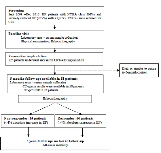
Each visits were performed by Annamaria Kosztin and Vivien Klaudia Nagy, which included a physical examination, assessment of the NYHA functional class, transthoracic echocardiography, detailed laboratory tests, 6 minute walk test and EQ5D quality of life measurements extended with device interrogations after the implantation. Investigations were performed at the baseline visit and 6 months after CRT/ICD implantation. Beyond regular outpatient visits, patients were contacted via telephone and the Hungarian National Database was used to obtain vital information at 3 years after CRT implantation (Figure 1) .
Figure 1. Flowchart of patient enrolment and follow up of optimal patient selection by biomarkers
4.2.1.2 Follow up in the BUDAPEST CRT upgrade study
Eligible patients undergo a baseline evaluation including clinical history, physical examination, NYHA class, 12-lead ECG with paced (paced VVI or DDD 70 bpm and non-paced QRS complexes using VVI 40 bpm settings), transthoracic echocardiography, device interrogation (RV pacing percentage and Holter data), quality of life assessment (EQ5D), 6 minute walk test and optional NT-pro-BNP measurement. Patients are followed up for 12 months after randomization. Regular, in-office follow-ups will be performed at 1, 6 and 12 months (Table 2), which are performed by Annamaria Kosztin (each investigations except for echocardiography) and Attila Kovacs (echocardiography).
While Semmelweis University is responsible for the maintenance of echocardiography core lab and Biobankok server, each PM interrogation files, ECGs and echocardiographic images that are performed during the patient follow ups from active centers, are uploaded to our Biobankok Server and will be analysed centrally.
4.2.2 ECG
By performing a 12-lead analog ECG, the assessment of QRS width and morphology were mandatory in each study. After all of the ECGs has been recorded, the same person assessed the data and fill our electronical database retrospectively.
LBBB was defined on 12-lead ECG as QRS duration >120 ms; QS or rS in lead V1; broad R waves in leads I, aVL, V5, and/or V6; and absent q waves in leads V5 and V6. RBBB required QRS duration >120 ms; rsr, rsR, rSR, or qR in leads V1 or V2; and occasionally, wide R waves and wide S waves in leads I, V5, and V6. Intraventricular conduction delay was defined as QRS >120 ms without typical features of LBBB or RBBB.
During BUDAPEST CRT study, the assessement of presence of intrinsic LBBB is based on the physicians’ discretion. The digital formation of ECGs are uploaded to the Biobankok server and will be analysed retrospectively.
4.2.3 Echocardiography
Echocardiography was performed according to current standards in a left lateral position using Philips iE33 echocardiography system equipped with an S5-1 transducer (Philips Healthcare, Best, The Netherlands). Image acquisition was performed according to current recommendations (76). Measurements were performed by the same person offline using the QLAB software (Philips Healthcare). Left ventricular end-systolic and end- diastolic volumes were measured and ejection fraction was calculated by the biplane Simpson method (76).
Table 2. Follow up visits of the BUDAPEST CRT upgrade study
Visit/evaluation Patient Enrolment
Visit Day 0
Device Implantation
and Programming
Within 14 days
1 month FU visit
Day 30
6 months FU visit
Day 180
12 months FU visit
Day 365
Inclusion criteria x Exclusion criteria x Signed Informed
Consent x
clinical history x x x x
physical
examination x x x x
assessment of
NYHA class x x x x
12-lead ECG
(paced) x x
12-lead ECG (at
VVI 40 bpm) x x
Echocardiography x x
device interrogation
(print, save, upload) x x x x
blood test (NP-pro-
BNP) x1 x1
6 minute walk test x2 x
Randomization x
Assessment of
clinical end-points x3 x3 x3
Assessment of post- implantation complications
x
SAE, AE, UADE,
USADE x x x x x
Quality of life assessment using
EQ-5D
x2 x
1: Optional, 2: After the randomization but before implantation, 3: Clinical end-points;
SAE= Serious Adverse Event; AE= Adverse Event; UADE= Unanticipated Adverse Device Effect; USADE= Unanticipated Serious Adverse Device Effect
4.2.4 Serum biomarker measurements
Human CT-apelin was measured by Annamaria Kosztin using C-terminus Enzyme Immunoassay competitive ELISA method (RayBiotech, Inc., Norcross, USA) which is designed to target the C-terminus of the 77-aminoacid apelin peptide. By this kit all active forms (apelin-13, -31, -28, and apelin-36) of the pre-prohormone 77-aa apelin peptide can be measured. NT-proBNP was measured with Cobas proBNP II kit (Roche Diagnostics Gmbh, Mannheim, Germany). Serum samples were stored at -80 oC until sample collection was completed.
Rutine laboratory measurements (ions, renal function, haematology parameters) were performed by Eva Forizs using automatic kits (Roche kit, Roche Diagnostics Gmbh, Mannheim, Germany) as routine clinical practice in our hosital.
4.3 Device implantation and programming 4.3.1 Device implantation procedure in Part 1
Device implantations were performed according to current standards by using a transvenous approach. By performing a coronary sinus angiogram LV lead implantation was tailored during device implantation. After positioning of each leads, pacing, sensing and impedance parameters were measured. In patients with intraoperative LV lead dislocation or phrenic nerve stimulation, coronary sinus stent implantation was performed in the CS side branch after repositioning of the LV lead (77,78). Right ventricular lead was primarily implanted into a septal position, while left ventricular lead into a posterolateral or lateral side branch. LV and RV lead positions were assessed by the implanting physician based on the right and left anterior oblique (RAO and LAO) views.
4.3.2 Upgrade procedure in BUDAPEST CRT Upgrade study
Upgrade procedures need to be performed within 14 business days after randomization.
(Table 2). During the procedure, duration time of the upgrade, X-ray dosage, details of
the implanted leads, adverse events and RV-LV AD are mandantory to report. Patients with an existing ICD, who are randomized to the ICD arm, may not need a procedure unless a generator replacement, a system revision is necessary or the PI decides on upgrading to CRT-D in RV only pacing mode. The optional study interventions are listed in Table 3. Decisions about lead extraction are based on the physicians’ discretion by actual recommendations. (79) Use of Boston Scientific Corporation (Marlborough, MA, USA) ICDs or CRT-D is preferred, but not mandatory. In the ICD arm, choosing single or dual chamber device is left to the physician decision. In the CRT-D arm, the left ventricular lead is recommended to be implanted in the lateral or postero-lateral side branch of the coronary sinus. Transvenous implantation is strongly preferred; however, alternative methods are also accepted if the transvenous attempt fails.
Table 3. Optional study interventions in BUDAPEST CRT upgrade study
CRT-D group ICD group
1. Existing PM
Addition of RV defibrillator lead Addition of RA pacing lead (unless already has one or has permanent AF) Addition of LV pacing lead
Extraction of old RV PM lead optional (physician’s judgment)
Any revision of the old lead(s) and device pocket, as necessary
Generator change to CRT-D 2. Existing ICD
Addition of RA pacing lead (unless already has one or has permanent AF) Addition of LV pacing lead
Any revision of the old lead(s) and device pocket, as necessary
Generator change to CRT-D
1. Existing PM
Addition of RV defibrillator lead Addition of RA pacing lead optional (physician’s judgment, unless already has one or has permanent AF)
Extraction of old RV PM lead optional (physician’s judgment)
Any revision of the old lead(s) and device pocket, as necessary
Generator change to VVI or DDD ICD, 1. Existing ICD
Continue with existing device
Addition of RA pacing lead and upgrading to a DDD ICD is optional (physician’s judgment, unless already has one or has permanent AF)
AF= Atrial Fibrillation; CRT-D= Cardiac Resynchronization Therapy with Defibrillatior;
ICD= Implantable Cardioverter Defibrillator; PM= Pacemaker; RA= Right Atrium; RV=
Right Ventricle
4.3.3 RV-LV AD measurement at implantations
After positioning both ventricular leads, intraoperative RV-LV activation delay measurements were performed by connecting to an electrophysiology system (Biotronik pacemaker interrogation device, Berlin, Germany). The right to left interventricular sensed delay was measured by the time delay of the peak activation in the right and left ventricular sensed signals phrased in milliseconds (Figure 2).
Figure 2. RV-LV AD measurement by assessment of the time delay between the peak activation in the right and left ventricular sensed signals
4.3.4 Device programming during BUDAPEST CRT UPGRADE study
Regarding bradycardia parameters DDD(R) or VVI(R) mode is required with base rate setting between 40-70 bpm. In order to achieve the optimal AV-delay, SMART AV (63) or echocardiographic optimization or fixed values (sensed AV delay 120-140 ms/ paced AV delay 140-160 ms) can be used. Regarding antitachycardia parameters, two zones are recommended: VT1 as a monitor zone between 170-200 bpm without programmed therapy and VF zone over 200 bpm with a 2.5 sec delay, ATP during charging (8 pulses at 88% of the tachycardia cycle length) and subsequent shocks (first : DFT + 10J or 30 J, subsequent shocks should be maximum energy shocks).
4.4 Endpoints
4.4.1 Endpoints of Part 1.
4.4.1.1 Endpoints in assessing the predictive value of NT-proBNP and a novel biomarker, CT-apelin
The primary endpoint of the study was non-response to CRT defined as an absolute increase of less than 4% in ejection fraction (80) at 6 months, compared to baseline measurements. Secondary endpoint was all-cause mortality during the three years follow- up.
4.4.1.2 Endpoints in evaluating the effect of RV-LV AD specified by QRS morphology
The primary composite endpoint was heart failure hospitalization or all-cause mortality.
Secondary endpoint was death from any cause.
Heart failure events were defined as symptoms and signs of heart failure that required intravenous diuretic treatment during an in-hospital stay. All-cause mortality was assessed by the National Health Fund Death Registry.
We also evaluated the clinical outcome as changes of ejection fraction, distance walked during the 6-minute walk test and NT-proBNP serum levels after 6-month.
First the recent endpoints were assessed by RV-LV AD as a continuous variable in the total patient cohort, then patients were dichotomized by the lower quartile of RV-LV AD (86 ms)
1) patients with RV-LV AD < 86 ms 2) and those with RV-LV AD ≥ 86 ms
Thereafter they were further grouped by their baseline LBBB morphology:
1) patients with RV-LV AD < 86 ms and LBBB 2) patients with RV-LV AD ≥86 ms and LBBB 3) patients with RV-LV AD < 86 ms and non-LBBB 4) patients with RV-LV AD ≥ 86 ms and non-LBBB
Finally we also investigated the outcomes of two subgroups: patients with LBBB and RV-LV AD < 86 ms together with patients with non-LBBB (“expected CRT non- responders”) and compared them to patients with LBBB but RV-LV AD ≥ 86 ms (“expected CRT responders”).
Our analyses were extended by RV-LV AD to QRS duration (RV-LV AD /QRS), moreover in order to further assess the effects of RV-LV AD as a continuous parameter on NT-proBNP and clinical outcome of HF/death, we evaluated the changes in NT- proBNP at 6-month by RV-LV AD quartiles along with the incidence of HF/death.
4.4.2 Endpoints of Part 2.
4.4.2.1 Endpoints in the meta-analysis of patients after CRT upgrade compared to de novo CRT implantation
We report data about all-cause-mortality, heart failure events, echocardiographic (LVEF, EDV), clinical (change of NYHA functional class) and ECG (change of QRS width) parameters of reverse remodeling.
4.4.2.2 Endpoints in the BUDAPEST-CRT upgrade study
The primary endpoint of the study is a composite endpoint of heart failure events, all- cause mortality, or less than 15% reduction in echocardiography determined left ventricular end-systolic volume from baseline to 12-month.
Secondary end points are the composite of heart failure events and all-cause mortality, all-cause mortality alone, the changes of echocardiographic parameters (left ventricular end-diastolic volume or left ventricular ejection fraction) from baseline to 12 month.
Tertiary endpoints are the success and safety of implantation procedures, the change of NYHA class, quality of life assessed by EQ-5D questionnaire, 6-minute walk test and the changes of NT-pro-BNP serum levels from baseline to 12 months.
4.5 Statistics and methods for analyses 4.5.1 Statistical analysis
Statistical analyses were performed by Graph Pad version 6.0 and 7.0 (Graph Pad Inc., CA, USA), SPSS version 9 (IBM, NY, USA) or Comprehensive Meta-Analysis 3.3 (Biostat, Inc., USA).
Continuous variables with normal distributions are expressed as mean±SD, while those with non-normal distributions as medians with interquartile range (IQR). Categorical variables are shown with numbers and percentages (n, %). Baseline clinical characteristics of Part 1 were compared by unpaired t-test for normally distributed continuous variables, the Mann–Whitney U-Test for non-normally distributed variables, while 2 - test or Fisher exact test was used for dichotomous variables, as appropriate.
Time-to-event data were presented by Kaplan-Meier curves. Unadjusted hazard ratios (HR) with 95 confidence intervals (95% CI) were calculated for mortality in Cox proportional hazards models, while adjusted HR in forward stepwise Cox proportional model adjusting for relevant clinical parameters as appropriate. A two-sided p-value of
<0.05 was considered as statistically significant.
Univariate and multivariable receiver-operating characteristic (ROC) curve analyses were also used to determine the discriminatory capacity of biomarkers on non-response and were shown as the area under curve (AUC) and p values. In case of a significant p value, an optimal cutoff was assessed for the continuous variable based on maximal sensitivity and specificity. Using these cutoffs, patients were separated to low and high biomarker level groups for logistic regression analyses. Multivariate logistic regressions were performed with variables showing a p value less than 0.05 in univariate analyses.
In the meta-analyses heterogeneity between individual trial estimates was assessed by the Q statistic and I2 statistic (81). Since, there was significant heterogeneity in the design and patient’s characteristics of the studies included into the meta-analyses, it was assumed that the true effect size varies from one study to the next, and hence the random-effect model was used(82). A forest plot was created with individual trials and the pooled estimates. Publication bias was assessed using the funnel plot, the trim and fill method of Duval and Tweedie (83) and an adjusted rank-correlation test according to Begg and Mazumdar(84). Since we did not have access to individual patient data from all studies reviewed, the median of delta values for LVEF, EDV, NYHA and QRS were calculated and compared between the two patient groups separately by using the Mann-Whitney U test. Methodological quality of all studies was assessed using the Methodological Index for Non-Randomized Studies (MINORS)(85). Studies were defined to be low, moderate and high quality studies based on their MINORS scores of <8, <16, and ≥16 points (data are not shown).
4.5.2 Study selection for systematic review and meta-analyses
The systematic review was performed according to the PRISMA Statement (86) and a predefined review protocol was published in the PROSPERO database under the registration number of CRD42016043747. A comprehensive search of PubMed, Research Gate, and Google Scholar databases was performed from January 2006 to June 2016
focusing on full-sized, peer-reviewed, English language papers reporting data on patient outcomes after upgrade CRT vs. de novo implantations as a comparator group. Abstracts were only included when critically relevant and not available as full-text articles. In order to identify all potentially relevant articles, the search was performed by using the terms of 1. “upgrade” AND “CRT”; 2. “upgrade” AND “cardiac resynchronisation therapy”.
The search was also extended by using the name of the most frequently cited authors of the identified studies. In addition, references of relevant review articles were also searched to find appropriate manuscripts. Potentially relevant articles were evaluated by three independent reviewers and additional manuscripts were retrieved that either reviewer felt were potentially relevant. According to our review protocol studies were accepted for analysis if (i) including heart failure patients with reduced ejection fraction (HFrEF) with de novo and upgrade CRT implantations (ii) reporting all-cause-mortality data or heart failure events; (iii) reporting echocardiographic (i.e. LVEF, EDV) or clinical (NYHA class) or ECG (QRS width) parameters of reverse remodeling (Table 4). Heart failure events were defined as hospitalization due to progression of heart failure. In order to evaluate the heterogeneity of patients who were enrolled into each therapy groups, the most important baseline clinical characteristics were collected. Data on procedure related complications were also investigated if available.
Table 4. Searching methodology and eligibility criteria for the meta-analysis Eligibility criteria
Criteria Included Excluded
Participants wide QRS, NYHA II – ambulatory IV and EF≤ 35%
No indication for CRT Intervention CRT upgrade Unsuccessful LV lead
implantation
Comparator de novo CRT implantation No comparator group Primary
Outcome
All-cause mortality Only cause specific mortality data or composit endpoints provided
Secondary outcomes
Changes in NYHA class, Echocardiographic parameters of reverse remodeling, QRS narrowing
NA
Study Design Randomized controlled trials Non-randomized trials Observational cohort studies
Case reports Reviews Meta-analyses
Languages English Any other languages
Publication status
Published or accepted manuscripts or abstracts
Non peer-reviewed, unpublished
CRT= Cardiac Resynchronization Therapy; LV= Left Ventricle; NA= not applicable;
NYHA= New York Heart Association
4.5.3 Sample size calculation and statistical methods in the BUDAPEST CRT UPGRADE study
Altogether 360 patients are planned to enroll to the study. The main objective is to investigate the primary composite clinical and echocardiographic endpoint after CRT upgrade (superiority of CRT-D upgrade vs. ICD only). Analyses will be performed (i) on an intention-to-treat-basis (without regard to device actually implanted/revised), (ii) and on efficacy basis, censoring follow-up when a patient crosses over to a different device.
The primary analyses will be stratified by the percentage of baseline RV pacing as pre- specified in the study. The null hypothesis for the primary endpoint is that the hazard rate, which is assumed to be constant across all study intervals, is identical in the two groups (CRT-D v. ICD). The hypothesis will be tested in a study in which subjects are entered and followed up until (i) the primary composite endpoint occurs, (ii) the patient drops out of the study, (iii) or the study ends while the patient is still being followed, in which case the patient is censored.
Power was calculated a priori based on a hazard ratio of 0.7 and a primary composite endpoint event rate of 80% in the ICD group over 12 months. The power calculation was based on higher RV pacing rates, while no data is available <40%.
Although the risk seems to correlate with RV pacing, the exact correlation is unclear. The attrition (drop out) rate was assumed at 0.01/interval. An instantaneous hazard rate of 0.134 for the ICD group and 0.094 for the CRT-D group was assumed – this equals to a median survival time of 5.17 intervals in the ICD group and 7.38 intervals in the CRT-D group, a cumulative event free survival at 12 intervals of 0.2 for the ICD group and 0.32 for the CRT-D group. The two-tailed alpha was set at 0.05. A total of 144 patients will be entered into the ICD group and 216 into the CRT-D group to achieve a power of 80.1%
to yield a statistically significant result.
5 RESULTS
5.1 Part 1 – Optimization of patient selection and intraoperative techniques in order to achieve a more beneficial clinical response
5.1.1 Optimal patient selection by measuring NT-proBNP and a novel biomarker, serum CT-apelin
5.1.1.1 Baseline clinical characteristics
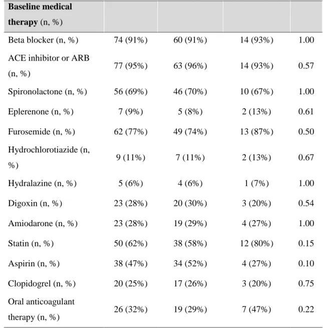
From those patiens who underwent a successful CRT implantation between September 2009 and December 2010, 81 patients were included in the current study. Mean age of the recruited patients was 64.9±10.5 years, with a mean ejection fraction of 28.5±6.5%, and mean QRS width of 167.7 ± 29.8 ms. Eighty-six percent of the patients had typical LBBB morphology and 59% had CRT-D device. Seventy-five percent of the patients were in NYHA class III functional state and 59 % had ischemic etiology before CRT implantation (Table 5a).
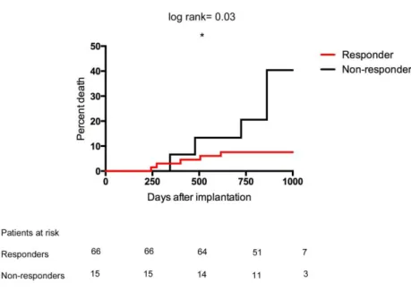
5.1.1.2 Response and prognosis
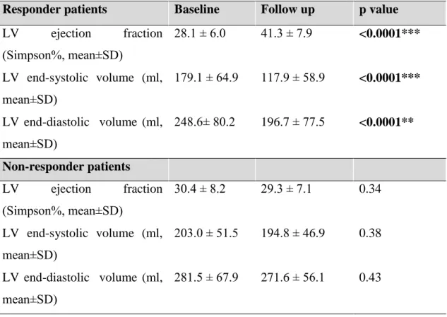
During the mean follow-up time of 795 ± 99 days, 7 (9%) patients died. Based on the pre- defined classification of response, 15 (18.5%) patients proved to be non-responders, of which 4 died during the follow up. Baseline clinical characteristics, medical therapy and echocardiographic findings were similar between responders and non-responders (Table 5a, 5b, 5c). In line with the definition of response, left ventricular volumes significantly decreased (ESV: 179.1 ± 64.9 vs. 117.9 ± 58.9, p<0.0001, EDV: 248.6± 80.2 vs. 196.7 ± 77.5, p<0.0001) and left ventricular function significantly improved (EF: 28.1 ± 6.0 vs.
41.3 ± 7.9) in responder patients after CRT implantation, while these parameters remained unchanged in the non-responder group after 6 months (Table 6).
Table 5a. Baseline clinical variables in the responder and non-responder patients
Baseline clinical variables
All patients (n=81)
Responders (n=66)
Non- responders
(n=15)
p value Age (yrs, mean±SD) 64.9 ± 10.49 64.1± 10.8 68.5 ± 8.4 0.14 Gender (female, n, %) 15 (18.5%) 14 (21%) 1 (7%) 0.28 Ischemic etiology (n, %) 48 (59%) 39 (59%) 9 (60%) 1.00
NYHA II. st (n, %) 11 (14%) 9 (14%) 2 (13%) 1.00
NYHA III. st (n, %) 61 (75%) 49 (74%) 12 (80%) 0.75
NYHA IV. st (n, %) 9 (11%) 8 (12%) 1 (7%) 1.00
QRS (ms, mean±SD) 167.7 ± 29.8 166.6 ± 28.8 172.0 ± 34.3 0.53 typical LBBB
morphology (n, %) 70 (86%) 57 (86%) 13 (87%) 1.00
not typical LBBB (n, %) 11 (14%) 9 (14%) 2 (13%) 1.00 6 minutes walk test (m,
mean±SD)
311.4
±117.1 307.3 ± 127.6 329.2 ± 54.1 0.56 RR systolic (mmHg,
mean±SD) 120.4 ± 18.8 121.1 ± 17.4 117.5 ± 24.8 0.51 RR diastolic (mmHg,
mean±SD) 76.2 ± 10.7 76.9 ± 10.2 73.1 ± 12.3 0.21
Heart rate (min-1,
mean±SD) 75.6± 14.5 75.6 ± 14.1 75.3 ± 16.5 0.93
Atrial fibrillation (n, %) 20 (25%) 14 (21%) 6 (40%) 0.18 Body mass index (BMI;
med, IQR)
27.0 (24 /
30) 27.0 (24 / 30) 29.0 (26 / 31) 0.16 LBBB = left bundle branch block; NYHA class = New York Heart Association class;
PCI= percutaneous coronary intervention; CABG = coronary artery bypass grafting; VF=
ventricular fibrillation