Late Sodium Current Inhibitors as Potential Antiarrhythmic Agents
Balázs Horváth1,2
*
, Tamás Hézso˝1, Dénes Kiss1, Kornél Kistamás1, János Magyar1,3, Péter P. Nánási1,4and Tamás Bányász11Department of Physiology, Faculty of Medicine, University of Debrecen, Debrecen, Hungary,2Faculty of Pharmacy, University of Debrecen, Debrecen, Hungary,3Division of Sport Physiology, University of Debrecen, Debrecen, Hungary,
4Department of Dental Physiology and Pharmacology, Faculty of Dentistry, University of Debrecen, Debrecen, Hungary
Based on recent fi ndings, an increased late sodium current (I
Na,late) plays an important pathophysiological role in cardiac diseases, including rhythm disorders. The article fi rst describes what is I
Na,lateand how it functions under physiological circumstances. Next, it shows the wide range of cellular mechanisms that can contribute to an increased I
Na,latein heart diseases, and also discusses how the upregulated I
Na,latecan play a role in the generation of cardiac arrhythmias. The last part of the article is about I
Na,lateinhibiting drugs as potential antiarrhythmic agents, based on experimental and preclinical data as well as in the light of clinical trials.
Keywords: voltage gated sodium channel, late sodium current, arrhythmias, antiarrhythmic drugs, sodium channel inhibitors
INTRODUCTION
During the non-pacemaker action potential (AP) in the heart, depolarization of the cell membrane opens voltage gated sodium channels (Na
v) for a short period of time (Scanley et al., 1990; Mitsuiye and Noma, 2002) giving rise to the early sodium current peak (I
Na,early). This I
Na,earlycauses the upstroke of the non-pacemaker AP. Through the course of the AP Na
vchannels may recover from inactivation and reopen, generating a sustained current component, called late sodium current (I
Na,late). I
Na,lateflows throughout the plateau phase of the AP therefore it significantly contributes to AP morphology, even though its magnitude is only a fraction of I
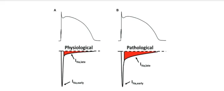
Na,early(Figure 1A).
If I
Na,lateis increased, it might play a pathophysiological role in acquired cardiac diseases (Figure 1B) such as myocardial ischemia (Maier and Sossalla, 2013) and heart failure (Coppini et al., 2013; Pourrier et al., 2014). In the cardiomyocytes, an upregulated I
Na,latehinders repolarization and causes a larger sodium entry, therefore increasing intracellular sodium concentration ([Na
+]
i). An increased [Na
+]
i, in turn, leads to a larger intracellular calcium content. These factors together can possibly cause contractile dysfunction (Sossalla et al., 2011), disturbed myocardial energetics (Liu and O'Rourke, 2008) and cardiac arrhythmias (Antzelevitch et al., 2014).
ELECTROPHYSIOLOGICAL IDENTIFICATION OF I
NA,LATEMammalian cardiac cells express a wide variety of Na
visoforms, differing in unit conductance, voltage sensitivity, kinetics, and drug sensitivity. In the majority of cardiac tissues, the dominant isoform of the pore-forming subunit is Na
v1.5, which is relatively insensitive to the sodium channel
Edited by:
Annamaria De Luca, University of Bari Aldo Moro, Italy Reviewed by:
Francesco Miceli, University of Naples Federico II, Italy Bin-Nan Wu, Kaohsiung Medical University, Taiwan
*Correspondence:
Balázs Horváth horvath.balazs@med.unideb.hu
Specialty section:
This article was submitted to Cardiovascular and Smooth Muscle Pharmacology, a section of the journal Frontiers in Pharmacology Received:31 December 2019 Accepted:18 March 2020 Published:20 April 2020 Citation:
Horváth B, Hézso˝ T, Kiss D, Kistamás K, Magyar J, Nánási PP and Bányász T (2020) Late Sodium Current Inhibitors as Potential Antiarrhythmic Agents.
Front. Pharmacol. 11:413.
doi: 10.3389/fphar.2020.00413
toxin tetrodotoxin (TTX) (Gellens et al., 1992; Catterall et al., 2005). Many of the TTX-sensitive (“non-cardiac”) Na
vchannels (Na
v1.1, Na
v1.2, Na
v1.3, Na
v1.4, and Na
v1.6) are also shown to be present in cardiac tissue (Maier et al., 2002; Haufe et al., 2005;
Valdivia et al., 2005; Biet et al., 2012; Yang et al., 2012). In nodal tissue Na
v1.1 and Na
v1.6 are expressed in the largest quantities.
Besides the pore-forming subunit, four auxiliary subunits (ß
1, ß
2, ß
3, and ß
4) and certain scaffolding proteins also participate in building up the whole complex, which also attaches to the cytoskeleton. These molecules can interact with each other and may modify the kinetics and voltage dependence of the actual channel (Malhotra et al., 2001).
Mechanisms that are discussed in the followings may contribute to the profile of I
N a , l a t eduring the AP.
Understanding these mechanisms better might be helpful in developing new antiarrhythmic therapeutic strategies targeting I
Na,late.
I
Na,lateIs Underlain by Different Channel Gating Modes
At the resting membrane potential, the vast majority of Na
v1.5 channels are in their closed state. Upon depolarization, Na
v1.5 channels open up within 1–2 ms after which they inactivate rapidly (Scanley et al., 1990; Mitsuiye and Noma, 2002). This produces I
Na,earlyand the upstroke of the non-pacemaker cardiac AP. During a sustained depolarization, Na
v1.5 channels can reopen with a small probability. In ventricular myocytes, three modes of Nav1.5 channel activity have been characterized in single-channel experiments: transient mode (TM), burst mode (BM), and late scattered mode (LSM) (Maltsev, 2006).
I
Na,earlyis mainly the result of TM activity, while BM and LSM are responsible for the sustained sodium current, I
Na,late(Figure 1A). The magnitude of the sustained current component is only about 0.5 – 1 % of I
Na,earlymeasured 50 ms after the onset of the depolarizing pulse (Maltsev, 2006). During a sustained
depolarization BM openings rapidly decline in the fi rst tens of milliseconds therefore leaving LSM as the gating mode being mainly responsible for I
Na ,la tetoward the end of the plateau phase.
Mutations of the channel protein and certain diseases can change the contribution of different Na
v1.5 channel activity patterns to the macroscopic current, therefore increasing I
Na,late(Bezzina et al., 1999; Valdivia et al., 2005; Wu et al., 2006;
Maltsev et al., 2007; Maltsev and Undrovinas, 2008; Song et al., 2008; Maltsev et al., 2009; Xi et al., 2009; Guo et al., 2010; Trenor et al., 2012) (Figure 1B). Apparently, each gating mode has a distinct drug sensitivity or drug af fi nity as well (Belardinelli et al., 2004; Ravens et al., 2004; Belardinelli et al., 2006). Based on this, selective pharmacological targeting of certain gating modes might have potential antiarrhythmic and/or cardioprotective effects (Belardinelli et al., 2006; Hoyer et al., 2011; Morita et al., 2011).
Window Sodium Current
The voltage dependence of the steady state activation and inactivation of most Na
vchannels overlaps with each other (Zaza and Rocchetti, 2013). This overlap provides a voltage range ( “ window ” ) where inactivated Na
vchannels are able to recover from inactivation and then might reopen. When the actual membrane potential falls within this “window” of overlap, a sustained current is evoked. Under physiological circumstances this “ window current ” mechanism likely plays a limited role in I
Na,late, because the Na
v1.5 voltage “ window ” is around − 70 mV, falling quite far from the AP plateau. Additionally, in the window voltage range, the current density is less than 5 % of the maximum current density in healthy myocytes (Maltsev et al., 1998; Wang et al., 2002; Liu et al., 2007). Hence, the “ window current ” mechanism is unlikely to be a major determinant of I
Na,latein healthy myocytes. Mutations of channel proteins or altered regulation in certain diseases may shift either the steady-state
A B
FIGURE 1 |The early and the late component of the sodium current under physiological(A)and pathological(B)conditions. Upper panels: membrane potential;
lower panels: sodium current. INa,early, early (peak) component of the sodium current; INa,late, late (sustained) component of the sodium current.
activation or inactivation curves of Na
vchannels to signi fi cantly change this voltage window, therefore increasing I
Na,lateunder these pathological conditions (Wang et al., 1996; Ruan et al., 2009).
Non-Equilibrium Channel Gating
During the AP of cardiac myocytes, the membrane potential changes continuously. Na
vchannels are incorporated into this dynamic system. It has been proposed by Clancy et al. (2003) that the voltage “history” of the cell membrane can modulate the transition between Na
vchannel states, termed “ non-equilibrium gating ” . As a result, recovery from inactivation is also modulated by the dynamics of voltage change. The theory is supported by experimental data showing that the application of repolarizing voltage ramps or AP shape voltage commands evoke a larger I
Na,latecompared to conventional square pulses or model simulations where “ non- equilibrium gating ” is not incorporated into the numerical model (Clancy et al., 2003; Magyar et al., 2004; Horvath et al., 2013).
Non-Cardiac Sodium Channel Isoforms in the Heart
Epilepsy (Alekov et al., 2000; Akalin et al., 2003) and certain skeletal muscle diseases (Komajda et al., 1980; Pereon et al., 2003) has been associated with pathological ECG recordings. Therefore it seemed possible that non-cardiac sodium channel mutations might cause electrical alterations in the heart. Later, Na
v1.1, Na
v1.2, Na
v1.3, Na
v1.4, Na
v1.6, and Na
v1.8 isoforms have been identified in cardiac tissue (Maier et al., 2002; Haufe et al., 2005; Valdivia et al., 2005; Biet et al., 2012; Yang et al., 2012). Based on the fi ndings of Biet et al., as much as 44 % of I
Na,lateis due to non-cardiac sodium channels (Biet et al., 2012) in canine ventricular cardiomyocytes. Furthermore, Yang et al. have shown that in mice and rabbit the TTX-resistant Na
v1.8 provides a substantial amount of I
Na,late(Yang et al., 2012).
Based on these experimental data, isoform speci fi c sodium channel modulators might provide a valid approach in pharmacological antiarrhythmic therapy (See Non-Cardiac Sodium Channel Inhibitors as Potential Antiarrhythmic Agent for further details).
ROLE OF I
NA,LATEIN CARDIAC PHYSIOLOGY
Role of I
Na,latein Cardiac Electrical Activity
Contribution of I
Na,lateto cardiac APs was questioned because of its small density. However, the plateau phase of the cardiac AP is shaped by a delicate balance between minuscule inward and outward current fl uxes. Therefore even a small change in these currents may signi fi cantly alter the duration of the AP (Horvath et al., 2006). Inhibition of I
Na,latesubstantially shortens the c ardiac AP in the c on duc ti ve sy ste m (Coraboeuf et al., 1979) and in ventricular cells (Kiyosue and Arita, 1989) as well, indicating that I
Na,latesigni fi cantly contributes to determining the duration of the non- pacemaker AP in cardiac myocytes. Recent AP voltage clamp experiments show that the density of I
Na,lateis of similar magnitude as the major potassium currents in g u i n e a p i g ( H o r v a t h e t a l . , 2 0 1 3) a n d r a b b i t
(Hegyi et al., 2018) ventricular myocytes. There is a characteristic interspecies difference in the shape of I
Na,lateas shown in the case of guinea pig, canine, and human ventricular myocytes (Horvath et al., 2020).
The sustained sodium current is also an important factor in determining electrophysiological properties of sinoatrial node cells (Maier et al., 2003; Lei et al., 2004). Tetrodotoxin, applied in lower than 1 µM concentrations, reduces the rate of spontaneous depolarization in sinoatrial node cells (Huang et al., 2015), clearly indicating that non-cardiac Na
visoforms also contribute to cardiac automaticity.
Cardiac Purkinje cells have the largest rate-dependence of their AP duration (APD) among cardiomyocytes with fast response APs. Purkinje cell APs are longer at lower stimulation rates, while shorter at higher rates than APs of ventricular cells. It has been shown that I
Na,latecontributes to this feature by possessing much slower decay and recovery kinetics in Purkinje cells than in ventricular cells. As a result Purkinje cell I
Na,lateis signi fi cantly larger at low heart rates, while smaller at high heart rates compared to ventricular cells. This unique feature predisposes Purkinje cells to serve as triggers in generating arrhythmias (Li et al., 2017).
I
Na,lateplays a role in forming the atrial AP as well (Burashnikov and Antzelevitch, 2013; Luo et al., 2014). I
Na,lateis expected to be larger in atria than in ventricles because I
Na, earlydensity is greater in atrial cells under similar conditions (Li et al., 2002; Burashnikov et al., 2007), suggesting a higher sodium channel expression in atrial cells. On the other hand, an overall more positive membrane potential, and a more negative steady-state inactivation voltage of the sodium current (Li et al., 2002; Burashnikov et al., 2007) in the atrial cells reduce the availability of the sodium channels (Burashnikov and Antzelevitch, 2008). In one set of experiments by Luo et al.
maximum I
Na,latedensity has been reported to be greater in rabbit left atrial myocytes than in ventricular cells (Luo et al., 2014) and in a different investigation the two cell types seemed to be similar in this matter (Persson et al., 2007). APs are shorter in the atria compared to the ventricles reducing the amount of Na
+in fl ux through I
Na,latein the former (Burashnikov and Antzelevitch, 2013).
I
Na,latePlays a Signi fi cant Role in the Sodium Homeostasis of Cardiomyocytes
[Na
+]
iis set by a dynamic equilibrium of the in fl ux of Na
+into the cell and ef fl ux of Na
+to the interstitial space. The [Na
+]
iof non-paced ventricular myocytes is around 4 – 8 mM in guinea- pig, rabbit, and canine; and about twice as high in rat and mouse (9 – 14 mM) (Despa and Bers, 2013). In non-paced human myocytes [Na
+]
iis thought to be in the 4 – 10 mM range.
Na
+can enter into the cell through Na
+channels, Na
+/Ca
2+exchanger (NCX) and Na
+/H
+exchanger (NHE). Na
+leaves the cell mainly via the Na
+/K
+pump (NKP), but the reverse mode NCX is also responsible for a moderate Na
+ef fl ux during the fi rst few milliseconds of the cardiac AP. Furthermore, Na
+/HCO
3−cotransport, Na
+/Mg
2+exchange, and Na
+/K
+/2Cl
−cotransport
can play a role in the sodium homeostasis of cardiomyocytes to a
small extent (Despa and Bers, 2013). It also has to be mentioned that Na
+concentrations between the cytosol and intracellular organelles are continuously balanced.
Upon pacing, [Na
+]
iincreases with increasing stimulation frequency, caused by the larger Na
+entry through Na
+channels and NCX. In paced, single cardiac cells approximately 25 % of the Na
+entry is mediated by Na
vchannels (Despa and Bers, 2013). The Na
+entry through Na
vchannels is about equally distributed between I
Na,earlyand I
Na,late(Makielski and Farley, 2006; Zaza and Rocchetti, 2013; Despa and Bers, 2013; Shryock et al., 2013), however this contribution can change at different heart rates (see Heart Rate and AP Duration In fl uences I
Na,latefor details). The higher Na
+in fl ux into paced cells is matched by an increased efflux through an elevated NKP activity. This is mainly caused by the increased [Na
+]
iitself, but nitric oxide-, and phospholemman-dependent mechanisms can also add to this effect (Despa and Bers, 2013).
Na
+and Ca
2+Homeostasis Is Linked in Cardiomyocytes
The Direct Connection Between Na
+and Ca
2+Homeostasis: Na
+/Ca
2+Exchanger
The NCX is a secondarily active transporter that carries 1 Ca
2+and 3 Na
+at the same time (Janvier and Boyett, 1996; Fujioka et al., 2000; Sipido et al., 2007; Despa and Bers, 2013; Ginsburg et al., 2013). The NCX function is determined by the relation of the actual membrane voltage and the sum of the actual electrochemical gradients of Ca
2+and Na
+. The main role of NCX is to remove Ca
2+from the cells by utilizing the potential energy present in the form of Na
+gradient ( “ forward mode ” ).
Besides this mode, in the first few milliseconds of the AP, NCX mediates Na
+extrusion from the cell and Ca
2+entry into the cytosol ( “ reverse mode ” ).
I
Na,lateFacilitates Ca
2+In fl ux via L-Type Calcium Channels
Being an inward current, I
Na,latedepolarizes the membrane, causing an increased membrane potential throughout the plateau phase and a longer AP. The more time the membrane spends in a depolarized state (above +40 mV) the higher the possibility that L-type calcium channels can open or re-open. It is well documented with AP voltage clamp technique that the L- type calcium current is fl owing throughout the AP plateau (Linz and Meyer, 1998; Linz and Meyer, 2000; Banyasz et al., 2003;
Fulop et al., 2004; Banyasz et al., 2012). Therefore a longer AP inevitably results in a larger Ca
2+entry to the myocyte.
Heart Rate and AP Duration In fl uences I
Na,lateHeart rate determines the magnitude of I
Na,late. Like many electrophysiological characteristics of cardiac cells (Banyasz et al., 2009), I
Na,lateis reverse-rate dependent, so the faster the stimulation rate the smaller the current density will be (Nagatomo et al., 2002; Wu et al., 2011). However, with increasing heart rate the density of I
Na,earlyand maximum rate of depolarization during the AP upstroke (V
max; an AP
parameter determined by I
Na,early) does not decrease that much (Nagatomo et al., 2002). This is because recovery of I
Na,lateis much slower than I
Na,early(Carmeliet, 2006). At higher heart rates this feature of the two sodium current components also results in a decreasing contribution of I
Na,lateto the overall Na
+influx. Under these conditions, the more frequent AP upstrokes cause a greater Na
+entry through I
Na,early, and there is a reduction of I
Na,latedensity because of the very slow I
Na,laterecovery kinetics. Moreover, rate-dependent changes of the AP length also influence Na
+entry. At high heart rates APs are shorter, therefore I
Na,lateis active for a shorter time, accounting for a further reduction of Na
+in fl ux through the already smaller I
Na,late. At the same time, extrusion of Na
+by the NKP is reduced at high pacing rates (Despa and Bers, 2013) leading to a rate- dependent [Na
+]
iloading in isolated cells. It must also be noted that this phenomenon is largely offset or may not occur at all during b -adrenergic stimulation because it augments NKP activity through phospholemman (Cheung et al., 2010).).
As it is described in the previous section, APD in fl uences I
Na,late: the shorter the AP the smaller the Na
+fl ux through I
Na,lateis. Therefore under any conditions that result in a shorter AP the contribution of I
Na,lateto the overall Na
+influx will be smaller.
This fact, together with signi fi cant differences in heart rate underlies differences in I
Na,latebetween species having short APs (e.g.: rats or mice) and long APs (guinea pig, rabbit, pig, human, etc.). In rats and mice both I
Na,lateand Na
+influx driven by I
Na,lateshould be much smaller than in species having long APs.
Modulation of I
Na,lateCytosolic Ca
2+Modulates I
Na,latein a Complex Way Ca
2+is the key player in the excitation-contraction coupling of cardiac cells and it also regulates many other cellular functions including sarcolemmal transport mechanisms. Na
vchannels are regulated by the individual and cooperative actions of Ca
2+, calmodulin (CaM), and Ca
2+-CaM dependent protein kinase II (CaMKII) as well (Bers and Grandi, 2009; Maier, 2011; Scheuer, 2011). Signaling through the Ca
2+—CaM—CaMKII pathway is thought to facilitate the sodium current, especially I
Na,late(Maltsev et al., 2008; Maltsev et al., 2009; Bers and Grandi, 2009).
Na
vChannels, Ca
2+and CaM
Motifs with Ca
2+binding (EF hand) as well as CaM binding (IQ motifs) capabilities are present in the Na
v1.5 channel structure.
Some groups have shown that Ca
2+alone can regulate sodium
channels (Wingo et al., 2004), while other results support that
Ca
2+is not capable of regulating Na
vchannels directly; the
regulation is mediated via Ca
2+-CaM complex (Tan et al.,
2002; Kim et al., 2004). Besides the exact regulatory
mechanism, the general agreement is that when Ca
2+is
elevated the SSI curve shifts toward more positive voltages
(Sarhan et al., 2012), although this is a largely negligible effect
at physiologically relevant Ca
2+concentrations in wild type
channels. However, under conditions when Ca
2+or CaM
sensing regions are mutated or when the Ca
2+sensitivity of
Na
vchannels are severely altered, diverse functional disturbances
may arise leading to an increased I
Na,late.
Ca
2+-CaM Dependent Protein Kinase II (CaMKII) Besides the direct regulation of Na
vchannels, the Ca
2+-CaM complex activates CaMKIId
Cthat also modulates these channels (Zhang and Brown, 2004; Anderson, 2005; Bers and Grandi, 2009). The active CaMKII is a Ser/Thr kinase that can phosphorylate Na
v1.5 channels on at least three amino acid residues (Grandi and Herren, 2014). While there is an ongoing debate about the exact role of these phosphorylation sites in channel gating, all the studies agree on that activation of CaMKII increases I
Na,late.
Complex Modulation by b -Adrenergic Stimulation In a meticulous set of AP voltage clamp experiments, Hegyi et al.
(Hegyi et al., 2018) showed how different downstream elements of the b -adrenergic pathway regulate I
Na,latein rabbit ventricular myocytes. Protein kinase A, CaMKII, Epac, nitrosylation, as well as reactive oxygen species (ROS) contributed to the upregulation of I
Na,lateduring different phases of the ventricular AP.
Cellular Metabolites
ROS and H
2O
2increase I
Na,late(Song et al., 2004; Song et al., 2006; Sossalla et al., 2008). Some results suggest that CaMKII can be involved in I
Na,latefacilitation observed in the presence of oxygen free radicals (Wagner et al., 2011), because ROS can also activate CaMKII (Erickson et al., 2008). See (Wagner et al., 2013) for a detailed review.
Acidosis also modulates Na
vchannels (Murphy et al., 2011;
Jones et al., 2011; Jones et al., 2013a; Jones et al., 2013b). Acidosis caused a rightward shift in steady-state activation, but not in steady-state inactivation in isolated canine ventricular myocytes therefore reducing I
Na,late(Murphy et al., 2011).
Many studies have found that hypoxia increases I
Na,late(Ju et al., 1996; Carmeliet, 1999; Harnmarstrom and Gage, 2002;
Wang et al., 2007; Shimoda and Polak, 2011; Tang et al., 2012).
Following a 15 minute hypoxic period, Wang et al. reported an increased BM channel activity, a plausible explanation of the increased I
Na,late.
Intermediary lipid metabolites shown to increase I
Na,late. Na
vchannels treated with lysophosphatidylcholine exhibited a sustained BM channel activity (Burnashev et al., 1991;
Undrovinas et al., 1992), while palmitoylcarnitine induced a slowly inactivating sodium current (Wu and Corr, 1994).
According to more recent data, poly-unsaturated fatty acids (docosahexaenoic acid and eicosapentaenoic acid) reduce both I
Na,earlyand I
Na,late(Pignier et al., 2007). According to the authors, the reduction is caused by a decreased overlap between the steady-state activation and inactivation voltage range.
Nitric oxide (NO) has been shown to enhance I
Na,late(Ahern et al., 2000). The neural NO synthase (nNOS) belongs to the huge macromolecular complex of Na
v1.5, with caveolin-3 and a 1-syntrophin among some additional proteins (Cheng et al., 2013).
Other Mechanisms Transcriptional Regulation
The possible promoter regions and their role in the regulation of human SCN5A gene transcription has already been reported.
(Yang et al., 2004; van Stuijvenberg et al., 2010) Recent studies have shown that the zinc- fi nger transcription factor, GATA4 (Tarradas et al., 2017), and the myocyte enhancing factor-2C (MEF2C) enhances SCN5A transcription (Zhou et al., 2018).
However, most likely many other transcription factors are involved in the transcriptional regulation of the SCN5A gene.
Glycosylation
Some amino acid motifs found in the Na
v1.5 protein are subject to N-glycosylation. Carbohydrates account for an about 5 % of the total mass of Na
vchannels in the rat heart (Cohen and Levitt, 1993). The lack of channel glycosylation caused shifts toward positive voltages in both steady state activation and inactivation curves when naturally sialic-acid deficient channels were used (Zhang et al., 1999), or when these carbohydrate residues were removed by enzymatic treatment (Ufret-Vincenty et al., 2001) Glycosylation also seem to be involved in channel traf fi cking (Mercier et al., 2015; Cortada and Brugada, 2019)
Protein Kinase C
Upon protein kinase C activation, Na
+channels are internalized from the plasma membrane (Hallaq et al., 2012). For the process, both channel phosphorylation on S1503 and ROS are required (Liu et al., 2017).
Phosphorylation on Tyrosine Residues
The “Fyn” tyrosine kinase phosphorylates Na
v1.5 channels on the Y1495 Tyr residue, located in the III – IV linker domain. This tyrosine residue helps with anchoring Ca
2+/CaM to the inactivation gate of the channel (Sarhan and Van Petegem, 2009). When Fyn phosphorylates the channel on Y1495, it increases the window voltage range by shifting the steady-state inactivation toward more positive potentials (Ahern et al., 2005), therefore resulting in an enhanced I
Na,late.
Arginine Methylation
There are three known arginine residues in Na
v1.5 (R513, R526, and R680), that are subject to methylation (Beltran-Alvarez et al., 2011). These residues are found in the domain I and domain II linker region. There are two known mutations of these arginines (namely R526H and R680H) that cause Brugada (Kapplinger e t a l . , 2 0 1 0 ) a n d L Q T 3 s y n d r o m e s (W a n g e t a l . , 2007), respectively.
Mechanosensitivity
Mechanical stimuli also affect channel gating in Na
v1.5 channels.
Beyder et al. investigated this phenomenon both in an expression
system (Beyder et al., 2010) and in isolated mouse ventricular
cells (Beyder et al., 2012). The pressure ramp applied by the
authors caused a 235 % increase in LSM Na
v1.5 channel
openings suggesting that I
Na,lateis enhanced by mechanical
stress. Similar mechanical effects can modify certain signal
transduction mechanisms like nNOS and CaMKII (Jian et al.,
2014), which can, in turn, increase I
Na,late.
THE ROLE OF SODIUM HOMEOSTASIS AND ELEVATED I
NA,LATEIN CARDIAC ARRHYTHMIAS
The pathophysiology of cardiac arrhythmias is based on the classical concept of “ arrhythmic triad ” ; combination of a proarrhythmic substrate, a trigger, and the modulating effect of the autonomic nervous system (Merchant and Armoundas, 2012).
The exact combination depends on etiology, cardiac-, and extracardiac comorbidities. Abnormal [Na
+]
ihomeostasis can play a role in creating an arrhythmia-prone substrate as well as in generating a trigger for the rhythm disorder. The discussed mechanisms are summarized on Figure 2.
[Na
+]
iIncreases in Many Cardiac Pathologies
Compared to non-failing myocytes, [Na
+]
iis about 2 – 6 mM larger in myocytes from failing hearts (Pieske et al., 2002; Despa et al., 2002; Schillinger et al., 2006; Louch et al., 2010). In a pressure- and volume-overload rabbit HF model, Despa et al.
have found an increased TTX-sensitive Na
+in fl ux (Despa et al., 2002). Interestingly, this larger in fl ux was present not only in electrically stimulated myocytes, but in non-paced cells as well.
In paced cells the most plausible candidate of this increased TTX-sensitive Na
+in fl ux is I
Na,late. However, the underlying mechanism of this in fl ux is not yet understood completely in resting myocytes.
I
Na,lateCan Contribute to the Elevated [Na
+]
iMany cardiac diseases are associated with an increased I
Na,late. The list contains cardiac myocytes originating from end-stage HF (Maltsev et al., 1998; Maltsev et al., 2007) and post- myocardial infarction (Huang et al., 2001) preparations as well as animal HF models (Valdivia et al., 2005; Maltsev et al., 2007).
The larger I
Na,latecan be caused by several pathophysiologic factors including oxidative stress (ROS (Song et al., 2006; Sossalla et al., 2008) and NO (Ahern et al., 2000) mainly by S- nitrosylation of the Na
v1.5 channels (Cheng et al., 2013)), hypoxia (Carmeliet, 1999; Tang et al., 2012), mechanical stress (Beyder et al., 2012), and certain ischemic metabolites, for example oxidized lipids (Burnashev et al., 1991). Looking at gating modes in single Na
v1.5 channels, enhanced I
Na,lateis likely underlain by an increased number of BM and LSM openings (Undrovinas et al., 2002; Maltsev, 2006) in HF.
The Ca
2+— CaM — CaMKII signal transduction pathway is upregulated in HF (Bers, 2010), and this pathway has been shown to increase I
Na,late(Tan et al., 2002; Wagner et al., 2006;
Ashpole et al., 2012; Ma et al., 2012). Oxidation activates CaMKII (Wagner et al., 2011) and keeps it constitutively active. The enhanced CaMKII-mediated Na
v1.5 phosphorylation, therefore, certainly takes part in increasing I
Na,lateunder oxidative stress.
Recent studies have found that Na
v1.8 expression is significantly up-regulated, while Na
v1.5 is reduced in human left ventricular hypertrophy (Ahmad et al., 2019) and HF (Dybkova et al., 2018).
FIGURE 2 |How can an impaired sodium homeostasis of cardiac myocytes lead to arrhythmias? AP, action potential; NHE, Na+/H+exchanger; [Na+]i, intracellular sodium concentration; NCX, Na+/Ca2+exchanger; [Ca2+]i, intracellular calcium concentration; SR, sarcoplasmic reticulum; CaMKII, calcium/calmodulin dependent protein kinase II.
The Vicious Circle of CaMKII — I
Na,late— [Na
+]
i— [Ca
2+]
i— CaMKII
When [Na
+]
iis elevated, it makes the NCX forward mode energetically less favorable, therefore a smaller amount of Ca
2+will leave the cell through NCX. This causes an increased [Ca
2+]
iload, and therefore further activates CaMKII, leading to enhanced phosphorylation of CaMKII targets such as Na
v1.5.
This, in turn, increases I
Na,late, which further elevates [Na
+]
ifinally creating an arrhythmogenic vicious circle (Grandi and Herren, 2014). By using genetic (LQT3 mutation) as well as pharmacological (anemone toxin-II, ATX-II) approaches to increase I
Na,late, and therefore achieve [Na
+]
iloading, Yao et al.
described this feedback (Yao et al., 2011). These conditions lead to the vicious circle described above, and as a result, arrhythmias can be generated because of an increase in the CaMKII- dependent phosphorylation of phospholamban and RyRs.
[Na
+]
i— Mitochondrial [Ca
2+] — Oxidative Stress — CaMKII — I
Na,late— [Na
+]
iFeedback
The mitochondrial NCX dynamically equilibrate concentrations of Ca
2+and Na
+of the mitochondrion and the cytosol. Ca
2+in the mitochondrion plays a role in determining the production of ATP and ROS by regulating the expression of enzymes involved in oxidative phosphorylation (Yang et al., 2014). If [Na
+]
iis elevated, it will impair Ca
2+accumulation in the mitochondrion at high pacing rates, leading to a decrease in NADH/NAD
+redox potential. This increases H
2O
2generation in the cells (Liu and O'Rourke, 2008), causing oxidative stress and thereby directly and indirectly (through CaMKII (Erickson et al., 2008)) activating I
Na,late. Finally, the process leads to a further increase in [Na
+]
i(Wagner et al., 2011). This shows that, similar to an elevated [Na
+]
i, CaMKII activation can be caused by and can also lead to an increased ROS production.
Arrhythmogenic Consequences of an Increased I
Na,lateand [Na
+]
iMany inherited and acquired diseases can lead to a longer ventricular repolarization, presented as long QT (LQT) syndromes (El-Sherif et al., 2019; Locati et al., 2019). The inherited LQT3 syndrome is caused by an increased I
Na,latebecause of a mutant, much slower inactivating Na
v1.5 channel.
Acquired LQTs include for example heart failure (Maltsev et al., 1998; Maltsev et al., 2007; Coppini et al., 2013), myocardial ischemia and post-infarction state (Huang et al., 2001; Rivera- Fernandez et al., 2016), and type 2 diabetes mellitus (Ninkovic et al., 2016).
Under physiological conditions there is a fi ne balance between the inward and outward currents during the AP plateau. During the plateau phase the impedance of the membrane is large, therefore even a small change in the delicate balance can lead to a marked change in AP duration. In this setting, the depolarizing drive caused by an increased I
Na,latecauses a longer AP (Studenik et al., 2001;
Horvath et al., 2013), as well as under a longer AP, I
Na,latewill generate a larger Na
+in fl ux. Even in normal hearts, both APD and I
Na,lateis greater in Purkinje fi bers and in “ M ” cells than in the rest of
the myocardium contributing to the physiological heterogeneity of repolarization. LQT syndromes increase both the spatial heterogeneity of repolarization (Maltsev et al., 2007) and the temporal variability of repolarization (El-Sherif et al., 2019) and therefore can present an arrhythmogenic substrate. This can be further exaggerated by bradycardia, where the APs are already long, and having larger heterogeneity (Szentandrassy et al., 2015). Cardiac diseases can also provide the proarrhythmic substrate in the form of temporal repolarization heterogeneity, “ repolarization alternans ” (Bonatti et al., 2014; Justo et al., 2016) which phenomenon is more pronounced in tachycardia.
The trigger is also highly rate-dependent. At low heart rates, where the cardiac APs are already long even under physiological conditions, an augmented I
Na,latecan further prolong repolarization therefore increasing the probability of early afterdepolarizations (EADs), and the risk for (fatal) ventricular arrhythmias (Wang et al., 1995; Wang et al., 1996; Makita et al., 2002; Hedley et al., 2009; Cardona et al., 2010; Yamamura et al., 2010; Lowe et al., 2012). Severe bradycardia together with an enhanced I
Na,lateand a long APD may also promote delayed afterdepolarization (DAD)-mediated triggered activities (Song et al., 2008; Coppini et al., 2013; Horvath et al., 2013). These triggered activities seem to heavily depend on an increased [Ca
2+]
i. As described previously, an increased [Na
+]
ioffsets NCX, decreasing Ca
2+removal from the cytosol (Bers, 2002; Nagy et al., 2004; Despa and Bers, 2013). This elevates diastolic [Ca
2+]
iand therefore increasing SR Ca
2+content; leading to spontaneous Ca
2+release events from the Ca
2+-overloaded SR (Gyorke and Terentyev, 2008). This can generate DADs and therefore possibly triggering arrhythmias. At high heart rates this can further be aggravated by the two feedback loops involving CaMKII, as described in the previous sections, resulting in an enhanced CaMKII mediated phosphorylation of RyR2 therefore increasing the probability of spontaneous SR Ca
2+release events.
It must be noted again that in vivo, there is no high heart rate without b -adrenergic stimulation. Adrenergic stimulation on one hand further activates CaMKII (Hegyi et al., 2018) but on the other hand, it also reduces or even diminishes [Na
+]
iloading of the cells by enhancing NKP activity (Cheung et al., 2010). This makes the role of I
Na,latein DAD-mediated arrhythmias occurring at high heart rates questionable.
In the diseased heart, however, rate-dependent properties of I
Na,lateand [Na+]i are quite poorly investigated. At high pacing rates, I
Na,latedecreases in LQT3 D KPQ mutant cells (Nagatomo et al., 2002) and an increased [Na
+]
iload was reported in hypertrophied feline cells (Mills et al., 2007) as well as in human cardiomyocytes from failing hearts (Pieske et al., 2002).
Pharmacologically enhanced I
Na,lateincreases repolarization
heterogeneity in intact, isolated rabbit and guinea pig hearts
(Restivo et al., 2004; Milberg et al., 2005), as well as in canine left
ventricular wedge preparations giving rise to TdP (Shimizu and
Antzelevitch, 1999a; Shimizu and Antzelevitch, 1999b). ATX-II
also induces AF in a wide range of experimental conditions (Lu
et al., 2012; Liang et al., 2016). Many gain-of-function SCN5A
mutations (including LQT3) have been associated with atrial
fi brillation (AF) (Benito et al., 2008). Also, in cases of chronic
(permanent) AF, larger I
Na,latewas found (Sossalla et al., 2010;
Poulet et al., 2015). These data suggest that enhancement of I
Na,latemight play a role in generating or maintaining AF most likely because of [Na
+]
ioverload dependent Ca
2+overload (Nattel and Dobrev, 2012).
I
NA,LATEAS AN ANTIARRHYTHMIC THERAPEUTIC TARGET
Sodium Channel Inhibitors
Natural products of peptide and non-peptide structure can inhibit sodium channels, although these compounds have negligible therapeutical relevance. Clinically relevant small- molecule sodium channel inhibitors include local anesthetics, anticonvulsants, and antiarrhythmic agents such as lidocaine, carbamazepine, phenytoin, lamotrigine, and mexiletine. These small-molecule inhibitors all bind to the so-called “ local anesthetic site ” of sodium channels where amino acid residues are highly conserved among different Na
vsubtypes (de Lera Ruiz and Kraus, 2015). Because of this, the “classic” Na
vblockers are not subtype speci fi c, they inhibit all subtypes to a certain extent.
Also, these compounds somewhat inhibit both I
Na,earlyand I
Na,late, usually having a higher inhibitory effect on I
Na,late. Therefore most Na
vblockers reduce excitability and impulse propagation (parameters associated with I
Na,early) together with the plateau sodium current (I
Na,late).
Selective I
Na,lateInhibitors
A few sodium channel blockers differ from the “ classic ” inhibitors, because they inhibit I
Na,latemore potently than I
Na, early. The molecular mechanism of the preferential I
Na,lateinhibition is still not completely understood. Even though ranolazine was used for most of the experimental and clinical studies, other selective I
Na,lateinhibitors also exist such as lidocaine, GS-458967, GS-462808, F15845, and GS-6615 (eleclazine). The half-maximal inhibitory concentration (IC
50) values of these inhibitors for the late and the early sodium current component are summarized in Table 1. For a more
thorough data summary on this, see Table 2 in the review of Antzelevitch et al. (2014).
For lidocaine, IC
50values of around 25 and 300 µM were determined for I
Na,lateand I
Na,early, respectively (Antzelevitch et al., 2014).
In case of ranolazine, the IC
50values are 17 µM for I
Na,lateand 1,329 µM for I
Na,earlyin rabbit (Belardinelli et al., 2013), whereas 6 µM for I
Na,late(Antzelevitch et al., 2004; Undrovinas et al., 2006) and 294 µM for I
Na,early(Undrovinas et al., 2006) in canine ventricular myocytes.
GS-458967 was found to have an IC
50of 333 nM for I
Na,lateinhibition while exhibiting smaller than 15% block of I
Na,earlyat the same concentration at 1 and 3 Hz pacing frequencies (Koltun et al., 2016a) measured on Na
v1.5 channels expressed in HEK-293 cells with automated patch-clamp. In rabbit ventricular cardiomyocytes, the IC
50was 130 nM for I
Na,late, and at 10 µM, GS-458967 caused an approximately 7.5 % reduction in I
Na,early. (Belardinelli et al., 2013).
Unfortunately for the developer, GS-458967 had a high brain penetration and a profound use dependent block on all the various sodium channel isoforms, making the compound prone for possible central nervous system side effects (Koltun et al., 2016a).
GS-462808 has an IC
50of 1.9 µM for I
Na,lateinhibition while blocking 10 % of I
Na,earlyat 10 µM and it is also more cardiac isoform selective than GS-458967 blocking only 8 % of the Na
v1.1 peak current. The problem with GS-462808 is that it caused liver lesions during the acute animal toxicity tests (Koltun et al., 2016b).
For GS-6615 the IC
50values of 0.62 and 51 µM were reported for I
Na,lateand I
Na,earlyblockade, respectively, in manual patch- clamp experiments, with practically no effect on Na
v1.1 channels (Zablocki et al., 2016).
F15845 has an IC
50of 3.25 µM for the inhibition of veratridine-induced I
Na,latewhile blocking 23 % of I
Na,earlyat 10 µM (Vacher et al., 2009). Last experimental data about F15845 were published in 2010, where it prevented ischemia-induced arrhythmias in rats (Pignier et al., 2010). Since then no new results came out regarding this agent.
Selectivity of these specific I
Na,lateinhibitors is usually voltage- dependent, these blockers have very little effect on I
Na,earlyat more negative (quite unphysiological, for example − 120 mV) holding potentials. As the holding potential gets closer to physiological resting membrane potentials, the selectivity of these compounds decrease, they start to inhibit I
Na,earlymore.
Also, most inhibitors block the sodium channels in a rate- dependent ( “ use-dependent ” ) fashion; the blockers are more effective at rapid than at slow heart rates. This is because most inhibitors preferentially bind to the open and/or inactivated channels rather than the closed channel. This effect is especially strong in sodium channel blockers having fast association and dissociation kinetics (Pless et al., 2011) (Vaughan-Williams class Ib agents).
In case of 1 µM GS-458967 for example, I
Na,earlydid not change in rabbit ventricular myocytes held at − 120 mV at pacing rates of 0.1, 1, or 3 Hz. When the holding potential was −80 mV, however, 1 µM GS-458967 reduced I
Na,earlyby 48 ± 7%, 50 ± 7%,
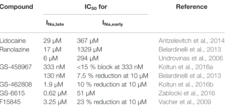
TABLE 1 |IC50values of selective late sodium current inhibitors for the late and the early sodium current component.
Compound IC50for Reference
INa,late INa,early
Lidocaine 29 µM 367 µM Antzelevitch et al., 2014
Ranolazine 17 µM 1329 µM Belardinelli et al., 2013
6 µM 294 µM Undrovinas et al., 2006
GS-458967 333 nM <15 % block at 333 nM Koltun et al., 2016a 130 nM 7.5 % reduction at 10 µM Belardinelli et al., 2013 GS-462808 1.9 µM 10 % reduction at 10 µM Koltun et al., 2016b
GS-6615 0.62 µM 51 µM Zablocki et al., 2016
F15845 3.25 µM 23 % reduction at 10 µM Vacher et al., 2009 Where the IC50value is missing, inhibition percentage at a given concentration was used instead.
![FIGURE 2 | How can an impaired sodium homeostasis of cardiac myocytes lead to arrhythmias? AP, action potential; NHE, Na + /H + exchanger; [Na + ] i , intracellular sodium concentration; NCX, Na + /Ca 2+ exchanger; [Ca 2+ ] i , intracellular calcium concen](https://thumb-eu.123doks.com/thumbv2/9dokorg/790471.36984/6.892.70.824.610.1004/homeostasis-arrhythmias-potential-exchanger-intracellular-concentration-exchanger-intracellular.webp)