Cardiac effects of long-term exercise training and acute exhaustive exercise in rat models
PhD Doctoral Dissertation
Attila Oláh, MD
Semmelweis University
Doctoral School of Basic Medicine
Tutors: Tamás Radovits, MD, PhD, assistant professor László Gellér, MD, PhD, associate professor Opponents: Tamás Csont, MD, PhD, associate professor
Tamás Ivanics, MD, PhD, associate professor Head of Examination Commission:
Emil Monos, MD, DSc, professor emeritus Members of Examination Commission:
Gábor Pavlik, MD, DSc, professor emeritus Zsuzsanna Miklós, MD, PhD, assistant professor
Budapest
2015
1
Contents
1. List of abbreviations ... 4
2. Introduction ... 7
2.1. Long-term exercise training-induced cardiac alterations, the athlete’s heart .... 8
2.1.1. Structural alterations in athlete’s heart ... 9
2.1.2. Functional consequences of exercise training ... 11
2.1.3. Molecular pathways related to physiological hypertrophy ... 12
2.1.4. Other characteristic changes of exercise-induced cardiac hypertrophy ... 13
2.1.5. Cardiac arrhytmogenic remodeling, sudden cardiac death ... 14
2.1.6. Differences between physiological and pathological cardiac hypertrophy ... 15
2.1.7. Animal models of athlete’s heart... 17
2.2. Acute exhaustive exercise-induced cardiac changes ... 20
2.2.1. Myocardial biomarkers of cardiac damage ... 20
2.2.2. Myocardial dysfunction after prolonged exercise ... 22
2.2.3. Acute exercise and oxidative stress ... 26
2.2.4. Animal models of acute exhaustive exercise induced cardiac injury ... 28
3. Aim of the work ... 30
4.Methods ... 31
4.1. Animals, experimental groups ... 31
4.2. Animal models, exercise protocols ... 33
4.2.1. Rat model of athlete’s heart ... 33
4.2.2. Rat model of acute exhaustive exercise-induced cardiac injury ... 34
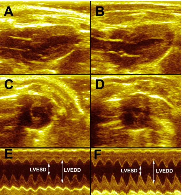
4.3. Echocardiography ... 35
4.3.1. Standard echocardiographic assessments ... 35
4.3.2. Speckle-tracking echocardiography ... 36
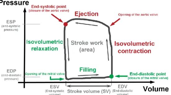
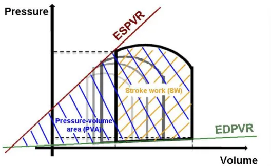
4.4. Hemodynamic measurements, left ventricular pressure-volume analysis ... 37
4.5. Blood and tissue sample collection ... 40
4.6. Biochemical measurements ... 40
4.6.1. Stress biomarkers ... 40
4.6.2. Cardionecrotic biomarkers ... 40
4.7. Histology ... 41
4.7.1. Hematoxylin-eosin (HE) staining ... 41
2
4.7.2. Masson’s trichrome staining ... 41
4.7.3. Dihydroethidium (DHE) staining ... 42
4.7.4. Nitrotyrosine (NT) staining ... 42
4.7.5. Terminal deoxynucleotidyl transferase-mediated dUTP nick-end labeling (TUNEL) staining ... 43
4.8. Cardiac mRNA analysis ... 43
4.9. Statistical analysis ... 44
5. Results ... 45
5.1. Athlete’s heart – exercise training-induced alterations ... 45
5.1.1. Body weight and heart weight ... 45
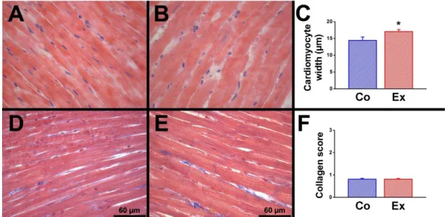
5.1.2. Histology ... 45
5.1.3. Markers of stress and pathological hypertrophy ... 46
5.1.4.Echocardiographic parameters... 47
5.1.5. Hemodynamic parameters ... 50
5.1.6. Reversibility of exercise-induced cardiac hypertrophy ... 55
5.2. Investigation of the correlation between strain values measured by speckle- tracking echocardiography and contractility parameters derived from pressure-volume analysis ... 57
5.2.1. Morphological markers of left ventricular hypertrophy ... 57
5.2.2. Baseline hemodynamics ... 58
5.2.3. Contractility indices derived from pressure-volume analysis at different preloads ... 59
5.2.4. Strain parameters derived from speckle-tracking analysis ... 61
5.2.5. Correlations between contractility and strain parameters ... 63
5.3. Cardiac effects of acute, exhaustive exercise ... 64
5.3.1. Body weight and heart weight ... 64
5.3.2. Biochemical parameters ... 64
5.3.3. Histology ... 65
5.3.4. Gene expression analysis ... 67
5.3.5. Hemodynamic parameters ... 70
6. Discussion ... 74
6.1. Long-term exercise-induced cardiac changes ... 74
3
6.1.1. Exercise-induced left ventricular hypertrophy ... 74
6.1.2. Systolic function and cardiac contractility ... 75
6.1.3. Diastolic function ... 76
6.1.4. Mechanoenergetics ... 76
6.1.5. Reversibility of exercise-induced cardiac hypertrophy ... 77
6.2. Correlation of contractility indices of pressure-volume analysis and speckle- tracking echocardiography ... 78
6.2.1. Left ventricular hypertrophy ... 78
6.2.2. Baseline hemodynamic data ... 78
6.2.3. Sensitive left ventricular contractility indices derived from pressure- volume analysis ... 79
6.2.4. Strain and strain rate measured by speckle-tracking echocardiography ... 79
6.2.5. Correlation between strain parameters and sensitive contractility indices .. 80
6.3. Acute exhaustive exercise-induced cardiac changes ... 81
6.3.1. Biomarkers of myocardial injury ... 81
6.3.2. Cardiac dimensions and baseline hemodynamics ... 81
6.3.3. Left ventricular contractility ... 82
6.3.4. Mechanoenergetics ... 83
6.3.5. Exhaustive exercise-induced oxidative stress ... 83
6.3.6. Oxidative stress induced apoptosis and dysregulation of matrix metalloproteinases ... 84
6.4. Limitations ... 85
7.Conclusions ... 87
8. Summary ... 88
9. Összefoglalás ... 89
10. References ... 90
11. List of publications ... 114
12. Acknowledgements ... 116
4
1. List of abbreviations
α-MHC - α-myosin heavy chain ACTH - adrenocorticotropic hormone AGE - advanced glycation end-products Akt - protein kinase B
ANF - atrial natriuretic factor AST - aspartate transaminase β-MHC - β-myosin heavy chain
Bax – B-cell lymphoma 2 associated X protein Bcl-2 - B-cell lymphoma 2
BNP - brain natriuretic peptide BW - body weight
CI - cardiac index CK - creatine kinase
CK-MB - myocardial band isoform of creatine kinase cMRI - cardiac magnetic resonance imaging
CO - cardiac output
CSr - circumferential strain rate cTn - cardiac troponin
cTnT - cardiac troponin T DHE - dihydroethidium
dP/dtmax - maximal slope of systolic pressure increment
dP/dtmax-EDV - maximal slope of systolic pressure increment – end-diastolic volume relationship
dP/dtmin - maximal slope of diastolic pressure decrement E/A - early-to-late diastolic filling rate
Ea - arterial elastance ECG - electrocardiogram
ECLIA - electrochemiluminescence immunoassay ECM - extracellular matrix
EDPVR - end-diastolic pressure-volume relationship Ees - end-systolic elastance
EF - ejection fraction Eff - mechanical efficiency
eNOS - endothelial nitric oxide synthase
5 ESPVR - end-systolic pressure-volume relationship FAC - fractional area change
FS - fractional shortening
G6PD - glucose-6-phosphate dehydrogenase
GAPDH - glyceraldehyde-3-phosphate dehydrogenase GCS - global circumferential strain
GLS - global longitudinal strain GPX-1 - glutathione peroxydase-1 GSR - glutathione reductase H2O2 - hydrogen peroxide
HCM - hypertrophic cardiomyopathy HE - hematoxylin-eosin
HR - heart rate HW - heart weight
IGF-1 - insulin-like growth factor 1
IGF-1R - insulin-like growth factor 1 receptor IVST - interventricular septal thickness LDH - lactate dehydrogenase
LSr - longitudinal strain rate LV - left ventricle, left ventricular
LVAWT - left ventricular anterior wall thickness LVEDD - left ventirular end-diastolic diameter LVEDP - left ventricular end-diastolic pressure LVEDV - left ventricular end-diastolic volume LVESD - left ventricular end-systolic diameter LVESP - left ventricular end-systolic pressure LVESV - left ventricular end-systolic volume LVPWT - left ventricular posterior wall thickness MAP - mean arterial pressure
MAPK - mitogen-activated protein kinases MMP - matrix metalloproteinase
MT - Masson’s trichrome
NADPH - nicotinamide adenine dinucleotide phosphate NO. - nitric oxide
NT - nitrotyrosine
6
NT-proBNP - N-terminal of the prohormone brain natriuretic peptide O2-.
- superoxide anion OH. -hydroxyl radical ONOO- - peroxynitrite
PI3K - phosphatidylinositol-3-kinase PRSW - preload recruitable stroke work P-V - pressure-volume
PVA - pressure-volume area ROS - reactive oxygen species RV - right venticle, right ventricular SCD - sudden cardiac death
SEM - standard error of the mean
SERCA2a - sarcoplasmatic reticulum Ca2+-ATPase 2a SOD-2 - superoxide dismutase 2
STE - speckle-tracking echocardiography SV - stroke volume
SVI - stroke volume index SW - stroke work
τ - time constant of left ventricular pressure decay TBARS - thiobarbituric acid reactive substance TDI - tissue Doppler imaging
TIMP - tissue inhibitor of metalloproteinases TGF-β - transforming growth factor beta TPR - total peripheral resistance
TUNEL - terminal deoxynucleotidyl transferase-mediated dUTP nick end-labeling VAC - ventriculo-arterial coupling
7
2. Introduction
It has been well documented that regular physical activity yields significant health benefits. Regular exercise reduces cardiovascular mortality approximately by 35% and all-cause of mortality by 33% and physical activity grants longer life for individuals (Nocon et al., 2008). By contrast, a lack of exercise can lead to many chronic diseases related to cardiovascular system (Roberts et al., 2013). Additional cardiovascular benefits of excercise include weight reduction, blood pressure reduction, increased insulin sensitivity and improved lipid profile (D’Silva and Sharma, 2014). However, recently the possible harmful effects of high-intensity prolonged exercise was also emphasized mainly because of increased vulnerability for atrial and ventricular arrhythmias and an increased risk of sudden cardiac death (La Gerche, 2007; La Gerche et Prior, 2013; Guasch and Nattel, 2013).
What remains unknown is precisely how much exercise is required to produce these beneficial effects and, more contraversially, how much exercise could cause harm?
The first studies suggested that more intensive the physical activity is undertaken, the greater the cardiovascular benefits (Tanasescu et al., 2002; Schnohr et al., 2013; Sesso et al., 2000). Thus for many years ’the more, the better’ concept has prevailed in evaluatuion of physical activity. Other data from large cohort studies have shown that increased exercise capacity, daily time of exercise and weekly run distances implies a large reduction in mortality until higher level of fitness (Kokkinos et al., 2008). After approaching this fitness level, the beneficial effect appeared to plateau: higher milage or more frequent running were not associated with better survival, thus a curvilinear relationship between mortality and level of fitness was supposed (Wen et al., 2011).
Controversially, some publications have proposed that a U-shaped curve exists for optimal exercise dosage and extreme levels of vigorous physical activity for prolonged periods could have harmful effects on the heart (La Gerche, 2007, O’Keefe and Lavie, 2013).Recent American Heart Association Guidelines recommend that 150 minutes of moderate-intensity aerobic exercise or 60 minutes of vigorous-intensity physical activity is necessary to promote and maintain health (Haskell et al., 2007). This guideline also recognize that ’[…] the shape of the dose–response curves, the possible points of maximal benefit, […] remain unclear’. Exercise training regimes planned to prepare for
8
prolonged races (i.e. marathon race) are several-fold greater than the recommendation, while the number of followers (also elite, trained as well as nonelite, amateur individuals) of extremely prolonged training and races has risen during the last decades and is still rising (Guasch and Nattel, 2013).
Hereinafter I present shortly the so far described cardiac effects of long-term exercise training and the acute, prolonged intense exercise as an introduction of my work.
2.1. Long-term exercise training-induced cardiac alterations, the athlete’s heart
Regular exercise training is accompanied by structural, funcional and electrical alterations of the heart to enhance performance. The resulting phenotype, the cardiac enlargement was already recognized more than hundred years ago based on clinical examination, with the recognition of cardiac enlargement and bradycardia among highly trained athletes (Darling, 1899; Henschen, 1899). In the 20th century our understanding has gradually expanded in parallel with the dynamic development of new invasive and non-invasive tools for understanding the effects of physical activity on the heart.
Initially, the chest x-ray and ECG demonstrated the enlargement of cardiac chambers and cardiac pathological examination were undertaken, which were followed by the use of modern imaging techniques, such as echocardiography and cardiac magnetic resonance imaging (cMRI) to deepen into the understanding of athlete’s heart. Recently, numerous studies have been undertaken about athlete’s heart in vivo as well as in vitro and the knowledge of these investigations were summarized in several expanded reviews (Pluim et al. 2000; Fagard, 2003, Maron and Pelliccia, 2006; Pavlik et al., 2010; Prior and La Gerche, 2012, Pavlik et al., 2013). Thus, during the last century there has been considerable interest on the constellation of exercise-induced cardiovascular alterations.
Investigations of athlete’s heart is important for a number of essential reasons.
First, to understand how the adaptation of the cardiovascular system contributes to improved athletic performance. The understanding of the mechanisms leading to enhanced function could be essential because pro-active therapeutic interventions -
9
based on the same stimuli and genes leading to physiological growth- may provide an additional strategy to prevent or treat heart failure (McMullen and Jennings, 2007).
The second is to enhance physical performance to optimise cardiac functional adaptation for elite athletes. This is important in terms of sports cardiology for elite, top- level athetes.
The third and maybe the most important is to allow the differentiation of normal athlete’s heart from pathological cardiac states, especially from hypertrophic cardiomyopathy and arrhythmogenic right ventricular cardiomyopathy. These are pathological conditions, which may share similar morphological features like exercise- induced hypertrophy, nevertheless these phenotypes are associated with increased risk of sudden cardiac death (Prior et La Gerche, 2012).
2.1.1. Structural alterations in athlete’s heart
A fundamental component of exercise-induced cardiac remodeling is physiological cardiac hypertrophy, a process leading to increased cardiac myocyte size.
During physical activity the loading conditions are altered in the cardiovascular system.
Different forms of exercise impose different loads of the cardiovascular system. These differential effects of pressure and volume loading during training was first documented in the essential work by Morganroth, in which he identified cardiac morphological differences between different kind of sports (Morganroth et al., 1975). It was hypothesized that these adaptations reflect differential haemodynamic loading during training. According to this comparative work, athletes involved in dynamic, isotonic exercise (e.g. running and swimming) had increased left ventricular mass with paralelly increased cardiac dimensions because of chronic volume overload. Thus endurance- trained athletes are presumed to demonstrate eccentric left ventricular hypertrophy, characterized by an unchanged relationship between left ventricular (LV) wall thickness, LV diameter and as a consequence, unaltered relative wall thickness (ratio of wall thickness to diameter). Participation in sports with a high static demand (isometric exercise, e.g. weightlifting or wrestling) is associated with increased LV wall thickness, which is increased in relation to cavity size like in similar disease states with chronic pressure loads. Thus, strength-trained athletes were presumed to demonstrate concentric LV hypertrophy, which is characterized by increased relative wall thickness values.
10
Sports such as cycling and rowing are examples of combined endurance and strength exercise: endurance training while working against an elevated resistance. These athletes display the greatest degree of LV dilatation and wall thickening (Pelliccia et al., 1991; Pluim et al., 2000; Barbier et al., 2006). Since that, after a large amount of echocardiographic studies and meta-analyses, we know that the development of an endurance-trained heart and a strength-trained heart is not to be considered an absolute and dichotomous concept and the geometric pattern of athlete’s heart is more complicated than expected by Morganroth (Pluim et al., 2010; Pavlik et al., 2010). Each sport can be classified by its dynamic (according to the estimated percent of maximal oxygen uptake) and static (according to estimated percent of maximal voluntary contraction) component, and the observed phyenotype of LV hypertrophy is dependent on these factors (Mitchell et al., 2005). Moreover, morphological manifestation is influenced by numerous other factors, such as age, gender, ethnicity and genetic factors (Pavlik et al., 2013).
The observation of training-induced structural changes of RV by echocardiography was not so clear for a long time because RV geometry and its location in the chest made it difficult to investigate. Thus the effect of exercise training on the right ventricle has been observed more recently, due to the technical development in cardiac imaging with the appearence of cMRI. An increased RV volume, mass and stroke volume have all been observed in athletes, largely based on measurements using cMRI. The balanced hypertrophy of the RV, similar as in the LV, has been described in endurance athletes (Scharhag et al. 2002; Scharf et al. 2010). However, recent works suggest that the RV undergoes remodelling process and dilate slightly more than the LV after endurance training (La Gerche et al., 2011).
In addition to the findings in ventricle morphological alterations, studies have shown atrial enlargement in trained athlete's heart, which predispose athletes to an increased risk of subsequent atrial fibrillation (Grimsmo et al., 2010; Wilhelm et al., 2012).
Detraining has been defined as the partial or complete loss of training-induced anatomical, physiological and performance adaptations, as a consequence of training reduction or cessation (Mujika and Padilla, 2001). Clinical studies have reported regression of the LV morphological changes characteristic of the athlete’s heart after
11
long-term detraining (Pelliccia et al., 2002), which property can be used to differentiate it from pathological conditions (Mitchell et al., 2013).
2.1.2. Functional consequences of exercise training
While structural changes within the LV has been very clearly and consistently demonstrated, the evidence regarding functional change has been less consistent (La Gerche et al., 2009). These studies demonstrate consistency in the fact, that athlete’s heart, in contrast to pathological hypertrophy, have preserved or even enhanced systolic and diastolic function (Utomi et al., 2013). These findings are based mainly on non- invasive functional studies that have used echocardiography, while a small subset have used cMRI.
Echocardiography has contributed most to our current understanding of cardiac morphology and function in athletes because of its low cost, widespread availablity and lack of ionizing radiation. LV systolic function has most often been studied by using echocardiography and expressed as fractional shortening of the LV internal dimension or ejection fraction. The meta-analyses on these conventional indices of LV systolic function were not different between athletes at rest and matched control subjects (Fagard et al. 1996, Pluim et al. 2000). cMRI studies also did not show any difference in systolic fuction between athletes and untrained individuals during resting conditions (Pluim et al., 1998; Scharf et al., 2010). However, the parameters used in human studies reflect chamber mechanics rather than intrinsic myocardial mechanics. Tissue Doppler and speckle-tracking imaging permits a more detailed and more accurate assessment of LV systolic function. Normal or even supernormal cardiac systolic and diastolic function was found in investigations using these methods, which can be used for the differential diagnosis of athlete’s heart from pathological conditions (D’Andrea et al., 2006; Richand et al., 2007, Simzek et al., 2013). The evaluation of contractility in humans is complicated and all of these above mentioned systolic parameters are influenced by cardiac loading conditions and heart rate. Whereas the harmless nature of physiological hypertrophy does not allow to perform invasive studies that can provide the most accurate and reliable, load-independent characterization of LV function.
LV diastolic function is commonly assessed by studying the pattern of ventricular filling through the mitral valve by Doppler echocardiography (Nishimura et al., 1989).
12
The ratio of the transmitral Doppler peak flow velocity during atrial contraction (A) to the peak flow velocity during rapid LV filling (E) was found to be normal or slightly enhanced in all type of sports (Fagard, 1997; D’Ascenzi et al., 2011). In some studies, however, the A wave was proportionately lower than the E wave. According to Pavlik et al. observations in young ages show no difference in early-to-late diastolic filling rate (E/A) ratio, while in older age E/A is definitely higher in athletic than in non-athletic subjects (Pavlik et al., 2010). Thus, it seems that regular physical training attenuates the age-associated impairment of diastolic function. These results should be interpreted with some caution, because E/A ratio is not only related to LV compliance but can be also influenced by other factors such as heart rate and loading conditions (Harrison et al., 1991). It is possible that the higher E/A is only a consequence of the training induced bradycardia and not an independent training effect. In general, studies of diastolic filling have confirmed that the structural remodelling seen as part of athlete’s heart is not associated with impairment of diastolic filling as observed in other kinds of LV hypertrophy and further examination is needed to observe load-independent parameters.
Right ventricular myocardial function has been described by echocardiography in athletes compared to healthy controls and patients with hypertrophic cardiomyopathy (HCM), and in these investigations improved systolic function (higher RV systolic velocities as well as strain values) was observed along with unaltered fractional area change (FAC, calculated as the ratio of area change in one cardiac cycle and end- diastolic area) in resting conditions (D’Andrea et al., 2003; D’Andrea et al., 2009).
Cardiac magnetic resonance imaging (cMRI) has excellent spatial resolution and the image quality is not influenced by body habitus. These features permit the very precise assessment of cardiac chamber size, particulary for the irregularly-shaped right ventricle (RV), where it is considered as gold standard. Studies using cMRI showed a balanced hypertrophy and unaltered ejection fraction in the right ventricle (Scharhag et al., 2002).
2.1.3. Molecular pathways related to physiological hypertrophy
There has been great interest to reveal molecular mechanisms leading to enhanced function in physiological hypertrophy. All of these studies are in agreement with the fact that the insulin-like growth factor 1 (IGF-1)- phosphatidylinositol-3-kinase (PI3K)-
13
protein kinase B (Akt) pathway plays a key role in mediating physiological cardiac growth (Ellison et al., 2012). It is well recognised that IGF-1 is released during development and in response to exercise training (Neri Serneri et al., 2001). IGF-1 produced also within the myocardium acts via the IGF-1 receptor (IGF-1R), a transmembrane receptor tyrosine kinase. IGF-1R transgenic mice displayed a greater hypertrophy with enhanced systolic function (McMullen et al., 2004), whereas cardiac selective knockout of IGF-1R showed a resistant phenotype against exercise-induced cardiac changes (Kim et al., 2008). Activation of IGF-1R leads to activation of PI3K(p110α), a lipid kinase composed of separate regulatory and catalytic subunits that phosphorylates lipids to form phosphatidyl-inositol 3,4,5-trisphosphate (PIP3), which acts as a second messenger to cause downstream signaling events, such as phosphorylation and activation of Akt. Transgenic and knockout mice studies have confirmed that PI3K(p110α) plays also a key part in exercise-induced hypertrophy and is also important for protecting the heart against pathological insults (McMullen et al., 2003; Lin et al., 2010). Akt, a serine/threonine kinase is a well characterized target of PI3K activity and it is well recognized that Akt1 is the predominant Akt isoform in the myocardium (Fujio et al., 2000). Akt1 is required for physiological heart growth as proved by an investigation revealing that Akt1 knockout mice showed a blunted hypertrophic response to exercise training (DeBosch et al., 2006). In the heart, the IGF1-PI3K pathway induces physiological hypertrophy with preserved or enhanced cardiac function, thus this pathway is important for maintaining cardiac function and its antifibrotic and anti-apoptotic effects can provide protection for the heart against pathological insults (McMullen, 2008). The gp130-JAK-STAT pathway and thyroid hormone receptor signaling contribute to the development of cardiac hypertrophy and postnatal heart growth, respectively, and have been associated with the IGF1-PI3K-Akt physiological hypertrophy pathway, although the exact mechanisms are still unclear (Bernardo et al., 2010). The specific activation of these pathways could be a potential target for the treatment of cardiovascular diseases, such as heart failure.
2.1.4. Other characteristic changes of exercise-induced cardiac hypertrophy In addition to structural and functional remodeling, it is recognised that there are electrical alterations in the athlete’s heart in response to training, resulting in distinct
14
changes in the ECG. Autonomous regulation of the physically trained subjects is characterized by an enhanced parasympathetic and a decreased sympathetic activity resulting in bradycardia during resting conditions. Resting bradycardia is the most common and characteristic electrical feature of the athlete’s heart: with this alteration the duration of diastole is elongated in athletic heart, while duration of systole remains relatively unaltered (Pavlik et al., 1999). This is more advantageous as coronary circulation occurs only in diastole, thus there will be a better blood supply of the myocardium between two cardiac contractions. Exercise-induced remodeling can result in other, well-described electrical alterations of the myocardium. It is really important for clinicians to be able to identify those changes, which are resulted from intense physical training and do not carry an increased risk of adverse cardiac outcomes (Pellicia et al., 2000). The European Society of Cardiology recently published guidelines for interpretation of the 12 lead ECG in the athlete. It provides classification of ECG abnormalities, which are considered as commonly related to training (such as sinus bradycardia, first-degree atriventricular block, incomplete right bundle branch block, signs of early repolarisation and isolated QRS voltage criteria for left ventricular hypertrophy) and those which are uncommon and should prompt examination for underlying cardiac pathology (Corrado et al. 2010).
Beside elongation of the diastolic phase as a consequence of bradycardia, an increased capillary density also contributes to the better blood supply of the myocardium and this improvement seems to be associated with training in younger ages (Tomanek, 1994). Athlete’s heart has also been characterized with a more effective metabolism. Positron emission tomography demonstrated, that trained athletes are able to perform the similar physical performance with lower myocardial perfusion, thus the heart needs less oxygen to provide the same performance (Laaksonen et al., 2007). The lower rates of glycolysis along with balanced enhancement in glucose and fatty acid oxidation suggests a protective phenotype (Burcelle et al., 2004).
2.1.5. Cardiac arrhytmogenic remodeling, sudden cardiac death
Despite the evident benefits of alterations in the heart of an athlete, numerous observational studies have raised concerns that high-level exercise training may be associated with increased risk of cardiac arrhythmia and even primary cardiac arrest.
15
Estimated incidence of sudden cardiac death (SCD) in young athletes varies in a wide range, from 1:25,000 to 1:300,000, mainly because of differences in the population studied (D’Silva and Sharma, 2014). The highest volume study, a long-term registry of italian athletes indicated 1.9 deaths/100,000 person yearly, with a 2.4-fold risk compared to untrained controls (Corrado et al., 2006). In athletes aged above 35 years atheromatous coronary artery is obviously the leading cause of SCD. In contrast, in young athletes suffered from SCD, hypertrophic cardiomyopathy, congenital anomalies in coronary arteries and arrhythmogenic right ventricular cardiomyopathy were found as most common underlying causes. Only about 2 percent of young athletes who die suddenly have normal cardiac structure at autopsy, and no definitive cause of death can be established (Maron, 2003). As most SCDs in young athletes occur during or immediately after exercise, it is hypothesized that exercise-driven mechanisms, such as dehydration, hyperpyrexia or electrolyte imbalances can trigger the operation of underlying arrhythmogenic substrate (Corrado et al., 2006). Several clinical studies suggested that long-term high level exercise might be associated with an increased risk of ventricular tachyarrhytmias, which have been shown to originate from the RV (Biffi et al., 2002; Heidbuchel et al., 2003). It is also observed that long-term endurance training may promote atrial enlargement and increased risk of atrial fibrillation in large epidemiological studies (Molina et al., 2008; Aizer et al., 2009). Confirming these concerns, an animal model of long-term intensive exercise-induced hypertrophy by Benito et al. showed an increase in ventricular arrhythmia vulnerability and atrial fibrillation incidence in exercised rats, while an increased interstitial collagen deposition was observed in the right ventricle and left atrium with a reversible character (Benito et al., 2011). Further investigations are needed to evaluate the relevance of these findings about cardiac arrhytmogenic remodeling in athletes.
2.1.6. Differences between physiological and pathological cardiac hypertrophy In response to an increased load, the heart must work harder than under normal conditions. To counterbalance the chronic increase in wall stress, cardiomyocytes enlarge leading to an increase in size and mass, which is called cardiac hypertrophy (Zak et al., 1979; Cooper, 1987). Pathological and physiological cardiac hypertrophy are caused by different stimuli, and are associated with distinct structural, functional and
16
molecular phenotypes (McMullen and Jennings, 2007). Both types of hypertrophy can be subclassified as concentric and eccentric based on changes in shape that are dependent on the initiating stimulus. This type of structural classification of athlete’s heart has been already discussed above. A pathological stimulus causing presure overload (e.g. hypertension, aortic stenosis) produces an increase in systolic wall stress that results in concentric hypertrophy, while volume overload of the heart (e.g. mitral regurgitation, arteriovenosus fistulas) leads to an increase in diastolic wall stress resulting in eccentric hypertrophy (Grossman et al., 1975).
The balanced increase of myocardial mass in hypertrophy following long-term exercise training results in preserved or enhanced cardiac function with increased cardiac output under stress condition (Ellison et al., 2012). In contrast, pathological hypertrophy due to pressure or volume overload is initially a compensatory mechanism, which may eventually decompensate, leading to left ventricle dilatation and heart failure with an increased risk of SCD (Zak et al., 1979; Opie et al., 2006).
Animal studies have demonstrated that physiological and pathological cardiac hypertrophy have distinct structural and molecular bases in the myocardium (Iemitsu et al., 2001; McMullen et al., 2003). In contrast to physiological heart growth, which is stimulated by IGF-1 and acts on the IGF-1R-PI3K-Akt pathway, the main triggers for pathological hypertrophy are the release of endothelin-1, angiotensin II and norepinephrine. These prohypertrophic hormones act by binding to Gq protein-coupled receptors and act on a fundamentally distinct pathway, where the main downstream effectors are calcineurin, protein kinase C and mitogen-activated protein kinases (MAPK) (Weeks and McMullen, 2011). In response to pathological stimuli, after loss of cardiomyocytes because of apoptosis and necrosis, extracellular matrix proteins accumulate disproportionately and this fibrotic replacement excessively leads to mechanical stiffness. In contrast, a normal network of collagen fibres surrounding cardiac myocytes could be observed in exercise-induced hypertrophy without apoptosis.
Models of pathological cardiac hypertrophy are associated with upregulation of fetal genes, including atrial natriuretic factor (ANF) and β-myosin heavy chain (β-MHC), which does not occur in models of physiological hypertrophy (McMullen et al., 2003).
Gene expression of proteins associated with contractile function, like α-myosin heavy chain (α-MHC) and sarcoplasmatic reticulum Ca2+-ATPase 2a (SERCA2a) are down-
17
regulated in pathological conditions, in contrast to physiological hypertrophy (Bernardo et al., 2010).
As mentioned before, exercise-induced cardiac hypertrophy is characterized by enhanced fatty acid and glucose oxidation. In contrast, pathological hypertrophy is associated with a decreases in fatty acid oxidation and increases in glucose metabolism (Christe and Rodgers, 1994). In case of progression to heart failure glucose metabolism decreases reducing the overall ability of the heart to generate sufficient energy (Neubauer, 2007).
Reversibility is a distinct feature between physiological and pathological hypertrophy. The most actual question for physicians in the field of cardiology is the differentiation of physiological LV hypertrophy from HCM, the leading cause of exercise related SCD in young athletes (Maron, 2003). The observed morphological alterations can overlap between the heart of highly trained athletes and structurally mild HCM (Basavarajaiah et al., 2008). Thus, the differentiation between the two entities may be challenging for even the most experienced cardiologist. Systematic evalutation containing a detailed physical and family history, 12-lead ECG and echocardiography and additional investigation with exercise testing, cMRI as well as screening for casual genetic mutations may be necessary for clear differentation (Rawlins et al., 2009). In doubtful cases the reversible nature of the observed hypertrophy after discontinuation of training can provide the definitive diagnosis.
2.1.7. Animal models of athlete’s heart
Because of research ethics and technical difficulties in humans, exercise models using animals are requisited for the future development of exercise physiology. The broader goals of animal models are to deepen our knowledge in exercise physiology and to uncover the molecular and cellular mechanisms (Seo et al., 2014).The use of animals in laboratory studies has been recommended by many researchers because of their homogeneity (genetically, physiologically) compared to human subjects and the tight control of exercise duration. Despite the number of advantages in animal exercise models, some limitations are also present. The tissue response to mechanical stresses and therapeutic treament may not be same to humans because of differences in the size and the movement patterns of the musculoskeletal system (Hasenfuss, 1998).
18
Animal exercise models have been developed to simulate the physical activities of humans. Indeed, there are no golden standard exercise modes or protocols for modeling athlete's heart since in addition to exercise mode the intensity, the timing and the duration are the critical determinants of physiological responses and various final outcomes and also enviromental factors (temperature), sex, age and strain affect the outcome (Wang et al., 2008).Additionally, a large amount of variation in animal studies was observed even between the same exercise modalities, because of the different apparatuses used with various training protocols. Thus animal exercise protocols should be well-designed to achieve research goals.
A number of animal models of exercise-induced cardiac hypertrophy have been developed and several types of endurance exercise training have been recognized to effectively induce physiological cardiac hypertrophy in experimental animals, such as treadmill running, voluntary wheel running and swim training (Wang et al., 2008).
Treadmill running is a widely used model because its relative simplicity and effectiveness. Moreover, both exercise intensity and duration are clearly controllable and several animals can be trained simultaneously. It can be performed either in a continuous manner with a fixed or progressively increased speed, or in an interval running mode. The disadvantages of treadmill running are the possibility of causing stress or limb injury and the need for expensive treadmill apparatus (Wang et al., 2008).
Forced exercise may be recognized as a type of stress, voluntary exercise is the most effective intervention in lessening this stress response. When compared to other types of exercise, spontaneous, voluntary wheel running has the strong advantage of voluntariness, that the exercise can be accomplished with minimal intervention by the investigator, thus it is comfortable for investigating long-term effects of increased physical activity. However, this type of exercise is not suitable for studies that require precise timing for training to achieve certain cardiac hypertrophy, since the investigator cannot easily regulate the duration or intensity of exercise sessions (Seo et al., 2014).
Regular swimming training appears to be effective for inducing relevant cardiac hypertrophy. Swimming exercise involves an inexpensive device as compared to treadmill running and the duration and load of exercise can be controlled to a greater extent than with voluntary wheel running. Rodents have the innate ability to swim, the animals already possess self-motivation, thus swimming exercise can be perfomed after
19
a short period of familiarization. Moreover, a relatively large number of animals can be exercised simultaneously and there is a negligible chance an animal to suffer injuries.
Unlike with the treadmill or wheel running, sedentary animals should be placed in the water to exclude the effect of the water itself (Wang et al., 2010; Seo et al., 2014).
20
2.2. Acute exhaustive exercise-induced cardiac changes
Ultraendurance athletes routinely complete repeated bouts of prolonged, strenuous exercise. Thus, it was not surprising that the first scientific papers concerning the cardiovascular consequences of prolonged exercise were published in this extreme group of athletes (Saltin and Stenberg, 1964). This kind of intensive physical activity such as ironman triathlon, cross country biking or ultramarathon and 24-h running races are becoming more popular. Despite the increased prevalence, these extremely prolonged exercise efforts tend to be limited to the exceptionally well trained and prepared individuals.
Similar changes have been noted after marathon races, where non-professional,
”amateur” runners can participate, hence even greater scientific and media interest was observed (Neilan et al., 2006c; Fortescue et al., 2007). Articles in the media such as
„Runners who don’t train well can have marathon miseries” (The Boston Globe, April 17, 2006) and ‘‘Ironman athletes put hearts at risk of fatal damage, experts warn’’ (The Times, January 22, 2007) have been published about an awkward paradox for both the scientist and endurance athlete. The number of participants in marathon-races have strongly increased for the last 20 years, resulting in more than 500,000 runners finished a marathon in 2012 (Guasch and Nattel, 2013). To date, numerous research groups have been investigated the short- and long-term consequences of exhaustive exercise regarding cardiovascular health.
2.2.1. Myocardial biomarkers of cardiac damage
The first reports of post-exercise elevations in serum concentrations of myocardial band isoform of creatine kinase (CK-MB) after the completion of endurance events led to the recognition that such activities could result in cardiac injury (Siegel et al., 1981).
Elevations in serum CK-MB after prolonged exercise is not enough specific for the detection of myocardial damage, as CK-MB levels could be increased in the skeletal muscle of distant runners (Apple et al., 1984).
Cardiac troponins, originated from myocardial troponin complex, as a part of myocardial sarcomeric unit, are highly specific and sensitive markers of myocardial insult and are considered as the gold standard for biochemical detection of myocardial cell damage (Alpert et al., 2000). Numerous reports on animals (Chen et al., 2000. Nie
21
et al., 2010) and humans (Koller et al., 1999; Rifai et al., 1999; Shave et al., 2007; Nie et al., 2008; Scharhag et al. 2008) reported cardiac troponin (cTn) elevation in serum after prolonged exercise that can transiently exceed clinical cut-off value for myocardial infarction. Troponin levels from participants in marathon-distance (Mousavi et al., 2009), ultra-distance running races (Shave et al., 2002), triathlons (Tulloh et al., 2006) and dedicated cycling events (Neumayr et al., 2005) have each been studied and according to a recent meta-analysis detectable troponin values occurred in approximately one-half of participants (Shave et al., 2007). Interestingly, incidence of cTn detection was growing as duration shortened; specifically, there was a higher occurrence of post-exercise cTn elevation in marathon-type events in contrast to ultra- marathon competitions. This inverse relationship between event duration and troponin elevation might be because shorter races are generally performed at higher exercise intensities. For unclear reasons the prevalence and absolute serum concentrations of the cTn increases vary considerably. Possible explanations include differences in the fitness levels of participants, the type or duration of exercise and the timing of the postexercise sample. Although numerous studies have reported troponin-release after exercise, there is no consensus regarding the aetiology, mechanisms and clinical relevance of exercise- induced troponin release. Increased membrane permeability and/or myocardial cell necrosis propagated by increased coronary artery shear stress, myocardial stretch, oxidative stress and ischaemia have been proposed as possible explanations (Shave et al., 2010).
Increased myocardial wall stress caused by volume or pressure overload elevate blood concentration of brain natriuretic peptide (BNP) and its cleaved inactive fragment NT-proBNP, due to myocyte stretch. As a marker of cardiac dysfunction, BNP and NT- proBNP can be a helpful tool in cardiovascular diagnostics and risk stratification. As a counter-regulatory hormone, BNP reduces myocardial wall stress by an increase in natriuresis, vasodilation and sympathoinhibitory effects as an opponent of the renin- angiotensin system (Panagopoulou et al., 2013). Physical exercise can acutely elevate serum or plasma BNP and NT-proBNP concentrations (Scharhag et al., 2008). After prolonged and strenuous endurance exercise, BNP and NT-proBNP levels were increased both in healthy elite and nonelite athletes above the upper reference limit (Neilan et al., 2006a; Ohba et al., 2001). The degree of BNP and NT-proBNP elevation
22
has been related to endurance exercise duration (Scharhag et al., 2005), analogous to the time-dependent increase in BNP expression in stretched cardiomyocytes in vitro (Wiese et al., 2000). A feasible explanation for the exercise associated increase in BNP and NT- proBNP can be derived from the physiological significance of the active hormone BNP, which reduces preload and afterload to diminish myocardial wall stress.
It is important to note that exercise-associated elevations of cTn and NT-proBNP typically decrease significantly within 24 h after exercise and usually reach normal values within this period (Herrmann et al., 2003). Although biomarker elevation can be detected also in hihgly trained elite athletes, a reverse relation between exercise- associated elevations and prerace endurance training could be observed (Neilan et al.
2006a).
The appearance of such biomarkers of myocardial damage in healthy subjects participating in ultraendurance races associated with prolonged, strenuous exercise has raised concerns about the cardiovascular consequences of such exercise and the theory that extreme exercise can induce harmful processes in the heart was widespread reported (La Gerche and Prior, 2007; Dangardt, 2013). The clinical consequences of elevations in biomarkers following an acute bout of exercise are dubious because the alterations reported are quite small and transitory (George et al., 2008). Therefore the impact of long-term prolonged, strenuous exercise should receive more attention. We should also mention that methodological variation as well as differences in exercise mode, duration, training status, age and gender in studies that have examined the possibility of postexercise myocardial injury, making it difficulty to study the factors involved or the mechanisms responsible for postexercise cardiac troponin and NT- proBNP elevation (Shave et al., 2007).
2.2.2. Myocardial dysfunction after prolonged exercise
The concept that prolonged exercise can lead to a depression in left ventricular function was first presented by Saltin and Stenberg in the mid-1900’s (Saltin and Stenberg, 1964). Since then there has been enormous interest to observe prolonged strenuous exercise-induced functional changes and these alterations have been called as
”exercise-induced cardiac fatigue” (Douglas et al., 1987). Over 50 studies have been published so far about this phenomenon, but the findings have often been inconsistent
23
because of differences in research design, subject heterogenity and mode of assessment of cardiac function (Oxborough 2010).
Despite the known limitations in the two-dimensional (2D) assessment of global left ventricular systolic function, numerous studies have utilized this modality before and after an acute bout of exercise. Short duration exercise seems to have insignificant impact on LV function and as a consequence of increased preload and sympathetic activity, ejection fraction have been shown to be either unchanged or improved (Neilan et al., 2006b). The impact of prolonged exercise on LV systolic function is considerably controversial with no alteration in EF after ultra-long duration intensive physical activity (Hassan et al., 2006), while other studies reported clearly reduced EF after brief prolonged exercise (Vanoverchelde et al., 1991). This disparity could be a consequence of the differences in training status, exercise intensity and duration and the small sample size involved in one study. Recently several substantial meta-analyses have been reported for unified conclusions (Middleton et al., 2006; Oxborough et al., 2010). An overall reduction in the EF immediately following prolonged endurance exercise was observed, suggesting a transient impairment of LV systolic function, which returns to baseline following a 24-48 h recovery period (Whyte et al., 2000). The subgroup analyses showed that exercise-induced cardiac fatique is dependent on the duration of exertion in elite athletes as the systolic impairment was observed only in the ultralong duration (i.e. ironman and ultraendurance events, 640-1440 min) group, but not in moderate (i.e. half marathon races, 60-150 min) or long duration (i.e. marathon races, 166-430 min) group. (Whyte et al., 2000). However, the mean alteration of EF was significantly related to the decrease in the LV diastolic diameter, suggesting that postrace reduction of preload influence this parameter. As EF is a load-independent systolic parameter, more evidence is needed to prove systolic LV dysfunction after prolonged exercise. It is important to note that an untrained subgroup was defined in the moderate duration group. These data suggest that exercise-induced cardiac fatigue could concern professional athletes in ultra-endurance races and untrained individuals participating in moderate or long duration competitions (Whyte et al., 2000).
Tissue Doppler imaging (TDI)-derived systolic velocities appears to be unchanged after marathon race (George et al., 2006). Myocardial strain may be a more representative parameter of contraction and relaxation. TDI derived strain and strain rate
24
showed a reduction in both LV and RV systolic and diastolic parameters after completion of a marathon race (Neilan et al., 2006c). Speckle-tracking echocardiography (STE) offers a more unique and reproducible assessment of myocardial contractility with less dependency on loading factors (Marwick, 2006). The few works using STE demonstrated individual heterogenity between myocardial segments and plane, thus no consistent consequence could be concluded (La Gerche et al., 2008; George et al., 2009; Scott et al., 2009).
Regarding LV diastolic function, E/A has been a widely used index to observe diastolic changes after races. An immediate postexercise E/A ratio reduction was observed due to a drop in E and rise in A waves, reflecting altered diastolic filling dynamics. This impairment was independent of exercise duration and did not correlated with changes in loading conditions. The few available data suggest that E/A ratio returns to baseline following a 24-h recovery period (Whyte et al., 2000; Shave et al., 2004), suggesting minimal clinical impact of this phenomenon. In accordance, color flow Doppler investigation observed a decrease in postexercise early diastolic flow propagation velocity, thus a decreased E value (Middleton et al., 2006). Findings from tissue velocities during diastole are consistent in demonstrating a reduced early diastolic LV myocardial velocity (E’) and E’/A’ ratio (George et al., 2005). These findings were complemented by pulmonary venous Doppler measurements which demonstrated a reduction in atrial filling during diastole. Therefore, the interpretation of these results is complex: impaired E/A ratio could reflect a true impairment in myocardial relaxation or a reduction in ventricular filling secondary to decreased preload (Oxborough et al., 2010). More recently STE after marathon races revealed that strain indices of diastolic function from radial, circumferential and longitudinal planes were significantly reduced, suggesting a global reduction of LV diastolic function (Dawson et al., 2008). Load- independent indices of active relaxation would improve our knowledge about exercise- induced cardiac fatigue.
Alterations in LV function after an acute bout of prolonged exercise are normally transient, with resumption of normal function typically observed after 24-48 h of recovery (Shave 2004).
The assessment of right ventricular volumes with 2D echocardiography, is extremely complicated because of the geometry, location and the trabeculation of RV.
25
Therefore FAC was used to measure RV systolic function after strenuous exercise, however the results were controversial. Studies also demonstrated increased RV FAC (Douglas et. al, 1990), as well as reduced RV FAC (La Gerche et al., 2008) after prolonged exercise. La Gerche et al. also observed a reduction in RV systolic tissue velocity after a triathlon which complemented their findings from 2D echocardiography.
MRI is maybe the most suitable noninvasive investigation for examining RV. function.
Mousavi et al. validated RV systolic and diastolic dysfuncion detected by echocardiography by cMRI in a small group after completion of a marathon race (Mousavi et al, 2009). They reported decreased RV ejection fraction, which was likely due to exercise-induced pulmonary hypertension (thus increased RV afterload), which normalized after one week. Surprisingly, RV diastolic dysfunction had not completely normalized after one week follow-up. A more recent cMRI study showed transient RV dilation and dysfunction after intense exercise (La Gerche et al., 2012). The observed fibrosis in RV is in line with the hypothesis that repetitive insults eventually lead to RV dilatation and chronic dysfunction, providing a substrate for ventricular arrhythmogenesis (La Gerche et al, 2008). This is in line with the observation that athletes diagnosed with ventricular arrhythmia had RV abnormalities which served as an arrhytmogenic focus (Heidbuchel et al., 2003; Ector et al., 2007). Marked RV dysfunction and dilation with subsequent fibrosis as a consequence of repeated long- term exercise sessions could play a key role in the origination of complex ventricular arrhythmias, thus in causing sudden cardiac death of athletes.
A number of investigators have coupled biochemical and functional testing after endurance events. These data suggest that there is a correlation between cardiac biomarkers and post-race diastolic dysfunction (Neilan et al., 2006c) as well as with RV dysfunction (Mousavi et al. 2009).
26 2.2.3. Acute exercise and oxidative stress
Oxidative stress is a condition, in which the delicate balance between production of pro-oxidant free radicals and their subsequent amelioration via the antioxidant defense system becomes skewed in favor of free radical generation. Production or formation of free radicals in vivo is primarily initiated by the consumption of molecular oxygen (Halliwell and Cross, 1994). Reactive oxygen species (ROS) are oxygen-based chemical species with high reactivity. They include free radicals, such as superoxide (O2-.
) and hydroxyl radical (.OH) and nonradicals capable of generating free radicals, such as hydrogen peroxide (H2O2). The antioxidant defense system of the body serves to protect the cells from excess ROS production and is composed of endogenous (superoxide dismutases, catalase, glutathione peroxidase) and exogenous (ascorbate, bioflavinoids, carotenoids) antioxidants. Oxidative stress is defined as an excess production of ROS relative to the levels of antioxidants.
Exercise-induced oxidative stress was recognized long time ago (Davies et al., 1982). Because of difficulties in measuring free radical production directly, most human studies have used indirect markers to demonstrate exercise-induced oxidative stress.
Most often used markers of lipid peroxidation (e.g. thiobarbituric acid reactive substance (TBARS) levels, oxidative modifications to DNA (8-oxo-7,8-dihydroxy-2’- deoxyguanosine), protein oxidation (plasma protein carbonyls), antioxidant markers of the glutathione system (ratio of reduced and oxidized glutathione) as well as plasma levels of antioxidants and antioxidant enzymes reflect increased oxidative stress after prolonged exercise (Vollaard et al., 2005). Even moderate exercise may increase ROS production exceeding the capacity of antioxidant defence (Alessio, 1993). Exhaustive exercise-induced nitro-oxidative stress was demonstrated by increased nitrotyrosine levels in serum and urine after ultra-endurance race (Radák et al., 2003). Regular endurance exercise training causes adaptation, such as elevated antioxidant enzyme activity, to reduce systemic oxidative stress following an acute bout of exhaustive exercise (Radak et al., 2001; Miyazaki et al., 2001).
Although xanthine oxidase and NADPH-oxidase enzyme systems in cardiomyocytes, as well as infiltrating neutrophil granulocytes can generate free radicals in the myocardium, the primary source of ROS is the electron transport chain of mitochondria (Ji, 1999). Cardiac muscle has a high oxygen uptake even at resting
27
conditions. During heavy physical exercise oxygen uptake from the blood by the heart is markedly increased. During maximal exercise, whole-body oxygen consumption increases up to 20-fold, for which the myocardium and the skeletal muscle are responsible. High oxygen uptake and utilization in cardiac mitochondria to provide sufficient energy during exercise may lead to increased formation of free radicals, especially superoxide anions. Thus, this increased oxygen metabolism can lead to increased oxidative stress in the myocardium during physical exercise (Frankiewicz- Jozko et al., 1996). Furthermore, superoxide anions can interact with nitric oxide (NO) spontaneously to form toxic peroxynitrite (ONOO-) (Pacher et al., 2007). NO is necessary for normal cardiac physiology in the regulation of cardiac function including coronary vasodilatation and modulation of cardiac contractile function (Takimoto and Kass, 2007). Therefore superoxide anions effect not only directly but also by inactivation of cytoprotective NO and formation of the reactive oxidant peroxynitrite.
Regarding oxidative status exercise has double-edge effects on myocardium. On the one hand, it results in increased formation of free radicals, on the other hand it may also induce antioxidant enzymes to minimize the effects of oxidative stress due to exercise (Gul et al., 2006). Heart is equipped with all the major antioxidant enzymes such as superoxide dismutases, catalase as well as glutathione peroxidase and reductase.
It has been well demonstrated that acute bouts of exercise activates Nrf2, a primary transcriptional regulator of major antioxidants, which results in alterations of the transcription of antioxidant genes such as catalase, glucose-6-phosphate dehydrogenase (G6PD), glutathione peroxydase-1 (GPX1), glutathione reductase (Muthusamy et al, 2012). This repeated exposure can lead to a favorable adaptation, an improved myocadial antioxidant defense system in trained heart, which can reduce potential damage from future acute bouts of exercise. This theory is underpinned by studies that shows reduced myocardial oxidative stress after preconditioning by exercise training (Gul et al., 2006; Okudan et al., 2012)
At physiological circumstances, ROS can act as second messengers in several cellular functions, because stimulation of DNA-synthesis and induction of growth- related genes are associated with free radicals (Grieve et al., 2004). This role of ROS appears to be essential for normal cell proliferation and growth.
28
Numerous experimental studies have demonstrated marked oxidative stress directly and indirectly in the myocardium after exhaustive exercise (Davies et al., 1982; Gul et al., 2006; Nie et al., 2010; Okudan et al., 2012). Under pathophysiological conditions, markedly elevated levels of ROS can be harmful to all cellular macromolecules, such as lipids, proteins and DNA and can lead to irreversible cell damage and death, which have been implicated in a wide range of pathological cardiovascular conditions such as coronary atherosclerosis and heart failure (Halliwell and Gutteridge, 1984). Specifically, in the myocardium excess ROS can cause remodeling, including contractile dysfunction by modifying proteins central to excitation-contraction coupling and structural alterations (Takimoto and Kass, 2007). Moreover, ROS activate a broad variety of hypertrophy signaling kinases and transcription factors and mediate apoptosis (Tsutsui et al., 2011). A recently published experimental study showed exhaustive-exercise induced apoptosis in LV myocardium, though the explanation of data is difficult because of the small sample size investigated and marked variability (Huang et al., 2009). ROS can stimulate cardiac fibroblast proliferation and activate matrix metalloproteinases (MMPs), playing a central role in physiological and pathological tissue remodeling processes. Activation of the MMP system [increased expression of MMPs or downregulation of their endogenous inhibitors, the tissue inhibitor of metalloproteinases (TIMPs)] might influence the structural properties of the myocardium by increased matrix turnover (Kandasamy et al., 2010). Although induction of apoptosis and activation of MMP system in skeletal muscle after exhaustive exercise are well documented (Koskinen et al., 2001; Phaneuf and Leeuwenburgh, 2001), limited information is available about these processes in the myocardium.
2.2.4. Animal models of acute exhaustive exercise induced cardiac injury
Animal experiments provide a much more controlled and integrated opportunity to investigate effects of exhaustive exercise, as well as the feasibility of directly measuring of a variety of oxidative stress biomarkers in biological tissues, such as myocardium. Briefly, treadmill and swimming acute exercise protocols are widespread to. Treadmill exhaustion protocols often use inclination and high final speed for exhaustion. In these protocols, exhaustion was defined as the animal is unable to upright itself when placed on its back (Gul et al., 2006; Lin et al., 2006; Huang et al., 2009).
29
There are two approaches of acute swimming exercise used by experimental researchers. The first perspective is to force rats to swim until exhaustion, which was determined by the inability of the rat to remain at the surface of water (Okudan et al., 2012; Zheng et al., 2012). The marked variability of swimming time until exhaustion makes the interpretation of data difficult. According to the other view an equal exertion imposed on animals is more suitable to investigate exhaustive exercise-induced alterations. Animals unable to complete the protocol should be removed from the investigation. To provide certain and effective exhaustion, these swimming protocols use workload attached to the animal (Chen et al., 2000; Nie et al., 2010).
30
3. Aim of the work
Sports cardiology received considerable attention recent years. Numerous research groups published multiple articles focusing on long-term exercise training and acute exhaustive exercise induced alterations of the heart in human subjects and in experimental animals. However the detailed LV functional aspects of athlete’s heart remained unclear.
The aims of the present study were:
1. Investigation of exercise training-induced changes of the LV in a rat model:
(i) Establishing the rat model of athlete’s heart induced by swim training.
Confirming physiological LV hypertrophy by imaging techniques, histology, molecular biology and biochemical measurements. Non-invasive investigation of morphological alterations of the LV and reversibility of the exercise-induced myocardial hypertrophy using echocardiography.
(ii) Providing a detailed characterization of in vivo LV hemodynamics (systolic function, contractility, active relaxation, LV stiffness as well as mechanoenergetics) by using LV pressure-volume analysis for a deeper understanding of functional aspects of athlete’s heart.
(iii) Correlating strain and strain rate values measured by non-invasive speckle- tracking echocardiography with sensitive, load-independent contractility parameters derived from pressure-volume analysis to prove its feasibility in experimental sports cardiology research.
2. In the rat model of exhaustive exercise-induced myocardial injury:
(i) Providing the first detailed in vivo description of LV hemodynamic alterations after an acute bout of exhaustive exercise using pressure-volume analysis to describe prolonged, strenuous exercise-induced LV dysfunction.
(ii) Determining key markers of cellular and molecular mechanisms leading to myocardial injury (nitro-oxidative stress, proapoptotic and profibrotic activation) as a consequence of excessive exercise.
31
4. Methods
4.1. Animals, experimental groups
All animals received humane care in compliance with the Principles of Laboratory Animal Care formulated by the National Society for Medical Research and the Guide for the Care and Use of Laboratory Animals prepared by the Institute of Laboratory Animal Resources and published by the National Institutes of Health (NIH Publication No. 86-23, revised 1996). All procedures and handling of the animals during the study were reviewed and approved by the Ethical Committee of Hungary for Animal Experimentation.
Young adult male Wistar rats (n=102,; m=275-375 g; from Charles River, Sulzfeld, Germany and Toxi-Coop, Dunakeszi, Hungary) were housed in a room with constant termperature of 22±2 oC with a 12/12 h light-dark cycle and fed a standard laboratory rat diet ad libitum and free access to water.
The detailed description of our research projects and experimental groups are summarized in Table 1.