AIM2-driven inflammasome activation in heart failure
Zso ´ fia Ono ´ di
1,2†, Miha´ly Ruppert
3†, Da´niel Kucsera
1,2, Alex Ali Sayour
3, Vikto ´ ria E. To ´ th
1,2, Ga´bor Koncsos
1, Julianna Nova´k
1,2, Ga´bor B. Brenner
1,4,5, Andra´s Makkos
1,4,5, Tama´s Baranyai
1, Zolta´n Giricz
1,4, Aniko ´ Go ¨ rbe
1,4,5, Przemyslaw Leszek
6, Mariann Gyo ¨ ngyo ¨ si
7, Iva´n G. Horva´th
8, Rainer Schulz
9,
Be´la Merkely
3, Pe´ter Ferdinandy
1,4,5†, Tama´s Radovits
3†, and Zolta´n V. Varga
1,2,4*
†1Department of Pharmacology and Pharmacotherapy, Semmelweis University, Budapest, Hungary;2HCEMM-SU Cardiometabolic Immunology Research Group, Budapest, Hungary;
3Heart and Vascular Center, Semmelweis University, Budapest, Hungary;4Pharmahungary Group, Szeged, Hungary;5MTA-SE System Pharmacology Research Group, Department of Pharmacology and Pharmacotherapy, Semmelweis University, Budapest, Hungary;6Department of Heart Failure and Transplantology, Cardinal Stefan Wyszynski National Institute of Cardiology, Warszawa, Poland;7Department of Cardiology, Medical University of Vienna, Vienna, Austria;8Heart Institute, Faculty of Medicine, University of Pe´cs, Pe´cs, Hungary; and
9Institute of Physiology, Justus Liebig University Giessen, Giessen, Germany
Received 15 April 2020; revised 24 February 2020; editorial decision 4 June 2021; accepted 10 June 2021; online publish-ahead-of-print 12 June 2021 Time for primary review: 20 days
Aims Interleukin-1b (IL-1b) is an important pathogenic factor in cardiovascular diseases including chronic heart failure (HF). The CANTOS trial highlighted that inflammasomes as primary sources of IL-1bare promising new therapeu- tic targets in cardiovascular diseases. Therefore, we aimed to assess inflammasome activation in failing hearts to identify activation patterns of inflammasome subtypes as sources of IL-1b.
...
Methods and results
Out of the four major inflammasome sensors tested, expression of the inflammasome protein absent in melanoma 2 (AIM2) and NLR family CARD domain-containing protein 4 (NLRC4) increased in human HF regardless of the aetiology (ischaemic or dilated cardiomyopathy), while the NLRP1/NALP1 and NLRP3 (NLR family, pyrin domain containing 1 and 3) inflammasome showed no change in HF samples. AIM2 expression was primarily detected in monocytes/macrophages of failing hearts. Translational animal models of HF (pressure or volume overload, and per- manent coronary artery ligation in rat, as well as ischaemia/reperfusion-induced HF in pigs) demonstrated activation pattern of AIM2 similar to that of observed in end-stages of human HF.In vitroAIM2 inflammasome activation in hu- man Tohoku Hospital Pediatrics-1 (THP-1) monocytic cells and human AC16 cells was significantly reduced by pharmacological blockade of pannexin-1 channels by the clinically used uricosuric drug probenecid. Probenecid was also able to reduce pressure overload-induced mortality and restore indices of disease severity in a rat chronic HF modelin vivo.
...
Conclusions This is the first report showing that AIM2 and NLRC4 inflammasome activation contribute to chronic inflammation in HF and that probenecid alleviates chronic HF by reducing inflammasome activation. The present translational study suggests the possibility of repositioning probenecid for HF indications.
䊏 䊏 䊏 䊏 䊏 䊏 䊏 䊏 䊏 䊏 䊏 䊏 䊏 䊏 䊏 䊏 䊏 䊏 䊏 䊏 䊏 䊏 䊏 䊏 䊏 䊏 䊏 䊏 䊏 䊏 䊏 䊏 䊏 䊏 䊏 䊏 䊏 䊏 䊏 䊏 䊏 䊏 䊏 䊏 䊏 䊏 䊏 䊏 䊏 䊏 䊏 䊏 䊏 䊏 䊏 䊏 䊏 䊏 䊏 䊏 䊏 䊏 䊏 䊏 䊏 䊏 䊏 䊏 䊏 䊏 䊏 䊏 䊏 䊏 䊏 䊏 䊏 䊏 䊏 䊏 䊏 䊏 䊏 䊏 䊏 䊏 䊏 䊏 䊏 䊏 䊏 䊏 䊏 䊏 䊏 䊏 䊏 䊏 䊏 䊏 䊏 䊏 䊏 䊏 䊏 䊏 䊏 䊏 䊏 䊏 䊏 䊏 䊏 䊏 䊏 䊏 䊏 䊏 䊏 䊏 䊏 䊏 䊏 䊏 䊏 䊏 䊏 䊏 䊏 䊏 䊏 䊏 䊏 䊏 䊏 䊏 䊏 䊏 䊏 䊏 䊏 䊏 䊏 䊏 䊏 䊏 䊏 䊏 䊏 䊏 䊏 䊏 䊏 䊏 䊏 䊏 䊏 䊏 䊏 䊏 䊏 䊏 䊏 䊏 䊏 䊏 䊏 䊏 䊏 䊏 䊏 䊏 䊏 䊏 䊏 䊏 䊏 䊏 䊏 䊏 䊏 䊏 䊏 䊏 䊏 䊏 䊏 䊏 䊏 䊏 䊏 䊏 䊏 䊏 䊏 䊏 䊏 䊏 䊏 䊏 䊏 䊏 䊏 䊏 䊏 䊏 䊏 䊏 䊏 䊏 䊏 䊏
Keywords Inflammation
•
Heart failure•
Cardiomyopathy•
Canakinumab•
Probenecid•
Drug repurposing...
*Corresponding author. Tel:þ36 1 210 4412; fax:þ36 1 210 4416, E-mail: varga.zoltan@med.semmelweis-univ.hu
†These authors contributed equally to this work.
VCThe Author(s) 2021. Published by Oxford University Press on behalf of the European Society of Cardiology.
This is an Open Access article distributed under the terms of the Creative Commons Attribution Non-Commercial License (http://creativecommons.org/licenses/by-nc/4.0/), which per- mits non-commercial re-use, distribution, and reproduction in any medium, provided the original work is properly cited. For commercial re-use, please contact
journals.permissions@oup.com
Downloaded from https://academic.oup.com/cardiovascres/advance-article/doi/10.1093/cvr/cvab202/6297397 by butz.henriett@med.semmelweis-univ.hu user on 24 September 2021
.. ..
.. ..
.. ..
.. ..
.. ..
.. ..
.. ..
.. ..
.. ..
.. ..
.. ..
.. ..
.. ..
1. Introduction .
Heart failure (HF) with reduced ejection fraction is associated with pathological structural, cellular, and molecular changes of the heart leading to impaired cardiac function. Maladaptive activation of the neurohormonal system ultimately induces detrimental effects on cardiac cells leading to cellular damage, remodelling, fibrosis, and cell death.1 Current therapies for HF aim the interruption of this maladaptive activation, which resulted in significant improvement in the outcome measures of HF.2
Inflammatory mediators such as interleukin-1b(IL-1b), interleukin-6, or tumour necrosis factor alpha (TNFa) has been considered so far as a biomarkers of HF, however, recent studies propose them as prognostic markers as well, raising the question whether inflammation represents a therapeutic target in HF.3,4Increased amounts of circulating proinflam- matory cytokines have been linked to impaired cardiac function and worse outcomes of patients with HF, suggesting that inflammation might
be an important common factor in the pathomechanism of HF.3Even though there are promising preclinical studies on targeting inflammation in HF, clinical trials have provided discouraging results so far.5–8 However, in the recent Canakinumab Anti-Inflammatory Thrombosis Outcomes Study (CANTOS) assessing the efficacy of canakinumab, a monoclonal antibody against IL-1b, promising outcomes for HF patients have been reported; as well as in patients having myocardial infarction or stroke.9,10
IL-1bis secreted mainly by immune cells as a part of the inflammatory reaction and acts both via autocrine and paracrine manner. The matura- tion and release of IL-1bis strictly achieved by inflammasomes, special cy- tosolic multiprotein complexes. Inflammasome activation is triggered by a series of pathogen- or danger-associated molecular patterns (DAMP) lead- ing to maturation of caspase-1 enzyme which ultimately cleaves pro-IL-1b to its mature form.11Additional mechanisms, e.g. the activity of pannexin- 1 channel (PANX1) play critical roles both in inflammasome assembly, IL- 1b release and even in priming of inflammasomes.12,13 Recent studies
Downloaded from https://academic.oup.com/cardiovascres/advance-article/doi/10.1093/cvr/cvab202/6297397 by butz.henriett@med.semmelweis-univ.hu user on 24 September 2021
.. ..
.. ..
.. ..
.. ..
.. ..
.. ..
.. ..
.. ..
.. ..
.. ..
.. ..
.. ..
.. ..
.. ..
.. ..
.. ..
.. ..
.. ..
.. ..
.. ..
.. ..
.. ..
.. ..
.. ..
.. ..
.. ..
.. ..
.. ..
.. ..
.. ..
.. ..
.. ..
.. ..
.. ..
.. ..
.. ..
.. ..
.. ..
.. ..
.. ..
.. ..
.. ..
.. ..
.. ..
.. .
suggest that inflammasome activation might play a role in various cardio- vascular events14–16; however, the role of inflammasome activation in chronic heart diseases such as HF remains unknown. Cardiovascular inflammasome research has so far focused mainly on the role of NLRP3, and revealed its activation in models of acute myocardial infarction, in ath- erosclerosis, in stroke, and in hypoxia and adrenergic stimuli-induced ad- verse remodelling giving a boost to the development of NLRP3 inhibitors.15,17,18However, recent studies pointed out that other inflam- masome pathways such as the absent in melanoma 2 (AIM2) and/or NLR family CARD domain-containing protein 4 (NLRC4) inflammasome may also play central role in disease development in stroke, atherosclerosis, and in diabetic cardiomyopathy.19–22
In this study, we intended to investigate activation of four major inflammasome types in human chronic HF. Additionally, to prove our concept in preclinical models, we examined failing hearts from rat and pig models to identify relevant translational models for HF with inflam- masome activation that reflects the human condition. Furthermore, we induced inflammasome activation in human monocytic THP-1 cells as well as in human AC16 cardiac cells to examine their interactions, as well as the pharmacological inhibition of PANX1 (with the clinically used uricosuric drug, probenecid). In addition, we studied the therapeutic po- tential of probenecidin vivoin a pressure overload-induced chronic HF model.
2. Materials and methods
The extended version of all the materials and experimental methods is described in theSupplementary material online.
2.1 Ethical approval
All experimental procedures were done in accordance with the ethical standards of the responsible institutional and national committee on hu- man experimentation, adhering to the Helsinki Declaration (1975).
Written informed consent was obtained from all patients involved in the study according to the protocol approved by the Local Ethics Committees of the Institute of Cardiology, Warszawa, Poland (IK-NP- 0021-24/1426/14).
The investigation conforms to the Guide for the Care and Use of Laboratory Animals published by the US National Institutes of Health (NIH publication No. 85-23, revised 1996), to the EU Directive (2010/
63/EU) and was approved by the animal ethics committee of the Semmelweis University, Budapest, Hungary (PE/EA/1784-7/2017, and PEI/001/2374-4/2015).
2.2 Human heart tissue collection
Human heart samples (n= 11–12) were collected in Department of Heart Failure and Transplantology, Cardinal Stefan Wyszynski National Institute of Cardiology, Warszawa, Poland, as previously described.23 Details on patients are summarized inSupplementary materialonline, Table S1.
2.3 Chronic heart failure animal models, echocardiography, and tissue collection
Animals were randomly assigned to the experimental groups, and the analysis of data was performed blinded by one to three experimenters.
Animals that died during or immediately after the surgery due to techni- cal reasons (e.g. excessive bleeding) or severe complications (e.g. ven- tricular arrhythmia, acute HF) were excluded from experiments.
Transverse aortic constriction (TAC), left artery descending (post-infarc- tion rat model) (LAD), infrarenal arterio-venous shunt (AVS), and porcine models were performed according to the previously described protocols with slight modifications.24–27Surgical procedures and echocardiographic measurements were performed under general anaesthesia induced by inha- lation of 5% isoflurane and maintained with 1.5–2% isoflurane mixed with 100% O2in rat experiments. After completion of the echocardiographic measurement, the abdominal aorta of the animals was cannulated and arte- rial blood was subsequently collected to euthanize the animals.
In porcine study,27,28anaesthesia was induced with an intramuscular in- jection of ketamine hydrochloride, xylazine, and atropine (12 mg/kg, 1 mg/
kg and 0.04 mg/kg, respectively), then maintained with isoflurane oxygen mix (2–2.5 vol% and 3 L/min). After the procedure, animals were adminis- tered by an antibiotic cocktail containing 100 mg benzathine benzylpenicil- lin, 100 mg procaine benzylpenicillin, 200 mg dihydrostreptomycin- sulphate before recovery, and intramuscular injections of 1 g metamizole for analgesia. Animals were euthanized under general anaesthesia induced by intramuscular injection of ketamine hydrochloride, xylazine, and atro- pine (12 mg/kg, 1 mg/kg and 0.04 mg/kg, respectively) with an intravenous injection of 10% potassium chloride solution.
2.4 Data analysis
All data are expressed asmean6SEMexcept inSupplementary material online,Table S1, where themean6rangesare shown. Comparisons of two groups were performed usingunpaired Student’s t-test. Experiments with more than two groups were evaluated byone-way Analysis of vari- ance (ANOVA)followed byTukey’s multiple comparisons testor two-way ANOVA followed by Bonferroni multiple comparisons test. Overall mortality was assessed byKaplan-Meier survival curvesandlog-rank (Mantel-Cox) test.
P< 0.05 were considered statistically significant. Statistical analysis was performed with GraphPad Prism 8 (GraphPad Software Inc.).
3. Results
3.1 Expression of AIM2 and NLRC4
inflammasome sensors increases in human failing hearts
Although the role of NLRP3 inflammasomes has been described in early- stage HF,29the expression of inflammasome components in the late- stage and in cases with different aetiologies of HF in humans has not been investigated so far. Therefore, the well-characterized inflamma- some sensors (NLRP3, NLRC4, AIM2 and NOD, LRR, FIIND, CARD domain and PYD domains-containing protein 1 aka, NALP1) were detected in left ventricular tissue (n= 11–12) harvested from healthy do- nor patients (CON) as well as from HF patients with history of ischaemic cardiomyopathy (ICM) or non-ischaemic cardiomyopathy (DCM) (see Supplementary material online, Table S1 for patient characteristics).
Interestingly, there was no difference in NLRP3 protein expression in the HF groups compared to control (Figure1A and B).
In contrast, the expression of AIM2 markedly increased both in ICM and DCM groups (Figure1A and B), and we also found a significant in- crease of NLRC4 protein level in left ventricular tissue of HF patients (Figure1A and B), This increased AIM2 expression was also observed among patients with hypertrophic cardiomyopathy (HCM;n= 5), but NLRC4 expression showed only a tendency towards increase in HCM patients (Supplementary material online,Figure S1). The expression of NALP1 protein was not altered in HF induced by any forms of
Downloaded from https://academic.oup.com/cardiovascres/advance-article/doi/10.1093/cvr/cvab202/6297397 by butz.henriett@med.semmelweis-univ.hu user on 24 September 2021
.. ..
.. ..
.. ..
.. ..
..
cardiomyopathies examined (Figure1A and B). Inflammasome activation was further confirmed by detection of cleaved fragments of caspase-1 and IL-1band by the detection of elevated IL-1blevels by ELISA in failing hearts (Figure 1A–C).
Inflammasomes are predominantly but not exclusively expressed and activated in the innate immune system e.g. in monocytes/macrophages
or granulocytes.30It is well known that adverse remodelling both on an ischaemic or non-ischaemic background is associated with chronic ex- pansion of macrophage populations and with IL-1bsecretion in the myo- cardium.31–33To assess the presence of macrophages in human failing hearts, immunohistochemistry was performed to stain ionized calcium binding adaptor molecule 1 (Iba1) and CD68, general markers of Figure 1AIM2 and NLRC4 are the major inflammasome components expressed in human failing hearts. Western blot analysis of the inflammasome sensors (NLRP3, AIM2, NLRC4, and NALP1) and downstream signalling (ASC, caspase-1, IL-1b) in left ventricle of patients with dilated (DCM,A) or ischaemic cardiomyopathy (ICM,B). *P< 0.05 vs. CON, Student’st-test;n= 11–12. (C) Quantification of IL-1bcontent in human left ventricular tissue by ELISA. *P< 0.05 vs. CON, Student’st-test;n= 7–8. (D) Identification of monocytes/macrophages in human heart tissue by immunohistochemical detec- tion of Iba1. Scale bar: 100mm. (E) Representative images of immunofluorescence detection of AIM2 (red) and Iba1 (green) proteins in failing heart har- vested from ICM and DCM patients. DAPI (blue) was used for counterstain. Scale bar: 30mm. (F) Representative images of immunofluorescence detection of double-stranded DNA (dsDNA, red) and AIM2 (green) protein in a failing heart harvested from a DCM patient. DAPI (blue) was used for counterstain. Scale bar: 20mm.
Downloaded from https://academic.oup.com/cardiovascres/advance-article/doi/10.1093/cvr/cvab202/6297397 by butz.henriett@med.semmelweis-univ.hu user on 24 September 2021
Figure 2AIM2 inflammasome expression increased in the late phase of chronic heart failure in rat and pig models. (A) Pressure-overload, post-infarc- tion, and volume-overload-induced rat models of chronic heart failure with representative histology (haematoxylin eosin, picrosirius red) and M-mode echocardiographic images, Western blot analysis of the inflammasome sensors and downstream signalling. Scale bar (echocardiography): 1 cm, timestamp:
1 s; scale bar (histology): 4 mm. *P< 0.05 vs. corresponding Sham, Student’st-test;n= 6–8. (B) Analysis of mRNA expression of macrophage marker Cd68andAif1by qRT-PCR. *P< 0.05 vs. corresponding Sham, Student’st-test;n= 6–8. (C) Analysis of mRNA expression of the M1 and M2 macrophage markers (Ccl2, Il23, Il6andCd206, Mrc2, Mgl1, respectively) by qRT-PCR. *P< 0.05 vs. corresponding Sham, Student’st-test;n= 6–8. (D) Representative images of immunofluorescence detection of AIM2 (red) and CD68 (green) proteins in a failing heart harvested from a TAC animal. DAPI (blue) was used for counterstain. Scale bar: 20mm. (E) Chronic ischaemia/reperfusion-induced pig heart failure model with western blot analysis of time-dependent AIM2 protein expression. *P< 0.05 vs. Sham, one-way ANOVA;n= 6–8.
Downloaded from https://academic.oup.com/cardiovascres/advance-article/doi/10.1093/cvr/cvab202/6297397 by butz.henriett@med.semmelweis-univ.hu user on 24 September 2021
.. ..
.. ..
.. ..
.. ..
.. ..
.. ..
.. ..
.. ..
.. ..
.. ..
.. ..
.. ..
.. ..
.. ..
.. ..
.. ..
.. ..
.. ..
.. ..
.. ..
.. ..
.. ..
.. ..
.. ..
.. ..
.. ..
.. ..
.. ..
.. ..
.. ..
.. ..
.. ..
.. ..
.. ..
.. ..
.. ..
.. ..
.. ..
.. ..
.. ..
.. ..
.. ..
.. ..
.. ..
.. .
monocyte-macrophage lineage (Figure1D,Supplementary materialon- line,Figure S2A),34and the number of cells were counted. We observed a mild but not significant increase in the total number of monocytes/mac- rophages in failings hearts (Supplementary materialonline,Figure S2B).
Despite of the growing interest regarding the role of inflammasomes in heart diseases, there is a general lack of reliable evidence on the cell-type specificity of inflammasome sensors in human hearts, and it is not known, whether resident myocardial cells are capable to express inflammasome components. To assess cell-type specificity of AIM2, indirect immunoflu- orescence staining was used to confirm the localization of AIM2 inflam- masomes by detecting AIM2 in combination with monocyte/
macrophage-specific markers Iba1 (Figure 1E,Supplementary material online,Figure S2C). Immunofluorescence staining showed that AIM2 is lo- calized predominantly in Iba1 positive cells, though weaker AIM2 signals can be found in other cell types, suggesting that primarily monocytes/
macrophages might be key players in the enhanced inflammasome activ- ity but their interactions with the surrounding non-myeloid cells might be also important in the development of the proinflammatory milieu in failing hearts (Figure1E). In addition, immunofluorescence assay revealed that not all Iba1 positive cells are characterized by increased AIM2 ex- pression indicating the presence of a heterogeneous macrophage popu- lation in the cardiac tissue during HF (Figure1E).
Controlled cell death may eventually lead to the release of nuclear double stranded DNA (dsDNA) to the cytosol that can be identified by the AIM2 inflammasome leading to the release of IL-1band interleukin- 18 (IL-18). We performed co-staining of dsDNA and AIM2 in sections from failing human hearts, and found that extranuclear dsDNA (Figure 1F,Supplementary materialonline,Figure S2C, red signal) shows tight co- localization with the AIM2 signal (Figure1F,Supplementary materialon- line,Figure S2C, green signal).
3.2 Inflammasome activation in animal models of chronic heart failure
It was previously demonstrated that in animal models of early-stage HF, NLRP3 inflammasome activation might play a significant role in initiating inflammatory reactions.15,29,35However, there is no data on the activa- tion of other inflammasome types, especially in a later stage of HF. To find suitable reverse translational animal models to study inflammasome activation, we assessed three pathologically different models of HF i.e.
pressure-overload (TAC), volume-overload (AVS), and the post- infarction HF rat model (LAD), as described previously (Figure2A).24–26 The detailed phenotypic and functional characterization of each model with transthoracic echocardiography is shown inSupplementary mate- rial online, Table S2. Increased lung mass and mRNA levels of failing markers [natriuretic peptide A (Nppa), natriuretic peptide B (Nppb)] in Supplementary materialonline,FigureS3indicated chronic HF at the pri- mary endpoint; however, marked differences were found in morphology and function. Pressure-overload-induced excessive myocardial hypertro- phy and fibrosis in TAC animals,24while volume-overload and ischaemic conditions promoted severe dilation as shown by the left ventricular dimensions and relative wall thicknesses (Supplementary materialonline, Table S2andFigure2A). Despite the observed morphological differences between the animal models, expression of NLRP3 did not increase in any of the HF groups as compared to corresponding sham groups, whereas the expression of AIM2 increased significantly in TAC and LAD, but not in AVS rats (Figure2A). In addition, a tendency towards elevation in the level of NLRC4 was observed in TAC and LAD animals (Figure 2A). In accordance with the elevation in the expression levels of
inflammasome sensors, the tissue level of IL-1bincreased in TAC animals and in AVS animals (Figure2A). Similarly, we found enhanced monocyte/
macrophage presence in rat failing hearts by assessing allograft inflamma- tory factor 1 (Aif1) and Cd68 mRNA expression with qPCR analysis (Figure2B) and by detecting Iba1 protein (encoded by theAif1gene) with immunohistochemistry as well (Supplementary material online,Figure S2D). Interestingly, detection of chemokine (C-C motif) ligand 2 (Ccl2), interleukin 23 (Il23), interleukin 6 (Il6) andCd206, macrophage mannose receptor 2 (Mrc2), macrophage galactose-type lectin 1 (Mgl1) mRNAs showed an M1 to M2 change in macrophage phenotype in TAC hearts while only minor changes were observed in LAD and AVS hearts (Figure 2C). Similar to the human tissue, AIM2 showed predominant co- localization with the pan-macrophage marker CD68 in myocardial sections from TAC animals (Figure2D,Supplementary materialonline, Figure S2C).
AIM2 inflammasome activation has been shown to play a significant role in acute ischaemia–reperfusion injury in the liver36and early post-in- farct HF in diabetic mice,37therefore, we aimed to further investigate inflammasome activation in late stage of chronic HF induced by ischae- mia–reperfusion injury in a translational pig model as well (Figure2E).
We assessed ischaemic left ventricular tissues collected from pigs ex- posed to ischaemia/reperfusion at three different time points: 3 h (acute), 3 days (subacute), or 2 months (chronic) after ischaemia/reper- fusion (Figure2E), representing the acute injury, the early inflammatory and the late remodelling phase, respectively. The detailed characteriza- tion of pig model was published previously by our research group.27,28 Surprisingly, the level of AIM2 protein in heart tissue was not altered at 3 h or 3 days, but it was markedly elevated at 2 months (Figure2E).
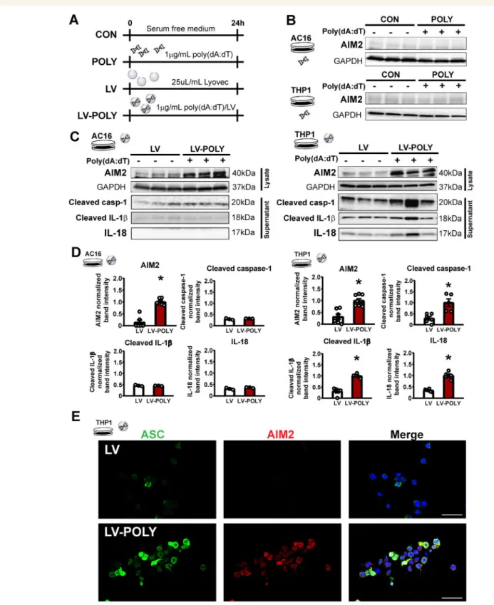
3.3 Poly(dA:dT) induces isolated AIM2 inflammasome activation in vitro
As our results suggest that AIM2 inflammasome may be a potential player of inflammation in HF, we speculated that inflammasome activa- tion might be a consequence of an interplay between immune cells and cardiac cells. To investigate inflammasome activationin vitro, AC16 hu- man cardiac and THP-1 human monocytic cell lines were stimulated with naked or cationic liposome encapsulated (LyoVecTM) poly(deoxya- denylic-deoxythymidylic) acid sodium salt [poly(dA:dT)], a specific AIM2 inducer, for 24 h (Figure3A). Naked poly(dA:dT) was unable to induce AIM2 inflammasome activation (Figure3B), however, liposome encapsu- lated poly(dA:dT) increased the expression of AIM2 in THP-1 cells (Figure3C), suggesting that vesicular uptake of dsDNA is critical in the in- duction of AIM2 inflammasome activation. Inflammasome activation was confirmed with detection of cleaved caspase-1, IL-18 and IL-1bfrom the supernatant, and immunofluorescence detection of the inflammasome adaptor protein apoptosis-associated speck-like protein containing a CARD (ASC) and AIM2 in THP-1 ASC-GFP reporter cell line (Figure 3C–E). Interestingly, poly(dA:dT) treatment also led to the induc- tion of AIM2 protein expression in the AC16 cells without significant in- terleukin release (Figure3C and D).
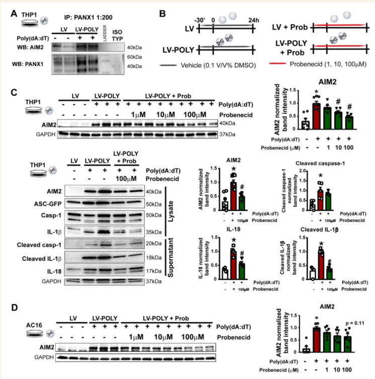
3.4 Pannexin-1 channel inhibition attenu- ates AIM2 inflammasome activation in THP1 cells
It has been shown that inflammasome activation by NLRP3 or NALP1 is strongly associated with the activation of purinergic signalling via P2X purinoreceptor 7 (P2X7) and hemichannel PANX1, however, it is un- known whether AIM2 inflammasomes and PANX1 have molecular
Downloaded from https://academic.oup.com/cardiovascres/advance-article/doi/10.1093/cvr/cvab202/6297397 by butz.henriett@med.semmelweis-univ.hu user on 24 September 2021
Figure 3Liposome encapsulated poly(dA:dT) induced the expression of AIM2 and inflammasome activationin vitro. (A) Experimental protocol for AIM2 induction in human AC16 cardiac and THP1 monocytic cell lines. (B) Representative western blot images for naked poly(dA:dT) stimulus on AC16 and THP1 cells. (C) Representative western blot images for liposome encapsulated poly(dA:dT) on AC16 and THP1 cell lines. (D) Quantification of western blot analysis on poly(dA:dT)-induced AIM2 inflammasome activation in AC16 and THP1 cells. *P< 0.05 vs. LV, Student’st-test;n= 4–6. (E) Representative images of immunofluorescence detection of AIM2 (red) and ASC (green) proteins in poly(dA:dT)-stimulated THP1 cells. DAPI (blue) was used for counterstain.
Scale bar: 50mm.
Downloaded from https://academic.oup.com/cardiovascres/advance-article/doi/10.1093/cvr/cvab202/6297397 by butz.henriett@med.semmelweis-univ.hu user on 24 September 2021
Figure 4Pannexin-1 channel inhibition attenuates AIM2 inflammasome activationin vitro.(A) Representative western blot images for co-immunopre- cipitation from control and poly(dA:dT)-stimulated THP1 cell lysate. PANX1 is shown as a loading control. Isotype anti-rabbit control was used as nega- tive control. (B) Experimental protocol for testing the PANX1 blocker probenecid in cell model for AIM2 inflammasome activation on human AC16 and THP1 cell lines. (C) Western blot analysis of AIM2 protein expression on poly(dA:dT)-stimulated THP1 cells in the presence or absence of different con- centration of probenecid, and detailed analysis of downstream signalling of AIM2 inflammasome activation in cell lysate and supernatant in the presence of 100mM probenecid. *P< 0.05 vs. control;#P< 0.05 vs. poly(dA:dT) without probenecid; one-way ANOVA;n= 5–6. (D) Western blot analysis of AIM2 protein expression and cell viability on poly(dA:dT)-stimulated AC16 cells in the presence or absence of different concentration of probenecid.
*P< 0.05 vs. control;#P< 0.05 vs. poly(dA:dT) without probenecid; one-way ANOVA;n= 5–6.
Downloaded from https://academic.oup.com/cardiovascres/advance-article/doi/10.1093/cvr/cvab202/6297397 by butz.henriett@med.semmelweis-univ.hu user on 24 September 2021
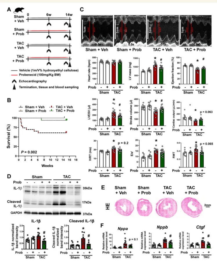
Figure 5Pannexin-1 channel inhibitor probenecid improves survival and cardiac functionin vivo. (A) Study design for investigating the effects of proben- ecid (Prob) in a rat model for chronic heart failure (TAC). (B) Kaplan–Meier analysis of overall mortality.P< 0.05, log-rank (Mantel-Cox) test;n= 11–23.
(C) Representative M-mode echocardiography images and assessment of cardiac function at week 14 after surgery. Scale bar: 1 cm; timestamp: 0.5 s.
*P< 0.05 vs. ShamþVeh,#P< 0.05 vs. TACþVeh, two-way ANOVA;n= 11–17. (D) Western blot analysis and representative images of IL-1band cleaved IL-1bin left ventricles. *P< 0.05 vs. ShamþVeh,#P< 0.05 vs. TACþVeh; two-way ANOVA;n= 6–8. (E) Representative histology images (hae- matoxylin eosin) at week 14. Scale bar: 2 mm. (F) Analysis of mRNA expression of hypertrophy and heart failure markers (Nppa, Nppb, andCtgf) by qRT- PCR. *P< 0.05 vs. ShamþVeh,#P< 0.05 vs. TACþVeh, one-way ANOVA;n= 7–8.
Downloaded from https://academic.oup.com/cardiovascres/advance-article/doi/10.1093/cvr/cvab202/6297397 by butz.henriett@med.semmelweis-univ.hu user on 24 September 2021
.. ..
.. ..
.. ..
.. ..
.. ..
.. ..
.. ..
.. ..
.. ..
.. ..
.. ..
.. ..
.. ..
.. ..
.. ..
.. ..
.. ..
.. ..
.. ..
.. ..
.. ..
.. ..
.. ..
.. ..
.. ..
.. ..
.. ..
.. ..
.. ..
.. ..
.. ..
.. ..
.. ..
.. ..
.. ..
.. ..
.. ..
.. ..
.. ..
.. ..
.. ..
.. ..
.. ..
.. ..
.. .
interactions. We performed co-immunoprecipitation on control and poly(dA:dT)-stimulated THP-1 cells, and saw that AIM2 was co- immunoprecipitated with PANX1 in activated cells indicating a potential interaction between the AIM2 inflammasome complex and PANX1 channels (Figure4A). As the opening of PANX1 channels is related to ap- optosis and release of ‘find me’ signals,38we tested the effects of proben- ecid, a potent PANX1 inhibitor, on AIM2 inflammasome activation in vitro(Figure4B). Probenecid showed a dose-dependent reduction in the protein expression of AIM2 in both THP-1 and AC16 cells without a significant effect on cell viability (Figure4C and D,Supplementary material online,Figure S4). Interestingly, the expression of PANX1 showed no sig- nificant differences in PANX1 levels between healthy and failing hearts with high individual variability (Supplementary materialonline,Figure S5).
3.5 Probenecid improves outcomes in pressure overload-induced chronic heart failure in rats
To test if probenecid improves cardiac functionin vivo, we investigated probenecid in the rat HF model induced by TAC (Figure 5, Supplementary materialonline,Tables S5andS6). In these rats, cardiac function was assessed at 6 weeks and, 14 weeks after TAC, while the rats were orally treated with probenecid (100 mg/kg body weight/day) or vehicle (hydroxyethyl cellulose) control. We evaluated mortality throughout the whole study. The group treated with vehicle and having TAC surgery showed a reduced survival rate compared with vehicle- treated sham operated rats (Figure5B). On the other hand, the group treated with a 100 mg/kg dose of probenecid showed significant amelio- ration of mortality compared with vehicle-treated TAC rats in Kaplan–
Meier analyses (Figure5B). As published previously39and shown above (Figure2A) TAC surgery resulted in HF development. 14 weeks after TAC, left ventricular ejection fraction (LVEF) was reduced compared with baseline from 69.2 ± 1.8% to 54.0 ± 2.0% and from 69.7 ± 0.9% to 60.2 ± 0.6% in rats allocated to vehicle or probenecid treatment groups, respectively (Figure5C,Supplementary materialonline,Tables S5andS6).
Thus, compared with vehicle, oral probenecid treatment of rats with TAC significantly prevented deterioration of LVEF. In accordance, at 14 weeks after TAC, left ventricular end-systolic volumes increased more in the vehicle group compared with the probenecid treated group (Figure5C,Supplementary materialonline,Tables S5andS6). In accor- dance with our previous observation above (Figure2), the protein levels of IL-1b and its mature form increased 14 weeks after TAC surgery, which was reduced by probenecid treatment (Figure5D). In addition, treatment with probenecid prevented development of left ventricular hypertrophy (Figure5C, E, andF). Fourteen weeks after TAC, in vehicle- treated TAC operated rats the left ventricular mass significantly in- creased (compared to sham) with a significant reduction after probene- cid treatment (Figure5C and E). This was further confirmed by analysis of pro-hypertrophic genes (Nppa and Nppb) and the pro-fibrotic factor connective tissue growth factor (Ctgf) (Figure5F). All these transcripts were significantly induced by TAC surgery, and their up-regulation was prevented by probenecid (Figure5F).
4. Discussion
We detected enhanced AIM2 inflammasome expression in failing hearts harvested from human patients as well as from different small and large animal models of chronic HF, highlighting the importance of chronic in- flammatory reactions in these conditions. In addition, increased NLRC4
expression was observed in human failing hearts as well. We assessed inflammasome activation in cardiac cells and macrophagesin vitro, and showed that dsDNA is capable of inducing the AIM2 inflammasome in both cell types, suggesting that necrotic DNA might be the major trigger of the AIM2 inflammasomein vivo. In addition, we showed that the AIM2 inflammasome associated PANX1 channels may play a role in inflamma- some activation, since the PANX1 inhibitor probenecid significantly re- duced IL-1bsecretion and maturation. Furthermore, chronic treatment with probenecid improved outcomes of pressure overload-induced HF.40These anti-inflammatory properties of probenecid could facilitate potential repurposing and use of this uricosuric drug to in chronic HF.40,41
The role of inflammatory mediators (such as interleukins and other cytokines) in cardiovascular diseases has been extensively studied over the last decades, nevertheless, clinical translation of these results was rather mixed and controversial.8,42Results of the CANTOS trial, however, pointed out that just by neutralizing IL-1b, with canakinumab, marked reductions can be achieved in incidence of major cardiovascular adverse events of post-infarction patients, highlighting the central role of IL-1b in these disease states.9,43 However, there are major limitations of the use of canakinumab (e.g. price, infectious adverse reactions), ruling it out from the rou- tine tools of current cardiovascular therapy. In light of these data, it is obvious that modulating new targets of IL-1b-related pathways might be of high therapeutic importance.
Our present human and translational animal data provides evidence for AIM2 and NLRC4 inflammasome activation in HF. We also show that co-activation of multiple types of inflammasomes is a possible phe- nomenon, suggesting that single inflammasome targeting may not be an optimal strategy in case of cardiovascular diseases including atheroscle- rosis22and chronic HF.
Bacterial and viral particles were considered as the primary triggers of inflammasome activation, however, it became evident that during sterile inflammatory conditions, DAMPs may also promote inflammasome ac- tivity. Among these, the AIM2 inflammasome is known to be activated by dsDNA.44It is reasonable to hypothesize that dsDNA was a major con- tributor to AIM2 inflammasome activation in our study as well, since the chronic remodelling process associates with a low degree of apoptotic/
necroptotic cell death resulting in a concomitant monocyte/macrophage infiltration and inflammasome activation.45A similar activation pattern has been described in case of chronic renal failure,46as well as in animal models of atherosclerosis.20The background of myocardial NLRC4 acti- vation in HF is even more surprising. Currently, the most characterized trigger of NLRC4 is flagellin of Gram negative bacteria.47,48It is presum- able that HF-induced hypoperfusion of the intestines leads to dysbiosis, and increased gut permeability,19promoting a low grade systemic inflam- matory state. This is supported by studies showing gut microbiome mod- ulation as a relevant target to alleviate the systemic inflammatory state during the course of human HF.49This hypothesis might provide an ex- planation for increased NLRC4 expression in human failing hearts; nev- ertheless, it is unknown whether significant gut hypoperfusion could have developed in our animal models. On the other hand, in animal mod- els of stroke a similar co-activation pattern of AIM2 and NLRC4 has been described previously,19,37suggesting that the activation of these two inflammasomes might be linked.
The complex pathways converging to inflammasome activation and signal- ling involve triggers that may influence inflammasome activity and assembly by mechanisms that associate with lysosomal membrane rupture,50as well as autoregulatory signalling by the products IL-1band IL-18. However, the
Downloaded from https://academic.oup.com/cardiovascres/advance-article/doi/10.1093/cvr/cvab202/6297397 by butz.henriett@med.semmelweis-univ.hu user on 24 September 2021
.. ..
.. ..
.. ..
.. ..
.. ..
.. ..
.. ..
.. ..
.. ..
.. ..
.. ..
.. ..
.. ..
.. ..
.. ..
.. ..
.. ..
.. ..
.. ..
.. ..
.. ..
.. ..
.. ..
.. ..
.. ..
.. ..
.. ..
.. ..
.. ..
.. ..
.. ..
.. ..
.. ..
.. ..
.. ..
.. ..
.. ..
.. ..
.. ..
.. ..
.. ..
.. ..
.. ..
.. ..
.. .
best characterized triggers are the classic mediators promoting inflamma- some priming [triggered by e.g. Toll-like receptors 4, 9 (TLR4, TLR9), and TNFa receptors], and inflammasome oligomerization (influenced by the purinergic receptors and the associated pannexin-1 channels), PANX1 chan- nels have so far been described as critical modulators of NALP1, NLRP3 as well as of non-canonical inflammasome activities via ATP release.38,51,52 Nonetheless, whether PANX1 is involved in the activation of AIM2 inflam- masomes has not been studied yet. By co-immunoprecipitation experi- ments, we have shown here first in the literature that PANX1 channels associate to the AIM2 inflammasome as well, and showed a prominent anti- inflammatory effect of the PANX1 channel inhibitor probenecidin vitro. The anti-inflammatory effect of probenecid was mediated by decreasing Il-1b level in a rabbit sepsis model.53We have seen a reduction in the expression of AIM2 and its downstream signallingin vitroin both dsDNA stimulated monocytes/macrophages and cardiac cells. In addition to AIM2 inflamma- some inhibition, PANX1 channels may play a role in leukocyte migration and in modulation of the NFjB pathway.54,55A recent study has also con- firmed that probenecid improves cardiac function at early phase of post-in- farction HF via inhibiting endothelial PANX1 channels and consequential leukocyte infiltration.56Therefore, we propose that probenecid might be a
‘broad-spectrum’ inflammasome inhibitor besides its well-characterized uri- cosuric properties. Probenecid has been previously demonstrated to im- prove outcome in an animal model of ischaemic HF with a shorter 4-week follow-up period by exerting positive inotropic effects via transient receptor potential vanilloid type-2 (TRPV2), and the positive inotropic effect was con- firmed in a small number of patients with HFrEF.40,57We now show that probenecid is able to prevent adverse cardiac remodelling upon a more pro- longed period of pressure overloadin vivo; however, the interplay between anti-inflammatory effects of probenecid and its action on TRPV2 as well as on myocardial contractility was not investigated within the frame of this study which should be acknowledged as a limitation. Nevertheless, these al- ready published beneficial effects (action on TRPV2 and contractility) and the novel anti-inflammatory effects might explain the recently observed clini- cal benefits of probenecid use in patients suffering from HF, as well as the ep- idemiological observation, that patients receiving probenecid therapy for gouty arthritis have better cardiovascular outcomes.40,41Thus, we believe that probenecid fulfils many of the characteristics desirable for a repurposed drug for the treatment of chronic HF.
4.1 Limitation
We have shown that probenecid has a significant inhibitory effect on AIM2 inflammasomein vitroand it improves survival and cardiac function in a rat model for HFin vivo. However, to identify precisely the contribu- tion of PANX1-mediated AIM2 inflammasome inhibition besides the other well-known effects of probenecid, furtherin vivostudies using ge- netically modified mice might be necessary.
5. Conclusion
We have shown with a series of experiments on human failing heart tis- sues as well as in various translationalin vivoanimal models (pressure or volume overload-induced rat HF models, and post-infarction rat and pig HF models) and inin vitrocell culture experiments, that inflammasome activation is primarily characterized by the activation of the AIM2 and NLRC4 inflammasome during chronic HF. We believe that our results highlight the importance of disease-, and disease-stage specific differen- ces of inflammasome activation patterns. IL-1band the upstream inflam- masome inhibition has been shown as an intriguing therapeutic target in
the CANTOS trial, therefore inhibition of AIM2 by probenecid may re- veal a promising new therapeutic concept promoting drug repurposing efforts in the treatment of chronic HF.
Data availability
The datasets used and/or analysed are available from the corresponding author upon request.
Supplementary material
Supplementary materialis available atCardiovascular Researchonline.
Authors’ contributions
Z.O. participated in study design and performedin vitro experiments, analysed data, and drafted the manuscript. M.R., D.K., A.A.S., and T.R.
designed and performedin vivorat experiments, analysed data and wrote the manuscript. P.L. collected human heart samples and provided clinical data. Z.G., G.B.B., A.M., I.H., and M.G. designed and performedin vivopig experiments and evaluated results. G.K. and V.E.T. performed in vitro experiments and evaluated results. P.F., R.S., A.G., T.R., and B.M. revised the manuscript, the intellectual content and provided professional ad- vice. Z.V.V. designed experiments, wrote manuscript, revised the intel- lectual content, and provided professional advice. All authors read and approved the final manuscript.
Conflict of interest:P.F. is the founder and CEO of Pharmahungary Group, a group of R&D companies. R.S. received honoraria for lecturing from Sanofi that is not related to the present study. The remaining authors declare no conflict of interest.
Funding
The work was supported by the European Union’s Horizon 2020 research and innovation programme under grant agreement No 739593. NVKP_16- 1-2016-0017 (‘National Heart Program’) has been implemented with the support provided from the National Research, Development and Innovation Fund of Hungary. The research was financed by the Thematic Excellence Programme (2020-4.1.1.-TKP2020) of the Ministry for Innovation and Technology in Hungary, within the framework of the Therapeutic Development and Bioimaging thematic programmes of the Semmelweis University, by grant VEKOP-2.3.2-16-2016-00002, VEKOP-2.3.3-15-2016- 00016, VEKOP-2.3.3-15-2016-00006 and by 2020-1.1.6-JO¨ VO-2021-00013} (‘Befektete´s a jo¨v}obe’ NFKIH). Prepared with the professional support of the Doctoral Student Scholarship Program of the Co-operative Doctoral Program of the Ministry of Innovation and Technology financed from the National Research, Development and Innovation Fund (to A.G.). This project was supported by grants from the National Research, Development and Innovation Office (NKFIH) of Hungary (K134939 to T.R., FK134751 to Z.V.V.). Z.O., M.R., Z.V.V., A.M., G.K., and Z.G. is supported by the New National Excellence Program of the Ministry of Human Capacities [U´ NKP- 18-3-I-SE-64, U´ NKP-19-3-I-SE-11; U´NKP-20-4-II-SE-20; U´NKP-20-5; U´NKP- 19-4-I-SE-18; U´ NKP-19-3-I-SE-60; U´NKP-18-3-III-SE-7]. Z.G. and Z.V.V. is supported by the Ja´nos Bolyai Research Scholarship of the Hungarian Academy of Sciences. B.G.B. and Z.O. was supported by EFOP-3.6.3- VEKOP-16-2017-00009, ‘Az orvos-, ege´szse´gtudoma´nyi- e´s gyo´gyszere´sz- ke´pze´s tudoma´nyos m}uhelyeinek fejleszte´se’, and B.G.B. was supported by Richter Gedeon Nyrt. scholarship.
Downloaded from https://academic.oup.com/cardiovascres/advance-article/doi/10.1093/cvr/cvab202/6297397 by butz.henriett@med.semmelweis-univ.hu user on 24 September 2021
.. ..
.. ..
.. ..
.. ..
.. ..
.. ..
.. ..
.. ..
.. ..
.. ..
.. ..
.. ..
.. ..
.. ..
.. ..
.. ..
.. ..
.. ..
.. ..
.. ..
.. ..
.. ..
.. ..
.. ..
.. ..
.. ..
.. ..
.. ..
.. ..
.. ..
.. ..
.. ..
.. ..
.. ..
.. ..
.. ..
.. ..
.. ..
.. ..
.. ..
.. ..
.. ..
.. ..
.. ..
.. . Acknowledgements
Z.O. dedicates this work to her beloved grandfather Csaba Ja´nos Sza´nto´, who passed away during the study. Z.V.V. would like to dedicate this study to his late father Tibor Varga, who died of complications from chronic heart failure. The authors thank Andrea Kova´cs, Krisztina Kecske´s, Vikto´ria Oravecz, Henriett Biro´, Edina Urba´n, and Petra Na´dasdi for their essential technical assistance. The authors wish to ac- knowledge SERVIER Medical Art (https://smart.servier.com/) for use of their medical art kits when making the illustrations in the article.
References
1. Ge Z, Li A, McNamara J, Dos Remedios C, Lal S. Pathogenesis and pathophysiology of heart failure with reduced ejection fraction: translation to human studies.Heart Fail Rev2019;24:743–758.
2. Ponikowski P, Voors AA, Anker SD, Bueno H, Cleland JGF, Coats AJS, Falk V, Gonzalez-Juanatey JR, Harjola VP, Jankowska EA, Jessup M, Linde C, Nihoyannopoulos P, Parissis JT, Pieske B, Riley JP, Rosano GMC, Ruilope LM, Ruschitzka F, Rutten FH, van der Meer P; Group ESCSD. 2016 ESC Guidelines for the diagnosis and treatment of acute and chronic heart failure: he Task Force for the diagnosis and treatment of acute and chronic heart failure of the European Society of Cardiology (ESC)Developed with the special contribution of the Heart Failure Association (HFA) of the ESC.Eur Heart J2016;37:2129–2200.
3. Testa M, Yeh M, Lee P, Fanelli R, Loperfido F, Berman JW, LeJemtel TH. Circulating levels of cytokines and their endogenous modulators in patients with mild to severe congestive heart failure due to coronary artery disease or hypertension.J Am Coll Cardiol1996;28:964–971.
4. Luscher TF. Inflammation: the new cardiovascular risk factor.Eur Heart J2018;39:
3483–3487.
5. Abbate A, Salloum FN, Vecile E, Das A, Hoke NN, Straino S, Biondi-Zoccai GG, Houser JE, Qureshi IZ, Ownby ED, Gustini E, Biasucci LM, Severino A, Capogrossi MC, Vetrovec GW, Crea F, Baldi A, Kukreja RC, Dobrina A. Anakinra, a recombinant human interleukin-1 receptor antagonist, inhibits apoptosis in experimental acute myocardial infarction.Circulation2008;117:2670–2683.
6. Kadokami T, Frye C, Lemster B, Wagner CL, Feldman AM, McTiernan CF. Anti-tu- mor necrosis factor-alpha antibody limits heart failure in a transgenic model.
Circulation2001;104:1094–1097.
7. Mann DL, McMurray JJ, Packer M, Swedberg K, Borer JS, Colucci WS, Djian J, Drexler H, Feldman A, Kober L, Krum H, Liu P, Nieminen M, Tavazzi L, van Veldhuisen DJ, Waldenstrom A, Warren M, Westheim A, Zannad F, Fleming T. Targeted anticytokine therapy in patients with chronic heart failure: results of the Randomized Etanercept Worldwide Evaluation (RENEWAL).Circulation2004;109:1594–1602.
8. Anker SD, Coats AJ. How to RECOVER from RENAISSANCE? The significance of the results of RECOVER, RENAISSANCE, RENEWAL and ATTACH.Int J Cardiol 2002;86:123–130.
9. Everett BM, Cornel JH, Lainscak M, Anker SD, Abbate A, Thuren T, Libby P, Glynn RJ, Ridker PM. Anti-inflammatory therapy with canakinumab for the prevention of hospitalization for heart failure.Circulation2019;139:1289–1299.
10. Ridker PM, Libby P, MacFadyen JG, Thuren T, Ballantyne C, Fonseca F, Koenig W, Shimokawa H, Everett BM, Glynn RJ. Modulation of the interleukin-6 signalling path- way and incidence rates of atherosclerotic events and all-cause mortality: analyses from the Canakinumab Anti-Inflammatory Thrombosis Outcomes Study (CANTOS).
Eur Heart J2018;39:3499–3507.
11. Martinon F, Burns K, Tschopp J. The inflammasome: a molecular platform triggering activation of inflammatory caspases and processing of proIL-beta.Mol Cell2002;10:
417–426.
12. Chen KW, Demarco B, Heilig R, Shkarina K, Boettcher A, Farady CJ, Pelczar P, Broz P. Extrinsic and intrinsic apoptosis activate pannexin-1 to drive NLRP3 inflammasome assembly. 2019;38:e101638.
13. Crespo Yanguas S, Willebrords J, Johnstone SR, Maes M, Decrock E, De Bock M, Leybaert L, Cogliati B, Vinken M. Pannexin1 as mediator of inflammation and cell death.Biochim Biophys Acta Mol Cell Res2017;1864:51–61.
14. Abulafia DP, de Rivero Vaccari JP, Lozano JD, Lotocki G, Keane RW, Dietrich WD.
Inhibition of the inflammasome complex reduces the inflammatory response after thromboembolic stroke in mice.J Cereb Blood Flow Metab2009;29:534–544.
15. Mezzaroma E, Toldo S, Farkas D, Seropian IM, Van Tassell BW, Salloum FN, Kannan HR, Menna AC, Voelkel NF, Abbate A. The inflammasome promotes adverse cardiac remodeling following acute myocardial infarction in the mouse.Proc Natl Acad Sci USA2011;108:19725–19730.
16. Miteva K, Pappritz K, Sosnowski M, El-Shafeey M, Mu¨ller I, Dong F, Savvatis K, Ringe J, Tscho¨pe C, Van Linthout S. Mesenchymal stromal cells inhibit NLRP3 inflamma- some activation in a model of Coxsackievirus B3-induced inflammatory cardiomyopa- thy.Sci Rep2018;8:2820.
17. Paramel Varghese G, Folkersen L, Strawbridge RJ, Halvorsen B, Yndestad A, Ranheim T, Krohg-Sorensen K, Skjelland M, Espevik T, Aukrust P, Lengquist M, Hedin U,
Jansson JH, Fransen K, Hansson GK, Eriksson P, Sirsjo A. NLRP3 inflammasome ex- pression and activation in human atherosclerosis.J Am Heart Assoc2016;5:e003031.
18. Xiao H, Li H, Wang JJ, Zhang JS, Shen J, An XB, Zhang CC, Wu JM, Song Y, Wang XY, Yu HY, Deng XN, Li ZJ, Xu M, Lu ZZ, Du J, Gao W, Zhang AH, Feng Y, Zhang YY. IL-18 cleavage triggers cardiac inflammation and fibrosis upon beta-adrenergic in- sult.Eur Heart J2018;39:60–69.
19. Denes A, Coutts G, Lenart N, Cruickshank SM, Pelegrin P, Skinner J, Rothwell N, Allan SM, Brough D. AIM2 and NLRC4 inflammasomes contribute with ASC to acute brain injury independently of NLRP3.Proc Natl Acad Sci USA2015;112:4050–4055.
20. Paulin N, Viola JR, Maas SL, de Jong R, Fernandes-Alnemri T, Weber C, Drechsler M, Do¨ring Y, Soehnlein O. Double-strand DNA sensing Aim2 inflammasome regulates atherosclerotic plaque vulnerability.Circulation2018;138:321–323.
21. Wang X, Pan J, Liu H, Zhang M, Liu D, Lu L, Tian J, Liu M, Jin T, An F. AIM2 gene si- lencing attenuates diabetic cardiomyopathy in type 2 diabetic rat model.Life Sci2019;
221:249–258.
22. Fidler TP, Xue C, Yalcinkaya M, Hardaway B, Abramowicz S, Xiao T, Liu W, Thomas DG, Hajebrahimi MA, Pircher J, Silvestre-Roig C, Kotini AG, Luchsinger LL, Wei Y, Westerterp M, Snoeck HW, Papapetrou EP, Schulz C, Massberg S, Soehnlein O, Ebert B, Levine RL, Reilly MP, Libby P, Wang N, Tall AR. The AIM2 inflammasome exacerbates atherosclerosis in clonal haematopoiesis.Nature2021;592:296–301.
23. Varga ZV, Pipicz M, Baa´n JA, Baranyai T, Koncsos G, Leszek P, Kusmierczyk M, Sa´nchez-Cabo F, Garcı´a-Pavı´a P, Brenner GJ, Giricz Z, Csont T, Mendler L, Lara- Pezzi E, Pacher P, Ferdinandy P. Alternative splicing of NOX4 in the failing human heart.Front Physiol2017;8:935.
24. Platt MJ, Huber JS, Romanova N, Brunt KR, Simpson JA. Pathophysiological mapping of experimental heart failure: left and right ventricular remodeling in transverse aortic constriction is temporally, kinetically and structurally distinct.Front Physiol2018;9:472.
25. Pacher P, Liaudet L, Mabley J, Komjati K, Szabo C. Pharmacologic inhibition of poly(a- denosine diphosphate-ribose) polymerase may represent a novel therapeutic ap- proach in chronic heart failure.J Am Coll Cardiol2002;40:1006–1016.
26. Wang X, Ren B, Liu S, Sentex E, Tappia PS, Dhalla NS. Characterization of cardiac hypertrophy and heart failure due to volume overload in the rat.J Appl Physiol (1985) 2003;94:752–763.
27. Baranyai T, Giricz Z, Varga ZV, Koncsos G, Lukovic D, Makkos A, Sarkozy M, Pavo N, Jakab A, Czimbalmos C, Vago H, Ruzsa Z, Toth L, Garamvolgyi R, Merkely B, Schulz R, Gyongyosi M, Ferdinandy P.In vivoMRI andex vivohistological assessment of the cardioprotection induced by ischemic preconditioning, postconditioning and remote conditioning in a closed-chest porcine model of reperfused acute myocardial infarction: importance of microvasculature.J Transl Med2017;15:67.
28. Brenner GB, Giricz Z, Garamvo¨lgyi R, Makkos A, Ono´di Z, Sayour NV, Gergely TG, Baranyai T, Petneha´zy O¨ , K}oro¨si D, Szabo´ GP, Vago H, Dohy Z, Czimbalmos C, Merkely B, Boldin-Adamsky S, Feinstein E, Horva´th IG, Ferdinandy P. Post-myocardial infarction heart failure in closed-chest coronary occlusion/reperfusion model in Go¨ttingen Minipigs and Landrace pigs.J Vis Exp2021;170:e61901.
29. Suetomi T, Willeford A, Brand CS, Cho Y, Ross RS, Miyamoto S, Brown JH.
Inflammation and NLRP3 inflammasome activation initiated in response to pressure overload by Ca(2þ)/calmodulin-dependent protein kinase II delta signaling in cardio- myocytes are essential for adverse cardiac remodeling.Circulation2018;138:2530–2544.
30. Kummer JA, Broekhuizen R, Everett H, Agostini L, Kuijk L, Martinon F, van Bruggen R, Tschopp J. Inflammasome components NALP 1 and 3 show distinct but separate expression profiles in human tissues suggesting a site-specific role in the inflammatory response.J Histochem Cytochem2007;55:443–452.
31. Chen B, Frangogiannis NG. Macrophages in the remodeling failing heart.Circ Res 2016;119:776–778.
32. Chen B, Frangogiannis NG. The role of macrophages in nonischemic heart failure.
JACC Basic Transl Sci2018;3:245–248.
33. Prabhu SD, Frangogiannis NG. The biological basis for cardiac repair after myocardial infarction: from inflammation to fibrosis.Circ Res2016;119:91–112.
34. Kohler C. Allograft inflammatory factor-1/Ionized calcium-binding adapter molecule 1 is specifically expressed by most subpopulations of macrophages and spermatids in testis.Cell Tissue Res2007;330:291–302.
35. Kawaguchi M, Takahashi M, Hata T, Kashima Y, Usui F, Morimoto H, Izawa A, Takahashi Y, Masumoto J, Koyama J, Hongo M, Noda T, Nakayama J, Sagara J, Taniguchi S, Ikeda U. Inflammasome activation of cardiac fibroblasts is essential for myocardial ischemia/reperfusion injury.Circulation2011;123:594–604.
36. Kim HY, Kim SJ, Lee SM. Activation of NLRP3 and AIM2 inflammasomes in Kupffer cells in hepatic ischemia/reperfusion. 2015;282:259–270.
37. Durga Devi T, Babu M, Ma¨kinen P, Kaikkonen MU, Heinaniemi M, Laakso H, Yla¨- Herttuala E, Rieppo L, Liimatainen T, Naumenko N, Tavi P, Yla¨-Herttuala S. Aggravated postinfarct heart failure in type 2 diabetes is associated with impaired mitophagy and exaggerated inflammasome activation.Am J Pathol2017;187:2659–2673.
38. Chekeni FB, Elliott MR, Sandilos JK, Walk SF, Kinchen JM, Lazarowski ER, Armstrong AJ, Penuela S, Laird DW, Salvesen GS, Isakson BE, Bayliss DA, Ravichandran KS.
Pannexin 1 channels mediate ‘find-me’ signal release and membrane permeability dur- ing apoptosis.Nature2010;467:863–867.
39. Ruppert M, Lakatos BK, Braun S, Tokodi M, Karime C, Olah A, Sayour AA, Hizoh I, Barta BA, Merkely B, Kovacs A, Radovits T. Longitudinal strain reflects ventriculoar- terial coupling rather than mere contractility in rat models of hemodynamic overload-induced heart failure.J Am Soc Echocardiogr2020;33:1264–1275.e4.
Downloaded from https://academic.oup.com/cardiovascres/advance-article/doi/10.1093/cvr/cvab202/6297397 by butz.henriett@med.semmelweis-univ.hu user on 24 September 2021
.. ..
.. ..
.. ..
.. ..
.. ..
.. ..
.. ..
.. ..
.. ..
.. ..
.. ..
.. ..
.. ..
.. ..
.. ..
.. ..
.. ..
..
40. Robbins N, Gilbert M, Kumar M, McNamara JW, Daly P, Koch SE, Conway G, Effat M, Woo JG, Sadayappan S, Rubinstein J. Probenecid improves cardiac function in patients with heart failure with reduced ejection fraction in vivo and cardiomyocyte calcium sensitivity in vitro.J Am Heart Assoc2018;7:e007148.
41. Kim SC, Neogi T, Kang EH, Liu J, Desai RJ, Zhang M, Solomon DH. Cardiovascular risks of probenecid versus allopurinol in older patients with gout.J Am Coll Cardiol 2018;71:994–1004.
42. Abbate A, Van Tassell BW, Biondi-Zoccai G, Kontos MC, Grizzard JD, Spillman DW, Oddi C, Roberts CS, Melchior RD, Mueller GH, Abouzaki NA, Rengel LR, Varma A, Gambill ML, Falcao RA, Voelkel NF, Dinarello CA, Vetrovec GW. Effects of interleukin-1 blockade with anakinra on adverse cardiac remodeling and heart failure after acute myo- cardial infarction [from the Virginia Commonwealth University-Anakinra Remodeling Trial (2) (VCU-ART2) pilot study].Am J Cardiol2013;111:1394–1400.
43. Ridker PM, Everett BM, Thuren T, MacFadyen JG, Chang WH, Ballantyne C, Fonseca F, Nicolau J, Koenig W, Anker SD, Kastelein JJP, Cornel JH, Pais P, Pella D, Genest J, Cifkova R, Lorenzatti A, Forster T, Kobalava Z, Vida-Simiti L, Flather M, Shimokawa H, Ogawa H, Dellborg M, Rossi PRF, Troquay RPT, Libby P, Glynn RJ; CANTOS Trial Group. Antiinflammatory therapy with canakinumab for atherosclerotic disease.
N Engl J Med2017;377:1119–1131.
44. Hornung V, Ablasser A, Charrel-Dennis M, Bauernfeind F, Horvath G, Caffrey DR, Latz E, Fitzgerald KA. AIM2 recognizes cytosolic dsDNA and forms a caspase-1- activating inflammasome with ASC.Nature2009;458:514–518.
45. Dorn GW 2nd. Apoptotic and non-apoptotic programmed cardiomyocyte death in ventricular remodelling.Cardiovasc Res2009;81:465–473.
46. Komada T, Chung H, Lau A, Platnich JM, Beck PL, Benediktsson H, Duff HJ, Jenne CN, Muruve DA. Macrophage uptake of necrotic cell DNA activates the AIM2 inflammasome to regulate a proinflammatory phenotype in CKD.J Am Soc Nephrol 2018;29:1165–1181.
47. Miao EA, Alpuche-Aranda CM, Dors M, Clark AE, Bader MW, Miller SI, Aderem A.
Cytoplasmic flagellin activates caspase-1 and secretion of interleukin 1beta via Ipaf.
Nat Immunol2006;7:569–575.
48. Mariathasan S, Newton K, Monack DM, Vucic D, French DM, Lee WP, Roose-Girma M, Erickson S, Dixit VM. Differential activation of the inflammasome by caspase-1 adaptors ASC and Ipaf.Nature2004;430:213–218.
49. Tang WHW, Li DY, Hazen SL. Dietary metabolism, the gut microbiome, and heart failure.Nat Rev Cardiol2019;16:137–154.
50. Hornung V, Bauernfeind F, Halle A, Samstad EO, Kono H, Rock KL, Fitzgerald KA, Latz E. Silica crystals and aluminum salts activate the NALP3 inflammasome through phagosomal destabilization.Nat Immunol2008;9:847–856.
51. Silverman WR, de Rivero Vaccari JP, Locovei S, Qiu F, Carlsson SK, Scemes E, Keane RW, Dahl G. The pannexin 1 channel activates the inflammasome in neurons and astrocytes.J Biol Chem2009;284:18143–18151.
52. Yang D, He Y, Mu~noz-Planillo R, Liu Q, Nu´~nez G. Caspase-11 requires the pannexin- 1 channel and the purinergic P2X7 pore to mediate pyroptosis and endotoxic shock.
Immunity2015;43:923–932.
53. He H, Liu D, Long Y, Wang X, Yao B. The pannexin-1 channel inhibitor probenecid attenuates skeletal muscle cellular energy crisis and histopathological injury in a rab- bit endotoxemia model.Inflammation2018;41:2030–2040.
54. Wu LY, Ye ZN, Zhou CH, Wang CX, Xie GB, Zhang XS, Gao YY, Zhang ZH, Zhou ML, Zhuang Z, Liu JP, Hang CH, Shi JX. Roles of pannexin-1 channels in inflammatory response through the TLRs/NF-kappa B signaling pathway following experimental subarachnoid hemorrhage in rats.Front Mol Neurosci2017;10:175.
55. Lohman AW, Leskov IL, Butcher JT, Johnstone SR, Stokes TA, Begandt D, DeLalio LJ, Best AK, Penuela S, Leitinger N, Ravichandran KS, Stokes KY, Isakson BE. Pannexin 1 channels regulate leukocyte emigration through the venous endothelium during acute inflammation.Nat Commun2015;6:7965.
56. Good ME, Young A, Wolpe AG, Ma M, Johnstone SR, Hall PJ, Duffy CK, Aronovitz M, Martin G, Blanton RM, Leitinger N, Wolf MJ, Isakson BE. Endothelial pannexin 1 regulates cardiac response to myocardial infarction.Circ Res2021;128:1211–1213.128:
57. Koch SE, Tranter M, Robbins N, Luther K, Singh U, Jiang M, Ren X, Tee T, Smith L, Varma P, Jones WK, Rubinstein J. Probenecid as a noninjurious positive inotrope in an ischemic heart disease murine model.J Cardiovasc Pharmacol Ther2013;18:280–289.
Translational perspective
Targeting interleukin-1band its release by the inhibition of inflammasomes may be a potential therapeutic approach in cardiovascular diseases in- cluding heart failure. Absent in melanoma 2 inflammasome activation was identified in human heart failure samples which was confirmed in various translational small and large animal models of chronic heart failure as well. Our findings suggest that NLRP3-independent inflammasome inhibitors (e.g. probenecid) might be novel agents in the treatment of chronic heart failure.
Downloaded from https://academic.oup.com/cardiovascres/advance-article/doi/10.1093/cvr/cvab202/6297397 by butz.henriett@med.semmelweis-univ.hu user on 24 September 2021