*Drs Tannous and Deloux contributed equally.
Key Words: acetyl coenzyme A
◼ cardiomyopathy, dilated ◼ energy metabolism ◼ glycolysis ◼ heart failure
◼ NAD ◼ nicotinamide-beta-riboside
◼ serum response factor Sources of Funding, see page 2271
Editorial, see p 2274
BACKGROUND: Myocardial metabolic impairment is a major feature in chronic heart failure. As the major coenzyme in fuel oxidation and oxidative phosphorylation and a substrate for enzymes signaling energy stress and oxidative stress response, nicotinamide adenine dinucleotide (NAD+) is emerging as a metabolic target in a number of diseases including heart failure. Little is known on the mechanisms regulating homeostasis of NAD+ in the failing heart.
METHODS: To explore possible alterations of NAD+ homeostasis in the failing heart, we quantified the expression of NAD+ biosynthetic enzymes in the human failing heart and in the heart of a mouse model of dilated cardiomyopathy (DCM) triggered by Serum Response Factor transcription factor depletion in the heart (SRFHKO) or of cardiac hypertrophy triggered by transverse aorta constriction. We studied the impact of NAD+ precursor supplementation on cardiac function in both mouse models.
RESULTS: We observed a 30% loss in levels of NAD+ in the murine failing heart of both DCM and transverse aorta constriction mice that was accompanied by a decrease in expression of the nicotinamide phosphoribosyltransferase enzyme that recycles the nicotinamide precursor, whereas the nicotinamide riboside kinase 2 (NMRK2) that phosphorylates the nicotinamide riboside precursor is increased, to a higher level in the DCM (40-fold) than in transverse aorta constriction (4-fold). This shift was also observed in human failing heart biopsies in comparison with nonfailing controls. We show that the Nmrk2 gene is an AMP-activated protein kinase and peroxisome proliferator-activated receptor α responsive gene that is activated by energy stress and NAD+ depletion in isolated rat cardiomyocytes. Nicotinamide riboside efficiently rescues NAD+ synthesis in response to FK866-mediated inhibition of nicotinamide phosphoribosyltransferase and stimulates glycolysis in cardiomyocytes.
Accordingly, we show that nicotinamide riboside supplementation in food attenuates the development of heart failure in mice, more robustly in DCM, and partially after transverse aorta constriction, by stabilizing myocardial NAD+ levels in the failing heart. Nicotinamide riboside treatment also robustly increases the myocardial levels of 3 metabolites, nicotinic acid adenine dinucleotide, methylnicotinamide, and N1-methyl-4-pyridone-5-carboxamide, that can be used as validation biomarkers for the treatment.
CONCLUSIONS: The data show that nicotinamide riboside, the most energy- efficient among NAD precursors, could be useful for treatment of heart failure, notably in the context of DCM, a disease with few therapeutic options.
© 2017 American Heart Association, Inc.
Nicolas Diguet, PhD Samuel A.J. Trammell,
PhD
Cynthia Tannous, PhD*
Robin Deloux, MSc*
Jérôme Piquereau, PhD Nathalie Mougenot, PhD Anne Gouge, MSc Mélanie Gressette, MSc Boris Manoury, PhD Jocelyne Blanc, MSc Marie Breton, MD Jean-François Decaux,
PhD
Gareth G. Lavery, PhD István Baczkó, MD, PhD Joffrey Zoll, PhD Anne Garnier, PhD Zhenlin Li, PhD Charles Brenner, PhD Mathias Mericskay, PhD
ORIGINAL RESEARCH ARTICLE
Nicotinamide Riboside Preserves Cardiac Function in a Mouse Model of Dilated Cardiomyopathy
http://circ.ahajournals.org
Circulation
by guest on May 22, 2018http://circ.ahajournals.org/Downloaded from by guest on May 22, 2018http://circ.ahajournals.org/Downloaded from by guest on May 22, 2018http://circ.ahajournals.org/Downloaded from by guest on May 22, 2018http://circ.ahajournals.org/Downloaded from by guest on May 22, 2018http://circ.ahajournals.org/Downloaded from by guest on May 22, 2018http://circ.ahajournals.org/Downloaded from by guest on May 22, 2018http://circ.ahajournals.org/Downloaded from by guest on May 22, 2018http://circ.ahajournals.org/Downloaded from by guest on May 22, 2018http://circ.ahajournals.org/Downloaded from by guest on May 22, 2018http://circ.ahajournals.org/Downloaded from by guest on May 22, 2018http://circ.ahajournals.org/Downloaded from by guest on May 22, 2018http://circ.ahajournals.org/Downloaded from by guest on May 22, 2018http://circ.ahajournals.org/Downloaded from by guest on May 22, 2018http://circ.ahajournals.org/Downloaded from by guest on May 22, 2018http://circ.ahajournals.org/Downloaded from by guest on May 22, 2018http://circ.ahajournals.org/Downloaded from by guest on May 22, 2018http://circ.ahajournals.org/Downloaded from by guest on May 22, 2018http://circ.ahajournals.org/Downloaded from by guest on May 22, 2018http://circ.ahajournals.org/Downloaded from by guest on May 22, 2018http://circ.ahajournals.org/Downloaded from by guest on May 22, 2018http://circ.ahajournals.org/Downloaded from by guest on May 22, 2018http://circ.ahajournals.org/Downloaded from by guest on May 22, 2018http://circ.ahajournals.org/Downloaded from by guest on May 22, 2018http://circ.ahajournals.org/Downloaded from by guest on May 22, 2018http://circ.ahajournals.org/Downloaded from by guest on May 22, 2018http://circ.ahajournals.org/Downloaded from by guest on May 22, 2018http://circ.ahajournals.org/Downloaded from by guest on May 22, 2018http://circ.ahajournals.org/Downloaded from by guest on May 22, 2018http://circ.ahajournals.org/Downloaded from
ORIGINAL RESEARCHARTICLE
D
espite progress in therapeutic advances, mortality remains high in congestive heart failure (HF), cre- ating a need for new evidence-based and cost-ef- fective treatments. Failure of cardiomyocyte bioenerget- ics is a key issue in HF that is not well addressed in current therapeutics.1,2 We focused our attention on pathways that regulate nicotinamide adenine dinucleotide (NAD+) homeostasis in the failing heart, which has emerged as a new avenue for the development of metabolic therapy of HF.3,4 NAD+ is the major hydride transfer coenzyme in fuel oxidation and mitochondrial ATP generation and is the precursor for NADPH, which is required for anabolic pathways and reactive oxygen species detoxification.5,6 In addition, NAD+ is an essential substrate of enzymes including sirtuins and poly(ADP-ribose) polymerases, which are important in responding to altered nutritionalstatus and genotoxic stress.6,7 Sirtuins perform NAD+-de- pendent deacylation of protein Lys residues modified by acetyl coenzyme A (Ac-CoA) derived acyl groups.8 Sirtu- ins regulate nuclear gene expression, and mitochondrial enzyme activities, as well, and can play a protective role in the heart,9 although chronic overactivation may be deleterious.10 Poly(ADP-ribose) polymerase 1 is activated by reactive oxygen species–induced DNA lesions, and could deplete myocardial NAD+ stores in HF.11 In situ- ations of physiological equilibrium, biosynthetic path- ways relying on dietary sources of tryptophan and NAD+ precursor vitamins counterbalance the consumption of NAD+ by signaling enzymes. However, dietary precursors may become insufficient to maintain the NAD+ metabo- lome in pathological conditions, thereby necessitating supplementation of NAD+ precursor.5,12,13
The homeostasis of NAD+ and regulation of NAD+ biosynthetic enzymes have not been studied in dilated cardiomyopathy (DCM), a leading cause of heart trans- plant.14 We previously generated a powerful model of nonischemic DCM in mice triggered by cardiac-specific inducible inactivation of Serum Response Factor (SRF-
HKO model).15–18 SRF is a major transcriptional regulator of genes and microRNAs involved in contractility, and in energy flux and calcium handling, as well.15,17,18 The SRF pathway is altered in the human failing heart19,20 and in animal models of DCM.21,22 The heart of SRFHKO mutant mice display an early downregulation of myofi- brillar creatine kinase (CK) activity encoded by the Ckm gene, which is followed by repression of enzymes of fatty acid β-oxidation and loss of ATP, thus recapitu- lating the transitional steps toward energy failure and cardiac structural remodeling in HF.18
Here, we show that NAD+ homeostasis is altered at an early stage in the heart of SRFHKO mice with a strik- ing induction of the nicotinamide riboside kinase 2 (NMRK2) pathway for NAD+ synthesis. Nmrk2 gene is 1 of 2 mammalian paralogs of the yeast NRK1 gene that was discovered as a eukaryotic route to NAD+ from nicotinamide riboside (NR).23 Given the activity of NR in promoting sirtuin activity and restoring metabolic bal- ance in a variety of model organisms,13,24–28 we aimed to test whether NR might treat HF in SRFHKO mice and in the pressure overload hypertrophy model induced by trans- verse aorta constriction (TAC). Here, we show that NR greatly protects cardiac function by virtue of improved citrate and Ac-CoA metabolism and antioxidant gene expression. Because NR is orally available in people,29 our data rationalize clinical testing of NR in human HF.
METHODS
The data, analytic methods, and study materials will be made available from the corresponding author on request to other researchers for purposes of reproducing the results or repli- cating the procedure. Detailed methods are available in the online-only Data Supplement.
Clinical Perspective
What Is New?
• Myocardial nicotinamide adenine dinucleotide (NAD) coenzyme levels are depressed in mouse models of heart failure.
• An expression shift occurs in murine and human failing hearts in which the normally predominant nicotinamide phosphoribosyltransferase enzyme using nicotinamide as a precursor for NAD+ synthe- sis is repressed, whereas the nicotinamide riboside kinase 2 enzyme using the nucleoside nicotinamide riboside (NR) is strongly upregulated.
• The nicotinamide riboside kinase 2pathway is acti- vated by energy stress in an AMP-activated protein kinase α/peroxisome proliferator-activated receptor α–dependent manner in cardiomyocytes.
• NR-supplemented diet administrated to murine models of dilated cardiomyopathy or pressure over- load-induced heart failure restores myocardial NAD levels and preserves cardiac function.
• NR increases glycolysis, and citrate and acetyl-coen- zyme A metabolism, as well, in cardiomyocytes.
What Are the Clinical Implications?
• NR is the most energetically favorable NAD precur- sor and was recently shown to be uniquely and orally bioavailable in humans in a phase I clinical trial.
• NR-supplemented diet could help patients who have heart failure to cope with limited myocardial ATP supply by restoring NAD coenzyme levels and associated signaling.
• NR-supplemented diet has been shown in sev- eral studies to enhance metabolic flexibility in the context of obesity and diabetes mellitus and to improve muscle function, justifying that NR is put in the front line for metabolic therapy of heart fail- ure syndrome in which these functions are altered.
by guest on May 22, 2018http://circ.ahajournals.org/Downloaded from
ORIGINAL RESEARCH ARTICLE
Patients
Left ventricular (LV) myocardium was obtained from terminally failing human hearts of 4 patients (mean age 54 years±7, SD) at the time of transplantation at the Hôpitaux Universitaires de Strasbourg as previously published with approval of the Hôpitaux Universitaires de Strasbourg ethics committee.30 All patients gave informed consent. Patients’ characteristics are detailed in Table I in the online-only Data Supplement. The investigations conformed to the principles of the Declaration of Helsinki. Control hearts were obtained from the depart- ment of Pharmacology and Pharmacotherapy, University of Szeged, Szeged, Hungary. Experimental protocols were approved by the Ethical Review Board of the Medical Center of the University of Szeged and by the Scientific and Research Ethical Committee of the Medical Scientific Board at the Hungarian Ministry of Health (ETT-TUKEB; No. 51–57/1997 OEj and 4991-0/2010-1018EKU).
Transgenic Mice
All experiments with animals conformed to the Directive 2010/63/EU of the European Parliament and were approved by the ethics committee Charles Darvin #5 (agreement 00369.01). See online-only Data Supplement for details on SRF inactivation protocol.
NR Supplementation
SRFHKO and control (Sf/Sf) mice were administered A04 rodent maintenance diet (Scientific Animal Food Engineering) sup- plemented with NR chloride provided by Chromadex. See online-only Data Supplement for details.
Transcriptomic Analysis
Transcriptomic data are available on NCBI Gene Expression Database, Accession number GSE84142.
Statistical Analysis
Animals were randomly assigned into different treatment groups. To assess significance, we performed Student t test for independent samples when the experimental design com- pared only 2 groups, or 1-way ANOVA for multiple group comparisons, or 2-way factorial ANOVA for independent fac- tors when appropriate. Tukey tests were used for comparison of specific experimental groups when 1-way ANOVA gave a P value of <0.05 or, in the case of 2-way ANOVA for interac- tion, a P value of <0.05. Values are expressed as mean±SEM.
Supplemental Information
Supplemental information includes supplemental methods, 9 supplemental figures, and 6 supplemental tables.
RESULTS
NMRK2 Pathway Is Activated in the Failing Heart of SRFHKO Mice
Transcriptome analyses in SRFHKO mice at baseline or af- ter phenylephrine α-adrenergic stimulation16 revealed
that the Nmrk2 gene was induced at an early stage in the SRFHKO heart and rose continuously during establish- ment of HF (Figure 1A and Table II in the online-only Data Supplement). The transcript for the ectoenzyme NT5E (CD73) that hydrolyzes extracellular NAD+ and nicotin- amide mononucleotide (NMN) to NR, the substrate of NMRK2 kinase31 also increased in SRFHKO hearts (Table II in the online-only Data Supplement). In contrast, the transcripts encoding nicotinamide phosphoribosyltrans- ferase (NAMPT), the enzyme converting nicotinamide (NAM) to NMN, and purine nucleoside phosphorylase (PNP), which converts NR to NAM,32 were depressed on phenylephrine treatment in SRFHKO hearts (Table II in the online-only Data Supplement). Genes involved in the tryptophan and nicotinic acid pathways for NAD+ synthesis were not altered (Figure 1A and Table II in the online-only Data Supplement).
Modulation of Nmrk2 and Nampt gene expression detected by transcriptomics was corroborated at the mRNA and protein level (Figure 1B through 1D). We observed a similar shift in human patients from pre- dominant expression of NAMPT in healthy hearts to in- creased NMRK2 and depressed NAMPT in failing hearts (Figure 1D, Table II in the online-only Data Supplement).
In mouse heart sections, NMRK2 protein localizes at the proximity of sarcolemma in controls (Figure 1E). The sig- nal was strongly enhanced and spread to cytoplasm in SRFHKO cardiomyocytes.
At day 15 after SRF inactivation, there was a 30%
loss of NAD (representing the sum of NAD+ and NADH) (Figure 1F, see Methods in the online-only Data Supple- ment for details). The gene expression pattern (Nt5e and Nmrk2 up with Pnp and Nampt down) suggested that cardiac tissue is attempting to mobilize and utilize NR as an NAD+ precursor while not increasing NAM us- age. We tested this hypothesis by intraperitoneal ad- ministration of NR and NAM to SRFHKO mice from day 8 to 15. Consistent with this hypothesis, myocardial NAD levels were preserved by NR but not by NAM (Figure 1F).
Srf and Nmrk2 expression levels were not changed by these treatments (Figure 1G and 1H). Because NR is orally available,26,29 we tested an NR-supplemented diet for 2 weeks. Myocardial NAD levels were reduced in this second series of SRFHKO mice in comparison with controls and rescued by NR administration (Figure 1I).
NR Preserves Cardiac Function in the SRFHKO Heart
We hypothesized that NR supplementation of food might be beneficial for cardiac function in the context of DCM. We fed control and mutant SRFHKO mice a standard chow diet or NR-supplemented diet to reach a dose of 400 mg/kg of body weight per day from day 5 to 50, a period in which untreated SRFHKO mice develop DCM and progress toward HF.15–18 NR diet induced a
by guest on May 22, 2018http://circ.ahajournals.org/Downloaded from
ORIGINAL RESEARCHARTICLE
Figure 1. Activation of the Nmrk2 gene in dilated cardiomyopathy.
A, Biosynthetic NAD+ pathways in mammalian cells. NA (nicotinic acid), NAM, NR, and TRP enter cells through special- ized transport systems. Extracellular NAD+ and NMN are hydrolyzed to NR by the ectonucleotidase CD73. In the de novo pathway, TRP is converted to kynurenin (KYN) by IDO and TDO enzymes, and then into quinolic acid (QA) in 4 steps (not detailed). QA, NA, and NAM are phosphoribosylated by QPRT, NAPRT1, and NAMPT enzymes, respectively, to form mononucleotides NAMN and NMN. NR is phosphorylated by NMRK enzymes to form NMN. NR can be converted to NAM by PNP. NAMN and NMN are adenylated by the NMNAT enzymes to form the dinucleotides NAAD+ and NAD+. NAAD+ is amidated by NADSYN1. Genes increased in SRFHKO heart are highlighted in green and those repressed in red (see accom- panying Table II in the online-only Data Supplement). Inhibitors are shown in blue. B and C, RT qPCR analysis of Nmrk2 and Nampt mRNA fold change (FC) in SRFHKO hearts at different days (D) after SRF inactivation. D, Western blot analysis of NMRK2 and NAMPT protein in control and failing human hearts and control and SRFHKO mouse hearts at D45. GAPDH is used as loading control. All human and mouse samples were analyzed on single blot, but 2 lanes separating that showed signs of protein degradation and aberrant migration pattern were cut out of the picture. Right, Ratio of NMRK2/
NAMPT in human samples. E, Control and SRFHKO mutant heart sections stained for NMRK2 (red) and vinculin (green) at D45. White bar in upper left panel=20 µm. F through H, NAM or NR (30 µmol) were injected intraperitoneally from D8 to 15 (400 mg/kg per day) to control and SRFHKO mice. Vehicle: saline solution. F, Myocardial NAD+ levels. G and H, RT qPCR analysis of Srf and Nmrk2 mRNA levels. I, Myocardial NAD levels in control and SRFHKO mice fed regular chow diet (CD) or NR-enriched diet (0.22%. 400–450 mg/kg per day) from D5 to 20. Throughout the figure, data are expressed as mean±SEM. Statistical analysis: One-way ANOVA (B, C) or 2-way factorial ANOVA for independent samples (F through I).
¶¶P≤0.01, ¶¶¶P≤0.001 for the genotype effect; §§P≤0.01 for the NR treatment effect; ii P≤0.01 for the interaction effect.
Post hoc Tukey test: asterisks indicate statistical significant difference between any group versus the control CD (or vehicle) group: *P≤0.05. **P≤0.01. ***P≤0.001. # P≤0.05 for the effect of NR within the SRFHKO group. A t test was used for the graph at the right of D: ** P≤0.01. FC indicates fold change over control group; IDO, indoleamine 2,3-dioxygenase;
NAAD, nicotinic acid adenine dinucleotide; NAD, nicotinamide adenine dinucleotide; NAM, nicotinamide; NAMN, nicotinic acid mononucleotide; NAMPT, nicotinamide phosphoribosyltransferase; NMN, nicotinamide mononucleotide; NMNAT, nicotinamide mononucleotide adenylyltransferease; NMRK, nicotinamide riboside kinase; NR, nicotinamide riboside; PNP, purine nucleoside phosphorylase; QPRT, quinolinate phosphoribosyltransferase; RT qPCR, quantitative reverse transcription polymerase chain reaction; SRF, Serum Response Factor; TDO, tryptophan 2,3-dioxygenase; and TRP, tryptophan.
by guest on May 22, 2018http://circ.ahajournals.org/Downloaded from
ORIGINAL RESEARCH ARTICLE
Figure 2. NR supplementation in diet prevents the onset of heart failure and dilatation.
A, CD- or NR-supplemented diet (0.22%) was given ad libitum to control and SRFHKO mice from day 5 after SRF inactivation to the end of the experiment. Body weight was monitored throughout the period. Data are expressed as mean % weight variation±SEM, in comparison with weight at day 5. A 2-factor ANOVA with repeated measures on 1 factor was used for sta- tistical analysis. §§§P≤0.001 for the time effect; iii, P≤0.001 for the interaction effect. Post hoc Tukey test: *P<0.05, **P<0.01,
***P<0.001 for any time point versus day 5 within a group; #P≤0.05 for the NR treatment in SRFHKO group. B through O, Cardiac parameters of control and SRFHKO mutant were analyzed in M-Mode echocardiography between day 45 and day 47.
B, Heart rate; C, left ventricle (LV) mass index; D, LV ejection fraction; E, fractional shortening; F and G, LV end-systolic diam- eter (F) and volume (G); H and I, LV end-diastolic diameter (H) and volume (I); J and K, LV posterior wall thickness in systole (J) and diastole (K); L and M interventricular septum thickness in systole (L) and diastole (M); N, H/R: LV thickness (H) to radius (R) ratio; and O, stroke volume. Dimensions were normalized by the body weight. Data are expressed as mean±SEM. Statisti- cal analysis: 2-way factorial ANOVA for independent samples. ¶¶P≤0.01, ¶¶¶P≤0.001 for the genotype effect; §P≤0.05,
§§P≤0.01, §§§P≤0.001 for the NR treatment effect; i P≤0.05, ii P≤0.01, iii P≤0.001 for the interaction effect. Post hoc Tukey test: asterisks indicate statistically significant difference versus the control CD group: *P≤0.05, **P≤0.01, ***P≤0.001.
#P≤0.05, ##P≤0.01 for the effect of NR within the SRFHKO group. CD indicates chow diet; CONT, control; EDV, end-diastolic volume; ESV, end-systolic volume; FS, fractional shortening; IVSThd, interventricular septum thickness in diastole; IVSThs, in- terventricular septum thickness in systole; LVEF, left ventricular ejection fraction; LVEDd, left ventricular end-diastolic diameter;
LVESd, left ventricular end-systolic diameter; LVMI, left ventricular mass index; NR, nicotinamide riboside; PWThD, posterior wall thickness in diastole; PWThS, posterior wall thickness in systole; and SV, stroke volume.
by guest on May 22, 2018http://circ.ahajournals.org/Downloaded from
ORIGINAL RESEARCHARTICLE modest 5% to 7% increase in body weight in control
mice and allowed SRFHKO mice to regain weight after an initial loss, whereas SRFHKO mice on a regular diet had lost weight at the end of the protocol (Figure 2A). Car- diac parameters were analyzed by echocardiography between days 45 and 47 (Figure 2B through 2O). NR did not change heart rate and LV mass index, but slightly it increased the LV thickness-to-radius ratio (H/R) in con- trol mice (Figure 2N). To assess a potential impact of NR treatment on vascular function, we compared vascular reactivity in isolated mesenteric arteries from NR-fed and control mice. We found no difference between the 2 groups neither in responses to K+ or agonist-induced contraction nor in the relaxation response to carbachol or a NO donor (Figure I in the online-only Data Supple- ment and Table III in the online-only Data Supplement).
SRFHKO mutant mice fed the standard diet displayed a severe decrease in LV ejection fraction and fraction- al shortening (Figure 2D and 2E). The NR diet clearly protected against this decline in cardiac function. It is important to note that NR fully protected SRFHKO mice against the dilatation and thinning of the LV wall (Fig- ure 2F through 2M), preserving the H/R ratio that nor- mally declines with DCM (Figure 2N). Changes in stroke volume (Figure 2O) and cardiac output (not shown) were not significant.
At the histological level, NR limited the appearance of very long cardiomyocytes (Feret > 100 µm) in SRFHKO mice, although the population of large cardiomyocytes (minFeret > 26 µm) remained larger than in controls (Figure II in the online-only Data Supplement).
NR-Enriched Diet Protects the Cardiac NAD+ Metabolome in HF
The impact of an NR-enriched diet on the cardiac NAD+ metabolome is not known (Figure 3A). Mice fed with or without NR were euthanized 3 days after the echocar- diography at day 50. Myocardial NAD was decreased in the SRFHKO mice at day 50 as measured by the NAD cycling assay, and the NR diet protected against this drop (Figure 3B). Myocardial NAD+ and NADH varied in the same direction in all conditions, leaving the NAD+/ NADH ratio unchanged (Figure III in the online-only Data Supplement). Liquid chromatography-mass spec- trometry analysis allowed us to provide the levels of NAD+ metabolites on a common scale with NAD+13,33 (Figure 3C through 3L). SRFHKO mice hearts did not have higher levels of adenosine diphosphate ribose (ADPR) or NAM (Figure 3D and 3E). Levels of NADP+ and NMN showed a pattern similar to NAD+ (Figure 3F and 3G).
NR increased the cardiac levels of nicotinic acid adenine dinucleotide, a sensitive biomarker of increased NAD+ metabolism,29 and methyl-NAM (MeNAM), as well (Fig- ure 3A, 3H through 3J). MeNAM oxidation by AOX1 gen- erates N1-methyl-4-pyridone-5-carboxamide (Me4PY)
and releases hydrogen peroxide (Figure 3A). Changes in unrelated metabolites were not significant (Figure 3K and 3L). As shown in the nematode,34 NR increased the ex- pression of the Nfel2 gene encoding NRF2, whereas its target genes Mt2 and Nox4 were already activated, and G6pdx, as well, in the heart of SRFHKO mice (Figure 3M through 3P). NR treatment in the SRFHK0 hearts limited the level of Myh7 induction, a signature of cardiac stress and metabolic remodeling in the failing heart (Figure 3Q).
The liver is a central organ for the regulation of nu- trient metabolism. We assessed the NAD+ metabolome response in the liver of control and SRFHKO mice (Table IV in the online-only Data Supplement). NR increased the MeNAM and Me4PY levels similarly in both genotypes.
It is interesting to note that the liver response to NR diet was more robust in the SRFHKO mice with regard to the increase in the steady-state level of NAD, nicotinic acid adenine dinucleotide, ADPR, and NAM. Because the liver is not targeted by the Srf deletion, it suggests that the HF condition indirectly modifies the balance of NAD synthesis and signaling in the liver.
NR-Enriched Diet Does Not Result in Global Cardiac Protein Deacetylation
The cardiac levels of NAM and ADPR in this HF model are low and show few changes with NR (Figure 3D and 3E), suggesting that the NR-driven boost in NAD+ syn- thesis is not necessarily translated into a higher level of NAD+ consumption by sirtuins and poly(ADP-ribose) polymerases. We performed antiacetyl-K and anti–poly- ADP-ribose Western blot analyses on cardiac proteins from control and SRFHKO mice fed with the chow diet or NR-enriched diets. Just as levels of ADPR were not modulated by SRFHKO or NR, neither were levels of PAR (Figure IVA in the online-only Data Supplement ). There was an increase in the acetylation level of some cardiac proteins in the hearts of NR-fed animals (Figure IVB in the online-only Data Supplement). Antibodies specific to the acetylated form of FOXO1 or p53 showed an in- crease in the acetylation level of these nuclear proteins in the heart in response to NR in controls as in SRFHKO mice (Figure 3R, Figure V in the online-only Data Supple- ment). By comparison, the acetylation level of the mito- chondrial aconitase 2 protein did not change in any of these conditions despite lower protein level in the SRFHKO hearts (Figure 3S and Figure VIA in the online-only Data Supplement). These data suggest that the bioenergetic improvement in NR-supplemented hearts may result in greater production of cytosolic Ac-CoA and consequent increased nucleocytosolic protein acetylation.
NR Improves Metabolism of Citrate in HF
Several regulators involved in mitochondrial biogenesis and oxidative metabolism, and lipid metabolism, as
by guest on May 22, 2018http://circ.ahajournals.org/Downloaded from
ORIGINAL RESEARCH ARTICLE
Figure 3. Impact of NR treatment on cardiac NAD+ metabolome.
A, NR to NAD+ pathway and NAD+ catabolism are depicted. NAD+ is cleaved into NAM and ADP-ribose by NAD+-consuming enzymes. NAM is either recycled by NAMPT or methylated by the nicotinamide N-methyltransferase (NNMT) enzyme and oxi- dized by the aldehyde deoxidase 1 (AOX1) giving rise to degradation products MeNAM and Me4PY, respectively. B, Myocardial NAD was quantified by colorimetric NAD+ cycling assay at day 50 in controls (n=6), Controls+NR (Continued )
by guest on May 22, 2018http://circ.ahajournals.org/Downloaded from
ORIGINAL RESEARCHARTICLE well, were repressed in SRFHKO hearts (Figure VIIA and
VIIB in the online-only Data Supplement). Simultaneous- ly, the expression of the glucose transporter Glut1 was increased and expression of Pdk2 was decreased, which can be seen as a trend of the failing heart to increase glycolysis (Figure VIIC in the online-only Data Supple- ment). NR did not alter the expression of these genes.
There was a trend toward a reduced mitochondrial to genomic DNA ratio in the SRFHKO LV (Figure 3T). The NADH-dependent complex I activity and Cox activity was not changed by the NR diet (Figure 3U and 3V).
Neither the complex I and II maximal respiration rates, nor the apparent Km for ADP, mitochondrial CK cou- pling, and ADP acceptor control ratio showed any al- teration at this stage (day 50) in the SRFHKO LV fibers.
These parameters were not altered by NR administra- tion (Figure VIID through VIIG in the online-only Data Supplement).
We noted that NR treatment resulted in a net increase in acetylation of FOXO1 and p53 (Figure 3R and Figure V in the online-only Data Supplement). Availability of cyto- solic Ac-CoA depends on mitochondrial citrate synthase (CS) production of citrate and conversion to Ac-CoA via the cytosolic enzyme, ATP-citrate lyase (ACL). CS activity was reduced to 65% of control levels in the SRFHKO LV myocardium (Figure 3W). It is striking that NR adminis- tration protected against the decline of CS activity in the failing heart. This rescue in activity occurred despite a net reduction in protein level in the SRFHKO myocardium that was not rescued by the NR treatment (Figure VIB in the online-only Data Supplement). It is interesting to that NR also increased cardiac ACL activity in both control and SRFHKO mice (Figure 3X). Taken together, these data sug- gest that NR improves CS and ACL activity, resulting in increased nucleocytoplasmic protein acetylation.
Nmrk2 Gene Is Induced by Inhibition of Alternative NAD+ Biosynthetic Pathways in Cultured Cardiomyocytes
To assess the impact of NMRK2 on NAD+ biosynthesis relative to the other pathways in cardiomyocytes, we used chemical inhibitors to target the alternative path- ways in neonatal rat cardiomyocytes (NRCs) grown in Dulbecco modified Eagle medium containing NAM as the vitamin B3. The glutamine analog azaserin inhibits the glutamine-dependent NAD synthase NADSYN1,35 which catalyzes the last step of NAD+ synthesis from tryptophan and NA (Figure 1A). FK866 is a specific in- hibitor of Nampt.36
Azaserin had a slowly repressive effect on NAD pools that resulted in a 50% reduction with respect to non- treated cells at 72 hours (Figure 4A through 4C) while progressively increasing Nmrk2 expression (Figure 4D).
FK866 treatment led to a rapid and severe loss of intra- cellular NAD+ but not NADH (Figure 4A through 4C).
Despite this severe loss, NRC could be maintained up to 72 hours in continuous layer with FK866 treatment.
FK866 treatment strongly increased Nmrk2 expression at 72 hours (Figure 4D). Combined azaserin and FK866 treatment had a similar impact than FK866 alone (Fig- ure 4A through 4D). Nmrk1 expression did not change in these conditions (not shown).
We tested whether NR could preserve NAD+ levels in the presence of FK866. NR slightly increased the baseline level of intracellular NAD+ in nontreated NRC, whereas exogenous NAD+ had no impact (Figure 4E).
Both compounds increased the NAD+/NADH ratio (Fig- ure 4F and 4G). NR fully protected NAD+ levels in NRCs treated with FK866, whereas the protection by exoge- nous NAD+ was partial (Figure 4E), although both treat- Figure 3 Continued. (n=5), SRFHKO (n=6), SRFHKO+NR (n=6). Data are expressed as means±SEM. C through L, Myocardial me- tabolites were analyzed by LCMS-based metabolomics. See abbreviations in the text. Controls (n=4), controls+NR (n=5), SRFHKO (n=6), +NR (n=5, except for NAM, MeNAM, and Me4PY, n=4, no peak was identified in 1 sample). Data are expressed as means±SEM. M through Q, Expression of genes related to oxidative stress signaling (M through O) and cardiac structural and metabolic remodeling (P and Q). n=5 in each group. R, Representative Western blot analysis of FOXO1 and acetyl-FOXO1 of 3 independent experiments realized in different duplicates for each group. GAPDH is used as a loading control. Right graphs, Quantification of total and acetyl-FOXO1 ratio on GAPDH and acetyl-FOXO1/total FOXO1 ratio in n=5 to 6 animals per group.
S, Acetylation level of mitochondrial aconitase 2. Aconitase 2 (75 kDa) was immunoprecipitated using a rabbit polyclonal antibody, and the immunoprecipitate was analyzed by Western blot using a mouse monoclonal anti-Ac(K103) antibody. Inputs were run in a parallel gel and immunoblotted with anti-aconitase 2 and anti-GAPDH for loading control. The ratio of acetyl- aconitase2/total aconitase2 and aconitase2/GAPDH are shown (right). See accompanying Figure VIA in the online-only Data Supplement for control immunoprecipitation with preimmune rabbit IgG. T through X, LV cardiac tissue was isolated at 50 days after tamoxifen injection in control and SRFHKO mice fed control diet (CD) or NR-enriched diet. DNA and proteins were extracted from parallel samples to quantify mitochondrial to genomic DNA ratio (T) and enzymatic activities: complex I (U), cy- tochrome oxidase (V), citrate synthase (W), and ATP citrate lyase (X). See associated Figure VII in the online-only Data Supple- ment. In M through S, data are expressed as mean fold change (FC)±SEM over control group CD. Statistical analysis: Two-way factorial ANOVA for independent samples was used for all panels. ¶P≤0.05, ¶¶P≤0.01, ¶¶¶P≤0.001 for the genotype effect;
§P≤0.05, §§P≤0.01, §§§P≤0.001 for the NR treatment effect; ii P≤0.01 for the interaction effect. Post hoc Tukey test: asterisks indicate statistically significant difference versus the control CD group: **P≤0.01. ACL indicates ATP-citrate lyase; ADP, ad- enosine diphosphate ribose; Cont, control; CS, citrate synthase; IMP, inosine monophosphate; LCMS, liquid chromatography- mass spectrometry; NAAD, nicotinic acid adenine dinucleotide; NAD, nicotinamide adenine dinucleotide; NAM, nicotinamide;
NAMPT, nicotinamide phosphoribosyltransferase; NMN, nicotinamide mononucleotide; and NR, nicotinamide riboside.
by guest on May 22, 2018http://circ.ahajournals.org/Downloaded from
ORIGINAL RESEARCH ARTICLE
Figure 4. Nmrk2 expression is activated by repression of alternative NAD+ biosynthetic pathways.
A through C, Intracellular levels of NAD+ (A), NADH (B), and NAD+/NADH ratio (C) in NRC after 10 µmol/L FK866 and 20 µmol/L azaserin treatment or no treatment (NT) for 24 to 72 hours as indicated. D, Nmrk2 mRNA level in NRC treated as in A through C. E through G, Same as in A through C in NRC treated for 72 hours with 10 µmol/L FK866 or not treated (NT) in normal culture medium (–) or in presence of 250 µmol/L NAD+ or 1 mmol/L NR. H, Nmrk2 mRNA level in NRC treated as in E through G. I, NAD+ levels in NRCs treated with 10 µmol/L FK866 for 72 hours, or not treated (NT) in the presence of increasing concentration of NR in culture medium. J, NAD+ content in nontreated (NT) or following 24-hour NR treatment (1 mmol/L) in NRC infected with Ad-GFP or HA-Nmrk2. Right, Western blot detection of HA-NMRK2 with anti-HA antibody. K through M, Mitochondrial stress test in Seahorse analyzer. NRC grown on Seahorse 96-well plates were analyzed for oxygen consumption rate (OCR) at day 8 after 5 days of treatment. K, Basal mitochondrial respiration is calculated from total cellular respiration minus nonmitochondrial respiration. L, ATP production is calculated from basal mitochondrial respiration minus respiration after oligomycin injection. M, Maximal respiration is measured after FCCP injection. See accompanying Figure IX in the online- only Data Supplement for other respiration parameters. N through P, Glycolysis stress test in Seahorse analyzer. NRC grown on Seahorse 96-well plates were analyzed at day 8 after 5 days of treatment. N, Glycolysis was measured as a function of extra- cellular acidification rate (ECAR) after injection of 10 mmol/L glucose. O, Glycolytic capacity as the maximum ECAR following injection of oligomycin. P, Glycolytic reserve as the difference between glycolysis and maximal glycolytic capacity. Throughout the figure, data are expressed as mean fold change (FC)±SEM over the control group, except when indicated. Statistical analy- sis: a 2-way factorial ANOVA for independent samples was used for A through J. ¶ and § symbols as indicated in the panels;
i P≤0.05, ii P≤0.01, iii P≤0.001 for the interaction effect. One-way ANOVA was used for K through P. ¶ P<0.05, ¶¶ P<0.01,
¶¶¶ P<0.001. §§ P≤0.01, §§§ P≤0.001. Post hoc Tukey test: *P<0.05, **P<0.01, ***P<0.001 between any (Continued )
by guest on May 22, 2018http://circ.ahajournals.org/Downloaded from
ORIGINAL RESEARCHARTICLE ments blocked Nmrk2 induction (Figure 4E). At least
100 µmol/L NR was required to rescue the NAD+ loss induced by 10 µmol/L FK866 (Figure 4I). To assess the functionality of NMRK2 in the absence of a stress like the FK866, we infected NRC with a recombinant ade- novirus expressing HA-tagged Nmrk2 cDNA (Figure 4J).
Overexpression of NMRK2 did not modulate the NAD+ levels in NRC. In presence of NR, Nmrk2 overexpression robustly increased the NAD+ level by a factor of 6. NR alone slightly increased the NAD+ level in control NRC infected with Ad-GFP (Figure 4J). In isolated primary cultures of adult rat cardiomyocytes, NR increased in- tracellular NAD nearly 3-fold, showing that the NMRK pathway is more active in adult rat cardiomyocytes than in NRCs (Figure VIIIA in the online-only Data Supple- ment). FK866 reduced NAD levels by a factor of 2 (Fig- ure VIIIA in the online-only Data Supplement) in adult rat cardiomyocytes and increased Nmrk2 expression by a factor of 4.5, whereas azaserin had no impact (Figure VIIIB in the online-only Data Supplement). We assessed the impact of NR on oxygen consumption rates in NRC following a mitochondrial stress protocol. NR had no impact on any mitochondrial respiration parameter in NRC at baseline (Figure 4K through 4M, Figure IX in the online-only Data Supplement). NR increased glycolysis at baseline in a dose-dependent manner (Figure 4N), but not the maximal glycolytic capacity (Figure 4O and 4P). We assessed the capacity of NR to rescue any impact of FK866 on metabolism. FK866 reduced basal oxygen consumption rates and maximal respira- tion and trended to reduce ATP production (adjusted P value=0.0547) (Figure 4K through 4M). Addition of NR at doses that we showed to fully rescue the drop in NAD+ levels triggered by the FK866 did not correct these alterations and even further reduced maximal res- piration at high doses (Figure 4K through 4M, Figure IX in the online-only Data Supplement). FK866 strong- ly reduced basal glycolysis and glycolytic capacities in NRC. NR strikingly preserved NRC glycolytic capacities to 100% control levels and more in FK866-treated cells and stimulated glycolysis at baseline in nontreated cells in a dose-dependent manner (Figure 4N through 4P).
SRF Deficiency-Induced Depression of the Nmrk2 Promoter Is Counterbalanced by the Sensitivity of Nmrk2 Gene to NAD+ Depletion and Energetic Stress
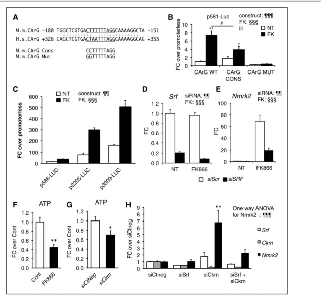
Because Nmrk2 expression is activated in vivo on Srf gene inactivation, we addressed the role of the SRF tran- scription factor in Nmrk2 gene regulation. We identified
an SRF binding site with 9/10 bp match to the consensus CC(A/T)6GG (CArG) sequence in a conserved 600-bp re- gion between the murine and human genes (Figure 5A).
We cloned the upstream region and 5ʹ-UTR of the mu- rine Nmrk2 gene (–581/+61) into a luciferase reporter vector and mutated the CArG-like sequence to a motif matching the consensus sequence (5ʹ-CC) or a motif un- able to bind SRF (5ʹ-GG) (Figure 5A). FK866 increased the expression of the WT construct 8-fold, whereas a consensus CArG reduced the extent of activation (Fig- ure 5B). The 5ʹ-GG mutation reduced baseline expression by 75% and abolished the response to FK866. The full intergenic 3009-bp sequence upstream of Nmrk2 had a much higher activity, especially in response to FK866 (Figure 5C). siRNA-mediated inhibition of Srf reduced, but did not abolish, the induction of endogenous Nmrk2 by FK866 in NRC, suggesting that FK866-triggered acti- vation of the Nmrk2 promoter is partially independent of SRF (Figure 5D and 5E). FK866 treatment reduces ATP to 50% of the control level in NRC (Figure 5F). SRF is major upstream regulator of Ckm gene involved in high- energy phosphate compounds balance. A siRNA-mediat- ed 5-fold knock-down of Ckm gene expression lowered the cellular ATP levels to 70% control level and triggered a 6.8-fold increase in Nmrk2 expression (Figure 5G and 5H). A 2-fold reduction in Srf expression resulted in a 2-fold reduction of Ckm expression leaving Nmrk2 ex- pression intact. Enforcing a more profound depression of Ckm expression in the context of Srf repression in NRC by cotransfection with both Srf and Ckm siRNAs resulted in a 2.3-fold increase in Nmrk2 expression (Figure 5H).
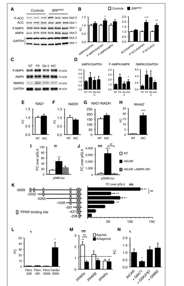
Nmrk2 Is Induced by the AMPK-PPARα Axis
In line with the previous results, we hypothesized that pathways related to energy failure might activate Nmrk2.
The level of phosphorylated AMP-activated protein kinase α (AMPKα), the energy stress–sensing AMP-activated ki- nase, and its target Ac-CoA carboxylase (ACC) was in- creased at an early stage when Nmrk2 induction began in the SRFHKO hearts (Figure 6A and 6B). 5-Aminoimid- azole-4-carboxamide ribonucleotide (AICAR) treatment stimulated AMPK phosphorylation in NRCs and increased NMRK2 protein level in NRCs (Figure 6C and 6D). The en- ergetic stress induced by glucose deprivation for 24 hours or FK866 for 48 hours also increased the NMRK2 protein.
AICAR treatment did not modulate NAD+/NADH but robustly induced Nmrk2 expression (Figure 6E through 6H). To assess whether the effect of AICAR took place at the level of the Nmrk2 promoter, we transfected Nmrk2-
Figure 4 Continued. group versus NT control cells; ##P<0.01, ###P<0.001 for indicated comparisons. Ad-GFP indicates adenovirus encoding green fluorescent protein; AZ, azaserin; FCCP, Carbonyl cyanide 4-(trifluoromethoxy)phenylhydrazone;
FK, FK866; HA, human influenza hemagglutinin tag sequence; NAD, nicotinamide adenine dinucleotide; NRC, neonatal rat cardiomyocyte; and NR, nicotinamide riboside.
by guest on May 22, 2018http://circ.ahajournals.org/Downloaded from
ORIGINAL RESEARCH ARTICLE
luciferase constructs into NRC and treated the cells with AICAR. The p586-Luc, and moreover, the p3009-Luc reporter, was highly responsive to AICAR treatment (Fig- ure 6I and 6J). Cotransfection of a plasmid overexpress- ing a dominant negative isoform of AMPK efficiently blunted induction by AICAR (Figure 6I and 6J).
Analysis of the 5ʹ-regulatory sequences of Nmrk2 re- vealed enrichment in putative peroxisome proliferator–
activated receptor (PPAR) binding sites (Figure 6K). The fragments –3009/–2552 and –1028/–228 contain most of the activity. By comparison, the activity of the Nmrk2 promoter was very low in cardiac fibroblasts (Figure 6L).
Figure 5. SRF is a component of Nmrk2 gene transcription.
A, CArG-like binding site for SRF in the conserved region between the murine (M.m) Nmrk2 promoter and human (H.s) NMRK2 intron 1. The CArG-like motif was mutated to a CArG consensus sequence (cons) and a CArG mutant (mut) site unable to bind SRF. B, Activity of p586-Firefly Luciferase construct bearing wild-type (WT), consensus (CONS), or mutated (Mut) CArG motif without (NT) or with FK866 treatment (10 µmol/L) in NRC. SV40-renilla luciferase was cotransfected with the Nmrk2-Firefly Luc constructs for normalization of transfection efficiency. Two-way factorial ANOVA for independent factors: ¶¶¶P<0.001 for con- struct effect, §§§P<0.001 for FK effect; iii P≤0.001 for the interaction effect. Post hoc Tukey-test: *P<0.05, **P<0.01 FK versus NT; #P<0.05 CArG construct versus CArG wild type. C, Various lengths of the murine Nmrk2 regulatory region were inserted into pGL4 vector and transfected in nontreated or FK866-treated NRC. Two-way factorial ANOVA for independent factors:
¶¶P<0.01 for construct effect, §§§P<0.001 for FK effect. D and E, Srf and Nmrk2 mRNA levels in NRC transfected with control scrambled siRNA (siScr) (white bars), or Srf or Nmrk2 siRNA (black bars), without (NT) or with FK866 treatment. Two-way facto- rial ANOVA for independent factors: ¶¶P<0.01 for siRNA effect, §§§P<0.001 for FK effect. F and G, ATP levels in NRCs treated for 72 hours with FK866 (F) or siCkm (G). t test: *P<0.05, **P<0.01, between FK-treated cells or siCkm-transfected cells versus control cells or siCtneg-transfected cells, respectively. H, Srf, Ckm, and Nmrk2 mRNA level in NRC transfected for 72 hours with siRNAs as indicated. A 1-way ANOVA was used. ¶¶¶P≤0.001. Post hoc Tukey test: **P<0.01, between any group versus siCtneg-transfected control cells. Throughout the figure, data are expressed as mean±SEM over control group, fold change over control group (FC) except for B and C, fold change over promoterless pGL4 plasmid and H FC over siCtneg. CArG indicates CC(A/T)6GG; NRC, neonatal rat cardiomyocyte; siRNA, small interfering RNA; and SRF, Serum Response Factor.
by guest on May 22, 2018http://circ.ahajournals.org/Downloaded from
ORIGINAL RESEARCHARTICLE
Figure 6. Nmrk2 expression is increased by AMPK and PPARα pathways.
A, Representative Western blot analysis of cardiac proteins in control and SRFHKO mice at day 9 using antibodies directed against ACC, phospho-ACC (Ser79), AMPKα, and phospho-AMPKα (Thr172). Phosphorylated and total proteins were ana- lyzed on 2 separate gels (gel 1 and 2) and GAPDH antibody was used for loading control. B, Quantification (Continued )
by guest on May 22, 2018http://circ.ahajournals.org/Downloaded from
ORIGINAL RESEARCH ARTICLE
To assess the role of PPAR factors in Nmrk2 gene regula- tion, we cotransfected NRC with the p3009-Luc report- er and PPAR expression vectors. Only PPARα increased the activity of the Nmrk2 promoter in the presence of its agonist GW7647 (Figure 6M). The PPARα antagonist G6471 repressed the Nmrk2 promoter (Figure 6M) and blunted the activating effect of AICAR (Figure 6N). Nei- ther the PPARβ nor the PPARγ antagonists had any effect on Nmrk2 promoter induction by AICAR. These results establish that Nmrk2 is an AMPK-PPARα–responsive gene that is induced by energy stress in cardiac cells.
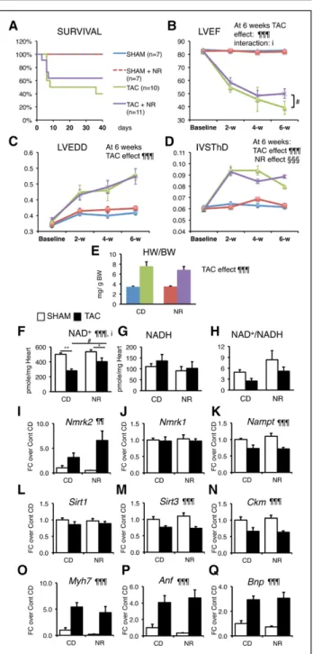
NR Preserves Cardiac Function in the TAC Model
To determine whether NR treatment could be useful in other forms of HF, we assessed its efficiency in the TAC model of pressure overload cardiac hypertrophy.
NR was administrated starting 2 days after the TAC or SHAM surgery. Kaplan-Meier survival curve analysis showed no significant difference, mainly because of a similar early mortality between the 2 groups within 1 week after TAC (Figure 7A). NR treatment reduced the final drop in LV ejection fraction that occurred between the fourth and sixth week after TAC (Figure 7B and
Table V in the online-only Data Supplement). NR did not attenuate the dilatation of the LV chamber and had only a transient effect on the thinning of the interven- tricular septum (Figure 7C and 7D). At euthanization, the cardiac hypertrophy index was similar between the chow diet and NR groups (Figure 7E).
The TAC-induced HF resulted in a decrease of the myocardial NAD+ pool, which was partially preserved by NR, with little impact on the NAD+/NADH ratio (Fig- ure 7F through 7H). TAC increased Nmrk2 expression level in the nontreated group and NR-treated group, whereas Nmrk1 expression remained unchanged and Nampt was repressed (Figure 7I through 7K). TAC re- pressed Sirt3 expression, Sirt1 and Ckm expression while increasing the expression of Myh7, Anf, and Bnp (Figure 7L through 7Q) but NR treatment had no impact on gene expression.
DISCUSSION
We report that depressed NAD+ homeostasis and acti- vation of NMRK2 kinase are early and persistent events in a mouse model of DCM leading to heart failure and that administration of NR, the substrate of NMRK2 and most energetically favorable NAD+ precursor, prevents Figure 6 Continued. of total and phosphoprotein signal from Western blot analyses. Data are normalized on GAPDH signal.
The Phos/Total ratio is calculated from GAPDH normalized levels for each individual. n=6 for each group. Data are expressed as mean fold change (FC)±SEM over control group. t test: *P≤0.05, **P≤0.01, ***P≤0.001 over control group. C, NRCs were treated with FK866 (10 µmol/L, 72 hours, FK), AICAR (500 µmol/L, 48 hours, AIC), or grown in the absence of glucose in the medium (Glc 0) for 48 hours, and proteins were extracted for Western blot analyses. Representative Western blot. D, Quan- tification on n=3 samples for each condition of NRCs treated as in C. Data are expressed as mean FC±SEM over NT group.
*P≤0.05 over control group. E through G, Intracellular NAD+ content (E), NADH (F), and NAD+/NADH ratio (G) in NRCs after 24 hours of treatment with AICAR (500 µmol/L). H, RT qPCR analysis of Nmrk2 mRNA level in NRCs treated with AICAR. t test:
***P≤0.001 over nontreated control group. I and J, NRCs were cotransfected with Nmrk2-luciferase constructs containing 586 (I) or 3009 base pairs (J) of upstream Nmrk2 regulatory region and a dominant negative (DN) AMPK expression vector. NRCs were transfected at day 3 after plating, followed by AICAR treatment (500 µmol/L) at day 4. Luciferase levels were analyzed at day 5. Normalized Fireflyl/Renilla values are expressed as in Figure 5B as FC±SEM over the promoterless pGL4 vector. One-way ANOVA: ¶¶P≤0.01, ¶¶¶P≤0.001 for treatment effect. Post hoc Tukey test: **P≤0.01, ***P≤0.001 over nontreated control group. ##P<0.01 for AICAR versus AICAR+AMPK-DN. K, Nmrk2 promoter deletion analysis by luciferase assay. Rectangular boxes show the position of the putative PPAR binding sites. Data are expressed as mean FC±SEM over the promoterless pGL4 plasmid. One-way ANOVA: ¶¶¶P≤0.001. Post hoc Tukey test: **P≤0.01, over the promoterless pGL4 vector. ##P<0.01 for over the immediately shorter construct. L, Neonatal rat cardiac fibroblasts and cardiomyocyte-enriched fractions were separated on a discontinuous Percoll gradient. Cardiac fibroblasts were transfected with the p-228, p-581, and p-3009-FLuc constructs.
Cardiomyocytes were transfected with the p-3009-FLuc. SV40-RLuc construct was cotransfected for normalization. Data are expressed as mean FC±SEM over the mean p228-FLuc activity in cardiac fibroblasts. One-way ANOVA: ¶P≤0.05. Post hoc Tukey test: *P≤0.05 Cardio-3009 over Fibro-30089. M, NRCs were cotransfected with the p3009-FLuc construct and the RXR expres- sion vector and with either PPARα, PPAR β/δ, or PPARγ expression vectors. NRCs were treated 24 hours later with the agonists GW7647 (0.6 µmol/L), GW501516 (0.6 µmol/L), and G1929 (0.6 µmol/L), for PPARα, PPARβ/δ, and PPARγ, respectively, or with their respective antagonists, GW6471 (10 µmol/L), GSK3787 (2 µmol/L), or GW9662 (2 µmol/L). Data are expressed as mean FC±SEM over normalized luciferase levels of NRCs transfected with the p3009-FLuc construct alone (dashed line) in the same experiment. A 1-way ANOVA was used because each group is independent of the other (different agonists and antagonists):
¶¶¶P≤0.001. Post hoc Tukey test: **P≤0.01 versus p3009-FLuc alone. ##P≤0.01 for comparison between agonist and antago- nist. N, NRCs were transfected with the p3009-FLuc construct. Transfected NRCs were treated 24 hours later with the antago- nists G6471, GSK3787, or G9662, 30 minutes before adding AICAR for a further 24-hour period. All concentrations were as in M. Data are expressed as mean FC±SEM over the p3009-luc construct treated with AICAR alone. One-way ANOVA: ¶P≤0.05.
Post hoc Tukey: *P≤0.05 versus AICAR treated cells. ACC indicates acetyl-coenzyme A carboxylase; AICAR, 5-aminoimidazole- 4-carboxamide ribonucleotide; AMPK, AMP-activated protein kinase; NRC, neonatal rat cardiomyocyte; NT, nontreated cells;
PPAR, peroxisome proliferator-activated receptor; and RT qPCR, quantitative reverse transcription polymerase chain reaction.
by guest on May 22, 2018http://circ.ahajournals.org/Downloaded from