doi: 10.3389/fimmu.2019.02570
Edited by:
Dietmar Fuchs, Innsbruck Medical University, Austria
Reviewed by:
Andrew L. Mellor, Newcastle University, United Kingdom Hajime Julie Yuasa, Kochi University, Japan
*Correspondence:
László Vécsei vecsei.laszlo@med.u-szeged.hu
Specialty section:
This article was submitted to Inflammation, a section of the journal Frontiers in Immunology
Received:11 April 2019 Accepted:16 October 2019 Published:06 November 2019
Citation:
Boros FA and Vécsei L (2019) Immunomodulatory Effects of Genetic Alterations Affecting the Kynurenine Pathway. Front. Immunol. 10:2570.
doi: 10.3389/fimmu.2019.02570
Immunomodulatory Effects of Genetic Alterations Affecting the Kynurenine Pathway
Fanni A. Boros1and László Vécsei1,2,3*
1Department of Neurology, Faculty of Medicine, Albert Szent-Györgyi Clinical Center, University of Szeged, Szeged, Hungary,
2MTA-SZTE Neuroscience Research Group of the Hungarian Academy of Sciences, University of Szeged, Szeged, Hungary,
3Department of Neurology, Interdisciplinary Excellence Centre, University of Szeged, Szeged, Hungary
Several enzymes and metabolites of the kynurenine pathway (KP) have immunomodulatory effects. Modulation of the activities and levels of these molecules might be of particular importance under disease conditions when the amelioration of overreacting immune responses is desired. Results obtained by the use of animal and tissue culture models indicate that by eliminating or decreasing activities of key enzymes of the KP, a beneficial shift in disease outcome can be attained. This review summarizes experimental data of models in which IDO, TDO, or KMO activity modulation was achieved by interventions affecting enzyme production at a genomic level. Elimination of IDO activity was found to improve the outcome of sepsis, certain viral infections, chronic inflammation linked to diabetes, obesity, aorta aneurysm formation, and in anti-tumoral processes. Similarly, lack of TDO activity was advantageous in the case of anti-tumoral immunity, while KMO inhibition was found to be beneficial against microorganisms and in the combat against tumors, as well. On the other hand, the complex interplay among KP metabolites and immune function in some cases requires an increase in a particular enzyme activity for the desired immune response modulation, as was shown by the exacerbation of liver fibrosis due to the elimination of IDO activity and the detrimental effects of TDO inhibition in a mouse model of autoimmune gastritis. The relevance of these studies concerning possible human applications are discussed and highlighted.
Finally, a brief overview is presented on naturally occurring genetic variants affecting immune functionsviamodulation of KP enzyme activity.
Keywords: kynurenine pathway, IDO, TDO, KMO, immunomodulation, genetic manipulation
INTRODUCTION
The kynurenine pathway (KP) is the main route of Trp metabolism. The enzymes of the pathway generate numerous metabolites, some of which are pro-inflammatory and/or generate free radicals, while others are known to be anti-inflammatory and/or scavenge free-radicals. Strong links between KP function and the immune system are demonstrated by extensive amounts of data on changes in the levels of KP metabolites and enzyme activities in diseases accompanied by alterations in immune function. Also, inflammatory cytokines are known to enhance the expression of a key KP enzyme, indoleamine 2,3-dioxygenase (IDO). Imbalances in the pathway can be detrimental,
as excessive production of either pro-, or anti-inflammatory metabolites can contribute to the development of autoimmunity and/or lead to inefficient immune response against pathogens.
Therefore, the understanding of how the KP changes in different immunological states, and, the reverse, how KP effects immunological responses, is cardinal both for understanding the true nature of specific diseases and for identification of therapeutic targets. Genetic manipulations leading either to enhancement or inhibition of the expression of KP enzymes might be a feasible way of restoring the imbalance of the pathway in various diseases. Naturally occurring genetic variations in the coding regions in several genes coding for KP enzymes have been identified [for a review see (1)]. In the majority of these, however, a causal relation between a specific gene variant and disease development has not been elucidated.
This review summarizes available data on the effects of expression modification of KP enzyme coding genes with specific attention to immune modulation. Following a brief overview
Abbreviations: KP, kynurenine pathway; IDO, indoleamine 2,3-dioxygenase (protein);IDO,indoleamine 2,3-dioxygenase (gene in human);Ido,indoleamine 2,3-dioxygenase (murine gene); TDO, tryptophan 2,3-dioxygenase (protein);
Tdo, tryptophan 2,3-dioxygenase (murine gene); TDO, tryptophan 2,3- dioxygenase (gene in human); KMO, kynurenine 3-monooxygenase (protein);
KMO, kynurenine 3-monooxygenase (gene in human); Kmo, kynurenine 3-monooxygenase (murine gene); CNS, central nervous system; L-KYN, L- kynurenine; KYNA, kynurenic acid; KATI-IV, kynurenine aminotransferases;
NMDAR, N-methyl-D-aspartate receptor; α7nAChRs, nicotinic acetylcholine receptors; KYNU, kynureninase; AA, anthranilic acid; 3-HK, 3-hydroxy kynurenine; XA, xanthurenic acid; 3-HAA, 3-hydroxyanthranilic acid; ACMS, 2-amino-3-carboxymuconate-semialdehyde; 3-HAO, 3-hydroxyanthranilate 3,4-dioxygenase; PIC, picolinic acid; ACMSD, aminocarboxymuconate- semialdehyde-decarboxylase; QUIN, quinolinic acid; ISRE, IFN stimulated response element; GAS, gamma-activated sequences; DRE, dendritic cell response element; NF-κB, nuclear factor kappa-light-chain-enhancer of activated B-cells;
TLRs, toll-like receptors; TGFBR, transforming growth factor beta receptor;
AHR, aryl hydrocarbon receptor; IFNBR, interferon beta receptor; IFNGR, interferon gamma receptor; TNFR, tumor necrosis factor receptor; SOCS3, suppressor of cytokine signaling 3; DC, dendritic cell; IFN, interferon; LPS, lipopolysaccharide; IL, interleukines; TNF, tumor necrosis factor; mTOR, mammalian target of rapamycin; NPC, nasopharyngeal carcinoma; AML, acute myeloid leukemia; AHR, aryl hydrocarbon receptor; ROS, reactive oxygen species;
siRNA, small interfering RNA; IDO−/−,IDOknockout; UPEC, Uropathogenic Escherichia coli;MuLV, murine leukemia virus; NK cell, natural killer cell; pDC, plasmocytoid dendritic cell; WT, wild type; ECMV, encephalomyocarditis virus;
sJIA, systemic juvenile idiopathic arthritis; sHLH, secondary hemophagocytic- lymphohistiocytosis; STING, Stimulator of Interferon Genes; DNP, DNA nanoparticle; EAE, experimental autoimmune encephalitis; MS, multiple sclerosis;
MOG, myelin oligodendrocyte glycoprotein; c-diGMP, cyclic diguanylate monophosphate; CIA, collagen induced arthritis; RA, rheumatoid arthritis;
AdIDO, adenoviral vector-mediated IDO gene delivery; NOD, non-obese diabetic; MRLlpr/lpr, Lupus-prone Murphy Roths large mice; SLE, systemic lupus erythematosus; MAS, macrophage activation syndrome; DR, diabetic retinopathy; AngII, angiotensin II; ApoE−/−, Apolipoprotein E knockout;
NAD(P)H, nicotinamide adenine dinucleotide phosphate; AAA, abdominal aortic aneurysm; VSMC, vascular smooth muscle cell;Ldlr−/−, low density lipoprotein—
receptor deficient; HFD, high fat diet; MMP2, matrix metallopeptidase 2; WAT, white adipose tissue; OGTT, oral glucose tolerance test; ITT, insulin tolerance test; IAA, indole-3 acetic acid; CCl4, carbon-tetrachloride; PMN, polymorph nuclear neutrophil; S.t.,Salmonella typhimurium; shRNA, short hairpin RNA; RT, radiotherapy; BER, base excision repair; MX, methoxyamine; TS, thymidylate synthase; IRI, ischemia-reperfusion injury; AKI, acute kidney injury; CTL, cytolytic T lymphocyte; PD, Parkinson’s disease; SSc, systemic sclerosis; CD, Crohn’s disease; BM, bacterial meningitis; CSF, cerebro-spinal fluid.
of the metabolites and enzymes of the pathway, we summarize observations which indicate links between KP and immune function. This is followed by an overview of findings obtained by the use of models with targeted ablation and up- or down- regulation of KP enzymes. With respect to diseases related to disorders of the immune system, such as infectious diseases, chronic inflammation, autoimmunity and cancer, these models have focused on three KP enzymes: IDO, tryptophan 2,3- dioxygenase (TDO) and kynurenine 3-monooxygenase (KMO) (Tables 1–5). These enzymes and, in particular, IDO are also targeted by several pharmacochemical interventions. Discussion of that field is out of the scope of this review, as we focus on gene level interventions. Readers interested in pharmacologic interventions of KP enzymes can find excellent summaries of the field in Ye et al. (45) and Lemos et al. (46). In the final section, we provide a summary of available data on those naturally occurring KP gene variants which are believed to be associated with different human diseases affecting immune function.
KYNURENINE PATHWAY—THE MAIN ROUTE OF TRYPTOPHAN METABOLISM
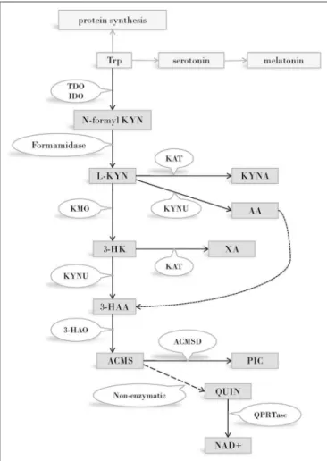
Disregarding protein synthesis, the KP is the main route of Trp metabolism, both in the peripheral and in the central nervous system (CNS) (Figure 1). In the CNS, 95 percent of the resident Trp is metabolizedviathe KP, and only the minority of the amino acid is transformed into serotonin and melatonin. In consecutive steps of the pathway, numerous metabolites possessing immune- and neuromodulatory properties are synthesized (47).
The first and rate limiting step of Trp metabolism is the conversion of the amino acid into N-formyl-L-kynurenine. This step is catalyzed by one of three enzymes: IDO (often referred to as IDO1), IDO2, or TDO. (Prior to the discovery of IDO 2, “IDO”
designation was used exclusively. Today IDO and IDO1 are used as synonyms and IDO2 is reserved for the enzyme recognized in 2007. In this review we will use IDO unless we are referring to IDO2). TDO is expressed mainly in the liver, thus plays a cardinal role in regulating the amount of available Trp throughout the body, outside the CNS. IDO is expressed in several human tissues, among them various cell types of the immune system (48). The enzyme plays a key role in reactions leading to the synthesis of immunoactive KP metabolites, consequently its role in immunomodulation is expected. IDO2 expression pattern and function is not known in detail. A strong argument against the role of this enzyme in Trp metabolism is the frequent occurrence of an IDO2 gene variant that gives rise to a non-functioning enzyme (49), and the high Michaelis Constant of the enzyme for Trp, which is 100-fold above the physiological concentrations of the amino acid (50).
N-formyl-L-kynurenine is converted to L-kynurenine (L- KYN) by formamidase. L-KYN is an important branch point of the KP as it can be alternatively metabolized into three different metabolites of which some are neurotoxic, while others possess neuroprotective and antioxidant properties (51, 52). Firstly, L-KYN can be metabolized into kynurenic acid (KYNA) by kynurenine aminotransferases (KATI-IV)
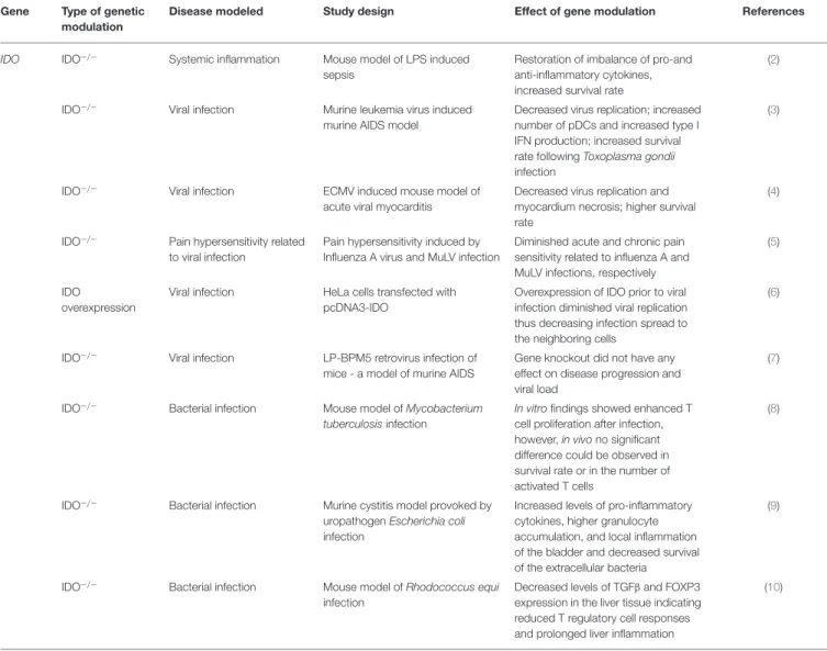
TABLE 1 |Effects of modulation of IDO function by genetic manipulation inin vivoandin vitromodels of systemic inflammation, viral, and bacterial infections.
Gene Type of genetic modulation
Disease modeled Study design Effect of gene modulation References
IDO IDO−/− Systemic inflammation Mouse model of LPS induced sepsis
Restoration of imbalance of pro-and anti-inflammatory cytokines, increased survival rate
(2)
IDO−/− Viral infection Murine leukemia virus induced murine AIDS model
Decreased virus replication; increased number of pDCs and increased type I IFN production; increased survival rate followingToxoplasma gondii infection
(3)
IDO−/− Viral infection ECMV induced mouse model of
acute viral myocarditis
Decreased virus replication and myocardium necrosis; higher survival rate
(4)
IDO−/− Pain hypersensitivity related to viral infection
Pain hypersensitivity induced by Influenza A virus and MuLV infection
Diminished acute and chronic pain sensitivity related to influenza A and MuLV infections, respectively
(5)
IDO
overexpression
Viral infection HeLa cells transfected with pcDNA3-IDO
Overexpression of IDO prior to viral infection diminished viral replication thus decreasing infection spread to the neighboring cells
(6)
IDO−/− Viral infection LP-BPM5 retrovirus infection of mice - a model of murine AIDS
Gene knockout did not have any effect on disease progression and viral load
(7)
IDO−/− Bacterial infection Mouse model ofMycobacterium tuberculosisinfection
In vitrofindings showed enhanced T cell proliferation after infection, however,in vivono significant difference could be observed in survival rate or in the number of activated T cells
(8)
IDO−/− Bacterial infection Murine cystitis model provoked by uropathogenEscherichia coli infection
Increased levels of pro-inflammatory cytokines, higher granulocyte accumulation, and local inflammation of the bladder and decreased survival of the extracellular bacteria
(9)
IDO−/− Bacterial infection Mouse model ofRhodococcus equi infection
Decreased levels of TGFβand FOXP3 expression in the liver tissue indicating reduced T regulatory cell responses and prolonged liver inflammation
(10)
IDO, Indoleamine 2,3-dioxygenase gene; IDO−/−, IDO knockout; pDC, plasmocytoid dendritic cell; IFN, interferon; ECMV, encephalomyocarditis virus; MuLV, murine leukemia virus;
TGFβ, transforming growth factor beta; FOXP3, forkhead box P3.
from which KATII plays the most important role in the human CNS (53, 54). Secondly, L-KYN is also a substrate of kynureninase (KYNU), an enzyme responsible for the formation of anthranilic acid (AA). Finally, the third route of L-KYN metabolism is catalyzed by KMO to form 3-hydroxy kynurenine (3-HK) which can be further transformed into xanthurenic acid (XA) by KATs. 3-HK and AA can both be metabolized into 3-hydroxyanthranilic acid (3-HAA), which, alongside with 3-HK, have free-radical generating properties, thus can lead to oxidative stress and neurodegeneration (55). However, depending on the redox properties of the cell, 3-HK and 3-HAA can also serve as antioxidant molecules (56).
Further down the pathway, the unstable 2-amino-3- carboxymuconate-semialdehyde (ACMS) is formed by 3-hydroxyanthranilate 3,4-dioxygenase (3-HAO). ACMS can be transformed either into picolinic acid (PIC) by an
aminocarboxymuconate-semialdehyde-decarboxylase (ACMSD) catalyzed reaction, or it can form the NAD+ and NADP+ precursor quinolinic acid (QUIN) via a non-enzymatic conversion. QUIN is a key figure in excitotoxicity mediated neurodegeneration (52,57,58).
In light of the numerous enzymes participating and metabolites generated, the involvement of the KP in various disorders is not surprising. Indeed, changes in KP enzyme activity and metabolite levels have been detected in inflammatory, autoimmune, neurodegenerative and psychiatric diseases, as well.
In the following sections of this review we will briefly consider observations that point to existing links between KP and immune function. Then we will overview results obtained by models in which the KP was modulated by interventions effecting gene activity. Finally, we list known genetic alterations in genes of KP enzymes that are believed to be associated with changes in immune functions.
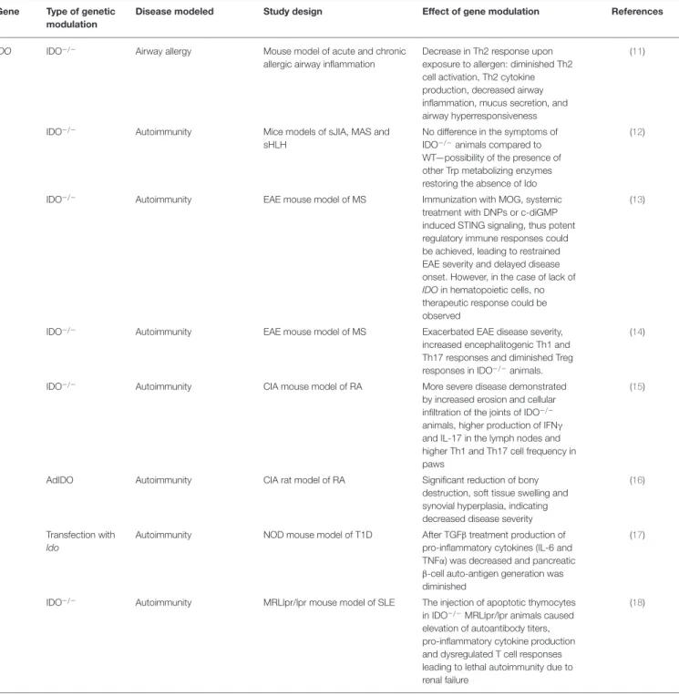
TABLE 2 |Effects of modulation of IDO function by genetic manipulation in animal models of allergy and autoimmunity.
Gene Type of genetic modulation
Disease modeled Study design Effect of gene modulation References
IDO IDO−/− Airway allergy Mouse model of acute and chronic
allergic airway inflammation
Decrease in Th2 response upon exposure to allergen: diminished Th2 cell activation, Th2 cytokine production, decreased airway inflammation, mucus secretion, and airway hyperresponsiveness
(11)
IDO−/− Autoimmunity Mice models of sJIA, MAS and
sHLH
No difference in the symptoms of IDO−/−animals compared to WT—possibility of the presence of other Trp metabolizing enzymes restoring the absence of Ido
(12)
IDO−/− Autoimmunity EAE mouse model of MS Immunization with MOG, systemic
treatment with DNPs or c-diGMP induced STING signaling, thus potent regulatory immune responses could be achieved, leading to restrained EAE severity and delayed disease onset. However, in the case of lack of IDOin hematopoietic cells, no therapeutic response could be observed
(13)
IDO−/− Autoimmunity EAE mouse model of MS Exacerbated EAE disease severity,
increased encephalitogenic Th1 and Th17 responses and diminished Treg responses in IDO−/−animals.
(14)
IDO−/− Autoimmunity CIA mouse model of RA More severe disease demonstrated
by increased erosion and cellular infiltration of the joints of IDO−/− animals, higher production of IFNγ and IL-17 in the lymph nodes and higher Th1 and Th17 cell frequency in paws
(15)
AdIDO Autoimmunity CIA rat model of RA Significant reduction of bony
destruction, soft tissue swelling and synovial hyperplasia, indicating decreased disease severity
(16)
Transfection with Ido
Autoimmunity NOD mouse model of T1D After TGFβtreatment production of pro-inflammatory cytokines (IL-6 and TNFα) was decreased and pancreatic β-cell auto-antigen generation was diminished
(17)
IDO−/− Autoimmunity MRLlpr/lpr mouse model of SLE The injection of apoptotic thymocytes in IDO−/−MRLlpr/lpr animals caused elevation of autoantibody titers, pro-inflammatory cytokine production and dysregulated T cell responses leading to lethal autoimmunity due to renal failure
(18)
IDO, Indoleamine 2,3-dioxygenase gene; IDO−/−, IDO knockout; sJIA, systemic juvenile idiopathic arthritis; MAS, macrophage activation syndrome; sHLH, secondary hemophagocytic- lymphohistiocytosis; WT, wild type; Trp, tryptophan; EAE, experimental autoimmune encephalitis; MS, multiple sclerosis; MOG, myelin oligodendrocyte glycoprotein; DNP, DNA nanoparticle; c-diGMP, cyclic diguanylate monophosphate; CIA, collagen induced arthritis; RA, rheumatoid arthritis; IFNγ, interferon gamma; AdIDO, adenoviral vector-mediated IDO gene delivery; NOD, non-obese diabetic; T1D, type 1 diabetes; TGFβ, transforming growth factor beta; MRLlpr/lpr, Lupus-prone Murphy Roths large mice; SLE, systemic lupus erythematosus.
OBSERVATIONS INDICATING LINKS BETWEEN THE KYNURENINE PATHWAY AND IMMUNE FUNCTIONS
Indoleamine 2,3-Dioxygenase
Interplay between several enzymes of the KP and immune function are well-demonstrated. In this respect IDO, a key
enzyme of the pathway, deserves particular attention. IDO is believed to exert its effects on immune function both by direct and indirect mechanisms. As an enzyme, IDO plays a role in Trp utilization and through this, in cellular metabolism via mTOR and GCN2 linked pathways. By converting Trp to KYN, IDO has a central role in determining concentrations of KP metabolites, many of which are direct or indirect regulators
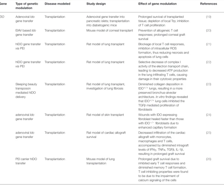
TABLE 3 |Effects of modulation of IDO function by genetic manipulation in transplant animal models.
Gene Type of genetic modulation
Disease modeled Study design Effect of gene modulation References
IDO AdenoviralIdo gene transfer
Transplantation Adenoviral gene transfer into pancreatic islets; transplantation into diabetogenic mice
Prolonged survival of transplanted tissue; depletion of local Trp; inhibition of T cell proliferation
(19)
EIAV basedIdo gene transfer
Transplantation Mouse model of corneal transplant Prevention of allogeneic T cell responses; prolonged corneal graft survival
(20)
hIDO gene transfer viaPEI
Transplantation Rat model of lung transplant Blockage of local T cell responses, inhibition of intracellular ROS formation, thus reducing necrosis and apoptosis of lung cells
(21)
hIDO gene transfer viaPEI
Transplantation Rat model of lung transplant Selective decrease of complex I activity of the electron transport chain, leading to decreased ATP production in the lung infiltrating T cells, causing damage in their cytotoxic properties
(22)
Sleeping beauty transposon mediated hIDO delivery
Transplantation Rat model of lung transplant, investigation of lung fibrosis
Diminished collagen deposition in IDO+/+lungs, resulting in a more preserved bronchus-alveolar architecture.In vitrofindings revealed that IDO+/+lung cells inhibited the TGFβmediated proliferation of fibroblasts
(23)
adenoviralIdo gene transfer
Transplantation Rat model of skin transplant Wounds with IDO expressing fibroblast healed faster than those with IDO−/−fibroblasts due to enhanced capillary formation
(24)
adenoviralIdo gene transfer
Transplantation Rat model of cardiac allograft survival
Decreased infiltration of the cardiac allograft with monocytes, macrophages and T cells,
accompanied by diminished intragraft levels of IFNγ, TNFα, TGFβ, IL-1β, resulting in prolonged graft survival
(25)
PEI carrier hIDO transfer
Transplantation Mouse model of lung transplantation
Prolonged graft survival due to inhibited early T cell responses and diminished memory T cell formation.
T cell inhibiting properties were found to be due to the impairment of calcium signaling of the cells
(26)
IDO, Indoleamine 2,3-dioxygenase gene; IDO−/−, IDO knockout; Trp, tryptophan; EIAV, Equine infectious anemia virus; PEI, polimer polyethilenimine; ROS, reactive oxygen species;
TGFβ, transforming growth factor beta; IFNγ, interferon gamma; TNFα, tumor necrosis factor alpha.
of immunofunction. Furthermore, IDO also acts as a signal protein. In concert with TGFβ, it regulates activation through non-canonical NF-κB response elements, thus affecting of its own production as well (45,46).
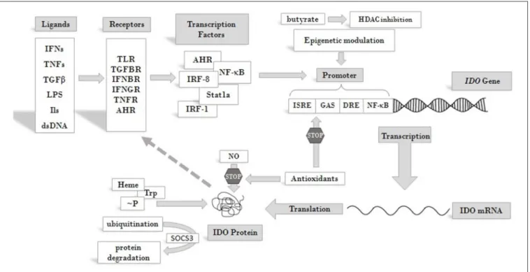
IDO production and activity is controlled at different levels, including both transcriptional and post-translational regulation [reviewed in (46, 59)] (Figure 2). At the protein level, both its substrate, Trp, and its co-factor, heme, enhance IDO activity (61, 62). NO was found to reversibly inhibit the enzyme by binding to the active site (63, 64). Antioxidants also inhibit enzyme activity, both at transcriptional and post-transcriptional levels (62). Phosphorylation of two tyrosine side chains also can modulate IDO activity and its halflife (65). Decrease in IDO enzyme levels can be the result of ubiquitylation of the protein by the suppressor of cytokine signaling 3 (SOCS3) factor and proteosomal degradation (66).
At the level of transcription several cis-regulatory elements in the IDO promoter transmit regulatory signals. These are IFN stimulated response elements (ISRE), palindromic gamma- activated sequences (GAS), dendritic cell response elements (DRE) and non-canonical NF-κB binding sites [see reviews (60, 65)]. A number of transcription factors have been identified so far, which bind to these elements and play roles in the transcriptional regulation of IDO. Among them are IRF-1, IRF- 8, Stat1a, NF-κB (67) and aryl hydrocarbon receptor (AHR).
Recently, epigenetic regulation of the gene through histone deacetylase activity has also been reported (68). Through these factors various receptor-ligand pathways converge to determine IDO gene expression. These transmit regulatory signals from activated toll-like receptors (TLRs), transforming growth factor beta receptors (TGFBRs), AHR, interferon beta and gamma receptors (IFNBR and IFNGR), and members of the tumor
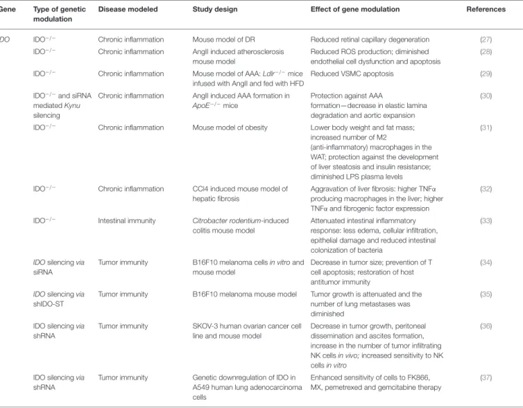
TABLE 4 |Effects of modulation of IDO function by genetic manipulation inin vitroandin vivomodels of chronic inflammation and cancer.
Gene Type of genetic modulation
Disease modeled Study design Effect of gene modulation References
IDO IDO−/− Chronic inflammation Mouse model of DR Reduced retinal capillary degeneration (27)
IDO−/− Chronic inflammation AngII induced atherosclerosis mouse model
Reduced ROS production; diminished endothelial cell dysfunction and apoptosis
(28)
IDO−/− Chronic inflammation Mouse model of AAA:Ldlr−/−mice infused with AngII and fed with HFD
Reduced VSMC apoptosis (29)
IDO−/−and siRNA mediatedKynu silencing
Chronic inflammation AngII induced AAA formation in ApoE−/−mice
Protection against AAA
formation—decrease in elastic lamina degradation and aortic expansion
(30)
IDO−/− Chronic inflammation Mouse model of obesity Lower body weight and fat mass;
increased number of M2
(anti-inflammatory) macrophages in the WAT; protection against the development of liver steatosis and insulin resistance;
diminished LPS plasma levels
(31)
IDO−/− Chronic inflammation CCl4 induced mouse model of hepatic fibrosis
Aggravation of liver fibrosis: higher TNFα producing macrophages in the liver; higher TNFαand fibrogenic factor expression
(32)
IDO−/− Intestinal immunity Citrobacter rodentium-induced colitis mouse model
Attenuated intestinal inflammatory response: less edema, cellular infiltration, epithelial damage and reduced intestinal colonization of bacteria
(33)
IDOsilencingvia siRNA
Tumor immunity B16F10 melanoma cellsin vitroand mouse model
Decrease in tumor size; prevention of T cell apoptosis; restoration of host antitumor immunity
(34)
IDOsilencingvia shIDO-ST
Tumor immunity B16F10 melanoma mouse model Tumor growth is attenuated and the number of lung metastases was diminished
(35)
IDO silencingvia shRNA
Tumor immunity SKOV-3 human ovarian cancer cell line and mouse model
Decrease in tumor growth, peritoneal dissemination and ascites formation, increase in the number of tumor infiltrating NK cellsin vivo;increased sensitivity to NK cellsin vitro
(36)
IDO silencingvia shRNA
Tumor immunity Genetic downregulation of IDO in A549 human lung adenocarcinoma cells
Enhanced sensitivity of cells to FK866, MX, pemetrexed and gemcitabine therapy
(37)
IDO, Indoleamine 2,3-dioxygenase gene; Ido−/−, Ido knockout; Kynu, kynureninase gene; siRNA, small interfering RNA; shIDO-ST, shRNA: short hairpin RNA; DR, diabetic retinopathy;
AngII, Angiotensin II; AAA, abdominal aortic aneurysm; Ldlr−/−, low density lipoprotein—receptor deficient; HFD, high fat diet; ApoE−/−, Apolipoprotein E knockout; CCl4, carbon- tetrachloride; ROS, reactive oxygen species; VSMC, vascular smooth cell; WAT, white adipose tissue; LPS, lipopolysaccharide; TNFα, tumor necrosis factor alpha; NK cell, natural killer cell; MX, methoxyamine.
necrosis factor receptor superfamily (TNFRs). Activation of any of these receptors by their ligands can trigger signaling pathways that promote or maintain the expression ofIDO. Consequently, inflammatory signals, such as IFNs, lipopolysaccharides (LPS), interleukins (ILs) (such as IL-1, IL-2, IL-27, IL-10) TNFs, TGFs, and prostaglandins, can induce IDO production (69,70).
Thus, induction of the enzyme can be very complex and cell type specific [reviewed in (71)]. Moreover, some inflammatory markers act synergistically to increase IDO production and the types of cytokines affecting gene expression may differ in various cell types. This might be reflected by seemingly contradictory reports on the roles of particular ligands in IDO induction. According to some data, IFNγ is one of the main inducers of IDO expression (72). On the other hand, results obtained in LPS induced systemic inflammatory rat model did not support the role of IFNγ in IDO induction in the CNS
and a more important role for other inflammatory cytokines, such as TNFα and IL-6, was proposed. Strengthening this conclusion, in LPS-stimulated glial cell cultures an increase of IDO expression was observed, accompanied by elevated levels of TNFα and IL-6, but no IFNγ expression. Based on these observations, it was concluded that IDO induction in the CNS by LPS is not mediated by IFNγ (73). However, recent findings strongly argue for the role of IFNs in the activation of IDO expression. It was found that not IFNγ but IFNα signaling was essential in enhancing IDO expression after B7 ligation of CTL4-Ig (74).IDOexpression up-regulationviaCpG oligodeoxynucleotide binding to TLR9 was also IFNαdependent (75). Futhermore,IDOexpression was found to be upregulated by cytosolic DNAviathe STING/IFNαβpathway (76). Thus, it seems firmly established that type I IFNs play a cardinal role in enhanced IDO gene expression with inflammatory signals
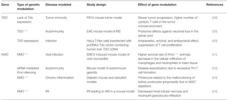
TABLE 5 |Effects of modulation of TDO and KMO function by genetic manipulation inin vitroandin vivomodels.
Gene Type of genetic modulation
Disease modeled Study design Effect of gene modulation References
TDO Lack ofTdo expression
Tumor immunity P815 mouse tumor model Slower tumor progression, higher number of cytolytic T cells in the tumor
microenvironment
(38)
TDO−/− Autoimmunity EAE mouse model of MS Protective effects against neuronal loss in the spinal cord
(39)
TDO expression Infection HeLa T-Rex cells transfected with pcDNA4-Tdovector containing human liverTDOcDNA
Antiparasitic, antiviral, and antibacterial effect;
suppression of T cell proliferation
(40)
KMO KMO−/− Viral infection EMCV induced mouse model of
viral myocarditis
Higher survival rate ofKmo−/−animals;
decrease in the cellular infiltration of marophages and neutrophiles in heart tissue
(41)
siRNA mediated Kmosilencing
Autoimmunity Mouse model of autoimmune gastritis
Disease exacerbation due to excessive Th17 cell formation
(42)
KMO−/− Chronic inflammation Diabetic mouse and zebrafish models
Proteinuria related to the malfunctioning of kidney podocytes (proposedly due to NAD+ depletion)
(43)
KMO−/− IRI IRI leading to AKI in a mouse model Decreased renal tubular necrosis and neutrophil granulocyte infiltration
(44)
TDO, Tryptophan 2,3-dioxygenase gene; TDO-/-, TDO knockout; KMO, Kynurenine 3-monooxygenase gene; KMO-/-, KMO knockout; EAE, experimental autoimmune encephalitis; MS, multiple sclerosis; EMCV, encephalomyocarditis virus; IRI, ischemia-reperfusion injury; AKI, acute kidney injury.
and thatIDOexpression following LPS treatment is induced by type I IFNs.
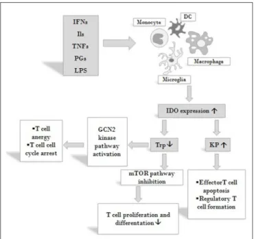
IDO is expressed by numerous cells of the immune system:
monocytes, dendritic cells (DCs), macrophages and microglia (48). It regulates immune responses in various direct and indirect ways (Figure 3). On the one hand, by decreasing the amount of available Trp, it causes an increase in free transfer RNA, thus activating the GCN2 stress-kinase pathway leading to T cell anergy and cell cycle arrest (77). On the other hand, a lack of the amino acid leads to the inhibition of the rapamycin (mTOR) pathway followed by a translational block (78). Moreover, via the formation of different immunologically active kynurenine metabolites, IDO also contributes to the apoptosis of effector T cells and promotes the formation of regulatory T cells (59,79).
Another important link between KP and the immune system is manifested by DCs, in particular in their role in inflammatory processes. Sepsis is a systemic inflammatory response syndrome which leads to hemodynamic shock accompanied by multi-organ failure. It is a major cause of mortality and morbidity among hospitalized patients. Sepsis is the consequence of microbial infection, in which Gram-negative bacteria outer-membrane components (LPS) trigger the uncontrolled production of pro-inflammatory cytokines, which leads to the imbalance of pro-and anti-inflammatory factors. DCs seem to play a cardinal role is sepsis development, as they are capable of producing pro- (IL-12) and anti-inflammatory (IL-10) cytokines, the balance of which was found to be altered during infection (80). In DCs, IDO expression is induced by LPS and the enzyme production contributes to the imbalance of anti- and pro-inflammatory cytokines (2).
A growing body of data shows the involvement of IDO in immune responses to tumors [see in (45,46,81)]. A pivotal role
of the enzyme is seen in establishing the immunosuppressive microenvironment of tumors by altering the functions of infiltrating T lymphocytes, thus promoting immune escape and progression of cancer cells (82,83). Upregulated expression of IDO has been reported in the microenvironment of laryngeal and esophageal carcinomas (84–87) and higher plasma enzyme activity was reported in lung-, gynecological-, breast- and colorectal cancers, and melanoma. Both local expression changes and elevated plasma IDO activity was reported in patients with nasopharyngeal carcinoma (NPC) (81). Interestingly, a significant difference in plasma IDO levels could be detected between healthy controls and NPC patients with metastasis, in contrast to patients without metastasis. Plasma IDO activity was also found to have a prognostic value, as patients with higher levels of enzyme activity had significantly lower rates of survival compared to those with lower IDO activity. Higher enzyme activity was shown to result from higher expression levels: Fukuno et al. reported that IDO mRNA expression in patients with acute myeloid leukemia (AML) was associated with a worse disease outcome (88). In light of the role IDO plays in immune responses to cancer, it is no wonder that IDO modulation is a hot topic in cancer research. Many therapeutic approaches are underway for pharmaceutical enzyme inhibition (65). This review will not discuss these in detail since our aim is to give an overview of findings on approaches targeting the KP by gene modulation.
Tryptophan-Dioxygenase
The first step of the KP can also be catalyzed by TDO, a functional ortholog enzyme of IDO. However, while IDO is mainly expressed by various immune cells, thus regulating the amount of locally available Trp, TDO is expressed in the liver, affecting
FIGURE 1 |The kynurenine pathway of tryptophan metabolism. Enzymes of the KP metabolise Trp into products possessing immune- and
neuromodulatory properties. By the utilization of Trp and generation of NAD coenzyme precursor the KP has profound effects on cellular protein and energy metabolism. Several internal metabolites of the pathway play role on redox regulation and have neuroprotective - or neurotoxic effects. Immune functions are modified by the KP both directly, via immuno modulatory metabolites and indirectly, via changing the metabolism of immune cells by altering amino acid availability, redox status and energy balance.
Abbreviations: Trp: tryptophan; TDO:tryptophan 2,3-dioxygenase;
IDO:indoleamine 2,3-dioxygenase; N-formyl KYN:N-formyl-kynurenine;
L-KYN:L-kynurenine; KAT:kynurenine aminotransferase; KYNA: kynurenic acid;
KYNU:kynureninase; AA:anthranilic acid; KMO:kynurenine 3-monooxygenase;
3-HK:3-hydroxy kynurenine; XA: xanthurenic acid; KYNU:kynureninase;
3-HAA: 3-hydroxyanthranilic acid; 3-HAO:3-hydroxyanthranilate 3,4-dioxygenase; ACMS: 2-amino-3-carboxymuconate-semialdehyde;
ACMSD: aminocarboxymuconate-semialdehyde-decarboxylase; PIC: picolinic acid; QUIN: quinolinic acid; QPRTase:quinolinate phosphoribosyltransferase;
NAD+: nicotinamide adenine dinucleotide; CNS: central nervous system.
the systemic level of the amino acid. The activity of the enzyme can be regulated by various mechanisms.TDOtranscription is enhanced by glucocorticoids and this is potentiated by glucagon, but inhibited by adrenaline and insulin (89). TDO can also be activated by its cofactor, heme, and its substrate, Trp [reviewed in (61)]. Recent evidence demonstrates TDO presence in rat skin and the CNS of humans (90, 91), thus broadening the
location and raising further questions on the exact role of the enzyme. As Trp stabilizes the TDO enzyme complex (92), and cortisone, a hormone with anti-inflammatory effects, enhances TDO expression (93, 94), one can expect the involvement of the enzyme in immune processes. This was reported first in the early 2000s (95,96): in 2000, Tatsumi et al. proposed a role for the enzyme in tolerance during embryonic implantation, based on finding upregulated expression of TDO mRNA in murine decidualized stromal cells surrounding the implanted embryo (96). In 2001 Suzuki et al. reported high TDO expression during early murine gestation, preceding the expression of IDO, thus revealing an important role of TDO in fetal tolerance (95). When regarding the immune modulator effects of the KP, research has been mainly focused on IDO, but a growing body of evidence is accumulating on the involvement of TDO as well. It indicates the presence of the enzyme in tumor immune resistance (38, 97) and parasite, viral and microbial infections (98) (Table 5).
Expression of TDO by several different tumor types—such as melanomas, bladder-, and hepatocarcinomas—drew attention to the possible role of the enzyme in tumor immunity. TDO was found to be constitutively expressed in glioblastomas and excessive production of the AHR agonist KYN was found to contribute to the immune escape, higher motility and survival of tumor cells (38).
Kynurenine Monooxygenase
A third enzyme with assumed immunomodulatory effects of the KP is KMO, which is situated at an important branch point of the pathway. KYN can be catalyzed by KATs into KYNA, representing a neuroprotective and antioxidant branch of the pathway. On the other hand, KMO can convert KYN to 3- HK, which can be further converted into PIC and QUIN. Both of these metabolites are known to have neurotoxic and free radical generating properties. Thus, KMO has a key role in determining the balance between pro- (QUIN, 3-HK, PIC) and anti-inflammatory (KYNA) kynurenine metabolites.
The substrate of the KMO enzyme, KYN, was shown to promote tumor formation and the generation of regulatory T cells via AHR (99) and adenylate- and guanylate-cyclase pathway activation (38). However, the mode of action of KYN on AHR raised questions, as the structure of the metabolite does not show the necessary features for high-affinity AHR binding. Recently two KYN condensation products have been identified, which are high affinity AHR ligands, active at low picomolar levels. Thus, KYN seems to be a pro-ligand that spontaneously converts to derivatives possessing AHR agonist properties (100). Theoretically, enhancing the metabolism of KYN via KMO upregulation could be protective against the development of tumors. However, upregulation of KMO also leads to the formation of metabolites with reactive oxygen species (ROS) generating properties, such as 3-HK, 3-HAA, and QUIN.
QUIN also exerts excitotoxic effects through the activation of NMDARs (38). Pharmacological inhibition of KMO enhances the production of KYNA, a metabolite with neuroprotective effects.
Besides its neuroprotective effects (101,102) KYNA also has an important role in immunomodulation, mainlyviathe activation of GPR35 receptors and AHRs [reviewed: (103)]. KYNA was
FIGURE 2 |Overview of pathways leading to IDO enzyme production and regulation. IDO activity is regulated at different levels. Its substrate Trp and cofactor heme are positive regulators of the enzyme, whereas antioxidants and NO act as inhibitors. Phosphorylation at tyrosine side chains and ubiquitination modulate IDO activity and half life. At the level of transcription several cis regulatory elements of the IDO promoter collect regulatory signals via binding of transcription factors and epigenetic regulators, which respond to signals arriving from receptors that are activated by cytokines and other immunomodulatory molecules. Extracellular signals produced by other cells or pathogens and intracellular signals, such as cytosolic dsDNA can both induce IDO expression and feed-back regulation of the production has also been described [see text for details and (60)]. Abbreviations: IFN: interferon; TNF: tumor necrosis factor; TGFβ: transforming growth factor beta; LPS: lipopolysaccharide;
dsDNA: double strand DNA; TLR: toll-like receptor; TGFBR: transforming growth factor beta receptor; IFNBR: interferon beta receptor; IFNGR: interferon gamma receptor; TNFR: tumor necrosis factor receptor; AHR: aryl hydrocarbon receptor; ISRE: IFN stimulated response element; DRE: dendritic cell response element; GAS:
gamma-activated sequences; HDAC: histone deacetylase;∼P: phosphorylation; IDO: indoleamine 2,3-dioxygenase; SOCS3: suppressor of cytokine signaling 3; Trp:
tryptophan; NO: nitrogen oxide.
found to attenuate inflammation under inflammatory conditions by several means: by reducing TNF expression in monocytes, IL-4 secretion of T-cell receptor stimulated variant natural killer-like T cells and LPS induced IL-23 formation of DCs [reviewed: (103)].
The expression of KMO was also found to be upregulated in the CNS of rats in LPS induced systemic inflammation, together with a significant increase in pro-inflammatory cytokines such as TNFαand IL-6 (73).
KMO expression and activity have been investigated in autoimmunity related diseases. A link seems to exist through AHRs as these receptors play an important role in the regulation of pro-inflammatory Th17 cell differentiation (42) and Trp metabolites act as agonists of AHRs. KP metabolites play roles in promoting the differentiation of naive T cells into effector Th17 cells (104), which are governors of autoimmune diseases (105).
Stephens et al. reported that Th17 cells highly express KMO, and that either the inhibition of the enzyme or the addition of 3-HK augmented the formation of effector T cells (42).
Since these three enzymes of the KP seem to be associated with immune functions, genetic approaches aimed at modifying immune responses are focused on altering their expression. So far, primarily gene knockouts, gene expression regulation by small interfering RNA (siRNA), short hairpin RNA (shRNA)
and different gene delivery techniques into model animals and tissue culture cells have yielded results regarding the immunomodulatory effects of these enzymes. The next section will review these results. We find it important to point out here that with the rapid progress of gene modulatory and gene editing techniques it is expected that the data summarized here will grow in the near future.
IMMUNOMODULATION VIA ALTERING THE EXPRESSION OF GENES THAT CODE FOR ENZYMES OF THE KYNURENINE
PATHWAY
Indoleamine 2,3-Dioxygenase
Effects of Genetic Modification of Indoleamine 2,3-Dioxygenase Related to Immune Responses in Infection
During the course of sepsis, induction of IDO by bacterial endotoxins plays a pivotal role in the disproportional production of pro- and anti-inflammatory cytokines. Consequently, the detrimental effects of excessive pro-inflammatory stimuli could be lessened by the genetic inhibition of IDO (2). Indeed, in IDO
FIGURE 3 |Effects on IDO activity on immune responses. IDO is expressed by various cells of the immune system in response to activation by inflammatory markers such as IFNs, ILs, TNFs, PGs and LPS. By decreasing the amount of available Trp IDO activates the GCN2 stress-kinase pathway leading to T cell anergy and cell cycle arrest, inhibits the mTOR pathway thus diminishing T cell proliferation. By increasing KP metabolite concentrations IDO also contributes to the apoptosis of effector T cells and promotes the formation of T cells of the regulatory subtype. Via these mechanisms IDO might exert profound effects on both systemic and local immune responses. Abbreviations: IFNs: interferons;
Ils: interleukins; TNFs: tumor necrosis factors; PGs: prostaglandines; LPS:
lipopolysaccharide; DC: dendritic cell; IDO: indoleamine 2,3-dioxygenase; Trp:
tryptophan; KP: kynurenine pathway; mTOR: mammalian target of rapamycin.
knockout mice (IDO−/−) the balance was shifted toward the production of the anti-inflammatory IL-10. IDO inhibition had beneficial effects on the survival rates as well: overall survival from LPS induced shock was higher among IDO−/− animals compared to wild-type mice (2) (Table 1).
In mouse model, Blumenthal et al. found upregulation of IDOexpression uponMycobacterium tuberculosisinfection both in vitro and in vivo. Though in vitro experiments indicated that genetic ablation of the enzyme resulted in enhanced T cell proliferation after infection, such changes were not observedin vivo, as there was no significant difference between the numbers of activated T cells in the lungs and lymph nodes of IDO−/−
and IDO+/+ animals. In accordance with this, the survival rate of Mycobacterium tuberculosis infected IDO−/− mice did not differ significantly from that of their IDO expressing counterparts (8). UropathogenicEscherichia coli (UPEC) was also found to elevate IDO expressionin vitroin human uroepithelial cells and polymorphonuclear leukocytes. In micein vivogenetic ablation of the enzyme (IDO−/−) resulted in increased levels of pro- inflammatory cytokines, such as granulocyte-colony stimulating factor, IL-6 and IL-17, leading to an increase in granulocyte accumulation and local inflammation in the bladder of animals and decreased survival of extracellular bacteria as compared to wild type (IDO+/+) mice. These observations led to the
conclusion that via IDO up-regulation the pathogen reduces host inflammatory responses thus enabling its own survival (9).
Similarly to UPEC, infection withRhodococcus equi, a facultative intracellular pathogenic bacterium also enhancedIDOexpression in DCs and alveolar macrophages. In the liver tissue ofIDO−/−
animals, infection with the pathogen decreased TGFβlevel and FOXP3 expression, indicating a reduction in T regulatory cell responses, in parallel with prolonged liver inflammation (10).
The IDO immunomodulatory role was also studied in chronic viral diseases. Infection of mice by LP-BM5 Murine Leukemia Virus (MuLV) resulted in the development of fatal immunodeficiency syndrome, also known as murine AIDS.
Similarly to acquired immunodeficiency in humans, murine AIDS is characterized by activation and proliferation of T and B cells with altered functions, a decrease in the number of function of natural killer (NK) cells and abnormal cytokine production.
Animals suffering from the disease are more prone to developing B cell lymphoma and to opportunistic infections. Genetic inhibition of IDO was found to evoke protective effects in MuLV infected animals: in IDO−/− mice an increase was observed in the levels of type I IFNs and the number of plasmocytoid dendritic cells (premature type of DCs, pDCs) accompanied by a decrease in virus replication as compared to wild type animals.
Interestingly, type I IFN neutralization in IDO deficient animals abolished the decrease in virus replication, suggesting a cardinal role of these IFNs in immune responses against viruses (3). The enhanced production of type I IFNs was attributed to pDCs, which were earlier reported to produce a number of different type I IFNs upon viral infection (106), and are chronically up regulated in HIV patients too (107). According to a further report from the same laboratory, genetic inhibition of IDO expression was also beneficial with the encephalomyocarditis virus (ECMV) murine model of acute viral myocarditis (4). Similarly to the case of MuLV infected mice, IDO−/− animals showed a significantly higher survival rate after ECMV infection compared to WT mice. In knockout animals, ECMV replication was inhibited, as demonstrated by the lower levels of viral genomic RNA in the heart and consequently the decreased levels of myocardial damage. The mechanism of the protective effect in the absence of the enzyme is believed to be the lack of KYN and 3-HK production. These metabolites are proposed to decrease the production of antiviral type I IFNs, key factors in myocardial damage protection. Indeed, treatment of IDO−/−animals with these KP metabolites decreased the otherwise elevated levels of type I IFNs and led to increased myocardial destruction and a prominent reduction in the survival rate. Based on the findings that bone marrow transplantation from IDO−/− animals to IDO+/+ mice resulted in significantly higher IFNβlevels than was in the case with IDO−/− animals receiving bone marrow from WT animals, it was proposed that type I IFN production is regulated in the bone marrow. It was concluded that inhibition of antiviral type I IFN production by IDO is the result of multiple mechanisms: on the one hand, the number of activated macrophages is suppressed by the formation of KP metabolites, and on the other hand, local Trp depletion also can contribute to a decrease in type I IFN production (4). In pDCs type I IFN production is regulated by the mTOR pathway (108) that can
be antagonized by amino acid starvation (109). Considering that IDO catalyses the metabolism of Trp, one can suppose that the enzyme inhibits mTOR signalingvialocally depleting Trp (4).
IDO overexpression was also found to be beneficial in the course of West Nile virus infections. Using IDO expressing HeLa cells, it was shown that overexpressing IDO prior to viral infection resulted in a significant decrease in viral replication.
Excessive enzyme expression restricted the spread of the infection to the neighboring cells (6).
In contrast with the findings of Hoshi and colleagues, O’Connor et al. reported that IDO deficient LP-BM5 infected mice displayed similar disease severity to their IDO expressing counterparts. These authors detected no differences in retroviral load between IDO−/−and WT animals, thus the lack of enzyme did not seem to affect viral replication or viral spread (7).
Similar results were reported by Huang et al., as they found no significant effect of either pharmacological or genetic inhibition of IDO on the outcome and severity of MuLV infection. On the other hand, this study identified IDO as a major factor in pain hypersensitivity related to acute influenza A and MuLV infection.
IDO was found to enhance hypersensitivityviathe production of KYN, and genetic inhibition of IDO resulted in the alleviation of acute and chronic pain related to infection (5).
Effects of Genetic Modification of Indoleamine 2,3-Dioxygenase on Immune Responses in Autoimmune and Allergic Diseases
Besides inflammation related to bacterial and viral infections, the role of IDO in autoimmune and allergic processes has also gained attention (Table 2). Studies on the role of IDO in mucosal allergic processes revealed that though the enzyme is not essential for antigen-induced airway immune tolerance, it plays a cardinal role in antigen-induced Th2 mediated immune responses (11).
Genetic inhibition of the enzyme in a mouse model of acute allergic airway inflammation led to a decrease in effector T cell formation and in the production of Th2 cytokines, such as IL- 4, IL-5, IL-9, and IL-13, which play important roles in asthma and other allergic diseases (110). As a consequence, attenuation in airway inflammation, mucus secretion, airway eosinophilia and hyperresponsiveness was observed in IDO−/−animals when compared to their WT counterparts. Investigation of a chronic asthma model yielded similar results with fewer DCs in the lymph nodes and a decrease in parameters indicating allergic airway inflammation. In accordance with this, IFNγ(a Th1 type cytokine) expression was elevated. In summary, IDO expression of infiltrating DCs seems to be essential in promoting Th2 type immune response upon exposure to airway allergens (11).
In contrast with the above, no effect of knocking out IDO was found in an auto-inflammatory disease, systemic juvenile idiopathic arthritis (sJIA) (12). In a number of sJIA cases secondary hemophagocytic-lymphohistiocytosis (sHLH) develops, a condition characterized by the over-activation of macrophages [macrophage activation syndrome (MAS)], thus leading to a potentially fatal disease state. The development of a cytokine storm—in which IL-1β, IL-6, and IL-18 play cardinal roles—is characteristic of both sJIA and sHLH (111). It has been supposed that both sJIA and sHLH were the consequences of
the lack of effective down regulation of an exaggerated immune response (112,113). Taking into account the immunomodulatory effects of IDO, assuming the involvement of the enzyme in these disease states seemed well-grounded. However, results of Put et al. did not support this hypothesis. Genetic inhibition of IDO in mice models of sJIA, MAS, and sHLH, did not indicate differences in the symptoms of IDO−/−animals as compared to WT mice. Though neither the level of IDO2 nor TDO was found to be elevated, they hypothesized that the absence of IDO was compensated by other Trp metabolizing enzymes of the KP (12).
According to results reported by Lemos et al., the presence of IDOis essential in provoking beneficial immune responses in a mouse model of experimental autoimmune encephalitis (EAE) (13). Cytosolic DNA leads to the activation of the Stimulator of Interferon Genes (STING) adaptor and results in the induction of interferon type I (IFNαβ) production. Continuous activation of STING provokes autoimmunity due to a failure in immune tolerance. In an EAE mouse model of multiple sclerosis (MS), it was found that following immunization with myelin oligodendrocyte glycoprotein (MOG), systemic treatment with DNA nanoparticles (DNPs) or cyclic diguanylate monophosphate (c-diGMP) induced STING signaling. In this way, potent regulatory immune responses could be achieved leading to restrained EAE severity and delayed disease onset. In accordance with this, reduced levels of effector T cell infiltration in the CNS and decreased immune responses to the administered MOG therapy in the spleen were observed. Interestingly, MOG treatment stimulated CNS neurons to express IDO, however, after DNP therapy no IDO expression could be detected in the CNS of immunized mice. The authors concluded that while immunization with MOG led to IDO expression in neurons, DNP induced the enzyme in tissues outside the CNS and paradoxically diminished MOG induced IDO expression in neurons. Based on these findings it was proposed that IDO induction in lymphoid tissues inhibited infiltration of effector immune cells in the CNS and consecutive neuronal IDO expression. In accordance with the above, for therapeutic responsesIDOgene function was essential only in hematopoietic cells and lack ofIDOin non-hematopoietic cells did not cause changes in the outcome of DNP therapy. The changes provoked by DNP administration were found to be highly dependent on intactIDOand IFNαβreceptor genes, as no therapeutic responses were observed in either STING-KO or IDO−/− animals. It was concluded, that attenuation of immune responses upon DNPs and c-diGMP application was due to the induction of T cell regulatory responses viathe STING-IFNαβ-IDO pathway.
This, accompanied by elevated Trp degradation and changes in the balance of pro-and anti-inflammatory metabolites and cytokines, results in better immune response outcome (13).
Earlier reports on diminished Treg responses, exacerbated EAE disease severity and increased encephalitogenic Th1 and Th17 responses in IDO−/−mice supports this notion. Administration of the Trp metabolite 3-HAA, besides inhibiting Th1 and Th17 cells, also enhanced Treg cell responses, thus improving disease outcome. It was concluded that IDO, by promoting the formation of Trp metabolites, such as 3-HAA, enhances Treg differentiation (14).
Investigation of IDO mRNA expression in mice with collagen induced arthritis (CIA), an animal model of rheumatoid arthritis (RA), revealed a significant increase in the level of the transcript in the lymph nodes of affected animals. Enhanced IDO expression was mainly limited to DCs in lymph nodes. By comparing disease progression in IDO deficient to WT mice, it was found that though the severity of the disease was similar at the early stages, in WT animals a plateau was observed 5 days after disease onset, while in IDO−/− mice arthritis progressed further leading to a more severe disease. Increased joint damage, higher production of IFNγand IL-17 in the lymph nodes and higher Th1 and Th17 cell frequency were observed in paws of IDO deficient animals. These observations led to the conclusion that IDO activation in lymph nodes is essential in reducing the accumulation of Th1 and Th17 cells in joints and thus restraining disease severity and progression in RA animal model (15).
In accordance with the findings of Criado et al., Chen et al.
reported that adenoviral vector-mediated intra-articular IDO gene delivery (AdIDO) into ankles of CIA rats ameliorated disease severity. In the ankle joints of CIA animals, a significant reduction was observed in bone destruction, soft tissue swelling and synovial hyperplasia. Furthermore, a significant decrease in CD4+ T cell infiltration accompanied by a higher apoptosis rate and reduced CD68 macrophage infiltration was detected in AdIDO treated CIA animals. Reduced Th17 cell activity was found as well, as was indicated by diminished IL-17, IL-6, IL-1β concentrations and RORγt expression in ankle joints and draining lymph nodes. The authors concluded thatIDO gene therapy reduced arthritisvia the up-regulation of the Trp degradation pathway, thus increasing kynurenine concentrations, leading to increased CD4+T cell apoptosis and diminished IL-17 production (16).
Type I diabetes is an autoimmune diseases, in which insulin-producing pancreatic cells are destroyed by activated T lymphoctyes (114). In pDCs IDO expression is triggered by TGFβ via the non-canonical NF-κB pathway. Besides its Trp catabolizing ability, in pDCs IDO acts as a signaling molecule as well: promoting its own and also TGFβexpression, it amplifies immune tolerance and enables the spreading of TGFβdependent tolerance (115–117).
In a study of non-obese diabetic (NOD) mice, the animal model of autoimmune diabetes, TGFβ failed to activate the non-canonical NF-κB pathway, thus no up-regulation could be achieved in Ido expression. However, after transfection of the Ido gene into NOD pDCs, TGFβ administration led to the activation of the NF-κB pathway. This enhanced IDO expression was accompanied by a decrease in IL-6 and TNFα pro-inflammatory cytokine production and an up-regulation of the anti-inflammatory TGFβ, ensuring a more immune- tolerant setting. Enhanced IDO expression also led to decreased production of pancreaticβ-cell auto-antigens. It was concluded, that immunoregulatory functions of TGFβ require a basal expression level of IDO, which could be achieved by the forced expression of the enzyme (17). The observation that enhancement of both the enzymatic and signaling activity of IDO proved to be beneficial in NOD mouse model, might allow us
to expect success from IDO modulation in other, autoimmune diabetes related disorder as well.
A study reported by Ravishankar et al. further strengthens the role of IDO in the course of autoimmune diseases. In a model of Lupus-prone Murphy Roths large (MRLlpr/lpr) mice—
an analog of systemic lupus erythematosus (SLE)—significant constitutive IDO expression was observed in the spleen of pre- symptomatic MRLlpr/lpranimals. In contrast, in normal mice little basal IDO activity was present. Treatment of MRLlpr/lpr mice with pharmacological IDO inhibitor, D1MT, yielded significantly elevated autoantibody levels and IgG immune- complex deposition in the skin and kidneys of affected animals, what is a manifestation of loss of self-tolerance. Injecting apoptotic thymocytes in IDO−/− MRLlpr/lpr animals resulted in an increase in autoantibody titers, pro-inflammatory cytokine production, and dysregulated T cell responses culminating in lethal autoimmunity. On the other hand, exposure of IDO+/+
MRLlpr/lprmice to apoptotic cells did not lead to pathogenic autoimmunity, as the response to thymocytes was low and self- limiting. Whether the presence of IDO enables the suppression of T cell responses to the antigens presented or whether it inhibits the antigen presentation itself to potentially autoreactive T cells, needs further elucidation. Nevertheless, the role of IDO in the maintenance of immune homeostasis and in the prevention of autoimmune progression is inevitable (18).
Effects of Genetic Modification of Indoleamine 2,3-Dioxygenase on Transplant Related Immune Responses
Besides studies on the role of IDO in immune responses to infections and autoimmune reactions, its involvement in transplant responses is also a focus of research (Table 3). A possible way of treating autoimmune (Type I) diabetes could be the restoration of insulin productionviathe transplantation of insulin producing pancreas cells (118). However, major concerns are the reappearance of autoimmunity and the rejection of the allograft (119). A study by Alexander et al. yielded promising results with respect of these issues. They found that transplantation of diabetic mice with pancreatic islets expressing IDO via adenoviral gene transfer resulted in prolonged graft survival (19).In vitroexperiments revealed a significant depletion of locally available Trp and inhibition of the proliferation of T cells obtained from diabetic animals. The extendedin vivograft survival was proposed to be due to local Trp depletion at the site of transplantation, in accordance with thein vitrofindings.
These results suggest that transplanted pancreatic cells expressing IDO due to ex vivo genetic editing are capable of inhibiting the proliferation of host diabetic T cells, thus preventing graft rejection (19). These findings open new possible avenues in the treatment of type I diabetes.
Enhanced IDO expression was also found to be beneficial in the case of transplantation of an immune-privileged tissue, the cornea (20). Over expression of IDO in donor corneal endothelial cells prior to the transplant resulted in increased formation of L-KYN in the allograft. As a consequence, the proliferation of allogeneic T cells was locally inhibited, thus permitting the prolonged survival of the graft when compared to