Review
Type I Interferon Production of Plasmacytoid Dendritic Cells under Control
Dóra Bencze1,2,†, Tünde Fekete1,†and Kitti Pázmándi1,*
Citation: Bencze, D.; Fekete, T.;
Pázmándi, K. Type I Interferon Production of Plasmacytoid Dendritic Cells under Control.Int. J. Mol. Sci.
2021,22, 4190. https://doi.org/
10.3390/ijms22084190
Academic Editor: Marcello Chieppa
Received: 11 March 2021 Accepted: 12 April 2021 Published: 18 April 2021
Publisher’s Note:MDPI stays neutral with regard to jurisdictional claims in published maps and institutional affil- iations.
Copyright: © 2021 by the authors.
Licensee MDPI, Basel, Switzerland.
This article is an open access article distributed under the terms and conditions of the Creative Commons Attribution (CC BY) license (https://
creativecommons.org/licenses/by/
4.0/).
1 Department of Immunology, Faculty of Medicine, University of Debrecen, 1 Egyetem Square, H-4032 Debrecen, Hungary; bencze.dora@med.unideb.hu (D.B.); fekete.tunde@med.unideb.hu (T.F.)
2 Doctoral School of Molecular Cell and Immune Biology, University of Debrecen, 1 Egyetem Square, H-4032 Debrecen, Hungary
* Correspondence: pazmandikitti@yahoo.de; Tel./Fax: +36-52-417-159
† These authors have contributed equally to this work and share first authorship.
Abstract: One of the most powerful and multifaceted cytokines produced by immune cells are type I interferons (IFNs), the basal secretion of which contributes to the maintenance of immune homeostasis, while their activation-induced production is essential to effective immune responses.
Although, each cell is capable of producing type I IFNs, plasmacytoid dendritic cells (pDCs) possess a unique ability to rapidly produce large amounts of them. Importantly, type I IFNs have a prominent role in the pathomechanism of various pDC-associated diseases. Deficiency in type I IFN production increases the risk of more severe viral infections and the development of certain allergic reactions, and supports tumor resistance; nevertheless, its overproduction promotes autoimmune reactions.
Therefore, the tight regulation of type I IFN responses of pDCs is essential to maintain an adequate level of immune response without causing adverse effects. Here, our goal was to summarize those endogenous factors that can influence the type I IFN responses of pDCs, and thus might serve as possible therapeutic targets in pDC-associated diseases. Furthermore, we briefly discuss the current therapeutic approaches targeting the pDC-type I IFN axis in viral infections, cancer, autoimmunity, and allergy, together with their limitations defined by the Janus-faced nature of pDC-derived type I IFNs.
Keywords: plasmacytoid dendritic cells; type I interferon; regulation; antiviral response; viral infection; cancer; autoimmunity; allergy; IFN gene signature; therapy
1. Introduction
Plasmacytoid dendritic cells (pDCs) are a specialized subset of dendritic cells (DCs), which despite their low frequency in the blood, play a crucial role in antiviral immunity and participate in the pathomechanism of several human diseases. PDCs represent a very heterogeneous and plastic cell population [1], which were initially described as a subset of cells with plasma cell-like morphology in lymph nodes in 1958, hence, the name plasmacytoid [2]. Later, in vitro studies showed that these cells share the developmental and functional features of DCs [3], and eventually were identified as professional type I interferon (IFN) producing cells (IPCs) due to their potential to produce large quantities of IFNαin response to viral stimuli [4].
Under physiological conditions, pDCs circulate in the blood or reside in secondary lymphoid organs but can hardly be found in peripheral non-immune tissues [5,6]. Never- theless, under pathological conditions such as microbial infection, chronic inflammation, or cancer, pDCs leave the circulation and accumulate in the inflamed tissues by following the route marked by different chemotactic factors [7]. PDCs infiltrate the mucosa or skin during viral infections [8,9], and their number is also increased in tissue lesions of patients suffering from different autoimmune diseases [10]. In addition, they are present in the nasal mucosa of allergic patients, and they are also associated with different tumor tissues [10].
Int. J. Mol. Sci.2021,22, 4190. https://doi.org/10.3390/ijms22084190 https://www.mdpi.com/journal/ijms
Under these pathological conditions, pDCs act as a double-edged sword in regulating immune responses. On the one hand, pDCs as professional IPCs are indispensable elements of antiviral immune responses, while on the other hand they can exacerbate inflammatory responses or symptoms of autoimmune diseases by the excessive production of type I IFNs, which are powerful cytokines with pleiotropic effects.
Proteins of the type I IFN family have a common helical structure composed of several longα-helices and are encoded by genes clustered on chromosome 9 in humans [11]. In humans, the multi-gene cytokine family of type I IFNs includes 13 subtypes of IFNα, only one subtype of IFNβand single subtypes of the poorly defined IFNε, IFNκand IFNω[12].
Human pDCs mainly express the IFNαand IFNβsubtypes, which act in an autocrine and paracrine manner to initiate cellular and intercellular processes to prevent the spread of viruses and promote the elimination of virus-infected cells [13]. Almost all cell types in the body can produce type I IFNs, mainly IFNβ, in response to viral infection, although to a much lower extent than pDCs. In addition, various microbial products and a diverse array of host factors such as cytokines and growth factors can trigger the production of type I IFNs in many cells [14].
Once secreted, type I IFNs signal through the heterodimeric transmembrane IFNα receptor (IFNAR), which is composed of the IFNAR1 and IFNAR2 subunits. The engage- ment of the receptor activates the tyrosine kinases Janus kinase 1 (JAK1) and tyrosine kinase 2 (TYK2), which phosphorylate the signal transducer and activator of transcription 1 (STAT1) and STAT2 transcription factors. Following that, STAT1 and STAT2 molecules dimerize and translocate to the nucleus to form the so-called IFN-stimulated gene factor 3 (ISGF3) trimolecular complex upon assembly with interferon regulatory factor (IRF) 9.
ISGF3 then binds to IFN-stimulated response elements (ISREs) and results in the transcrip- tion of several hundreds of IFN-stimulated genes (ISGs). ISG-encoded proteins induce the establishment of an antiviral state in infected and neighboring cells to prevent viral replication and the dissemination of the pathogen, thus type I IFNs are a powerful tool to tackle viral infections [14,15]. Among the IFN-induced proteins, many enzymes such as the RNA-dependent protein kinase (PKR), the 20,50-oligoadenylate (Oligo A) synthetase (OAS), the ribonuclease L (RNase L) and the myxovirus resistance guanosine triphosphatases (Mx GTPases) are upregulated and implicated in the protection against viral infection. In particular, PKR changes the translational pattern of the host cell by phosphorylating theα subunit of eukaryotic initiation factor 2 (eIF2α), which leads to the transient suppression of protein synthesis and the consequent prevention of viral replication [16]. Upon binding to dsRNA, OAS starts to synthesize Oligo A, which as a second messenger activates RNase L to degrade viral RNA [17]. Moreover, Mx protein GTPases self-assemble into oligomers and block the intracellular transport of viral nucleocapsid or nucleocapsid-like structures, thus these viral components become trapped and unavailable for the generation of new virus particles [18].
In addition to eliciting an antiviral state, type I IFNs as potent pleiotropic cytokines fine-tune innate immune responses by promoting antigen presentation, supporting nat- ural killer (NK) cell functions while limiting the excess production of inflammatory cy- tokines [14]. In particular, IFNαpromotes the recruitment of monocytes into the inflamed tissues and their differentiation into effective antigen presenting cells (APCs) [19,20]. More- over, type I IFNs induce DC maturation and activation [21]. At later stages of infection, type I IFNs activate adaptive T and B cell responses and promote the development of immunological memory. Via supporting the production of B lymphocyte stimulators such as B cell activating factor (BAFF) and A proliferation-inducing ligand (APRIL) by DCs and macrophages, type I IFNs have also been reported to improve B cell survival, maturation, differentiation, and class-switch recombination [22]. Furthermore, upon acute infections, type I IFNs support the activation and expansion of antigen-specific CD4+ helper T (Th) cells and CD8+ cytotoxic T cells, and contribute to the differentiation of follicular Th cells, which are critical to the induction of B cell responses [23,24].
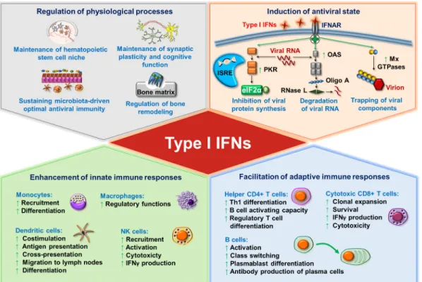
Besides regulating innate and adaptive immune cell activation, survival, and differen- tiation, type I IFNs also contribute to the control of various physiological processes such as the regulation of hematopoietic stem cell niche function, bone remodeling, the maintenance of synaptic plasticity and cognitive function of the healthy central nervous system, and the maintenance of immune homeostasis [25,26]. Furthermore, commensal microbiota-driven tonic levels of IFN signals at mucosal surfaces adjust the activation threshold of the innate immune system, which is essential for ideal antiviral responses [27–29] (Figure1).
Besides regulating innate and adaptive immune cell activation, survival, and differ- entiation, type I IFNs also contribute to the control of various physiological processes such as the regulation of hematopoietic stem cell niche function, bone remodeling, the mainte- nance of synaptic plasticity and cognitive function of the healthy central nervous system, and the maintenance of immune homeostasis [25,26]. Furthermore, commensal microbi- ota-driven tonic levels of IFN signals at mucosal surfaces adjust the activation threshold of the innate immune system, which is essential for ideal antiviral responses [27–29] (Fig- ure 1).
Figure 1. The pleiotropic effects of type I interferons (IFNs). Continuous baseline production of type I IFNs by various tissues and cells fine-tunes a wide variety of physiological processes including hematopoietic stem cell functions, synaptic plasticity, bone remodeling and immune homeostasis. In addition, the microbiota-induced basal IFN-signature prepares stromal and immune cells for upcoming infections (upper left panel). Upon viral infection, type I IFN signaling induces antiviral state in all nucleated cells via the upregulation of IFN-stimulated genes that inhibit the replication and spreading of viruses (upper right panel). Type I IFNs also control the cells of innate (lower left panel) as well as adaptive (lower right panel) immune system by shaping the activation, differentiation, effector functions and trafficking of these cells.
eIF2α: eukaryotic initiation factor 2α; IFN: interferon; IFNAR: interferon-alpha/beta receptor; ISRE: IFN-stimulated response ele- ment; Mx GTPase: myxovirus resistance guanosine triphosphatase; NK: natural killer; OAS: 2′-5′ oligoadenylate synthetase; Oligo A: 2′-5′-oligoadenylate; PKR: protein kinase R; Rnase L: ribonuclease L; Th: T helper.
Under physiological conditions, in the absence of acute infection, type I IFNs are con- stitutively secreted at a baseline level in many tissues, and it appears that lower or higher amounts than that might have pathologic consequences [25]. Despite their beneficial ef- fects, type I IFNs are detrimental to the host when their expression is dysregulated. While the acute and transient production of type I IFNs promote antiviral responses, their sus- tained and chronic secretion drives various autoimmune and non-autoimmune inflam- matory diseases. These diseases are characterized by the so-called IFN gene signature (IGS), which refers to the upregulation of IFN inducible genes in peripheral blood cells.
IGS was found to be correlated with disease severity in patients with autoimmune dis- eases such as systemic lupus erythematosus (SLE), rheumatoid arthritis (RA) or derma- tomyositis [30].
Owing to their potential to secrete large quantities of type I IFNs, pDCs have been identified as major players in a number of type I IFN-mediated inflammatory conditions.
Figure 1. The pleiotropic effects of type I interferons (IFNs).Continuous baseline production of type I IFNs by various tissues and cells fine-tunes a wide variety of physiological processes including hematopoietic stem cell functions, synaptic plasticity, bone remodeling and immune homeostasis. In addition, the microbiota-induced basal IFN-signature prepares stromal and immune cells for upcoming infections (upper left panel). Upon viral infection, type I IFN signaling induces antiviral state in all nucleated cells via the upregulation of IFN-stimulated genes that inhibit the replication and spreading of viruses (upper right panel). Type I IFNs also control the cells of innate (lower left panel) as well as adaptive (lower right panel) immune system by shaping the activation, differentiation, effector functions and trafficking of these cells.
eIF2α: eukaryotic initiation factor 2α; IFN: interferon; IFNAR: interferon-alpha/beta receptor; ISRE: IFN-stimulated response element;
Mx GTPase: myxovirus resistance guanosine triphosphatase; NK: natural killer; OAS: 20-50 oligoadenylate synthetase; Oligo A:
20-50-oligoadenylate; PKR: protein kinase R; Rnase L: ribonuclease L; Th: T helper.
Under physiological conditions, in the absence of acute infection, type I IFNs are constitutively secreted at a baseline level in many tissues, and it appears that lower or higher amounts than that might have pathologic consequences [25]. Despite their beneficial effects, type I IFNs are detrimental to the host when their expression is dysregulated.
While the acute and transient production of type I IFNs promote antiviral responses, their sustained and chronic secretion drives various autoimmune and non-autoimmune inflammatory diseases. These diseases are characterized by the so-called IFN gene signature (IGS), which refers to the upregulation of IFN inducible genes in peripheral blood cells. IGS was found to be correlated with disease severity in patients with autoimmune diseases such as systemic lupus erythematosus (SLE), rheumatoid arthritis (RA) or dermatomyositis [30].
Owing to their potential to secrete large quantities of type I IFNs, pDCs have been iden- tified as major players in a number of type I IFN-mediated inflammatory conditions. Since the discovery of pDCs, several regulatory factors and mechanisms have been identified,
which affect their type I IFN production and might serve as possible therapeutic targets.
In the present review, first we briefly introduce the molecular basis of the unique type I IFN producing capacity of pDCs. Following that, we summarize all those endogenous factors, which can modulate type I IFN production specifically in pDCs and outline the role of pDC-derived type I IFNs in antiviral response, cancer, autoimmunity, and allergy, and shortly discuss the potential therapeutic approaches in these diseases.
2. Professionalism of pDCs in the Production of Type I IFNs
Despite being discovered in the mid-20th century, pDCs were explicitly characterized many decades later due to their low frequencies in peripheral blood and rapid apoptosis under in vitro culture conditions. After the ability of these cells to secrete extreme amounts of type I IFNs in a relatively short time was recognized, they were defined as natural IPCs. According to the current model, IPCs are precursors of pDCs, and therefore the IFN-producing form of pDCs is regarded as plasmacytoid pre-DCs. Plasmacytoid pre-DCs are small, round cells with a plasma cell-like morphology and a well-developed endoplas- mic reticulum (ER), which enables the production of large quantities of proteins. Upon differentiation into mature pDCs, they lose their exceptional type I IFN-producing ability, take up DC-like morphology, express high levels of major histocompatibility complex (MHC) and costimulatory molecules, and become professional APCs being capable of stimulating naive T cells [31].
Plasmacytoid DCs have emerged as “the virus experts” of our body owing to their striking capacity to produce large amounts of type I IFNs. Within 6 h of viral exposure, pDCs devote 50% of their induced transcriptional activity to initiate type I IFN gene ex- pression [32]. Human pDCs express a wide repertoire of type I IFNs including 13 subtypes of IFNαand single subtypes of IFNβ, IFNω, and IFNτ[33]. In response to viral infection, pDCs are responsible for 95% of type I IFN production by mononuclear cells, as they are able to produce 200–1000 times more type I IFNs than any other white blood cell after microbial exposure [4]. Quantitatively, one single pDC can produce 3–10 pg of IFNαin response to a strong stimulus. While secreting high amount of IFNα, pDCs produce much lower levels of IFNβand additional type I IFNs [34]. All these data raise the question of how pDCs are capable of such a peak performance. In contrast to conventional DCs (cDC), pDCs selectively express endosomal Toll-like receptor (TLR) 7 and 9, which recognize viral RNA and DNA, respectively, and their activation is associated with a high production of type I IFNs by pDCs [35]. In contrast to other cell types, pDCs show a high degree of resistance to viral infections, and do not need to be infected with live viruses to induce the production of type I IFNs [36], since inactivated viruses can also stimulate pDCs if the viral envelope remains intact [37]. The internalized virus particles are degraded within endocytic vesicles and are sensed through TLR7 and TLR9, the engagement of which induces signaling through the adaptor protein MyD88. Upon association with downstream signaling components, MyD88 leads to the phosphorylation and nuclear translocation of the IRF7 transcription factor, which initiates the transcriptional activation of type I IFN genes. In contrast to cDC, in which IRF7 is inducible and requires prior stimulation, pDCs constitutively express IRF7, possibly due to the lack of the translational repressor eukaryotic translation initiation factor 4E (eIF4E)-binding protein (4E-BP), which allows the rapid onset of type I IFN production in pDCs [38]. However, constitutive IRF7 expression alone would not be sufficient to induce the production of large amounts of type I IFNs in pDCs. Honda et al. described a unique spatiotemporal regulation of the TLR9-MyD88 pathway in pDCs in response to a specific TLR9 ligand, CpG-A. Following recognition, CpG-A oligonucleotides form large multimeric aggregates, which are retained in the early endosomes of pDCs for about 30 min that allows prolonged IRF7 induction, whereas in cDCs, those are rapidly transferred to lysosomal compartments. Thus, CpG-A stimulation efficiently enhances the production of type I IFNs in pDCs via IRF7, whereas barely affects nuclear factor-κB (NF-κB) activity and thus the maturation of these cells. However, it is important to note that this effect highly depends on the structural properties of the
TLR ligand. Other subtypes of oligonucleotides such as the monomeric CpG-B is rapidly transported to late endosomes, where recognition through TLR9 leads to the activation of the NF-κB pathway, which induces the synthesis of inflammatory cytokines, chemokines, and costimulatory molecules; therefore, CpG-B is much less effective in the initiation of type I IFN production compared to CpG-A [39]. In terms of kinetic, pDCs secrete high amounts of IFNαwithin the first 12 h of exposure to CpG-A or live viruses, and then in the next 48 h, pDCs are able to produce only a small fraction of this quantity upon re-stimulation [33].
Previously, our research group proposed a model, in which type I IFN secretion by pDCs occurs in two waves under the coordinated regulation by different subtypes of pattern recognition receptors (PRRs) [40]. In the early phase of viral infection, pDCs are able to detect debris from virus-infected cells in the lymph nodes through constantly expressing TLR7 and TLR9 receptors that results in the secretion of high levels of type I IFNs and subsequent induction of a systemic antiviral state. The systemic effect is facilitated by the unique localization of pDCs, in which aspect they highly differ from cDCs that are generally located in peripheral tissues, i.e., at the sites of viral entry, whereas pDCs are found in the blood or in lymphoid tissues such as lymph nodes. Therefore, pDCs are initially not infected, but can detect virus-infected cell debris delivered to lymph nodes and produce a large amount of antiviral cytokines, which might reach every cell in the body through the blood or lymphoid circulation, and thus contributes to the development of a systemic antiviral state. In the later stage of viral infection, TLR-activated pDCs migrate to the site of virus entry, where due to the high viral load, they can also get infected. However, it is important to note that previous TLR stimulation induces the expression of cytosolic retinoic acid-inducible gene I (RIG-I)-like receptors (RLRs) in pDCs, the expression of which is only marginal in these cells without prior activation. Therefore, pDCs gain the ability to recognize replicating viruses even in the cytoplasm after TLR stimulation. Upon ligand engagement, RLRs recruit the mitochondrial antiviral signaling adapter protein (MAVS), which leads to the activation of MAVS-dependent IRF3/7 pathways and ultimately results in a second/late wave of type I IFN production. The RLR-mediated type I IFN production is lower in amounts than the TLR-induced secretion by pDCs; however, it still effectively supports potent local antiviral responses [40].
Based on single-cell genomic profiling several models have recently been proposed on the fate and functional plasticity of pDC [1,36,41]. According to these models, pDCs highly differ in their degree of differentiation and consequently in their capacity to produce type I IFNs or present antigens. A current study demonstrated that the same individual pDC first produces type I IFNs and then acquires the ability to present antigens to T cells during in vivo viral infections suggesting that pDCs exert different functions orchestrated in a spatiotemporal manner [42]. Other models suggest that only a small fraction of pDCs can produce type I IFNs, regardless of the stimulus. It is also plausible that while some pDCs get infected, others recognize the infected pDCs and start to produce the type I IFNs as a response [1]. The above data indicate that further studies are required to explain the observed discrepancies and clarify whether the functional differences of pDCs are due to terminal functional specialization or are subsequent stages of pDC maturation.
In conclusion, pDCs represent the number one source of type I IFNs, which cytokines impact an array of cellular events, physiological processes and both innate and adaptive immune responses. Therefore, fine-tuning type I IFN responses of pDCs by different endogenous factors or regulatory mechanisms is essential to keep a balance between protection and unwanted pathological events.
3. Regulation of Type I IFN Production at the Transcriptional and Posttranscriptional Level 3.1. Transcription Factors
The induction of type I IFN secretion is primarily controlled at the transcriptional level, where IRFs play an essential role. In pDCs, IRF7 and IRF3 are the master regulators of type I IFN responses [40,43]. As we previously described, the MyD88-dependent TLR7/9
signaling cascade activates IRF7 to induce robust production of type I IFNs, whereas the MAVS-dependent RLR signaling pathway is able to activate both IRF3 and IRF7 to promote the secretion of type I IFNs in pDCs [40,44]. In addition, other members of the IRF family have been implicated in the regulation of type I IFN production [45].
Several studies demonstrated that IRF5 and IRF8 also contribute to the regulation of type I IFN production. In mouse pDCs, IRF5 was found to only partially affect the induction of IFNα, whereas it seems to be critical for IFNβgene induction [46,47]. In the human CAL-1 pDC cell line, IRF5 silencing resulted in an 80% reduction in CpG-B- triggered gene activation as compared with controls [48]. Interestingly, the same study identified IRF8 as a negative regulator, since silencing of IRF8 led to a 60% increase in gene activation upon CpG stimulation [48]. Moreover, the authors also found that IRF5 and IFR8, which colocalize within the cytoplasm of resting pDCs, rapidly translocate to the nucleus after CpG triggering, and thus hypothesize that IRF8 interacts with IRF5 to control TLR9 signaling in human pDCs [48]. By contrast, another study demonstrated that the depletion of IRF8 decreased the IFNαsecreting capacity of mouse pDCs upon CpG stimulation indicating its importance in mouse pDCs [49]. Thus, the above data suggest that IRF8 controls the magnitude of IFN responses and exerts positive or negative regulatory effects depending on the origin of the cell.
Besides IRFs, several other transcription factors were identified as regulators of pDC functions. Runt-related transcription factor 2 (RUNX2) is essential for the optimal produc- tion of type I IFNs through the modulation of IRF7 expression in mice. In the absence of RUNX, both resting and CpG-activated mouse bone marrow (BM)-derived pDCs showed decreased IRF7 expression, which resulted in a significantly reduced production of IFNα and IFNβ[50]. Further, mouse pDCs highly express the Ets family transcription factor, Spi-B, which can transactivate the promoters of type I IFN in synergy with IRF7 [51].
BM-derived and splenic pDCs from Spi-B-deficient mice showed defective induction of IFNαgenes following CpG-B, polyuridylic acid (polyU) and vesicular stomatitis virus (VSV) stimulation. The authors also concluded that constitutive high expression of Spi-B contributes to the ability of pDCs to produce high amounts of type I IFNs in response to TLR7 and TLR9 ligands [51]. The nuclear factor of activated T cells C 3 (NFATC3) was also found to enhance IRF7-mediated IFN release by both mouse and human pDCs [52]. In BM-derived pDCs from NFATC3-deficient mice TLR7/9-mediated IFNαproduction was greatly reduced compared to wild type mice [52]. In the Gen2.2 human pDC cell line, the knockdown of NFATC3 also led to a substantial reduction of IFNαproduction in response to CpG-A. Furthermore, it was demonstrated that NFATC3 forms a complex with IRF7 and binds to IFN promoters to augment maximal production of type I IFNs upon TLR9 stimulation in GEN2.2 cells [52].
In contrast to the above mentioned transcription factors, the pleiotropic transcription factor MYC negatively regulates the TLR-mediated antiviral response of human pDCs [53].
The knockdown of MYC increased the CpG-B triggered induction of IFN-stimulated genes in the human GEN2.2 pDC cell line. In particular, MYC is shown to interact and form a complex with the nuclear receptor co-repressor 2 (NCOR2) and histone deacetylase 3 (HDAC3) to occupy, and thus repress the promoter region of IRF7. These data imply that MYC suppresses IRF7 promoter activity to ensure the optimal levels of type I IFN production and prevent the development of autoimmune diseases [53].
Another critical regulator of various signaling pathways, the CXXC type zink finger protein 5 (CXXC5) is suggested to act as a transcription factor as well as an epigenetic modifier [54]. It is highly expressed in mouse and human pDCs, where, as an epigenetic regulator it controls DNA methylation and histone modifications [55]. Following stimu- lation with herpes simplex virus-1 (HSV-1) or CpG-A, pDCs from CXXC5-deficient mice expressed lower levels of IRF7 and produced much less type I IFN compared to control pDCs. Mechanistically, CXXC5 recruits the Tet2 DNA demethylase, which maintains hy- pomethylation of CpG-island containing genes such as IRF7, and thus contributes to the rapid and robust type I IFN production of mouse pDCs [55]. Similarly, the knockdown of
CXXC5 in the human GEN2.2 pDC cell line led to reduced IRF7 expression and decreased mRNA levels of IFNαand IFNβupon exposure to the TLR7 ligand R848 and HSV-1 [55].
Furthermore, E2-2 is also a specific regulator of mouse as well as human pDCs, since it can directly activate the expression of multiple genes involved in pDC-mediated type I IFN responses, namely the TLR7 and TLR9 receptors as well as the IRF7, IRF8 and Spi-B transcription factors [56]. The deletion of E2-2 in mouse BM-derived pDCs abolished type I IFN release in response to CpG-A that could be explained by the reduced expression of the aforementioned TLR pathway components [56]. Similarly, E2-2 knockdown in the human GEN2.2 cell line downregulated the expression of pDC signature genes and diminished IFNαproduction in response to CpG-B. Interestingly, E2-2 silencing in GEN2.2 cells upreg- ulated a set of cDC specific genes, including the anti-inflammatory TLR10 and inhibitory Siglec-6 receptors that could explain the abrogated IFNαproduction in response to TLR9 stimulation [57].
The above data indicate that several transcription factors act in concert to coordinate the optimal expression of type I IFNs in both human and mouse pDCs (Table1).
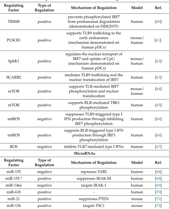
Table 1.Regulation of type I IFN production at the transcriptional and posttranscriptional level.
Transcription Factors Regulating
Factor
Type of
Regulation Mechanism of Regulation Model Ref.
IRF5 positive induces the expression of type I
IFN genes mouse [46,47]
IRF5 positive induces the expression of type I
IFN genes human [48]
IRF8 negative inhibits IRF5 human [48]
IRF8 positive - mouse [49]
RUNX2 positive induces IRF7 expression human [50]
Spi-B positive transactivates the promoters of
type I IFNs mouse [51]
NFATC3 positive
binds to type I IFN promoters in synergy with IRF7 (mechanism demonstrated on
human pDCs)
mouse/
human [52]
MYC negative represses IRF7 promoter activity human [53]
CXXC5 positive
maintains constitutive transcription of IRF7 (mechanism demonstrated on
mouse pDCs)
mouse/
human [55]
E2-2 positive supports the expression of TLR7,
TLR9, IRF7, IRF8 and Spi-B mouse [56]
E2-2 positive downregulates the expression of
TLR10 and Siglec-6 human [57]
Adaptor Proteins and Other Intracellular Regulators Regulating
Factor
Type of
Regulation Mechanism of Regulation Model Ref.
Opn-i positive supports the nuclear translocation
of IRF7 mouse [58]
PACSIN1 positive - mouse/
human [59]
Table 1.Cont.
Adaptor Proteins and Other Intracellular Regulators Regulating
Factor
Type of
Regulation Mechanism of Regulation Model Ref.
TRIM8 positive
prevents phosphorylated IRF7 from proteasomal degradation (demonstrated on HEK293T)
human [60]
PLSCR1 positive
supports TLR9 trafficking to the early endosomes (mechanism demonstrated on
human pDCs)
mouse/
human [61].
SphK1 positive
regulates the nuclear transport of IRF7 and uptake of CpG (mechanism demonstrated on
human pDCs)
mouse/
human [62]
SCARB2 positive mediates TLR9 trafficking and the
nuclear translocation of IRF7 human [63]
mTOR positive
supports TLR-mediated IRF7 phosphorylation and nuclear
translocation
mouse/
human
[64]
mTOR positive supports RLR-mediated TBK1
phosphorylation human [65]
mtROS negative
suppresses TLR9-triggered type I IFN production through inhibiting
IRF7 phosphorylation
human [66]
mtROS positive
supports RLR-triggered type I IFN production through IRF3
phosphorylation
human [66]
ROS negative inhibits TLR7-mediated type I IFNs human [67]
MicroRNAs Regulating
Factor
Type of
Regulation Mechanism of Regulation Model Ref.
miR-155 negative represses TAB2 human [68]
miR-155 * positive suppresses IRAK-M human [68]
miR-146a negative targets IRAK-1 human [69]
miR-618 positive - human [70]
miR-21 positive suppresses PTEN mouse [71]
miR-126 positive targets TSC1 mouse [72]
Abbreviations: CXXC5: CXXC-type zinc finger protein 5; IFN: interferon; IRAK: interleukin 1 receptor associated kinase 1;
IRF: interferon regulatory factor; miR: microRNA; mtDNA: mitochondrial DNA; mTOR: mammalian target of rapamycin;
mtROS: mitochondrial ROS;NFATC3: nuclear factor of activated T cells 3; Opn-i: intracellular osteopontin; Ox-mtDNA:
oxidized mitochondrial DNA; PACSIN1: protein kinase C and casein kinase substrate in neurons 1; PLSCR1: phospholipid scramblase 1; PTEN: phosphatase and tensin homolog; RLR: RIG-I-like receptor; RUNX2: Runt-related transcription factor 2; SCARB2: scavenger receptor class B member 2; SphK1: sphingosine kinase 1; TAB2: TGFβactivated kinase 1 binding protein 2; TBK1: TANK-binding kinase 1; TLR: toll-like receptor; TRIM8: tripartite motif containing protein 8; TSC1:
tuberous sclerosis complex 1.
3.2. Adaptor Proteins and Other Intracellular Regulators
Besides the common downstream signaling components of TLR and RLR pathways, numerous adaptor proteins and intracellular molecules act as positive or negative regula- tors of type I IFN secretion by pDCs.
Among them, the glycoprotein osteopontin (Opn) seems to be essential to the type I IFN responses in pDCs. Many cell types are able to express Opn, which is involved in
various pathophysiological events when secreted [73]. However, novel studies found that a portion of newly synthesized Opn is retained in the cytoplasm (referred as Opn-i), where acting as an adaptor protein controls signal transduction pathways downstream of innate immune receptors [73]. In splenic mouse pDCs, Opn deficiency significantly reduced the production of IFNαin response to CpG-A or CpG-B stimulation [58]. Opn-i colocalizes with MyD88 and TLR9 that is essential to the nuclear translocation of IRF7, and thus the optimal induction of IFNαgene expression [58]. Furthermore, the cytoplasmic phosphoproteins Protein kinase C and casein kinase substrate in neurons 1 (PACSIN1) was also identified as a pDC-specific adaptor molecule critical for the TLR7/9-mediated type I IFN responses in both human and mouse pDCs [59]. The knockdown of PACSIN1 in human GEN2.2 cells inhibited IFNαresponses to CpG-A stimulation. In addition, the depletion of PACSIN1 in mouse BM-derived pDCs considerably reduced the levels of IFNαproduction in response to CpG-A, influenza virus (Flu), VSV and HSV-1. Since Flu and VSV are recognized via TLR7, whereas HSV-1 and CpG are sensed by TLR9, the authors concluded that PACSIN1 affects IFNαproduction by both TLR7 and TLR9 signaling pathways in pDCs [59].
Several members of the tripartite motif (TRIM)-containing proteins have also emerged as important modulators of innate immune signaling cascades including the type I IFN pathway of pDCs. In primary human pDCs, TRIM20, TRIM22, TRIM28, and TRIM36 were identified as negative regulators of type I IFN responses [60]. On the contrary, TRIM8, which is constitutively expressed in resting pDCs, was revealed as a strong positive regulator of type I responses of pDCs and therefore its regulatory mechanisms were further investigated [60]. The silencing of TRIM8 in pDCs led to a profound decrease in IRF7 phosphorylation and abolished type I IFN production in response to HIV or Flu virus infection. The authors also showed with mechanistic studies on HEK293T cells that TRIM8 prevents the proteasomal degradation of phosphorylated IRF7 through inhibiting its recognition by the peptidyl-prolyl isomerase Pin1 [60].
Another positive regulator is the phospholipid scramblase 1 (PLSCR1) protein, which interacts directly with several plasma membrane receptors, and acting as a scramblase, is involved in multiple biological processes [74]. Silencing of PLSCR1 in human GEN2.2 cells led to a significant reduction of IFNαresponses following CpG-A or CpG-B stimulation [61].
Similar results were also obtained with BM-derived pDCs from PLSCR1-deficient mice when stimulated with CpG-A, Flu virus and HSV-1. In one human pDC cell line, PLSCR1 plays an important role in TLR9 trafficking from the ER to the early endosomes, and thus supports its engagement with synthetic or viral ligand [61].
Recently, it was reported that the lipid converting enzyme Sphingosine kinase 1 (SphK1), which catalyzes the formation of the lipid signaling molecule sphingosine-1- phosphate (S1P), plays a critical role in pDC functions as well [62]. The depletion of ShpK1 in mouse pDCs or pre-treatment of human CAL-1 cells and mouse splenic pDCs with ShpK1-specific inhibitors significantly impaired TLR7/9-mediated type I IFN produc- tion [62]. Mechanistically, ShpK1 was found to regulate the nuclear transport of IRF7 as well as the uptake and trafficking of CpG to endosomes, where TLR9 activation occurs.
Further, pharmacological inhibition of SphK1 or its genetic deletion in a mouse model of SLE decreased pDC activation and ISGs expression [62].
In addition, the lysosomal membrane protein Scavenger receptor class B member 2 (SCARB2) is highly expressed in resting pDCs, and its expression can be further upregulated by CpG stimulation [63]. Interestingly, the silencing of SCARB2 in human GEN2.2 cells significantly reduced the CpG-B-mediated IFNαproduction, whereas did not affect it upon CpG-A triggering. Further studies revealed that SCARB2 localizes in the late endosome, where it mediates the endosomal transport of TLR9 as well as nuclear translocation of IRF7, and thus regulates the expression of IFNαin pDCs [63].
Besides the aforementioned regulatory factors, the major drivers of immunometabolic changes can also impact type I IFN production of pDCs. Immunometabolism is one of the hottest topics and a dynamically growing field of immunology, which can provide new therapeutic approaches for the treatment of immune-related diseases. Immune cell
functions can be shaped both by intracellular metabolic processes and external metabolites derived from pathogens, microbes, or tumor cells [75]. Most importantly, several papers reported that alterations in energy and lipid metabolism modulate the type I IFN producing ability of pDCs [76–78]. The mammalian target of rapamycin (mTOR) has emerged as the master regulator of cellular metabolism by controlling a myriad of cellular functions in response to environmental factors and intracellular signals. It was revealed first in 2008 that the phosphatidylinositol 3-kinase (PI3K)-Akt-mTOR signaling pathway is crucial for the TLR-mediated type I IFN responses of pDCs [64]. The inhibition of mTOR complex 1 by rapamycin significantly decreased the TLR9-triggered production of IFNαboth in mouse and human pDCs. Moreover, the inhibition of the PI3K upstream molecule or p70S6 kinase downstream target also substantially suppressed the CpG-A-induced IFNα secretion by mouse pDC. Furthermore, the TLR7-induced IFNαsecretion was also impaired in murine splenic pDCs. The findings also indicated that blockade of mTOR signaling results in the disruption of MyD88-TLR9 complex and impairment of IRF7 phosphorylation and nuclear translocation [64]. Another group further confirmed these data by showing that suppressing the signaling components of the PI3K-Akt-mTOR pathway by various approaches significantly reduced IFNαsecretion in primary human pDCs [79]. A different study also demonstrated that rapamycin potently inhibits TLR7/9-induced IFNαsecretion in peripheral blood pDCs [80]. In addition, our research group demonstrated for the first time that mTOR signaling is also essential to the RLR-triggered antiviral immune responses of pDCs. We described that mTOR blockade by rapamycin or the dual kinase inhibitor AZD8055 inhibited the type I IFN production in primary human pDCs and GEN2.2 cells upon RLR stimulation [65]. Further, we found that mTOR blockade decreased the RLR- triggered phosphorylation of Tank-binding kinase 1 (TBK1), which based on literature data is a requirement for IRF3/7 activation [81]. Thus, we hypothesize that mTOR might support the RLR-initiated type I IFN production of pDCs via regulating at the level or upstream of TBK1.
It has long been recognized that several metabolic processes are linked to the enhanced generation of mitochondrial reactive oxygen species (mtROS), which are also essential mediators of immune responses. MtROS are constitutively generated during physiological conditions and their production can be further increased under pathological states. As important signaling molecules, mtROS participate in various processes of immune cell functions such as activation or inflammatory cytokine production [82]. Interestingly, BM- derived pDCs of aged mice show defective IRF7 upregulation upon TLR9 activation as compared with cells from younger counterparts [83]. Both resting and TLR9 activated aged pDCs displayed elevated levels of reactive oxygen species (ROS), the reduction of which by antioxidant pre-treatment restored IFNαproduction during TLR9 activation. These results suggest that age-induced oxidative stress impairs the antiviral capacity of pDCs in response to TLR9 stimulation, which might explain the increased susceptibility of older individuals to viral infections. In addition, our research group described that mtROS can influence the type I IFN producing capacity of human pDCs [66]. The TLR9 agonist CpG-A triggered the type I IFN production of human GEN2.2 cells was markedly reduced by high mtROS levels, which inhibited the phosphorylation of IRF7. On the contrary, elevated mtROS increased RIG-I-stimulated type I IFN expression and augmented the phosphorylation of Akt and IRF3, which are essential components of RLR signaling [66]. Interestingly, in a separate study, we demonstrated that exogenous ROS also have a negative effect on the TLR7-dependent activation of pDCs [67]. Thus, our data suggest that the effects of mtROS on pDCs depend on which viral sensing pathways are stimulated. Namely, the early TLR7/9-mediated type I IFN response is abrogated, whereas the late RLR-mediated production of type I IFNs is supported by elevated mtROS levels. (Table1)
Although, the above listed regulatory molecules have multifunctional roles in the regulation of various physiological cellular functions, those are also important in fine- tuning the extent of type I IFN responses by pDCs as well. It is also noteworthy that through
feedback mechanisms secreted type I IFNs might also act on diverse cellular functions including metabolic processes, which highly influence the activation of immune cells.
3.3. MicroRNAs
MicroRNAs (miRs) are short, non-coding RNAs, which upon binding to target mRNAs lead to their degradation or translational suppression. By controlling gene expression at the posttranscriptional level in various cell types, miRs have emerged as key coordinators of both innate and adaptive immune responses [84], and several of them have also been linked to the regulation of type I IFN responses in pDCs [85].
MiRs profiling of human pDCs revealed that TLR7 stimulation highly induced the expression of both the guide (miR) and passenger strands (miR*) of 19 different miRNAs, among which miR-155 and miR-155* were the most highly induced ones. Interestingly, miR- 155 inhibited the TLR7-triggered production of IFNαand IFNβexpression by targeting TGFβactivated kinase 1 binding Protein 2 (TAB2), whereas miR-155* augmented it by suppressing interleukin-1 receptor-associated kinase (IRAK)-M [68].
Similarly, miR-146a expression was also induced upon TLR7/9 stimulation in primary human pDCs [69]. However, the silencing of miR-146a increased the percentage of IFNαex- pressing pDCs upon stimulation with CpG-A, thus it was identified as a negative regulator of type I IFN production [69]. Further studies with the CAL-1 pDC cell line also revealed that miR-146a targets IRAK-1, which is an essential element for IRF7 activation [69].
Interestingly, increasing evidence has revealed that the dysregulation of miR ex- pression can contribute to the development and maintenance of various autoimmune diseases [86]. So far, two studies revealed association between miR expression and type I IFN production of pDCs in patients with IFN signature. One study found that pDCs of systemic sclerosis patients upregulated miR-618, the overexpression of which in primary human pDCs resulted in a higher secretion of IFNαin response to the TLR9 stimulation compared to pDCs derived from healthy individuals [70]. Another study reported that pDCs of SLE and antiphospholipid syndrome (APS) patients with high IFN signature displayed reduced expression of miR-361-5p, miR-128-3p and miR-181a-2-3 p compared to pDCs from patients without an IFN signature or healthy controls [87]. Circulating pDCs from Sjögren’s patients with an IFN signature also showed decreased expression of several miRs, however, no strong correlation was found between the miR profile of pDCs and IFN signature of patients [88].
The role of miRs in the regulation of type I IFN responses by pDCs was also proven in mouse models. In mouse BM-derived pDCs, both TLR7 and TLR9 stimulation increased the expression of different miRs such as miR-21, which was found to be an essential positive regulator for IFNα. The phosphatase and tensin homolog (PTEN) was identified as the target of miR-21. By suppressing PTEN, miR-21 promotes the TLR7/9-mediated activation of PI3K-Akt-mTOR signaling, which is essential for IRF7 protein expression and subsequent IFNαproduction [71]. Furthermore, miR-126, which is mostly associated with the regulation of vasculogenesis and angiogenesis, is also able to control IFNαand IFNβ production of mouse pDCs. MiR-126 deficient mice produced less IFNαthan wild-type mice in response to TLR7 (R848) and TLR9 (CpG-A) agonists [72]. Splenic and lymph node-derived pDCs from miR-126-/- mice were also defective in their TLR9-mediated IFNαproducing capacity compared to wild type pDCs [72]. Further data indicate that tuberous sclerosis complex 1 (TSC1), which is a negative regulator of mTOR, is the target for miR-126 in pDCs [72]. It was also hypothesized that miR-126 acts through the upregulation of vascular endothelial growth factor receptor 2 (VEGFR2), which also negatively regulates TSC1, thus increases mTOR activity and type I IFN production in pDCs [72] (Table1).
Altogether, miRs can impact the type I IFN release of pDCs both in a positive or negative manner and might represent potential biomarkers for the diagnosis and prognosis of certain type I IFN-mediated autoimmune diseases.
4. Regulation of Type I IFN Production by Receptor Interactions 4.1. Activating Receptors of Type I IFN Production
Receptor-mediated regulatory mechanisms influence various events in a cell’s life.
PDCs express a wide range of cell-surface and intracellular receptors, which upon in- teraction, might affect the outcome of pDC activation [1]. Interestingly, only a few of these immunoregulatory receptors have the ability to support pDC functions, and more specifically their type I IFN producing capacity.
The first identified positive regulator of type I IFN responses of pDCs is represented by the immunoregulatory CD300a/c molecule. It was found that the cross-linking of CD300a/c increased IRF7 expression and IFNαsecretion by primary human pDCs af- ter TLR7/9 activation [89]. Later, it was reported that pDC specific triggering receptor expressed on myeloid cells (PDC-TREM), which expression requires TLR7/9 stimula- tion, also positively affects the type I IFN production of pDCs. Upon stimulation of mouse BM-derived pDCs with CpG-A, PDC-TREM forms a complex with endogenous Plexin-A1 and its endogenous ligand Sema6D, which leads to the phosphorylation of PI3K, extracellular-signal-regulated kinase 1/2 (ERK1/2) and inhibitory kappa B kinaseα(IKKα), and eventually results in a robust production of type I IFNs by pDCs [90].
Interestingly, despite the presence of the inhibitory cytoplasmic immunoreceptor tyrosine-based inhibition motif (ITIM), Ly49Q is a positive regulator of type I IFN re- sponses in mouse pDCs as well. In mice, the interaction of Ly49Q receptor with its ligand, the classical MHC-I molecule, was found to be required for IFNαsecretion by pDCs, since splenic pDCs from Ly49Q-deficient mice displayed lower levels of IFNαupon CpG-A challenge [91]. Moreover, Ly49Q-/- mice were defective in systemic IFNαproduction.
Blockade of either Ly49Q or its ligand by monoclonal antibodies (mAB) almost com- pletely abrogated the IFNαsecretion by pDCs. These findings suggest that Ly49Q and MHC-I linkage positively regulates TLR9 mediated type I IFN production in mice [91].
As a mechanism, the research group demonstrated that Ly49Q controls the intracellular trafficking of TLR9/CpG-A containing vesicular compartments [92]. These data were supported by another study showing that Ly49Q deficient mouse BM-derived pDCs pro- duced lower amounts of IFNαand IFNβrelative to control pDCs in response to CpG-B and Sendai virus [93]. As another mechanism, later it was revealed that Ly49Q enhances TLR9-mediated IRF7 nuclear translocation, and thus type I IFN gene expression in an ITIM-dependent manner in mouse pDCs as well [94].
In addition, the signaling lymphocyte activation molecule family 9 (SLAMF9) re- ceptor is highly expressed on pDCs and plays and important role in their differentiation and function. In mice, SLAMF9 deficiency results in the accumulation of pDCs in the lymph nodes, where they exhibit reduced costimulatory potential, and show a decreased capacity to secrete IFNαin steady-state condition and during experimental autoimmune encephalomyelitis (EAE). Moreover, SLAMF9-/- pDCs derived from EAE mice and CpG-A stimulated mice show a reduction in IFNαlevels compared with the wild type controls suggesting that SLAMF9 might impact pDC functionality under different inflammatory conditions. A gene expression analysis of SLAMF9 deficient pDCs further revealed that the levels of the SpiB transcription factor was strongly downregulated that might contribute to the impaired functionality of pDCs in the periphery [95].
Furthermore, pDCs express the multifunctional receptor for advanced glycation end- products (RAGE), which is able to bind multiple ligands via recognizing a common struc- tural motif in them, and thus it is regarded as a PRR [96]. An important ligand for RAGE is the nuclear protein high mobility group box 1 protein (HMGB1), which when released by dying cells acts as a proinflammatory factor. In 2005, it was demonstrated that purified hu- man pDCs express RAGE, and upon TLR9 stimulation secrete HMGB1, which, acting in an autocrine manner, supports the maturation and type I IFN secretion of pDCs [97]. Another study demonstrated that HMGB1 released by pDCs and NK cells triggers IFNαsecretion of pDC in the context of HIV infection in humans [98]. By supporting IFN secretion, HMGB1 contributes to the upregulation of tumor necrosis factor-related apoptosis-inducing ligand
(TRAIL) on pDCs, which makes them able to kill death receptor 5 (DR5) expressing CD4+ T cells. Moreover, HMGB1 released from necrotic cells can bind to DNA containing immune complexes, which then positively regulate IFNαsecretion by pDCs. HMGB1 in complex with CpG-A oligonucleotides significantly increased the IFNαproduction in mouse BM- derived pDCs compared to CpG-A alone through inducing the association of RAGE with MyD88-TLR9 [99]. In obese individuals, by transporting extracellular DNA through RAGE to TLR9, adipose tissue derived HMGB1 triggers pDCs to produce IFNα, which then drives the proinflammatory polarization of resident macrophages and contributes to the development of systemic insulin resistance [100] (Table2).
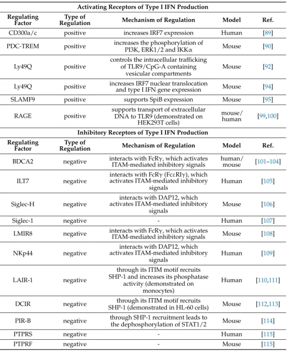
Table 2.Regulation of type I IFN production by receptor interactions.
Activating Receptors of Type I IFN Production Regulating
Factor
Type of
Regulation Mechanism of Regulation Model Ref.
CD300a/c positive increases IRF7 expression Human [89]
PDC-TREM positive increases the phosphorylation of
PI3K, ERK1/2 and IKKα Mouse [90]
Ly49Q positive
controls the intracellular trafficking of TLR9/CpG-A containing
vesicular compartments
Mouse [92]
Ly49Q positive increases IRF7 nuclear translocation
and type I IFN gene expression Mouse [94]
SLAMF9 positive supports SpiB expression Mouse [95]
RAGE positive supports transport of extracellular DNA to TLR9 (demonstrated on
HEK293T cells)
mouse/
human [99,100]
Inhibitory Receptors of Type I IFN Production Regulating
Factor
Type of
Regulation Mechanism of Regulation Model Ref.
BDCA2 negative interacts with FcRγ, which activates
ITAM-mediated inhibitory signals human/
mouse [101–104]
ILT7 negative interacts with FcRγ(FcεRIγ), which activates ITAM-mediated inhibitory
signals
Human [105]
Siglec-H negative interacts with DAP12, which activates ITAM-mediated inhibitory
signals
Mouse [106]
Siglec-1 negative - Human [107]
LMIR8 negative interacts with FcRγ, which activates
ITAM-mediated inhibitory signals Mouse [108]
NKp44 negative
interacts with DAP12, which activates ITAM-mediated inhibitory
signals Human [109]
LAIR-1 negative
through its ITIM motif recruits SHP-1 and increases its phosphatase
activity (demonstrated on monocytes)
Human [110,111]
DCIR negative through its ITIM motif recruits
SHP-1 (demonstrated in HL-60 cells) Mouse [112,113]
PIR-B negative through SHP-1 recruitment leads to
the dephosphorylation of STAT1/2 Mouse [114]
PTPRS negative - Human [115]
PTPRF negative - Mouse [115]
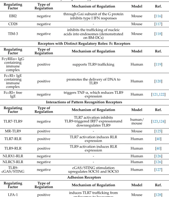
Table 2.Cont.
Inhibitory Receptors of Type I IFN Production Regulating
Factor
Type of
Regulation Mechanism of Regulation Model Ref.
EBI2 negative through Gαi subunit of the G protein
inhibits type I IFN responses Mouse [116]
CD28 negative - Mouse [117]
TIM-3 negative inhibits the trafficking of nucleic acids into endosomes (demonstrated
on BM-DCs)
Mouse [118]
Receptors with Distinct Regulatory Roles: Fc Receptors Regulating
Factor
Type of
Regulation Mechanism of Regulation Model Ref.
FcγRIIα+ IgG containing
immune complex
positive supports TLR9 trafficking Human [119]
FcεRI+ IgE containing immune complex
positive promotes the delivery of DNA to
TLR9 Human [120]
FcεRI+ free
IgE negative triggers TNF-α, which reduces TLR9
expression Human [121,122]
Interactions of Pattern Recognition Receptors Regulating
Factor
Type of
Regulation Mechanism of Regulation Model Ref.
TLR7-TLR9 negative TLR7 activation inhibits TLR9-triggered IRF7 expressionand
downregulates TLR9
human/
mouse [123,124]
MR-TLR9 positive - Mouse [125]
TLR7-RLR positive TLR7 activation induces RLR
expression Human [40]
TLR9-RLR positive TLR9 activation induces RLR
expression Human [40]
NLRX1-RLR negative - Human [126]
NLRC5-RLR negative - Human [126]
TLR9-
cGAS/STING negative cGAS/STING stimulation
upregulates SOCS1 and SOCS3 Human [127]
Adhesion Receptors Regulating
Factor Type of
Regulation Mechanism of Regulation Model Ref.
LFA-1 positive induces TLR7 trafficking from
endosomes to lysosomes Mouse [128]
Abbreviations: BDCA2: blood dendritic cells antigen 2; BM-DC: bone marrow-derived dendritic cell; CD: cluster of differentiation; cGAS: cyclic GMP-AMP synthase; DAP12: DNAX activating protein of 12 kDa; DCIR: dendritic cell immunoreceptor; EBI2: Epstein-Barr virus-induced G-protein-coupled receptor 2; ERK1/2: extracellular signal-regulated kinase 1/2; FcRγ:γsubunit of the Fc receptor; FcγRIIα: Fc gamma receptor II alpha; FcεRI: Fc epsilon receptor I; FcεRIγ:γ subunit of the Fc epsilon receptor; Gαi: Gi alpha subunit; IFN: interferon; IgE: immunoglobulin E; IKKα: IκB kinase (IKK) complexα; ILT7: immunoglobulin-like transcript 7; IRF: interferon regulatory factor; ITAM: immunoreceptor tyrosine-based activation motif; ITIM: immunoreceptor tyrosine-based inhibition motif; LAIR-1: leukocyte-associated immunoglobulin-like receptor 1; LFA-1: lymphocyte function-associated antigen 1; LMIR8: leukocyte mono-immunoglobulin-like receptor 8;
MR: mannose receptor; mTOR: mammalian target of rapamycin; NKp44: natural killer cell p44-related protein; NLRC5:
NOD-like receptor family CARD domain containing 5; NLRX1: nucleotide-binding domain and leucine-rich repeat–
containing protein X1; p70S6K: P70 S6 kinase; PDC-TREM: plasmacytoid dendritic cell—triggering receptor expressed on myeloid cells; PI3K: phosphatidylinositol 3-kinase; PIR-B: paired immunoglobulin-like receptor B; PTPRF: Protein tyrosine phosphatase receptor type F; PTPRS: Protein tyrosine phosphatase receptor type S; RAGE: receptor for advanced glycation endproducts; RLR: RIG-I-like receptor; SHP-1: Src homology 2 domain-containing protein tyrosine phosphatase 1; Siglec:
sialic acid-binding immunoglobulin-type lectin; SLAMF9: signaling lymphocytic-activating molecule family 9; SOCS:
suppressor of cytokine signaling; STAT: signal transducer and activator of transcription; STING: stimulator of IFN genes;
TIM-3: T cell immunoglobulin and mucin domain-containing protein 3; TLR: toll-like receptor; TNF: tumor necrosis factor.
4.2. Inhibitory Receptors of Type I IFN Production
Many studies have reported that the cross-linking of regulatory cell surface receptors on pDCs efficiently suppresses their ability to produce type I IFNs. Several of these regu- latory receptors associate with immunoreceptor tyrosine-based activation motif (ITAM) containing adapter proteins such as DNAX activation protein 12 (DAP12) and Fc receptor (FcR)γ-chain (FcRγ) or contain ITIM motifs themselves, which mediate inhibitory sig- nals [129]. FcRγis often referred to as FcεRIγsince it was first discovered as the third subunit of FcεRI [130]. Later, it was revealed that it is a common subunit of various FcRs and is also able to associate with a number of immune receptors such as blood dendritic cell antigen 2 (BDCA2) and immunoglobulin-like transcript 7 (ILT7) [131].
BDCA2 (also known as CD303) is a type II C-type lectin receptor, which is exclusively expressed by human pDCs. In 2001, it was demonstrated that the ligation of BDCA2 with a specific antibody suppresses the ability of peripheral blood-derived human pDCs to produce IFNαin response to CpG-A [101]. Later, co-immunoprecipitation experiments revealed that BDCA2 forms a complex with the transmembrane adapter FcRγ, which interferes with the TLR7/9-mediated type I IFN producing ability of purified human pDCs [102]. A separate study with primary human pDCs revealed further details of BDCA2 and FcRγinteraction by showing that upon association with FcRγBDCA2 signals through a B cell receptor (BCR) signalosome like complex consisting of Lyn, spleen tyrosine kinase (Syk), Bruton tyrosine kinase (Btk), Src homology 2 (SH-2) domain-containing leukocyte protein of 65 kDa (Slp65) and phospholipase C-gamma 2 (PLCγ2) [103]. Moreover, a subsequent study with GEN2.2 cells and primary human pDCs declared that the BCR- like signaling activates the mitogen-activated protein kinase (MAPK) kinase (MEK)1/2- ERK pathway, which then upregulates c-Fos, and thus results in the inhibition of CpG-A mediated type I IFN production [132]. Further data indicate that the CD2-associated adaptor protein (CD2AP), which is specifically expressed by pDCs, forms a complex with SH-2 domain-containing inositol-5-phosphatase 1 (SHIP1) and inhibits the Casitas B cell lymphoma (Cbl)-mediated ubiquitination and degradation of FcRγ[104]. Thus, via supporting BDCA2/FcRγreceptor signaling, CD2AP negatively controls TLR9-induced type I IFN responses in pDCs [104]. Interestingly, some studies identified a few possible virus-derived ligands for BDCA2, in particular, hepatitis B virus surface antigen (HBsAg) and HIV-1 glycoprotein 120 (gp120). The binding of HBsAg [133] or gp-120 [134] to BDCA2 on the surface of human pDCs contributed to the suppression of IFNαsecretion in response to TLR9.
The above data indicate that targeting BDCA2 by blocking or depleting antibodies might be a promising approach to manage type I IFN-mediated pathologies including autoimmune diseases [135–137]. Importantly, certain mAbs are already under clinical testing for the treatment of SLE and cutaneous lupus erythematosus (CLE) [138].
Similar to BDCA2, the ILT7 (also known as LILRA4) protein, which is exclusively expressed by pDCs, can also form a complex with FcRγ, and thereby leads to the inhibition of pDC functions. The cross-linking of ILT7 inhibited the production of IFNαin both CpG- and Flu-activated pDCs. The rapid phosphorylation of Src family kinases and Syk indicates that ILT7 cross-linking leads to the activation of ITAM mediated inhibitory signals in pDCs [105]. The bone marrow stromal cell antigen 2 (BST2), which is expressed only in low amounts on the surface of human pDCs, was identified as the biological ligand for ILT7. Following incubation with recombinant BST2, human pDCs were impaired in their ability to express type I IFNs upon challenge with CpG-A or Flu [139]. Interestingly, the Vpu HIV-1 protein hijacks this interaction by acting as an antagonist of BST2 and inhibiting TLR7-mediated type I IFN production by pDC. Vpu downregulates BST2 to enable virion assembly, while relocates remaining BST2 molecules to the surface of infected cells to maintain sufficient inhibitory signals for pDCs [140]. The ILT7-mediated negative feedback on type I IFN production is also hijacked by human cancer cells. Many human cancer cell lines constitutively express ILT7 ligands, thus inhibit TLR9-triggered IFNαproduction by human peripheral blood pDCs [141]. Interestingly, a study demonstrated that in vitro
culturing of human peripheral blood mononuclear cells (PBMCs) leads to a spontaneous loss of ILT7 on the surface of pDCs. Consequently, the blocking of BST2 by mABs had no effect on the IFNαproduction of TLR7/9-triggered pDCs. Therefore, the authors propose that BST2-mediated ILT7 as a homeostatic regulator limits the activity of immature pDCs and does not serve as a negative feedback mechanism to restrict the functions of mature pDCs [142].
Another modulator of pDC functions is the transmembrane sialic acid binding im- munoglobulin type lectins H (Siglec-H) receptor, which was identified as a specific marker of mouse pDCs. First it was demonstrated that cross-linking of Siglec-H with specific antibodies reduced TLR9-mediated type I IFN production of pDCs through the DAP12 adaptor protein [106]. In mice infected with murine cytomegalovirus (MCMV), Siglec-H deficiency induced elevated serum IFNαlevels compared with wild type mice, whereas viral clearance was not affected [143]. Further, it was revealed that Siglec-H protects mice from developing MCMV virus-triggered lupus-like syndrome by preventing the induction of type I IFN signature [144].
According to a recent study, the Siglec-1 positive pDCs might represent the human counterpart of the Siglec-H positive pDCs in mice [107]. In humans, Siglec-1 is expressed in a subset of blood pDCs, which display a semi-mature phenotype, express lower levels of BDCA2 and interleukin (IL)-3 receptorα, and do not respond to TLR7/9 engagement.
In vitro, its expression is inducible in Siglec-1 negative pDCs upon exposure of whole blood to Flu. Interestingly, the proportion of Siglec-1 expressing pDCs is higher in SLE patient and correlates with disease severity compared to healthy individuals; nevertheless, their functional role in SLE needs further investigations [107].
Another FcRγ-coupled regulator is the leukocyte mono-immunoglobulin-like receptor 8 (LMIR8), which is selectively expressed by mouse pDCs residing in the BM, spleen, or lymph nodes. It was found that LMIR8 cross-linking with mABs attenuates the CpG-A- mediated production of IFNαin BM-derived pDCs through the ITAM-containing adaptor protein FcRγ [108]. An additional pDC inhibitory receptor is NKp44, the expression of which is inducible on the surface of human tonsil and blood-derived pDCs upon culture with IL-3. It was reported that cross-linking of NKp44 inhibits IFNαsecretion in CpG-activated pDCs through association with the ITAM-containing adaptor molecule DAP12 [109]. In contrast to NKp44, the expression of leukocyte-associated Ig-like receptor- 1 (LAIR-1) is high on resting human pDCs, whereas it is downregulated in the presence of IL-3. The cross-linking of the inhibitory ITIM-containing LAIR-1 receptor impairs IFNαproduction by pDCs in response to TLR stimulation, and thus acts synergistically with Nkp44 to inhibit IFNαrelease [110]. Human pDCs also express the inhibitory C- type lectin receptor DC immunoreceptor (DCIR), which contains one ITIM motif. The cross-linking of DCIR inhibits TLR9-induced IFNαproduction, while it promotes antigen uptake and efficient antigen presentation by pDCs [112]. In mice, the ITIM-bearing paired immunoglobulin-like receptor B (PIR-B) is an inhibitory MHC-I receptor, which suppresses CpG-A triggered type I IFN production in mouse pDCs. In particular, PIR-B recruits the SH- 2 domain-containing phosphatase 1 (SHP-1), which then leads to the dephosphorylation of STAT1/STAT2, and thereby prevents the autocrine type I IFN-mediated positive feedback loop [114].
In contrast to the aforementioned inhibitory receptors the following ones do not con- tain ITAM or associate with ITAM-containing adaptors, and the specific mechanism by which they regulate type I IFN responses of pDCs remained largely unknown. Protein tyrosine phosphatase receptor type S (PTPRS) is an evolutionarily conserved pDC specific inhibitory molecule, which is expressed by both murine and human pDCs, whereas protein tyrosine phosphatase receptor type F (PTPRF) is detectable only in mice. PTPRS cross- linking decreased type I IFN production in primary human pDCs and GEN2.2 cells upon CpG-A stimulation. In mice, quiescent pDCs co-express PTPRS and PTPRF, the knockdown of which enhanced TLR9-induced pDC activation. These receptors are downregulated on activated pDCs that seems to be a requirement for efficient type I IFN production. In