PART III
THE MOLECULAR BASIS
OF CANCER CHEMOTHERAPY
P. EMMELOT Department of Biochemistry,
Antoni van Leeuwenhoek-Huis: The Netherlands Cancer Institute, Amsterdam, The Netherlands
I. Introduction . . . . . . . . . . . 55
II. How is Cytotoxicity Brought About? . . . . . . . 59
III. Chemotherapeutic Targets . . . . . . . . . 65
1. Nucleic Acids and Proteins. . . . . . . . . 65
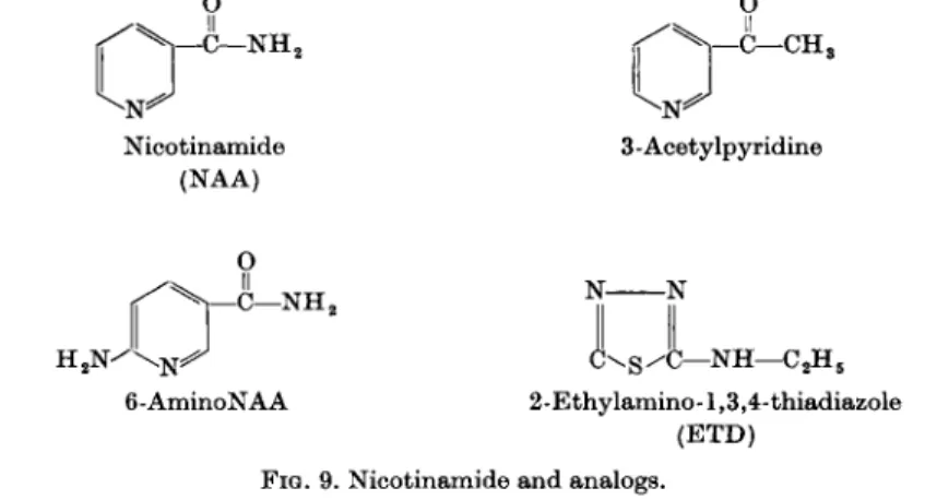
1.1. Synthesis of Ribonucleotides and R N A ; Inhibitors: Azaserine, DON, Aminopterin, Amethopterin, 6-Mercaptopurine, 6-Thioguanine, 2,6- Diaminopurine, 8-Azaguanine, 6-Uracil Micthylsulfone, 6-Azauracil, and 5-Fluoroorotic Acid . . . . . . . . 70
1.2. Synthesis of Deoxyribonucleotides and DNA; Inhibitors: 6-Uracil IMethylsulfone, 5-Fluorouracil, 5-Bromouracil, 5-Iodouracil, and 5- Mercaptouracil . . . . . . 92
1.3. Feedback Controls by Antimetabolites in Nucleic Acid Metabolism . 98 1.4. Alkylating Agents as Inhibitors of Nucleic Acid and Protein Synthesis and Function . . . . . . . . . . 99 1.5. Miscellaneous Compounds Interfering with Nucleic Acid and Protein
Metabolism . . . . 1 1 1 53
2. Glycolysis and Respiration. . . . 1 1 4 2.1. Antimetabolites . . . . 1 1 4
2.2. Alkylating Agents 119 2.3. Miscellaneous Compounds . . . . 1 2 4
2.4. Hormones 128 2.5. The Theory of Warburg 130
2.6. The Hexose Monophosphate Shunt . . . . 1 3 2 3. Membrane Function . . . . 1 3 2 4. Phospholipids and Lipids . . . . 1 3 6
IV. Selective Toxicity 137 V. Biological Alkylating Agents; Attempts at Improvement of Selective Toxicity 143
1. Chemical Reactivity . . . . 1 4 5 2. Chemical Reactivity and Carcinostatic Effect . . . . . 1 4 6 2.1. Nitrogen Mustards . . . . 1 4 6
2.2. Diepoxides 148
2.3. Ethylenimines . . . . . . . . . . 149
2.4. a,co-(Dimethanesulfonoxy)alkane Series . . . . . 149 3. Carcinostatic Effect as a Function of Parameters Other than Chemical
Reactivity. . . . . . . . . . . 152
4. Design of Alkylating Agents More Selective in Action . . . . 1 5 3 4.1. Molecular Structure of the Drug in Relation to Cellular Specificity
(Transport and Uptake) . . . . 1 5 3 4.2. Conversion of Inactive Drug to Active Drug in Situ . . . 1 6 0
5. Conclusion . . . . . . . . . . . 169
VI. Drug Resistance . . . . 1 7 0 1. Natural and Acquired Resistance . . . . 1 7 0 2. Biochemical Mechanisms Responsible for Drug Resistance in Tumors . 171 3. Attempts to Control Resistance . . . . 1 7 3 4. Drug Combinations and Increased Therapeutic Effect . . . . 1 7 5
References . . . . . . . . . . . . 179
Note Added in Proof 198
I. INTRODUCTIO N
Cancer is a group of allied diseases which in the absence of therapy lead to the death of the host except in extremely rare cases of spontaneous regression. The cancer cells seem to proliferate freely in the host, forming metastases and invading the normal tissues; the normal mechanisms of growth control appear to be lacking. It is generally assumed that cancer cells originate from normal cells by one or a number of somatic mutations, or heritable alterations, which are formally equivalent to a mutation, involving the disappearance of growth control. The mutation may be considered to be one by loss rather than by gain of a special device which confers autonomy to the cell. This conclusion is based, among other reasons, upon the fact that every normal cell type is, either at some stage (postmitotics) or during the whole of its lifetime, endowed with a potential capacity to divide. The latent power of growth appears to be a general biological phenomenon (422). However, instead of dividing indefi- nitely, normal cells are regulated by some homeostatic mechanism of unknown nature (probably of a complex repressive type) exercized by the organism as a whole. Any interference from outside which challenges the internal environ- ment of control may release the latent growth, but as long as an irreversible change has not been induced by this interference the growth stops as soon as the original situation has been restored. In autonomous cancer cells, the sup- pressed growth potential is permanently liberated (de-repressed), whereas in hormone-dependent tumors, growth is dependent on the endocrine imbalance which caused the original tumor cells to grow out. The latter tumors may suddenly become autonomous.
The problem of chemotherapy is, generally speaking, one of selective toxicity. The parasite, whether exogenous (bacterium; virus) or endogenous in origin (neoplastic cell), has to be destroyed completely while inflicting only a minimum of damage upon the host. The interaction of the administered drug with a receptor, present in the parasite and essential for its survival but absent in the host, would constitute an ideal situation. Penicillin's interference (498) with the synthesis and maintenance of the cell wall and the sulfonamides' interference (271) with the folic-acid-synthesizing enzyme systems of certain bacteria, illustrates this rather well.
The cancer cell situation is, however, wholly different. As compared with the normal cell, the cancer cell appears to have been "de-differentiated" to a less-specialized somatic entity. Tissue-specific functions (enzymes) may be lost and, generally speaking, a more primitive type of metabolism seems to emerge, aiming at the mere reproduction of new cells. This way of regarding the cancer cell should, however, not be generalized too much. Many exceptions to the principle of metabolic uniformity among tumors are now known; the metabolic characteristics of the original normal cells are retained to varying
55
extents. The so-called minimal deviation hepatomas, recently established and studied in detail (e.g. 3, 4), are notable examples of tumors which share many properties with their tissue of origin.
As yet, no convincing evidence has been presented for the view that cancer cells have acquired exclusive or new functions not present in the tissue of origin. It has recently been reported by Woolley (685, 686, 687) that female mice carrying spontaneous mammary carcinomas contain higher amounts of vitamin B12 than do normal animals. This finding has led the author to con- clude that the vitamin is synthesized by the cancerous tissue, but this con- clusion is in need of more direct evidence.* An analogous situation appears, however, to exist in the crown gall tumors of plants which synthesize their own indole-acetic acid. If cancer cells have gained a new function which is absent in the normal cells, a sound basis for chemotherapy may be present. It is of interest that some permanent cures of spontaneous mammary carcinomas in Swiss mice with antimetabolites of the B12-precursor, dimethyldiamino- benzene, have been obtained by Woolley (687). Compelling evidence that the drugs really inhibit the synthesis of the vitamin by the tumor is, however, lacking.
In our present state of knowledge, cancer chemotherapy must still be con- cerned with differences in quantity and not in quality. This situation may even continue indefinitely, especially if tumor cells originate by a loss-mutation.
Therefore, differences in the concentrations of enzymes, coenzymes, meta- bolites, and cofactors, which govern the rate of important metabolic reactions, have to be traced and exploited. If all tumors were to possess either a much higher or a much lower concentration of a certain component as compared with all normal tissues, and if the particular concentration of this component were essential for the maintenance of the tumor, then it might be relatively easy to inhibit or even destroy neoplastic cells preferentially. This simple situation does not, unfortunately, exist, for the following reasons:
1. Selectivity: The particular processes of the tumor which seem, at first sight, to be most suitable for a rational chemotherapeutic attack, e.g., ribo- nucleic-acid (UNA) and deoxyribonucleic-acid (DNA) synthesis, are in many cases operating with a similar intensity in dividing normal tissues. Altern- atively, it has been shown that antitumor drugs, for which the action mechan- ism is not (or not definitely) known, may damage both the tumor and a number of normal tissues. The exploitable metabolic differential between normal and cancer cells may be small.
2. Heterogeneity and resistance: Since every tumor constitutes a new biological species (343, 344), which may have its own distinct metabolic back- ground, it may be doubted whether a drug active against all cancers can ever be found. Specific drugs against the various types of cancer, molded upon the
* A selective uptake of vitamin ( C o5 8) B12 by human and mouse cancer cells has recently been reported (137).
III. CHEMOTHERAPY OF CANCER 57 enzymatic parameters of the particular tumor types, have to be designed.
Primary cancer offers great difficulties in this regard since it bears relatively the closest resemblance to the homologous normal tissue. Transplanted tumors, on the other hand, may, by mutational adjustment and selection, acquire quantitatively far more different parameters and lose certain enzyme activities altogether. However, many of these tumors still show a natural resistance towards certain cancer drugs.
Selection of pre-existing or de novo variants in the presence of a drug may give rise to resistant cell populations (acquired resistance). The development of drug resistance hampers successful chemotherapy and must, accordingly, be prevented or otherwise exploited in some way for the success of therapy.
For prevention, suitable and fast-acting combinations of drugs are required.
The discovery of the actual mechanism by which resistance is conferred may serve the second aim since the knowledge thus gathered may be used in favor- able cases to design drug derivatives which are still active.
3. Tumor-host relations: Many biochemical and biological data show that tumors exert systemic effects by changing the metabolism of the host tissues.
The latter changes may make the tumor-bearing host more sensitive to chemo- therapeutic treatment than the healthy animal. The search for an exploitable metabolic difference between normal and cancer tissue should thus be carried out on the normal tissues of the tumor-bearer. Complications as to the general applicability of such results may then arise since the systemic effects might be conditioned by the nature and the particular condition of the tumor under investigation.
Since the measure of effect of a given chemotherapeutic treatment in vivo may, furthermore, be influenced by the dietary regimen, route of administra- tion of the drug, environmental conditions, site and volume of the tumor, etc., it follows that no general rules for obtaining a selective antitumor effect exist.
The study of cancer chemotherapy has been directed along two main lines.
The first is the method of trial-and-error which involves the empirical screening of a large number of compounds. A notable example of an antitumor drug thus detected is azaserine. In the second approach, some peculiarity of the cancer cell is exploited in a rational (e.g. 5-fluorouracil) or semirational (e.g., purine analogs and derivatives) attempt to exploit the differential in regard to the normal cells with the hope that it will be large enough to gain success. In many cases, a combination of the two principles has been involved in that the original compound was a chance discovery, but that more active and selective derivatives have been prepared by a rational or semirational approach (e.g., biological alkylating agents). The whole field may, in the words of G. H.
Hitchings, be described as one of "rational empiricism." Both lines of research have and will continue to have their merits; they should be regarded as com- plementary. As long as the rational approach is hampered by lack of sufficient basic knowledge, the empirical approach should be continued. Not only is
cancer an acute medical problem that may profit from an accidental dis- covery, but the finding of new drugs, even of limited practical significance, may be expected to lead—as it actually has done before, e.g., azaserine and ribo- nucleotide synthesis—to a deeper insight into the basic chemical mechanisms of the cell and thus, in turn, provide information for the rational approach.
The search for more effective anticancer agents, by more or less tentatively modifying the structure of drugs of known biological activity, is fully war- ranted on the basis of the results which have been obtained already (467; see also 610a). It will, however, be evident that basic biochemical information about both normal and neoplastic tissues is essential for ultimate control of cancer by chemical means. Hopefully, this information would provide clues both to the design of new drugs and to more fruitful ways of modifying the structure of the present drugs.
The limited successes scored as yet are due to the fact that the existing knowledge of the biochemical mechanisms underlying normal growth, differentiation, and their control, is poor. Although many biochemical studies on the metabolism of normal and cancer tissues have been, and are being, carried out, no unique difference has as yet been observed. Moreover, primary animal tumors have been relatively little investigated. Rational cancer chemotherapy is, therefore, in its very infancy. This applies particularly to the chemotherapy of human cancer since there is a general lack of information about the biochemical properties of normal and cancer tissues of man. Little is known in regard to the enzyme concentrations, integration, and relative importance of metabolic pathways in the human. Extrapolation from animal experiments is not generally possible, as shown, for instance, by the finding that the de novo pathway of pyrimidine biosynthesis appears to be of limited significance in man in contrast to the situation in other species (674). Moreover, the genetically very heterogeneous population constituted by men should show a wide range of phenotypic expressions at the molecular level. Indeed, the biochemical individuality of man has been well documentated (678a). The enormous difficulties confronting the chemotherapy of human cancer are apparent if one considers the high degree of individuality that may already be inherent to tumors of one and the same tissue of the inbred animal. This may lead one to expect that the choice of the drug to be applied to human tumors will in many cases have to be made on the basis of a study of biopsy material.
Though the first chemotherapeutic agent for cancer, potassium arsenite or Fowler's solution, was introduced as early as 1865 (420a) and used since then with some success in the treatment of chronic leukemia (232a), it was only after World War II, following research on war gases (nitrogen mustard was found active against mouse lymphosarcoma and Hodgkin's disease in 1942), antibiotics, and the introduction of the antimetabolite concept of Woods and Fildes (in 1940), that systematic research was initiated. The first practical application of the latter concept in human cancer chemotherapy occurred in
ΠΙ. CHEMOTHERAPY OF CANCER 59 1948 when S. Farber and his associates used folic acid antagonists for the treatment of leukemia (224a).
In this chapter, an attempt will be made to discuss the present status of cancer chemotherapy from the experimental point of view. As regards the number of drugs to be described, the discussion will be limited and illustrative rather than exhaustive and will include also drugs of little practical import
ance. The nature of the receptors with which the drugs interact, or may interact, and the processes in which the receptors are involved will receive our attention in particular. For other aspects, such as the biological assay of the antitumor effect and the physiological "disposition" of the drugs, the reader is referred to recent reviews (443, 624). The present emphasis on biochemical detail may serve to illustrate the enormous difficulties involved in unraveling the mode of action of antitumor drugs and the intricacies of such action.
II. HO W IS CYTOTOXICITY BROUGHT ABOUT?
The carcinostatic or carcinolytic effect of a drug must be mediated by an effect upon some essential function of the cancer cell. In view of the many active agents which exist, it can hardly be doubted that toxicity becomes manifest in many different ways. The induction of mitotic abnormalities* may serve to illustrate this. X-rays, many chemical substances (including the alkylating agents, purine and pyrimidine antimetabolites, and quinones), malnutrition or even simple exposure to hypo- and hypertonic media cause chromosome abnormalities. The question thus arises as to the nature of the primary effect by which these agents interfere with the integrated processes and the con
nection between the primary biochemical effect and the biological end-effect.
The nature of the primary lesion appears to differ from compound to com
pound ; it is not necessarily connected with the DNA component but may also involve other receptors including protein, RNA, and the thiol groups of the mitotic apparatus (54), or even the building up of the energy reservoir which is required for mitosis (634). The fact that different prime lesions are funnelled into closely related or similar end-effects appears to be an expression of the integrated metabolic and structural mechanisms of the cell. Though multiple effects may thus be considered to lead to toxicity, a rather specific change must be involved in the induction of the neoplastic process. Even in the latter case, the multitude of agents—physical, biological as well as chemical—cap
able of inducing cancer suggests that many different primary mechanisms must be involved. In this connection it may be pointed out that the carcino
genic and carcinostatic activity displayed by one and the same compound does not necessarily involve the same sites of interaction (see p. 144).
For quite a number of antitumor agents, for instance, the 4-aminostilbenes,
* For recent reviews on the experimental and therapeutic modification and the bio
chemistry of mitosis, see (54a) and (454a).
nothing is known about the biochemical mechanism of their action. All that is known is that the active compounds of the aminostilbene series have a planar structure (in contrast to some of the related styrylquinolines) and that they give rise to nuclear and cytoplasmic abnormalities (378). The amino- stilbenes do not appear to be of practical importance; they are, however, of considerable theoretical interest because there is a close parallel between the carcinostatic and carcinogenic properties of the various members of the series (274).
Until recently, the mechanism of action of the carcinostatic and highly toxic styrylquinolines was also completely unknown; an interference with the energy metabolism of the cell now seems responsible for at least part of the toxicity produced by these agents (see III.2.3.B).
In regard to the class of alkylating agents much more evidence is available.
It is, however, still impossible to describe their antitumor effect in precise metabolic terms. On account of their chemical reactivity, the alkylating agents may interact with a number of receptors of both normal and neoplastic tissues. The interactions appear to be funneled into effects on glycolysis (see section III.2.2), nucleic acid and protein synthesis and function (see section III.1.4.).
Multiple metabolic effects may be induced by compounds bearing a struc- tural analogy to, but lacking the functional activity of, the corresponding metabolites (see sections III. 1.1-1.4 on antimetabolites). From the very nature of their chemical constitution it may follow that the prime sites at which these analogs link up with the metabolism of the tumor cell, are known. The mechan- ism of action of the antifolics and of azaserine is relatively the most simple, since these compounds appear to act, either exclusively or predominantly, as competitors of folic acid and glutamine, respectively. However, since the folic acid system and glutamine function in a number of important metabolic reactions, the biological end-effect will be attained by inhibition of a number of reactions of which the relative contribution to the end-effect may vary in different biological systems. The situation may become complicated when- ever the antimetabolites are incorporated into more complex molecules as, for instance, into those concerned with nucleic acid metabolism. First, anormal intermediates (nucleotides) which are inhibitory to the normal reaction sequences of coenzyme and nucleic acid synthesis, may be formed. Second,
"fraudulent" products may be formed such as nucleotide coenzyme-analogs;
being afunctional or inhibitory, they may paralyze the reactions dependent upon the corresponding normal coenzymes. When the coenzymes or cofactors are concerned with glycolysis and respiration, such as nicotinamide-adenine dinucleotides (NAD and NADP), flavoadenine dinucleotide, coenzyme A, and the adenine and guanine nucleotides which function in the phosphorylative processes, energy production may be impaired. In addition, synthetic reac- tions dependent upon such coenzymes or cofactors, (e.g., guanine nucleotides
III. CHEMOTHERAPY OF CANCER 61 in amination processes and microsomal protein synthesis, coenzyme A and cytosine nucleotides in fatty-acid or phospholipid synthesis) may also become inhibited. Such an interference by purine analogs may especially affect the metabolism of those neoplastic tissues which are known to contain low levels of the coenzymes. In addition, some antimetabolites have also been shown to become incorporated into high molecular weight products such as RNA and DNA, and thus to give rise to afunctional or fraudulent templates.
Though the purine and pyrimidine antimetabolites are metabolized by the same enzymes which handle the normal metabolites—pointing to a lack of specificity on the part of these biocatalysts—the creation of abnormal types of mono-, di, or polynucleotides might thus conceivably affect metabolic performances, since anywhere along the metabolic sequences and at various levels of molecular complexity the lack of specific structure may become manifest. However, in this complex of possibilities created by the initial enzymic blunder, it is very difficult to establish which of the affected reactions is responsible for the biological end-effect. It may even be asked whether in certain cases any simple inhibition can at all be singled out as the cause of the end-effect. Since low molecular compounds usually have a higher turnover rate than macromolecular ones, the effects of analogs which interfere with the former may become manifest earlier in time and may be more readily reversible with time than in the case of the latter. It may thus be possible that a single injection of, say, a purine antagonist may lead to an inhibition of the respira- tion of the tumor within a short time by interfering at the nucleotide level with coenzyme and cofactor synthesis or function; any effect on tumor growth in the intact animal cannot, of course, be observed during this period. Without further administration of the drug, the respiration may become quickly repaired and no effect on the tumor be observed. Continued administration, however, may lead to an observable inhibition of tumor growth. In that case, the antimetabolite may, in addition, have been incorporated into more (including very complex) products and may have produced a sufficient alteration or inhibition of RNA or DNA metabolism to disturb the main- tenance of the integrated cellular functions irreversibly. In such a case, it is hard to say whether the respiratory impairment, the creation of more complex faulty products, or both, results in the biological end-effect. In fact, by intro- ducing the antimetabolite many different biochemical reactions may be affected with mutually potentiating effects. Accordingly, it may be very difficult to establish whether a certain biochemical lesion is the direct cause of the interference of the drug with a particular receptor or the effect of another lesion produced by the drug.
The connection between metabolic disarrangement and drug-effect may be demonstrated by the method of inhibition analysis. Such an analysis can be carried out in vitro with isolated tissue or cell-free preparations while the result is expressed in metabolic terms. Since one has, however, to deal in
reality with a tumor growing in situ, the inhibition analysis must ultimately be carried out in vivo, both on the metabolic level by measuring reaction rates, coenzyme concentration, etc., and on the biological level by measuring tumor growth and host survival. The counteraction by nicotinamide of the antitumor effect of some alkylating agents in combination with the measurement of the NAD-level in treated and untreated tumors [see section III.2.2.A], the counteraction of antifolics by folic acid and its derivatives (250) or by certain products of biosynthetic reactions dependent upon folic acid (280), and that of mercaptopurine by adenine nucleotides [see sections III.1.1.B(3) and III.2.LA] illustrates that the method of inhibition analysis is applicable to mammalian cells growing in vivo. Tissue culture may be of considerable help in this respect. However, it should be kept in mind that, when more complex compounds (e.g., NAD; coenzyme A; nucleic acids) are administered to integrated biological systems (in vitro or in vivo), positive effects might actually result from smaller products derived from the administered ones. Negative effects, on the other hand, might be due to permeability barriers.
The comparative study of susceptible and resistant tumors may yield insight into the mechanism of drug action. Thus, if a drug affects a certain biochemical reaction in both types of tumors to the same extent, the particular inhibition might not be connected with the growth inhibition of the susceptible tumor. However, such a finding should be considered as an indication rather than as evidence since the resistant tumor might have developed a concurrent metabolic pathway which nullifies the effect of drug action on the former pathway. If so, the inhibition of that pathway in the susceptible tumor might still be connected with the growth inhibition. On the other hand, a decreased or lack of inhibition of a certain reaction by a certain drug in the resistant tumor, as compared with the susceptible tumor, may indicate that the particular reaction is directly or indirectly involved in drug action in the susceptible tumor. The same considerations apply to the metabolism of the drug itself. Thus, the finding that drugs are anabolized (certain antimeta- bolites) or activated (latent drugs) in the susceptible but not in the resistant tumor, suggests that a drug metabolite is the actual inhibitor.
The unknown role played by the host's defences provides a complicating factor in establishing the correlation between the effect of a given chemo- therapeutic agent on tumor metabolism and the biological end-effect. Since spontaneous regression is known to occur in a certain percentage of highly anaplastic rat tumors growing in their present hosts (but in origin from foreign hosts) it may be concluded that the "antigenic simplification" may not be complete and that under certain circumstances the host defences may overcome the homograft. Recent findings may indicate that primary tumors and isologous transplants contain tumor-specific antigens (16a, 163a, 580a, 698a). If so, it may follow that the host defences also operate in these cases.
m . CHEMOTHERAPY OF CANCER 63 Some evidence is accumulating that the compatibility of the transplanted tumor with its host diminishes when the tumor is damaged by chemical treat- ment. Cortisone, which inhibits the general immunological response, has been found to decrease significantly the number of regressions or to prevent the tumor inhibition induced by treatment with several anticancer agents, e.g., mercaptopurine (638) and polycyclic hydrocarbons (266). It may be that the more pronounced antitumor activity of several drugs on anaplastic rat tumors, as compared with that on strain-specific mouse tumors, is due partly to an immunological effect. These topics have recently been discussed by Klein (377).
The increased incidence of metastases following cortisone treatment of tumor- bearing animals, is also in line with the operation of an immunological defence of the host against the tumor.
A situation analogous to the one described for the anaplastic rat tumor may exist in the human female suffering from postpartum choriocarcinoma. This choriocarcinoma has its origin in placental tissue and thus owes its existence, in part at least, to the germ plasm of the consort; the tumor may be considered as a graft from the offspring to the mother and, in principle, to be able to elict an antibody response. About two-thirds of those patients with disseminated disease achieve complete remissions by chemotherapy. This unique response has been attributed to the possibility that the chemotherapeutic treatment, by causing only partial destruction of the tumor, liberates substances from the tumor which initiate an antibody response, by which destruction of the tumor is obtained (118a). Chemotherapy has been a failure in the treatment of the male choriocarcinoma which is entirely derived from autologous tissue.
The point of view that immunology may contribute to the success of chemo- therapeutic drugs (or vice versa) has received support from experiments in which mice that survived systemic leukemia following treatment with halo- genated derivatives of amethopterin [see section III.1.1.B(2)] showed im- munity on reinoculation of the leukemia (252). Conceivably, chemothera- peutics may so alter or influence tumor cells that homograft characteristics are required or unmasked, thereby facilitating therapy (253). It has also been found that resistance to therapy does not necessarily imply abrogation of the effectiveness of the immune response (254).
The general experience that the success of experimental cancer chemo- therapy often depends upon the time interval between transplantation of the tumor and treatment has also been regarded as an indication that an alteration of the host-transplant relationship rather than an antitumor effect per se constitutes an important effect of drug treatment (375). On the other hand, a drug-induced immunological tolerance has been observed (587) in that mercaptopurine suppressed the antibody response to human serum albumin in the rabbit; an inhibition of antibody synthesis by azaguanine in vitro has also been reported (181). The administration of suboptimal concentrations of
certain carcinostatic agents to tumor-bearing mice has actually been found to lead to an increase of lung metastases (380). This might indicate that under such conditions host resistance factors are impaired. Furthermore, it would be interesting to know whether incorporation of anormal constituents (drugs) into biological macromolecules may confer antigenicity. It follows from the present discussion that the tumor-host relationship may be of paramount importance to the chemotherapeutic response. In the following discussion attention will be confined to the interaction of certain drugs with tumor cells.
This should, however, not distract from the fact that a number of antitumor agents, especially hormones, may act indirectly, that is, via an effect on the host other than an immunological response. This is certainly so in the treatment of hormone-dependent tumors by means which suppress the internal secretion of those hormones on which tumor growth depends.
Tissue culture techniques (54, 322) allow a study of the direct effect and mode of action of drugs on cancer cells, not complicated by host mechanisms.
However, the biological end-effects in vitro do not always correspond with those obtained in vivo. Possible reasons for these differences have been dis- cussed (322) (see section IV).
It should finally be pointed out that most anticancer drugs only retard or inhibit the growth of transplanted tumors. Regression of well-established experimental tumors by purely chemical means is seldom seen and a definite cure is even more exceptional. As already mentioned, carcinostasis is a func- tion of the volume of the tumor: small tumors are more susceptible than large ones. This phenomenon might be of value in administering postoperative chemotherapy as an adjunct to surgery in the cure of microscopic metastases and circulating tumor cells resulting from the inadvertent "seeding" of tumor cells in the wound fields. For this purpose, probably nontoxic doses could be used (598). Preliminary clinical results have, however, shown that this procedure yields no general success (325).
In conclusion, and disregarding indirect mechanisms of action either as cause (certain hormones) or result (immunological response) of drug treatment, it may appear that the course of events lying between the primary interaction of an antitumor drug with cellular metabolism and the final death of the cell is, in general, complex. Evidence in cancer chemotherapy for drug interaction with one particular metabolic event or receptor leading to the immediate death of the affected cell, is scarce if not lacking. The primary interaction may be difficult to distinguish from the results of drug action. Drug action may also be diffuse in that various primary interactions are involved. The integrated metabolic state of the cell, the primary interaction(s) of a drug with the former, and their results on metabolism should be known in order to arrive ultimately at a rational chemotherapy of cancer, i.e., to obtain a basis for the development of more successful drugs.
III. CHEMOTHERAPY OF CANCER 65
III. CHEMOTHERAPEUTIC TARGETS
III A. Nucleic Acids and Proteins
The significance of nucleic acids in cellular proliferation makes the inhibition of their synthesis or function a logical point at which to attack the cancer cell.
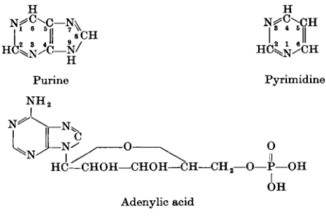
DNA and RNA are polynucleotides consisting of nucleotide subunits. Four ribonucleotides and 4 deoxyribonucleotides are arranged in a special sequence to form the RNA- and DNA-strands, respectively. In addition, very small amounts of nucleotides containing so-called " o d d " bases (e.g., in s-RNA) may be present. Each nucleotide consists of a purine or pyrimidine base attached, via N-9 in the case of the purines and N-l in the case of the pyrimidines, to the C-l of ribose (R) or 2'-deoxyribose (in RNA and DNA, respectively), the latter containing a 5'-phosphate group which, in the intact polynucleotide, forms an
Η Η
H C2^ / C — g / H C ^ C H
Purine Pyrimidine N H2
- N ^ ^ Ο ^ Ο HC—CHOH—CHOH—CH—CH2—Ο—Ρ—OH
I OH Adenylic acid
FIG. 1. Constituents of nucleic acids.
ester bond with the hydroxyl at the 3'-position of the sugar of the next nucleo
tide. The 4 bases in RNA are the 2 purines, adenine (A) and guanine (G), and the 2 pyrimidines, cytosine (C) and uracil (U). In DNA, uracil is replaced by thymine (T). The ribonucleotide containing adenine is abbreviated as AMP or ARP (adenylic acid, adenosine-5'-phosphate). Adenosine stands for the nucleoside of adenine, which is the nucleotide less its phosphate group, and is represented by AR (Fig. 1 and Table I).
Ribonucleic acid (RNA) has been shown to be abundantly present in rapidly dividing cells [Caspersson, in Brachet (67)]. RNA appears to be concentrated both in the nucleus and in the cytoplasm. Part of this RNA is present in ribonucleoprotein particles (ribosomes or Palade granules) which are either attached to the membranes of the endoplasmic reticulum or are free. In addi
tion, a low-molecular-weight RNA is present in the soluble fraction (s-RNA).
The latter and the ribosomal RNA play an important role in protein synthesis (98,109,113,131,696). Amino acids are activated in the soluble fraction with
TABLE I
NUCLEIC-ACID BASE CONSTITUENTS AND PRECURSORS
Base Type Substituents Nucleoside Nucleotide
Adenine Purine 6-Amino Adenosine Adenosine-5'-phosphate or adenylic acid (AMP) Guanine Purine 2 - Amino - 6 -hydroxy Guanosine Guanosine-5'-phosphate or guanylic acid (GMP) Cytosine Pyrimidine 2-Hydroxy-4-amino Cytidine Cytidine-5'-phosphate or cytidylic acid (CMP) Uracil Pyrimidine 2,4-Dihydroxy Uridine Uridine-5'-phosphate or uridylic acid (UMP) Thymine Pyrimidine 2,4-Dihydroxy-5-methyl Thymidine ThyTnidine-5'-phosphate or thymidylic acid (TMP) Hypoxanthine Purine 6-Hydroxy Inosine Inosine-5'-phosphate or inosinic acid (IMP) Xanthine Purine 2,6-Dihydroxy Xanthosine Xanthosine-5'-phosphate or xanthylic acid (XMP) Orotic acid Pyrimidine 2,4-Dihydroxy-6-carboxy Orotidine Orotidine-5'-phosphate or orotidylic acid (OMP)
P. EMMELOT
ΙΠ. CHEMOTHERAPY OF CANCER 67 adenosine triphosphate (ATP) as energy source and then bound to s-RNA, each amino acid being bound to a specific s-RNA. This complex functions as a carrier of the amino acids to the ribosomes where a guanosine triphosphate (GTP)-dependent incorporation of amino acid into the peptide linkage occurs, thus:
amino acid-f- ATP > amino acyl ~ AMP + pyrophosphate (la) amino acyl ~ AMP + s-RNA > amino acyl ~ s-RNA + AMP (lb)
GTP
amino acyl ~ s-RNA > ribosomal polypeptide (lc)
It is thought that the base sequence of the RNA present in the ribosomes bears the coding (template) which directs the specificity of the newly formed protein, with the s-RNA functioning as the adaptor.
The DNA molecules of the chromosomes (genes) bear the genetic informa
tion of the cell. The information must be transmitted to the cytoplasm in order to be available there for protein synthesis. Although indications for a release of nuclear RNA into the cytoplasm have been obtained for some time, it is only recently that convincing evidence has been presented for the role of RNA as a transmitter of the information residing in DNA (40). A DNA-dependent synthesis of RNA with a base sequence complementary to that of DNA has been demonstrated (105). A RNA-dependent synthesis of protein with a RNA-specific amino acid sequence has also been obtained (405, 406, 486).
Accordingly, the genetic information can be transmitted from DNA to RNA to protein, the last carrying the phenotypic expression of the genetic information.
Since in the mature mammalian cell much of the protein is synthesized in the extranuclear ribosomes, it has long been thought that the ribosomal RNA per se contained the coding information. However, recent findings on induced enzyme synthesis in bacteria have shown that a so-called messenger, or informational, RNA with a short life-time is synthesized on the DNA and, after release, attaches* itself to the ribosomes [Gros, in (132)]. As yet, however, no indication for the existence of a short-living messenger-RNA in resting mammalian cells has been obtained. It may be that most nuclear RNA's are transmitted to the ribosomes early in the life-span of these cells and remain there permanently; only following a change in environmental conditions, might this information be abolished or supplemented. Mechanisms for the induction and repression of enzymes involving RNA and DNA templates have been proposed (132, 349). Similar mechanisms may be operative in cell differentiation leading to the expression of a specific part of the potential genetic information. Once expressed, this information appears to be stable in situ. Growth regulation and the action of trophic hormones may be con
sidered to be mediated by short-living messenger-RNA's which replenish the more permanent cell-specific information.
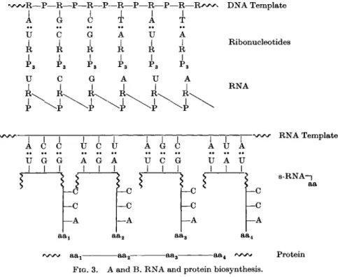
In DNA duplication, transcription of the information from DNA to RNA and
* To form the so-called polysomes.
translation from RNA to protein, the sequence of the individual building blocks of the newly-formed macromolecules is determined by a common prin
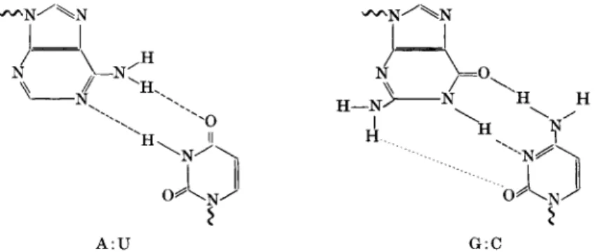
ciple. Nucleic acid bases—in the form of either deoxyribonucleoside or ribo- nucleoside triphosphates or s-RNA-amino-acid complexes—combine via hydrogen bonds with the bases of the "parent" template nucleic acid, adenine pairing with thymine or uracil and guanine pairing with cytosine (Fig. 2), followed by polymerization of the nucleotides or amino acids to yield the com
plementary nucleic acid strand or the protein. After unwinding of the DNA double-helix (Watson-Crick model), which consists of two complementary strands, two new strands are synthesized, complementary to each original strand. On the DNA strand a complementary RNA strand may be formed (Fig. 3). In protein synthesis, part of the sequence of each specific s-RNA orients itself complementary to the template RNA; it seems that a triplet
FIG. 2. Nucleic acid base-pairing. Left: adenine with uracil. Right: guanine with cytosine.
base sequence contains the information for one amino acid, viz., determines its relative position in the peptide chain (Fig. 3).
Arrest of DNA synthesis does, and chemical interaction with DNA templates may prevent DNA duplication in the premitotic cell and consequently abolish mitosis. Interference with RNA or DNA (synthesis and structure) will affect protein synthesis and, accordingly, enzymic function. Nucleic acid anti
metabolites have been shown to inhibit the adaptive (induced) enzyme formation in bacteria and mammalian cells; the inhibition of antibody synthesis by similar compounds in the mammal (181, 587) may be considered as an analogous phenomenon.
It is noteworthy that tumor cells contain large nuclei with hypertrophic nucleoli. A large part of the metabolic economy of the tumor cell must be directed to the maintenance and synthesis of the nuclear materials; 40% of the protein of the tumor cell and more than 30% of the RNA has been re
covered in the nuclear fraction of both animal and human cancers (390).
Tumor cells frequently contain a higher amount of DNA, corresponding to their aneuploid chromosome number, than do normal cells. Tumor nuclei show
Η
A : U G:C
III. CHEMOTHERAPY OF CANCER 69 also an enhanced protein synthesis, especially of histones (11, 94) and an active nucleic acid synthesis. These and related findings warrant the search for anti
tumor agents in the nucleic acid area. For a discussion of nucleic acid synthesis in the neoplastic cell, the impact of nuclear changes on the biochemistry of tumor tissue and the nucleolus of cancer cells, the reader is referred to the reviews of Goldthwait (257), Kitt (376) and Busch et al. (95b).
In comparing the effect of drugs on normal and tumor cells the following consideration may be of interest in the present context. Normal differentiated
v w R — Ρ — R — Ρ — R — Ρ — R — P - - R -
A G ύ Τ A I
U C
I G
| A
I U
k
R 1 R 1 R 11
Œ
Ps 1 Pa 1 1υ C G A
1 υ
1 1 1
R^
^ P
- R — R > w . I Τ
R I
p3
A R \ I
DNA Template
Ribonucleotides
RNA
I A U G I I
c
G U A
-C
*s aa.
I U
-c -c
-A G I U I
—c
—c
— A
υ I
υ
I A U
-C
—C
— A aa4
FIG. 3. A and B . RNA and protein biosynthesis.
RNA Template
s-RNA-|
aa
Protein
cells in the resting state exert many specific functions which are nonessential to the survival of the cell per se, that is, these cells contain a certain amount of nonvital machinery (enzymes; RNA and DNA templates). There are also indications that at least some of the vital machinery (e.g., respiratory enzymes) may be present in excess to the actual needs. By contrast, in tumors, especially in many transplanted ones, much of the nonvital machinery may be lacking (or repressed in expression), whereas the vital machinery is operating at top capacity. In proliferating cells, an active synthesis of nuclear RNA and trans
port of the latter to the cytoplasm is likely to occur in order to equip the ribo
somes with vital information. In differentiating and differentiated cells much of the information (RNA) molded on DNA, transferred and expressed
(protein), will be nonvital for the cell itself. These fundamental differences should be kept in mind whenever metabolic inhibitions produced by antitumor drugs in cells, or the binding of antitumor drugs to cells of normal and tumor tissues, are compared. An equal degree of binding of, for instance, a biological alkylating agent to normal and cancer cells may impair the survival of the tumor cells to a greater extent than that of the normal cells. Moreover, growth regulation can be considered to be mediated by a mechanism operating as a result of differentiation, and whatever the exact mechanism may be, it is evident that at least part of the mechanism (e.g., one loop of a feedback mechanism) must be anchored to the cells which are being regulated. When the damage inflicted upon normal tissue by a drug is confined to specific enzymatic functions, including the growth regulatory mechanism of the cells, a compensatory synthesis may result which actually leads to formation of new cells due to the disturbance of the growth regulatory mechanism in these cells. Given the time to differentiate, the equilibrium will be restored.
Also, if a certain proportion of the cells were killed by the drug, a regeneration would set in. The increased incorporation of RNA precursors and amino acids into the liver 12 hr after administration of the alkylating agent aminouracil mustard (162, 337) may be taken as representative of the regenerative process.
It should, however, also be noted that the performance of the organism as a whole allows only a critical amount of cells, or their specific functions, to be eliminated by drug action.
We shall now proceed to discuss how antimetabolites of glutamine, folic acid, purines and pyrimidines, and the alkylating agents may interfere with the metabolism (synthesis and function) of the nucleic acids and the nucleotide coenzymes and cofactors. To this end, some knowledge of the patterns of nucleic acid synthesis is essential.
The biosynthesis of the ribo- and deoxyribonucleotides, the constituents of RNA and DNA, may be summarized as follows:
De novo and constituent precursors > Ribonucleotides (AMP, GMP, CMP, UMP) (2a )
Constituent precursors > Deoxyribonucleotides (2/-deoxyAMP,
—GMP, —CMP, —TMP) (2b) III. 1.1. SYNTHESIS OF RIBONUCLEOTIDES AND RNA; INHIBITORS : AZASERINE,
DON, AMINOPTERIN, AMETHOPTERIN, 6-MERCAPTOPURINE, 6- THIOGUANINE, 2,6-DIAMINOPURINE, 8-AZAGUANINE, 6-URACIL METHYLSULFONE, 6-AZAURACIL, AND 5-FLUOROOROTIC ACID
Ribonucleotides are formed via 2 concurrent routes:
1. Synthesis de novo from small precursor molecules, involving 2 distinct pathways:
ΠΙ. CHEMOTHERAPY OF CANCER 71 a. De novo synthesis of purine-containing nucleotides,
b. De novo synthesis of pyrimidine-containing nucleotides.
2. Incorporation of constituent molecules (preformed purines and pyrimidines):
a. By the nucleoside phosphorylase-catalyzed reaction of base plus ribose-1 - phosphate to form inorganic orthophosphate (Pj) and the corresponding nucleoside, followed by a phosphorylation of the latter to the nucleotide by a nucleoside phosphokinase,
~p
A + R-l-P > Pj + A R > AMP
b. By the pyrophosphorylase-catalyzed reaction of base plus 5-phosphori- bosylpyrophosphate (5-PRPP) to form pyrophosphate (PPj) and the corre
sponding nucleotide,
A + 5-PRPP > PPt + AMP
The metabolic pathways can be schematically represented as shown in reactions 3 and 4.
A
<
2>|
AMP Ua) /
Denovo > IMP{ (3) (2)
I
GMPΗ ( 2 ) | G C
(2)1
CMP (4) Ub) /
De novo > UMP ( 2 ) |
u
The relative rates of the various reactions may vary among different biological systems. For instance, enzymes which convert the free bases to the nucleosides or nucleotides may be absent or very little active. The bases may be degraded by other enzymes before the latter reactions may take place. A preformed base may depress the de novo synthesis of its own nucleotide by some feedback mechanism [uracil and de novo UMP synthesis in bacteria (690,
692 )1
3. The ribonucleotides are converted to the corresponding di- and tri
phosphates (e.g., AMP->ADP^ATP) and the latter are either used as cofactors or built into coenzymes and polymeric RNA. A polynucleotide phosphorylase is known which converts nucleoside diphosphates to RNA
(Ochoa), the triphosphates are incorporated into microsomal and soluble RNA, whereas the net, DNA-dependent, synthesis of RNA occurs also with the 4 nucleoside triphosphates (105).
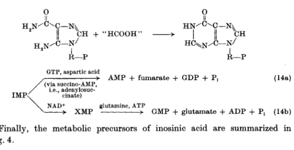
III. 1.1. A. De Novo Synthesis of Ρ urine-Containing Ribonucleotides. The elucidation of this pathway is largely due to the investigations of Buchanan and Greenberg and their co-workers (87,103, 314). The following sequence of reactions* has been found to operate:
R-l-P — R-5-P + ATP — Glutamine + 5-PRPP — Glycine + PRA + ATP —
H2C — N H2
I + H2N 0 = C — O H £
R-5-P
5-PRPP + AMP
PRA + glutamic acid + PPi GAR + ADP + P.
(5) (6) (7) (8)
R—Ρ GAR + 'HCOOH'-folic acid derivative
' H C O O H " — 0 = C - N H I
LR —p
F G AR (9) H2C — N H2
0 = C — N H I +
U
L R —Ρ
F G AR + glutamine + A TP + H 20 - H2C — N H
I ^CHO + N H2
0 = C — N H <
LR - P >
F G AM + A TP
H2C — N H I M3HO - H N = C — N H
LR — Ρ
A I R + CO 2 + A T P + aspartic acid -
H2C — N H ' > I ^CHO
0 = C — N H I
LR — Ρ
FGAM + ADP + Pi + glutamic acid (10) H2C - N H
> I ^CHO
~ N H
U
H N = C
^-R—Ρ AIR + ADP + ^
HC—N.
> II HoN—C
(11) - N ^ *CH
R—Ρ I
AICAR + fumaric acid + ADP + P, (12) H9Nx =0
H C — Ν ii χ H2N — C — Ν
I
5CH C 02; ATP + aspartic acid H2N - R—Ρ
AICAR + "HCOOH"-folic acid derivative
Ο I C—N^
II ^ C H - C — N ^
I R—Ρ
IMP + H20 (13)
* In the reactions, RP stands for ribose phosphate; PRPP for phosphoribosyl pyro
phosphate; PRA for 5-phosphoribosylamine; GAR for glycinamide ribotide; FGAR for α-iV-formylglycinamide ribotide; FGAM for α-iV-formylglycinamidine ribotide; AIR for 5-aminoimidazole ribotide; AICAR for 5-amino-4-imidazolecarboxamide ribotide.
III. CHEMOTHERAPY OF CANCER 73
H 2 N ^ C - N^
N / C —/ N
CH + " H C O O H ' Η
R—Ρ
I II
H C ^N/ C — N / R—Ρ GTP, aspartic acid
-> AMP + fumarate + GDP + P, IMP<
(via succino-AMP, i.e., adenylosuc
cinate) NAD+
(14a)
glutamine, ATP
X M P > GMP + glutamate + ADP + Pt (14b)
Finally, the metabolic precursors of inosinic acid are summarized in Fig. 4.
aspartic acid (amino N)
0
\ II f H N ^ ' h C -
co2
. formate
y •
Lformate ^ glycine glutamine
(amide N)
FIG. 4. Precursors of inosinic acid.
III.l.l.B. Inhibitors.
III.l.l.B(l). Glutamine antagonists. Azaserine (1953) and DON (1956) have been detected in culture filtrates of 2 species of Streptomyces because of the ability of such filtrates to retard the growth of a mouse sarcoma {537, 538).
H- COOH - C - N H2
I C H2 C H2
<U
I
COOH I Η — C — N H2
I C H2
<U
Ο Η — C = N = I +COOH I Η — C — N H2
C H2 C H2 C= 0
I + - H — C = N = N
=N
(I) (II) (III) L-Glutamine O-diazoacetyl- 6-diazo-5-oxo-
L-serine L-norleucine (azaserine) (DON)
The antibiotics were isolated from the fermentation broths and found to be markedly inhibitory to a number of tumors, DON being approximately 50 times more active than azaserine on a dose basis.
In the de novo synthesis of the purine skeleton, glutamine functions as amino donor in 2 animation reactions (compare reactions 7 and 10). It was established by Buchanan et al. (86, 297) that both these reactions were inhibited by the two drugs in cell-free systems of pigeon liver, especially the reaction 10 leading to a-i^-formylglycinamidine ribotide (267, 312, 468, 608, 609, 641). DON exerted an equivalent inhibitory response on the latter reaction at one- fortieth the concentration required for azaserine; 100 times higher concentra
tions of the two drugs were necessary to obtain a 50% inhibition of reaction 7.
During short periods of incubation of the enzyme preparations with drug and limiting amounts of a-iV^-formylglycinamide ribotide, the inhibition of reaction 10 is of a competitive type in respect to glutamine. However, when either one of the 2 drugs is preincubated with the enzyme preparation, even for a very short period of time, noncompetitive inhibition results since no amount of glutamine suffices to reverse the inhibition. Results of experiments on reaction 10, carried out with excess substrate over longer periods of incuba
tion with the drugs (no preincubation), showed also that glutamine rather delayed than protected the enzyme from inactivation. The latter findings may indicate that a direct chemical reaction between drug and enzyme takes place.
Diazoketones may react with amino, hydroxyl, and mercapto groups in a reaction involving rearrangement by anionic migration, thus:
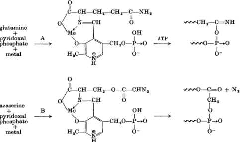
This reaction is an acylation rather than an alkylation; DON, for example, might introduce an α-aminoadipic acid residue into the enzyme. A mechanism of action for the enzymic activation of the amide-NH2 group of glutamine as an aminating agent has recently been proposed (24), with pyridoxal phosphate acting as the coenzyme (Fig. 5A). Azaserine and DON were considered to replace glutamine noncompetitively following an irreversible alkylation of the phosphate group of the pyridoxal-phosphate-drug complex (Fig. 5B).
The actual course of the two reactions illustrated by reaction 15 and Fig. 5B is that the diazoketone loses nitrogen to form a ketocarbene, containing a free pair of electrons, which may either react directly according to reaction 16a (Fig. 5B) or isomerize first and then react according to reaction 16b ( = reaction 15). More recent evidence shows (233a) that azaserine (as diazo- nium salt) alkylates a cysteine-SH group of the FGAR amidotransferase.
RCOCHN2 + R ' N H2 >
(drug) (enzyme receptor)
RCH2CONHR' + N2
(drug-receptor complex) (15)
RCOCHN2 •» N2 + RCOCH
R C O C H2O P = (reaction 16a)
R C H2C = 0 I N H R ' R ' N H2
(reaction 16b)
ΠΙ. CHEMOTHERAPY OF CANCER 75 The specificity of action of DON is illustrated by the finding that 4-diazo-4- oxonorvaline (DON less one methylene carbon) is not inhibitory to the aminating enzyme and lacks antitumor activity. In tracer studies, a marked inhibition of de novo purine synthesis in vitro and/or in vivo was demonstrated for variety of normal and tumor cells; the inhibition was accompanied by an accumulation of FGAR. Although the block of the conversion of FGAR to FGAM appears to be the most critical one, the inability to achieve complete reversal of the drug effect with purines in various systems suggests that another site of inhibition must be involved. Inhibition of the amination of uracil to cytosine derivatives (186) (pathway lb, reaction 24; see section
glutamine pyridoxal + phosphate
metal +
Ο II ,C—CH
°(
1\ Ο H3C
CH 2— C H 2— C — N H 2
II
N — C H Ο
ι—CHoO- OH
I - P — (
A-
^ C H2- C = N H
ATP I Ο
>- w ^ O — P- * 0 I
o-
azasenne pyridoxal + phosphate
metal +
Ο II
C—CH- C H2 CH
- C — C H N2
II
ο
H3c J ^ .
i—CH.O- OH - P- * 0
I
o-
^ • ^ o — C= 0 + N2
C H2
ο
- Ρ-
Α -
FIG. 5. A. Glutamine activation according to Baker (24), involving formation of an azomethine (Schiff's base). B. Azaserine replaces glutamine noncompetitively, following irreversible alkylation.
III.LLC) and of IMP to GMP (38) (reaction 14b), both involving glutamine, has been observed; in Novikoff hepatoma cells, this reaction was 10 times less sensitive to inhibition by DON than the de novo purine synthesis (470).
Amylase synthesis by mouse pancreas is reported to be strongly inhibited by azaserine (567); however, no marked inhibition of protein synthesis was seen in various tumors and spleen (304, 408). For instance, Ehrlich-ascites cells showed a complete inhibition of glycine uptake for the de novo purine synthesis 10 min after addition of azaserine, whereas no effect on glycine uptake into the proteins was evident.
Transamination reactions in which glutamine participates may also be inhibited by the drugs. Transamination reactions in which an azomethine between amino acid and pyridoxal phosphate is also formed, are stereospecific