ION CHANNELS, RECEPTORS AND TRANSPORTERS
[Ca 2+ ] i -induced augmentation of the inward rectifier potassium current (I K1 ) in canine and human
ventricular myocardium
Norbert Nagy&Károly Acsai&Anita Kormos&Zsuzsanna Sebők&
Attila S. Farkas&Norbert Jost&Péter P. Nánási&Julius Gy. Papp&
András Varró&András Tóth
Received: 25 March 2013 / Revised: 7 June 2013 / Accepted: 7 June 2013
#Springer-Verlag Berlin Heidelberg 2013
Abstract The inward rectifier K+current (IK1) plays an im- portant role in terminal repolarization and stabilization of the resting potential in cardiac cells. Although IK1was shown to be sensitive to changes in intracellular Ca2+ concentration ([Ca2+]i), the nature of this Ca2+sensitivity—in spite of its deep influence on action potential morphology—is controver- sial. Therefore, we aimed to investigate the effects of a nonadrenergic rise in [Ca2+]ion the amplitude of IK1in canine and human ventricular myocardium and its consequences on cardiac repolarization. IK1, defined as the current inhibited by 10μM Ba2+, was significantly increased in isolated canine myocytes following a steady rise in [Ca2+]i. Enhanced IK1was also observed when [Ca2+]i was not buffered by ethylene glycol tetraacetic acid, and [Ca2+]Itransients were generated.
This [Ca2+]i-dependent augmentation of IK1was largely atten- uated after inhibition of CaMKII by 1μM KN-93. Elevation of [Ca2+]oin multicellular canine and human ventricular prep- arations resulted in shortening of action potentials and accel- eration of terminal repolarization. High [Ca2+]oenhanced the action potential lengthening effect of the Ba2+-induced IK1
blockade and attenuated the prolongation of action potentials following a 0.3-μM dofetilide-induced IKrblockade. Block- ade of IKsby 0.5μM HMR-1556 had no significant effect on APD90in either 2 mM or 4 mM [Ca2+]o. It is concluded that high [Ca2+]ileads to augmentation of the Ba2+-sensitive cur- rent in dogs and humans, regardless of the mechanism of the increase. This effect seems to be at least partially mediated by a CaMKII-dependent pathway and may provide an effective endogenous defense against cardiac arrhythmias induced by Ca2+overload.
Keywords Canine/human myocardium . Inward rectifier K+ current (IK1) . Cytosolic Ca2+. Action potential duration . Ventricular repolarization . Ba2+
Introduction
Cardiac repolarization, and consequently the duration of the ventricular action potential (APD), is tightly controlled by interactions and fine balance among various transmembrane ion currents [6,43]. Plenty of these currents were shown to be sensitive to shifts in [Ca2+]i. A few of them (e.g., the L-type Ca2+current, ICaLand the Na+/Ca2+exchanger current, INCX) are predominantly inward, favoring depolarization [1, 2], while others (e.g., the Ca2+-activated Cl− current, ICl) are outward, facilitating repolarization [45, 47]. Even many
“Ca2+-independent”current, like the transient outward current (Ito) or the slow and fast components of the delayed rectifier K+current (IKrand IKs, respectively) were also found to be [Ca2+]i-sensitive to some extent, or at least being modulated by Ca2+-dependent signaling pathways [16,34,40,42].
The inward rectifier K+ current (IK1), an important repolarizing current, active in ventricular, atrial, and Purkinje A. Kormos
:
Z. Sebők:
J. G. Papp:
A. Varró:
A. Tóth (*)Department of Pharmacology and Pharmacotherapy, University of Szeged, Dóm tér 12, P.O. Box 427, 6720 Szeged, Hungary e-mail: toth.andras@med.u-szeged.hu
N. Nagy
:
K. Acsai:
N. Jost:
J. G. Papp:
A. Varró:
A. Tóth Division of Cardiovascular Pharmacology, Hungarian Academy of Sciences, University of Szeged, Szeged, HungaryA. S. Farkas
Second Department of Internal Medicine and Cardiological Centre, University of Szeged, Szeged, Hungary
P. P. Nánási
Department of Physiology, University of Debrecen, Debrecen, Hungary
DOI 10.1007/s00424-013-1309-x
cells, has long been known to play a crucial role in terminal repolarization. It is also a major contributor to the repolari- zation reserve of the heart [5]. Recent studies firmly support the idea that IK1is subject to complex regulation by multiple interacting signaling pathways [13, 17, 20, 29, 36, 44].
Furthermore, all related studies agree that the current is [Ca2+]i-sensitive; however, experimental data on the direc- tion and details of this Ca2+dependence are highly contro- versial [7,10, 13,17, 19,22, 28–31, 36,44, 46]. For in- stance, IK1was shown to be decreased by elevation of [Ca2+]i
in guinea pig [10,28,46] and rat [13] cardiac myocytes due to the [Ca2+]i-induced enhancement of the inward rectifica- tion of the channel. On the other hand, the increased activity of CaMKII kinase, which may also be a consequence of the elevated [Ca2+]i, reduced the amplitude of IK1in mice, while IK1 was enhanced by CaMKII kinase in rabbit ventricular cells, but reduced in rats [44]. Since multiple controlling mechanisms converge on IK1, the effect of a [Ca2+]i-induced shift on the current can hardly be predicted. Indeed, IK1was shown to be subject to complex modulation by both inhibi- tory (via PKA/PKC) [13,20] and activating (via CaMKII) Ca2+-dependent pathways [44].
Considering the contradictory nature of the available ex- perimental data, the primary aim of the present study was to elucidate the effect of [Ca2+]ion the amplitude of IK1and to evaluate the [Ca2+]idependence of the relative contribution of IK1to ventricular repolarization in canine and human ventric- ular myocardium—as being the best known model for human cardiac tissues in terms of electrophysiology [38,39].
IK1 is generally considered as a composite current of the Kir2.x and a few more, yet to be identified, background chan- nels, all functioning between−80 and−30 mVand inhibited by Ba2+when applied at low concentrations. Indeed, 10μM BaCl2
is considered as a relatively selective inhibitor of IK1 [5].
Therefore, in this study, IK1was defined as the current blocked by 10μM BaCl2. [Ca2+]i was increased via nonadrenergic ways to avoid the marked activation of PKA (and probably PKC) to minimize their modulatory effects on IK1. In order to prevent any decline in the repolarization reserve, possibly caused by the enzymatic digestion process, all action potential measurements were performed in intact multicellular prepara- tions excised from either a canine or an undiseased human heart. The results support a Ca2+-induced and CaMKII- translated increase of the Ba2+-sensitive current, which may contribute to adaptation of APD to conditions of elevated [Ca2+]i—independently of activation of the adrenergic pathway.
Methods
Experiments were performed in compliance with the Guide for the Care and Use of Laboratory Animals (USA NIH
publication no 86–23, revised 1985). All experimental pro- tocols were approved by the Ethical Committee for Protec- tion of Animals in Research of the University of Szeged, Hungary (permit no. I-74-9/2009). Investigations performed in human cardiac samples conform to the principles outlined in the Helsinki Declaration. All experimental protocols were approved by the Regional and National Human Medical and Biological Research Ethics Committee, University of Sze- ged (permit no. 63/1997).
Canine preparations
Adult mongrel dogs of either sex, weighing 10–20 kg, were anesthetized with thiopental (30 mg/kg). The hearts were rapidly removed through right lateral thoracotomy and im- mediately rinsed with ice-cold Tyrode solution containing (in millimolar) the following: NaCl 144, NaH2PO40.33, KCl 4, MgCl21, glucose 5.5, HEPES 5, and CaCl22. The pH of the solution was adjusted to 7.4 with NaOH. A wedge- shaped section of the left ventricular wall, supplied by the left anterior descending coronary artery, was dissected and cannulated for isolation of single myocytes using the seg- ment perfusion technique. Papillary muscles from the right ventricle were excised for simultaneous recording of action potentials and [Ca2+]itransients.
Isolation of ventricular myocytes
The excised left ventricular segments were perfused through the anterior descending coronary artery using a gravity flow Langendorff apparatus. The perfusate was a modified MEM solution (Minimum Essential Medium Eagle, Joklik modifi- cation, Sigma, M-0518), supplemented with 1.2 mM CaCl2, 10 mM HEPES, 2.5 g/l taurine, 0.175 g/l pyruvic acid, and 0.75 g/l ribose (pH=7.2). After removal of blood, the per- fusate was switched for 10 min to nominally Ca2+-free MEM. Dispersion of cells was achieved by an application of 0.5 g/l collagenase (Sigma type I) for 40 min in the presence of 50 μM CaCl2. During the isolation procedure, the solutions were gassed with 100 % oxygen, and the temperature was maintained at 35 °C. Finally, the tissue was minced and gently agitated. The cells, freshly released from the tissue, were stored at room temperature before use.
At least 60 % of the cells were rod-shaped and showed clear striation when the external Ca2+was restored.
Human samples
Undiseased hearts (n=5) obtained from organ donors were explanted to obtain pulmonary and aortic valves for trans- plant surgery. Before cardiac explanation, the donors did not receive medication except furosemide, dobutamine, and
plasma expanders. Right ventricular papillary muscles were used in the experiments.
Measurement of IK1in single cardiomyocytes
IK1was recorded using the whole-cell version of the patch clamp technique. A drop of cell suspension was placed into a lucid chamber mounted on the stage of an inverted micro- scope. All measurements were performed at 37 °C. The cells were allowed to adhere for at least 10 min before starting superfusion with Tyrode solution. Micropipettes were fabri- cated from borosilicate glass capillaries (Clark Electromedical Instruments) using a microprocessor-controlled horizontal puller (Model P-97, Sutter Instruments). These electrodes had resistances of 1.5–2.5 MΩ, when filled with pipette solution, containing (in millimolar) the following: K- aspartate 100, KCl 40, K2ATP 5, MgCl21, and HEPES 10, at pH=7.2. Pipette solutions were either unbuffered to allow [Ca2+]itransients or were Ca2+buffered by the addition of an appropriate mixture of ethylene glycol tetraacetic acid (EGTA) or BAPTA plus CaCl2. The unbuffered solution was made using nominally Ca2+-free distilled water, pur- chased from Sigma. Actual values of free Ca2+in the pipette solutions ([Ca2+]pip) were calculated using WinMaxC [35].
Furthermore, the pipette solution containing high [Ca2+] was also verified by measuring the concentration of free [Ca2+] with a Ca2+-sensitive electrode (World Precision Instruments Inc.). Ionic currents were recorded using an Axopatch 1D amplifier (Axon Instruments). Gigaseal has been established via gentle suction. The cell membrane was disrupted by either further suction or application of several short electrical pulses.
Membrane currents were digitized under software control (pClamp 10.0, Axon Instruments) following low-pass filtering at 1 kHz with an analog-to-digital converter (Digidata 1440A, Axon Instruments). Sampling rate was set to 5 kHz. The applied voltage protocols are detailed in theResultssection and shown in the figures as pertinent.
Recording of action potentials in multicellular preparations Action potentials were recorded at 37 °C from the surface cell layer of ventricular papillary muscles using conventional microelectrode techniques. The preparations were mounted in a Plexiglas chamber, allowing continuous superfusion with O2-saturated Tyrode solution. The muscles were stim- ulated by rectangular current pulses, having durations of 2 ms and amplitudes of twice the diastolic threshold, at a constant rate of 1 Hz. These pulses were delivered to the preparations through a pair of bipolar platinum electrodes coupled to an electrostimulator (Hugo Sachs Elektronik, model 215/II). Sharp microelectrodes, having tip resistance of 10–20 MΩwhen filled with 3 M KCl, were connected to an amplifier (Biologic Amplifier, model VF 102). Voltage
output from the amplifier was sampled using an A/D con- verter (NI 6025, Unisip Ltd). In order to optimize data processing during measurements, dual sampling rates were applied: in the initial 50 ms of the action potential, the sampling rate was set to 40 kHz, while the second, slow phase was digitized at 1 kHz. No further filtering was used.
Action potential duration, determined at 90 % level of repolarization (APD90), was obtained using Evokewave v1.49 (Unisip Ltd). To ensure the physiological conditions of the preparations, ventricular muscle samples having ac- tion potential amplitudes less than 100 mV or showing a drift in APD during the control period were discarded. Efforts were made to maintain the same impalement throughout the whole experiment. When the impalement was dislodged, an adjustment was attempted. The measurement was continued if the action potential characteristics of the reestablished impalement deviated less than 5 % from the original values.
Monitoring [Ca2+]itransients in multicellular samples and single cells
Multicellular samples were loaded with 25 μM Fluo 4-AM (Molecular Probes Inc.) for 50 min at room temperature.
Isolated cells were loaded with 2μM Fluo 4-AM for 15 min.
Both were mounted in a low-volume imaging chamber (RC47FSLP, Warner Instruments) and field-stimulated at a rate of 1 Hz while continuously superfused with Tyrode solution at 37 °C. Fluorescence measurements were performed using an Olympus IX 71 inverted fluorescence microscope. Optical signals were recorded by a photon counting photomultiplier module (Hamamatsu, model H7828) sampled at 1 kHz. The dye was excited at 480 nm, and the emitted fluorescence was detected at 535 nm. Data acquisition and analysis were performed using the Isosys software (Experimetria, Hungary).
Fluorescence traces recorded from multicellular samples were corrected for nonspecific background and bleaching. Alter- ations in [Ca2+]i were expressed as changes in normalized fluorescence (F/F0). Fluorescence traces recorded from single cells were only used to validate the absence/presence of [Ca2+]i
transients in the patch clamped cells. On these traces, no further processing, except smoothing, has been performed.
Drugs
All chemicals were purchased from Sigma, except for other- wise indicated. BaCl2 (10 μM) was used to dissect IK1. Dofetilide (gift from Gedeon Richter Ltd, Hungary) and HMR-1556 (Aventis Pharma) were dissolved in dimethyl sulfoxide. BaCl2was dissolved in distilled water performing stock solutions of 1–1 mM and 100 mM, respectively. KN- 93 was purchased from Calbiochem and was also dissolved in dimethyl sulfoxide in a stock solution of 1 mM. Its final concentration was 1 μM, the same as that used in other
studies [9]. Following its application, time was allowed to reach at the maximal inhibition of CaMKII. For negative control, its inactive analog, KN-92, was used at the same concentration. All stock solutions were stored at 4 °C. Solu- tions were freshly made prior to the measurement.
Statistics
All values presented in this study are arithmetic means ± SEM. Statistical significance of the differences when making multiple comparisons was evaluated using repeated mea- sures ANOVA + Bonferroni post hoc test. All other data were analyzed using Studentst test for paired or unpaired data, as relevant. Differences were considered significant whenpwas less than 0.05.
Results
Ca2+-dependent modulation of IK1in isolated canine ventricular cells
To characterize its Ca2+dependence, IK1was determined in isolated canine left ventricular myocytes using the whole cell configuration of the patch clamp technique. In the first set of experiments, Ca2+concentration in the pipette solution was adjusted to low (∼160 nM) or high (∼900 nM) levels by adding the appropriate mixture of Ca2+and BAPTA to the pipette solution. Steady-state IK1 amplitudes were deter- mined at the end of 300 ms of voltage pulses usually clamped to potentials ranging between −90 and −30 mV from the holding potential of−90 mV. IK1was determined by repeat- ing the protocol in the presence of 10μM BaCl2. As shown in Fig.1a, b, within the voltage range of−70 to−40 mV, the use of high [Ca2+]pip (and presumably elevated [Ca2+]i) significantly enhanced the magnitude of the Ba2+-sensitive current compared to that recorded with low [Ca2+]pip
(p<0.05,n=7 for both groups, randomized from three dogs).
Average values of membrane capacitance were also deter- mined for the low and high [Ca2+]pipgroups: 145.11±8.3 and 138.77±10.21 pF were obtained, respectively. This differ- ence was not significant statistically.
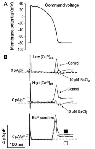
Ca2+-dependent activation of IK1during the action potential Activation of IK1 during the AP was analyzed using the action potential clamp technique (i.e., by applying typical ventricular action potential waveforms as a command poten- tial), as demonstrated in Fig. 2a. As previously, IK1 was determined as the current component dissected by 10μM BaCl2. In the presence of high [Ca2+]pip, the magnitude of peak IK1was significantly higher than that in the presence of low [Ca2+]pip, without changing the time course of activation
(Fig.2b, c). As shown in Fig.2d, the I–V relationship (phase- plane trajectory), obtained for IK1by plotting the average current against the respective membrane voltage, reveals that the maximum of the current was shifted towards less nega- tive membrane potentials (from −65±1.1 to −57±1.3 mV, p<0.05,n=7) in response to elevation of [Ca2+]i.
Effects of [Ca2+]itransients on IK1
To better characterize the beat-to-beat modulation of IK1by changes in [Ca2+]i, we also tested the hypothesis that an unbuffered increase in [Ca2+]i (e.g., during the normal [Ca2+]i transient, generated by the cell in the absence of internal EGTA) may contribute to a higher amplitude of IK1. In these experiments, IK1 was determined in voltage- clamped myocytes, both in the absence and presence of [Ca2+]itransients, evoked using pipette solutions either con- taining 5 mM EGTA or without any calcium chelator, re- spectively. In order to trigger the onset of [Ca2+]itransients prior to activation of IK1, the voltage protocol started with a short prepulse stepping from the−80 mV holding potential to 0 mV for 50 ms. As expected, neither a [Ca2+]itransient, nor the elevation of IK1could be observed in the presence of 5 mM EGTA. In contrast, when EGTA was omitted from the pipette, the presence of [Ca2+]i transients significantly in- creased the amplitude of IK1(Fig.3a, b). With EGTA in the pipette, the steady-state IK1was relatively moderate (similar to the situation when normal [Ca2+]owas applied; see Fig.1);
however, when EGTA was omitted from the pipette, the magnitude of the current was significantly increased in the
−70 to −30-mV membrane potential range and also after hyperpolarizing the membrane to −90 mV (p<0.05, n=5, Fig.3c). Small time-dependent changes of the current profile could be observed during these [Ca2+]itransients; however, they remained within the experimental variance.
Mechanism of the Ca2+-induced enhancement of IK1
Since CaMKII was previously shown to increase the inten- sity of IK1in rabbit ventricular myocytes [44], the relatively selective CaMKII inhibitor, KN-93 was used to test the involvement of this mechanism in canine cardiac cells. Three groups of myocytes were established: [Ca2+]pipwas set low (∼160 nM) in the first (control) group and 1μM of KN-92, the inactive analog of KN-93, was used; in the second and third groups, high [Ca2+]pip (∼900 nM) was applied in the presence of either the inactive KN-92 or the active KN-93 (1–1μM each). Steady-state currents were determined at the end of 300 ms of voltage pulses ranging between−90 and
−30 mV. IK1 was dissected using 10 μM BaCl2. Without CaMKII inhibition (i.e., in the presence of KN-92), a signif- icant difference could be observed between the low and high [Ca2+]pipgroups (n=8 in each group, randomized from four
dogs). However, this difference was significantly decreased (p< 0.05) following the inhibition of CaMKII by KN-93 (Fig.4a, b).
To test the assumption that CaMKII activity changes during the Ca2+ transient, we performed a few additional experiments (n=3), in which the effect of Ca2+influx via L-type Ca2+ channels on IK1activation during the AP has been estimated using a Ca2+ channel blocker (Nifedipine, 10 μM). Data from these preliminary experiments (not shown) support some contribution of the Ca2+influx to IK1
activation, but this contribution seems to be limited.
Effect of [Ca2+]oon [Ca2+]itransients and action potential configuration in multicellular canine ventricular preparations Ca2+-dependent alterations in action potential morphology were studied in canine right ventricular papillary muscles exposed to either 2 or 4 mM extracellular Ca2+concentra- tions. This strategy was chosen in order to minimize the
possible modulation of action potentials through the known intracellular signaling pathways, including adrenergic acti- vation. On the other hand, multicellular preparations are known to display more stable action potentials than single cells; furthermore, the lack of enzymatic digestion may war- rant a more“physiological”set of ion channels in the cell membrane of these cells. Elevation of [Ca2+]o from 2 to 4 mM resulted in a significant (35±11 %) increase in the amplitude of [Ca2+]itransients, without alterations in diastol- ic [Ca2+]i (Fig.5a, b). Elevation of [Ca2+]itransients fully paralleled with the shortening of action potentials: APD90
decreased from 202.4±3.5 to 185.2±8.2 ms (p<0.05,n=5).
This shortening of APD was associated with a markedly accelerated terminal repolarization (Fig. 5c, d). Indeed, the amplitude of −dV/dtmin was increased by more than 40 % (from−1.13±0.06 to−1.59±0.05 V/s,p<0.05,n=5) after the elevation of [Ca2+]o. The action potential lengthening effect of the Ba2+-induced suppression of IK1was tested at both normal and elevated [Ca2+]o(2 and 4 mM, respectively). As shown in Fig. 1 [Ca2+]pipdependence of the steady-state IK1in isolated canine
ventricular myocytes determined as the current blocked by 10μM BaCl2.a Representative sets of superimposed Ba2+-sensitive current records andbthe current–voltage relation curves obtained for IK1with [Ca2+]pipbuffered to∼160 or to∼900 nM (openandfilled symbols, respectively). Steady-state IK1currents were determined at the end of
300 ms of test potentials ranging in amplitude from−90 to−30 mV.
Statistical analysis (repeated measures ANOVA + Bonferroni ad hoc test) revealed that the difference between the two curves is significant (p<0.05). Theasterisksindicate significant (p<0.05) differences be- tween low and high [Ca2+]pipresults, obtained in seven cells isolated from three dog hearts
Fig.6a, b, the BaCl2-induced prolongation was significantly greater (nearly doubled) at 4 mM [Ca2+]o. APD90 was in- creased by 10μM BaCl2from 185.2±8.2 to 231.2±13.1 ms at 4 mM [Ca2+]o(Δ=19.6±1.9 %,p<0.05)—in contrast to the moderate, only 10.8±0.8 % lengthening effect of BaCl2
obtained at 2 mM [Ca2+]o(from 202.4±3.5 to 222.9±2.8 ms, p<0.05). This effect of BaCl2was fully reproducible, since following the application of BaCl2in the presence of 2 mM [Ca2+]oBaCl2was washed out, and the Ba2+challenge could be repeated in the presence of 4 mM [Ca2+]ousing the same preparation (Fig.6c).
Triangulation (defined as a difference between APD90and APD25) was also estimated to further quantify the Ca2+-in- duced changes in action potential morphology. Statistical analysis revealed that triangulation by 10μM BaCl2 when tested at 2 mM [Ca2+]o was significantly increased (from 84.4±4.3 to 102.4±4.4 ms,p<0.05). A significant and rela- tively even greater BaCl2-induced increase in triangulation could be observed at 4 mM [Ca2+]o (from 112.3 ±8.1 to 155.3±13.5 ms,p<0.05).
Ca2+dependence of action potential morphology in undiseased human myocardium
Ca2+-dependent effects of 10μM BaCl2 were different in human and canine right ventricular papillary muscles. In human preparations, APD90was not lengthened by 10μM BaCl2at 2 mM [Ca2+]o(353.8 ±12.2 versus 362.4±14.3 ms, Δ=2.36±1 %, N.S.,n=5 from two hearts). In contrast, the Ba2+-induced lengthening of APD90 was significant when [Ca2+]o was elevated to 4 mM (from 313.8 ± 19.16 to 332.8±20.91 ms,Δ=6.02±0.5 %,p<0.05, Fig.7a, b). Fur- thermore, elevation of [Ca2+]osignificantly accelerated the terminal phase of repolarization (from −0.44 ± 0.02 to
−0.49 ± 0.03 V/s, p< 0.05, Fig. 7c). Compared to canine papillary muscles, however, this effect was also weaker.
AP triangulation values were also calculated for human samples. Statistical analysis revealed that application of 10 μM BaCl2did not change significantly triangulation at 2 mM [Ca2+]o(control: 188.8±11.6 ms; BaCl2: 175.8±14.6 ms;
n=5, N.S.). In contrast, at 4 mM [Ca2+]o, triangulation was Fig. 2 Effects of increased [Ca2+]pipon IK1during a canine ventricular
action potential.aA typical ventricular AP waveform has been applied as command voltage. IK1current was dissected by the application of 10μM BaCl2. bA set of representative original recordings of IK1
current profiles and the calculated Ba2+-sensitive current (openand filled symbols, respectively). cPeak IK1currents evoked by the AP command in the presence of low (∼160 nM) and high (∼900 nM)
[Ca2+]pip. Theasterisksindicate significant (p<0.05) differences be- tween low and high [Ca2+]pipresults, obtained in seven cells, (from three hearts). dCurrent–voltage relationships, generated by plotting the magnitude of the (Ba2+-sensitive) IK1current against the corresponding membrane potential at high and low [Ca2+]i(openandfilled symbols, respectively). Thearrowsindicate IK1current maximums
significantly increased (control: 194.8 ± 11.7 ms; BaCl2: 212.2±10.6 ms;n=5,p<0.05). Human and canine triangulation data were also compared, and a significant difference was re- vealed between all four corresponding pairs of experimental groups.
[Ca2+]-dependent contribution of IK1to the repolarization reserve in canine myocardium
If the magnitude of the Ba2+-sensitive current was increased by elevation of [Ca2+]o, its relative contribution to the repo- larization reserve should also be enhanced. In order to char- acterize the [Ca2+]-dependent redistribution of repolarizing currents, the contribution of IKrand IKsto repolarization was estimated at normal and high [Ca2+]olevels. As summarized in Fig. 8, prolongation of APD90 induced by 300 nM dofetilide was significantly less at 4 than 2 mM [Ca2+]o(at
2 mM [Ca2+]o; control: 205.2±6.5 ms; Dofetilide: 253.2±9.3 ms, p<0.05; at 4 mM [Ca2+]o, control: 185.8±8.1 ms, Dofetilide 208.4±9.2 ms, N.S.; for the relative increase 23.5±1.8 versus 12.2±1.1 %, p<0.05, n=5). Selective inhibition of IKs by 0.5μM HMR-1556 had no significant APD90lengthening effect in either 2 or 4 mM [Ca2+]o (at 2 mM [Ca2+]o: control:
206.3±5.9 ms, HMR-1556: 211.5±6.1 ms, N.S.; at 4 mM [Ca2+]o: control: 191.4±6.7 ms, HMR-1556: 193±4.8 ms, N.S.) (Fig.8b, c) congruently with a very limited contribution of IKs
to cardiac repolarization under control conditions. These find- ings demonstrate that the APD-shortening effect of high [Ca2+]ocannot be attributed to accelerated activation of either IKr or IKs due to the concomitant elevation of the plateau potential. These results are just the opposite of those observed with inhibition of IK1(Figs.6and7). Since the elevation of [Ca2+]isignificantly alter the contribution of the Ba2+-sensi- tive current to terminal repolarization, this effect may have Fig. 3 Enhancement of IK1in the presence of regular [Ca2+]itransients in
isolated canine ventricular myocytes.a Representative set of 10μM Ba2+-sensitive current records andbFluo-4 fluorescence recordings (raw, uncorrected traces) obtained with 5 mM EGTA (low [Ca2+]pip) and without 5 mM EGTA (high [Ca2+]pip) in the pipette solution (left andright panels, respectively). [Ca2+]itransients were evoked by 50 ms of prepulses clamped to 0 mV for 50 ms preceding the 500-ms test pulses—as shown in the insert.cCurrent–voltage relationships were
obtained by plotting IK1amplitudes, measured at the end of these test pulses, as a function of the respective test potential with (open symbols) and without (filled symbols) 5 mM EGTA in the pipette solution. Statis- tical analysis (ANOVA + Bonferroni post hoc) revealed significant dif- ference between the low and high [Ca2+]pipcurves (p<0.05). Theaster- isksindicate significant (p<0.05) differences between low and high [Ca2+]pipresults, obtained in five myocytes isolated from three dog hearts
also a considerable influence on the magnitude of repolariza- tion reserve.
Discussion
Ca2+dependence of IK1and AP morphology
IK1is an essential repolarizing current playing important role in terminal repolarization and in stabilizing the resting mem- brane potential [5,25]. As such, it is an important component of cardiac repolarization reserve [5]. The regulation of IK1is complex because of the multiple interacting signaling path- ways converging on the current [13, 17, 20, 29, 36, 44]—many of those are known to be [Ca2+]i-sensitive. Nev- ertheless, experimental data on the nature and details of the Ca2+dependence of IK1itself are quite controversial. In some studies, the effect of a [Ca2+]irise on IK1(or on Kir2 chan- nels) was an inhibition [28–30], while in other studies, IK1
was found to increase with rising [Ca2+]i [7,17, 19, 36].
Possible reason for this scattering of data may be the marked differences in experimental conditions and the wide variety
of the animal models used. However, even under very similar experimental conditions, the results may often be divergent.
Due to the special importance of IK1 in shaping action potential morphology (especially in case of Ca2+overload), the aim of the present study was to elucidate the effects of [Ca2+]irise on IK1under conditions when the majority of the other Ca2+-sensitive regulatory mechanisms are relatively inactive. For this reason, we selected nonadrenergic ways to increase [Ca2+]i, namely elevation of [Ca2+]oin multicel- lular preparations and direct modulation of [Ca2+]i when using buffered or unbuffered pipette solutions in single cell experiments. In these cases, although a number of Ca2+- dependent signaling pathways may simultaneously be acti- vated, the elevation of [Ca2+]iis most probably not related to activation of PKA/PKC pathways, allowing thus a relatively consistent interpretation of the results.
Convincing data support the observation that cardiac AP morphology is modulated by a large number of [Ca2+]i- dependent and [Ca2+]i-independent signaling pathways. Ear- ly clinical studies demonstrated that changes in serum Ca2+
level induced alterations in QT interval [8,14,33]. [Ca2+]i- dependent shifts in AP configuration are involved in heart Fig. 4 Sensitivity of the [Ca2+]pip-induced augmentation of IK1to inhibi-
tion of CaMKII.aRepresentative IK1current sets evoked by 300 ms of pulses to test potentials ranging between−90 and−30 mV. Ba2+-sensitive (10μM) current was determined at low [Ca2+]pipplus KN-92 (left panel), high [Ca2+]pipplus KN-92 (middle panel), and high [Ca2+]pip+ KN-93 (right panel, CaMKII inhibition).bSteady-state current–voltage relation- ships obtained for the Ba2+-sensitive currents under various experimental
conditions (eight cells isolated from four dog hearts in each group).
Statistical analysis (ANOVA + Bonferroni) revealed significant difference between the low and high [Ca2+]pip, as well as the low [Ca2+]pipand high [Ca2+]pip+ CaMKII inhibition curves (p<0.05). Theasterisksindicate significant (p<0.05) differences between the high [Ca2+]pipand [Ca2+]pip+ CaMKII inhibition groups
failure and ischemia–reperfusion injury and may increase the incidence of cardiac arrhythmias. In spite of its crucial role, the impact of perturbations in [Ca2+]i homeostasis on AP morphology is still poorly understood. Even novel, sophis- ticated mathematical models are unable to explain the in- verse relationship between the length of the QT interval and [Ca2+]i[15].
ICaLand INCXare the primary factors believed to translate [Ca2+]ishifts to membrane potential changes. [Ca2+]i-depen- dent acceleration of ICaLinactivation, resulting in reduction of inward current during the action potential plateau, could ex- plain the concomitant AP shortening [15]. Small conductance Ca2+-activated (SKCa) channels also could provide a direct link between cellular Ca2+ handling and repolarization [41, 45].
However, our recent study demonstrated that a significant contribution of the apamin-sensitive SK current to ventricular repolarization is quite unlikely in canine and human hearts [12]. IKscould be another candidate to transfer [Ca2+]ichanges to APD. Lowering [Ca2+]oincreased IKs[21], while increasing the level of PKA also augmented IKsand enhanced the rate- dependent shortening of action potentials [24, 27]. Finally,
Ca2+-activated Cl− current might also contribute to Ca2+-in- duced adaptation of APD; however, investigation of its poten- tial role is hampered by the lack of selective inhibitors. Since all these currents are quasi-simultaneously activated, the Ca2+- induced APD adaptation is a highly complex process making the reliable dissection of the individual currents extremely difficult. In the present study, [Ca2+]i-dependent contribution of IK1 to repolarization was directly evaluated in isolated canine ventricular myocytes and papillary muscle preparations isolated from canine and undiseased human hearts.
[Ca2+]idependence of IK1in isolated cardiomyocytes In isolated cardiomyocytes, four sets of experiments were performed using the whole cell patch clamp technique. In the first set, steady-state IK1current was determined at the end of depolarizing voltage steps as the Ba2+ sensitive current (Fig.1). Secondly, IK1current profiles were recorded during actual action potentials (Fig. 2). Effects of an unbuffered increase in [Ca2+]pipon steady-state IK1was investigated in the third set of experiments (Fig. 3), and the final set of Fig. 5 [Ca2+]o dependence of [Ca2+]itransients (a, b) and action
potential morphology (c,d) in multicellular canine ventricular prepara- tions (papillary muscles).a Representative [Ca2+]itransients and b corresponding, superimposed action potentials in the presence of 2 or 4 mM [Ca2+]o(openandfilled symbols, respectively). [Ca2+]itransients were recorded using the fluorescent [Ca2+]iindicator, Fluo-4. Fluores- cence traces were corrected for nonspecific background and bleaching
prior to quantitative analysis.cAverage values obtained for the ampli- tude of [Ca2+]itransients and diastolic [Ca2+]ilevels.dAverage action potential duration (APD90) and minimal rate of terminal repolarization (dV/dtmin) values obtained from five papillary muscles (isolated from five dogs) in the presence of 2 or 4 mM [Ca2+]o. Theasterisksindicate significant (p<0.05) differences observed between the low [Ca2+]oand high [Ca2+]oresults
experiments revealed the mechanism involved in the [Ca2+]i- dependent activation of IK1 (Fig. 4). Independent of the experimental protocol applied, our data reflect a highly con- sistent upward shift in the I–V relationship obtained for IK1in the physiologically relevant outward current range following the elevation of [Ca2+]i, of which response seems to be mediated to a large extent by the enhanced CaMKII activity.
Our results obtained in single cardiomyocytes are in full agreement with those of earlier studies in Purkinje fibers reporting increased IK1and a higher contribution of IK1 to AP shortening following an increase in [Ca2+]o[17]. These data also support previous studies dealing with the mechanism of hypoxia-induced APD shortening, where substantial contri- bution of [Ca2+]i-dependent elevation of IK1to the early phase of shortening was demonstrated [31,36,37], in addition to the proposed changes in a few more [Ca2+]i-modulated currents,
like ICaL, INCX, or ICl. On the other hand, the present data, demonstrating an augmentation of IK1in response to elevation of [Ca2+]i, seem to contradict to findings of some other studies [10,13,28,29,46] reporting a reduced steady-state IK1 fol- lowing a rise of [Ca2+]i. In the case of the first three reports, the major reason for the discrepancy may be the highly unphysiological experimental conditions applied. In the study by Zaza et al. [46], the increased IK1following the reduction of [Ca2+]icould only be observed at membrane potentials corre- sponding to the plateau phase of the AP, leaving the terminal repolarization unaltered. Finally, while Fauconnier et al. [13]
attributed the low density of IK1 in cardiomyocytes isolated from failing hearts to elevated diastolic [Ca2+]ilevels caused by an increased diastolic leak from the SR, their results could also be interpreted in a different way. In myocytes from failing a heart, an increased NCX activity together with decreased Fig. 6 [Ca2+]odependence of the action potential lengthening effect of
IK1blockade induced by 10μM BaCl2in canine papillary muscles.a Representative superimposed action potentials recorded in the presence of 2 or 4 mM [Ca2+]oprior to and after the application of 10μM BaCl2
(openandfilled symbols, respectively).bMean APD90values measured
under conditions specified above in five papillary muscles isolated from five dogs. Theasterisksdenote significant (p<0.05) changes induced by exposure to 10μM BaCl2.cTime course of development of the Ba2+-induced APD lengthening in the presence of 2 and 4 mM [Ca2+]o, in a representative experiment from four performed
pumping rate of SERCA2 is known to result in a substantial loss of cellular Ca2+content [4] leading subsequently to large decay in systolic [Ca2+]i levels. Therefore, it may be quite probable that the average [Ca2+]iseen by the Kir2.x channels was, indeed, decreased in these failing cells.
Ca2+-induced changes of AP morphology in multicellular preparations
In papillary muscles isolated from canine and undiseased human hearts, [Ca2+]i was increased by the elevation of [Ca2+]o. Increased [Ca2+]oresulted in enhanced Ca2+influx, and due to the autoregulative nature of cardiac Ca2+cycling, a new equilibrium developed with elevated [Ca2+]i and higher amplitude of [Ca2+]itransients [11]. Indeed, as shown in Fig.5a, b, switching to 4 mM [Ca2+]othe amplitude of the [Ca2+]itransient significantly increased with only a minor shift in diastolic [Ca2+]i. As expected [22], increased [Ca2+]i
was paralleled with a significant shortening of APD in both canine (Fig.5c, d) and human (Fig.7a, b) papillary muscles, indicating a [Ca2+]i-induced imbalance of the inward/outward current ratio. More importantly, the rate of terminal
repolarization—considered to be proportional with the density of IK1—was also significantly increased at high [Ca2+]o
(Figs.5dand7c).
In experiments shown in Figs.5and6c, the recording of APs in 2 mM [Ca2+]oalways preceded the exposure to 4 mM [Ca2+]o. In principle, this sequence may carry the risk of a systematic error if the samples are unstable. In a set of addi- tional time control experiments, however, no significant shifts in either AP morphology or APD90during a 60-min control time period could be observed (n=3; data not shown).
Triangulation was increased by elevation of [Ca2+]o to 4 mM, which represents a stronger shortening of APD25than APD90. Since the AP is a highly complex phenomenon, in principle, both increased and decreased triangulation may be feasible, since the momentary level of AP triangulation may also be modulated by concomitant changes in several further ion currents (e.g., IKr, IKs, IK1, ICa, and INCX). Furthermore, the overall effect of the interdependence may also be enhanced if Ca2+iis substantially elevated since a few of these currents are also known to be modulated by Ca2+i. Consequently, one may speculate that the final level of triangulation can be, indeed, increased if IK1is augmented by Ca2+i, since in the presence of Fig. 7 [Ca2+]odependence of the Ba2+-induced prolongation of action
potentials in human papillary muscles isolated from undiseased hearts.
aRepresentative superimposed pairs of action potentials recorded be- fore and after the exposure to 10μM BaCl2(openandfilled symbols, respectively) in the presence of either 2 or 4 mM [Ca2+]o.bAverage APD90 values showing the effect of 10μM BaCl2 in five human preparations isolated from two undiseased hearts in the presence of 2
or 4 mM [Ca2+]o.cEffect of [Ca2+]oon the minimum rate of repolar- ization (dV/dtmin) in human papillary muscles (n=5). Theasteriskinb indicates that in the presence of 4 mM [Ca2+]othe application of 10μM BaCl2induced a significant (p<0.05) change in APD90. Theasteriskin cindicates significant (p<0.05) differences in (dV/dtmin) between low and high [Ca2+]oconditions
high Ca2+i, ICaLis substantially reduced (consequently APD25
is also reduced), while at the same time, forward INCX is probably enhanced, providing larger depolarizing currents in the range of APD90. Our results are in line with the proposed primary role of ICaLin the Ca2+-induced shortening of action potentials [15]; however, the increased rate of terminal repo- larization, and the more pronounced triangulation obtained with BaCl2in the presence of 4 mM [Ca2+]omay rather be consistent with an additional mechanism, namely the contri- bution of enhanced IK1.
Increased density of the repolarizing K+currents have also been proposed to modulate APD under conditions of elevated [Ca2+]i, especially when the shortening of APD was paralleled by elevated plateau potentials. Since all K+currents (IKr, IKs, IK1) are known to contribute to some extent to terminal repo- larization, their putative role in Ca2+-induced APD shortening was further studied. The dofetilide-induced selective blockade of IKrresulted in a smaller prolongation of APD in high than in low [Ca2+]o(Fig.8a, d), while no significant changes could be
observed following the application of the selective IKsinhib- itor HMR-1556 (Figs. 8b, c). The finding that the APD lengthening effect of dofetilide was reduced at high [Ca2+]o
levels and no changes could be observed in the effect of HMR-1556 excludes the possibility of a Ca2+-dependent aug- mentation of IKror IKs. Although the compromised effects of these K+channel blockers could partially be ascribed to the [Ca2+]o-induced shortening of APD [3], allowing shorter time for activation of both delayed rectifiers, this argumentation fails to account for the marked enhancement of the APD lengthening effect of BaCl2. Thus the reduced effect of the IKrand IKsblockade on APD is more likely a consequence of the increased contribution of another outward current, IK1, providing this way an enhanced repolarization reserve capac- ity in response to elevation of [Ca2+]i. The present results provide direct experimental support for the complex mathe- matical model of Grandi et al. [15], who correctly predicted limited capability of the delayed rectifiers to accelerate repo- larization when [Ca2+]iis high.
Fig. 8 In canine papillary muscles elevated [Ca2+]osignificantly attenu- ated the contribution of IKrto repolarization (i.e., its action potential lengthening effect) with no apparent effect on IKs. aRepresentative superimposed action potential pairs recorded in the presence of 2 or 4 mM [Ca2+]o, before and after the application of 0.3μM dofetilide to block IKr(openand filled symbols, respectively).b Representative superimposed action potential pairs recorded in the presence of 2 or 4 mM [Ca2+]o, before and after the exposure to 0.5μM HMR-1556 to block IKs(openandfilled symbols, respectively).cSummary of action
potential durations determined in the presence of 2 and 4 mM [Ca2+]oin these experiments (control,open symbols; blockade,filled symbols); the asterisksindicate significant (p<0.05) changes from control.dCompar- ison of the effect of 0.3μM dofetilide on action potential duration in the presence of 2 and 4 mM [Ca2+]o. Data are average values obtained from five preparations, each isolated from different dogs. Theasterisksindicate significant (p<0.05) differences between the effect of dofetilide in low and high [Ca2+]o
Ca2+-induced changes—canine vs. human
By comparing Figs.6and7, it is evident that the effect of BaCl2on both the rate of terminal repolarization and APD was considerably weaker in human than in canine multicel- lular preparations (e.g., in human papillary muscles, 10μM BaCl2failed to lengthen APD at 2 mM [Ca2+]o). This differ- ence is likely related to a significantly smaller contribution of IK1to ventricular repolarization in humans. This assumption is supported by our results in a parallel study (under publi- cation) demonstrating that the contribution of IKr to the repolarization reserve is significantly larger in human than in canine ventricle. Furthermore, the full repolarization re- serve in humans is substantially weaker than that in dogs, presumably also for the much smaller contribution of IK1. These data may explain the moderate effect of Ba2+, the relatively small extent of the Ca2+-induced augmentation of the Ba2+-sensitive current, and also the reduced speed of repolarization in human samples.
The substantially weaker IK1(and other repolarizing K+ currents) in human hearts may also well explain the large differences found between the triangulation data determined in canine and human samples. The significantly weaker repolarization force may be the primary cause of the largely elevated control levels in human samples at both low and high [Ca2+]o. Furthermore, it may also be the reason for the significantly reduced effect of 10μM BaCl2on AP triangu- lation in human compared to dog samples.
Multifactor regulation of IK1?
Beyond its direct [Ca2+]idependence, IK1is also known to be sensitive to several factors modulating the phosphorylation/dephosphorylation state of channel proteins. IK1was shown to be simultaneously targeted by PKA, PKC, and CaMKII with distinct roles. PKA and PKC reduced IK1 during adrenergic stimulation [20], while the current was enhanced following acute CaMKII activation [44]. In line with these results, our present data underline the proposed significant role of CaMKII activation in the Ca2+-induced enhancement of the Ba2+-sensitive cur- rent. It seems quite feasible that these distinct modulatory pathways (i.e., PKA, PKC, and CaMKII), by concomitantly targeting IK1, may also jointly fine-tune it and subsequently improve the adaptation of APD to changing conditions. While sympathetic activation of PKA and PKC exert its crucial role in defining action potential waveform by modulating other ion currents, like ICaL, IKs, and ICl[18], their proposed inhibiting effect on IK1might be highly arrhythmogenic, due to subse- quent reduction of the repolarization reserve. In this context, the rise in [Ca2+]iand subsequent activation of CaMKII lead- ing to augmented IK1 may counteract and limit this PKA/PKC-induced suppression, thus may largely contribute to the prevention of cardiac arrhythmias by normalizing APD.
Substantially differing experimental conditions or pathologic states (e.g., heart failure) may result in “loss of balance”
between these opposing pathways, resulting in significantly compromised IK1and a consequently increased incidence of cardiac arrhythmias. The above-discussed enhancement of IK1
may be helpful when trying to interpret the contradictory results on Ca2+dependence of the current.
Limitations of the study
IK1versus Ba2+-sensitive current
Since IK1is a composite current flowing through a number of channel types not yet clarified, it should be emphasized that the magnitude of the actually measured IK1may substantially depend on the experimental conditions applied. Similarly to most previous studies, we defined IK1as the current sensitive to 10 μM Ba2+. This dose of Ba2+was shown to have no effect on other major repolarizing currents, like Ito, IKr, and IKs. [5]. However, we have very limited information about its possible effect on background leak channels [26], or on cardiac Ca2+ activated K+ channels. Therefore, we should not assign the significant Ca2+sensitivity of IK1to a single channel type. In addition, we cannot rule out the possibility that putative apamin-insensitive SK channels [32] are, at least partially, responsible for the observed Ca2+dependence found in the present study. Considering the complexity and nonlinear feature of the action potential, the relative contri- bution of the Ba2+-sensitive and other currents to high Ca2+- induced APD changes are not fully predictable and require further studies.
Low versus high concentrations of [Ca2+]i
In patch clamp studies, it is widely accepted that ion concen- trations in the pipette shortly equilibrate with those in the cytosol. This assumption is rather feasible, especially for monovalent ions, but not necessarily true for [Ca2+]. Some of the possible reasons are as follows: (1) the inherently high spatial and temporal heterogeneity of [Ca2+], (2) subsequent lack of a true average cytosolic [Ca2+], (3) slower intracellular diffusion of bivalent ions, and (4) uneven spatial/temporal activity of Ca2+transporters etc. Consequently, a much sim- plified“average”[Ca2+]i, although should be rather close, is probably not identical to actual [Ca2+]pip. We have to mention, however, that in a few cases of high [Ca2+]pip, a minor con- tracture of the cell could also be observed as a sign of sub- stantially enhanced intracellular Ca2+content.
Steady-state [Ca2+]iversus [Ca2+]itransient?
Data presented in this study clearly demonstrate that, in addi- tion to membrane potential changes, IK1is, at least partially,
![Fig. 8 In canine papillary muscles elevated [Ca 2+ ] o significantly attenu- attenu-ated the contribution of I Kr to repolarization (i.e., its action potential lengthening effect) with no apparent effect on I Ks](https://thumb-eu.123doks.com/thumbv2/9dokorg/1107681.77120/12.892.119.767.76.546/papillary-elevated-significantly-contribution-repolarization-potential-lengthening-apparent.webp)
