ARTICLE
Different effects of amiodarone and dofetilide on the dispersion of repolarization between well-coupled ventricular and
Purkinje fi bers
Fn1
1
Tamás Árpádffy-Lovas, Zoltán Husti, István Baczkó, András Varró, and László Virág
Abstract:Increased transmural dispersion of repolarization is an established contributing factor to ventricular tachyar- rhythmias. In this study, we evaluated the effect of chronic amiodarone treatment and acute administration of dofetilide in canine cardiac preparations containing electrotonically coupled Purkinjefibers (PFs) and ventricular muscle (VM) and com- pared the effects to those in uncoupled PF and VM preparations using the conventional microelectrode technique. Disper- sion between PFs and VM was inferred from the difference in the respective action potential durations (APDs). In coupled preparations, amiodarone decreased the difference in APDs between PFs and VM, thus decreasing dispersion. In the same preparations, dofetilide increased the dispersion by causing a more pronounced prolongation in PFs. This prolongation was even more emphasized in uncoupled PF preparations, while the effect in VM was the same. In uncoupled preparations, ami- odarone elicited no change on the difference in APDs. In conclusion, amiodarone decreased the dispersion between PFs and VM, while dofetilide increased it. The measured difference in APD between cardiac regions may be the affected by electro- tonic coupling; thus, studying PFs and VM separately may lead to an over- or underestimation of dispersion.
Key words: dispersion of repolarization, chronic amiodarone, dofetilide, electrotonic coupling, cardiac Purkinjefibers.
Résumé :Il est établi que l’augmentation de la dispersion de la repolarisation à travers la paroi constitue un facteur contri- butif aux tachyarythmies ventriculaires. Dans cette étude, nous avons évalué l’effet de l’administration d’amiodarone à long terme et de l’administration aiguë de dofétilide dans des préparations canines de cœur contenant desfibres de Pukinje (FP) et du muscle ventriculaire (MV) couplés électrotoniquement, ainsi que comparé les effets à ceux de préparations de FP et de MV découplés, et ce, à l’aide de la technique de microélectrode classique. La dispersion entre les FP et le MV était déduite de la différence entre les durées des potentiels d’action (DPA) respectives. Dans les préparations couplées, l’amio- darone entraînait une diminution de la différence entre la DPA des FP et du MV, et donc une diminution de la dispersion.
Dans les mêmes préparations, le dofétilide entraînait une augmentation de la dispersion par une prolongation plus mar- quée dans les FP. Cette prolongation était encore plus prononcée dans les préparations de FP découplées, tandis que l’effet était le même dans le MV. Dans les préparations découplées, l’amiodarone n’entraînait pas de changement dans la différ- ence entre les DPA. En conclusion, l’amiodarone entraînait une diminution de la dispersion entre les FP et le MV, tandis que le dofétilide entraînait une augmentation de celle-ci. La différence mesurée entre la DPA des régions cardiaques pour- rait être affectée par le couplage électrotonique; l’étude des FP et du MV séparément pourrait donc mener à une sous- ou une surestimation de la dispersion. [Traduit par la Rédaction]
Mots-clés : dispersion de la repolarisation, amiodarone à long terme, dofétilide, couplage électrotonique,fibres de Purkinje cardiaques.
Introduction
Dispersion of repolarization affects various mechanisms in car- diac arrhythmogenesis. Increased transmural dispersion is an established contributing factor to ventricular tachyarrhythmias (VTs), such as Torsades de Pointes arrhythmias (Antzelevitch et al. 1998). Transmural dispersion has been reduced by amioda- rone ex vivo in canine wedge preparations (Sicouri et al. 1997) as well as human transmural slice preparations (Drouin et al. 1998).
In vivo, transmural dispersion is derived from QT/QTc or JTc dis- persion in the ECG, which have all been decreased by chronic amiodarone in a clinical setting (Cui et al. 1994,1998), whilst dofe- tilide has increased global electrical heterogeneity (Stabenau et al. 2020).
Another region characterized by high dispersion is the junc- tion between Purkinjefibers (PFs) and the ventricular myocar- dium (VM), also known as the Purkinje–muscle junction (PMJ).
The PMJ has been described as a contributor to local heterogeneity
Received 29 April 2020. Accepted 26 June 2020.
T. Árpádffy-Lovas and Z. Husti.Department of Pharmacology and Pharmacotherapy, Faculty of Medicine, University of Szeged, Szeged, Hungary.
I. Baczkó, A. Varró, and L. Virág*.Department of Pharmacology and Pharmacotherapy, Faculty of Medicine, University of Szeged, Szeged, Hungary;
Department of Pharmacology and Pharmacotherapy, Interdisciplinary Excellence Centre, University of Szeged, Szeged, Hungary; MTA-SZTE Research Group of Cardiovascular Pharmacology, Szeged, Hungary.
Corresponding author:András Varró (email: varro.andras@med.u-szeged.hu).
1This paper is part of a Special Issue of selected papers from the Joint North American/European IACS 2019.
*These authors share senior authorship.
Copyright remains with the author(s) or their institution(s). Permission for reuse (free in most cases) can be obtained fromcopyright.com.
of ventricular action potential duration (APD) (Walton et al. 2014;
Martinez et al. 2018). Based on this, if PF repolarization lengthening is markedly stronger than that of the surrounding VM at the PMJ, early afterdepolarization (EAD) may develop in PFs, which under certain conditions can evoke propagating extra beats in VM (Nattel and Quantz 1988; Varró et al. 1990). As such, PF repolarization lengthening can serve as a trigger for arrhythmias; in addition, increased dispersion of repolarization itself can also enhance the risk of tachyarrhythmia in the PMJ as a substrate for arrhythmia (Nogami 2011a,2011b)
AQ1
. While transmural dispersion may be directly measured ex vivo and closely estimated in vivo, studying the dis- persion of repolarization between PFs and VM is currently only pos- sible in preparations containing electrotonically well coupled PFs and VM. Studying the effects of antiarrhytmic agents or agent can- didates in such preparations may be beneficial, since decreasing dispersion could be a valid goal. Screening for drug candidates that do not increase dispersion may also increase cardiac safety. Even though the general electrophysiological effects of dofetilide (selec- tiveIKrinhibitor) and amiodarone (complex mechanism) are well understood, their effect on the dispersion between PsF and VM may only be estimated based on measurements from individual, uncoupled preparations. The aim of this study was to assess the effects of these two widely used antiarrhythmic drugs with estab- lished class III actions in preparations containing well-coupled PFs and VM to uncover additional features of these agents in the con- text of local dispersion.Materials and methods
Animals
All experiments were carried out in compliance with the Guide for the Care and Use of Laboratory Animals (USA NIH publication No. 85-23, revised 1996) and conformed to the Directive 2010/63/
EU of the European Parliament. The protocols have been approved by the Ethical Committee for the Protection of Animals in Research of the University of Szeged, Szeged, Hungary (ap- proval No. I-74-24-2017) and by the Department of Animal Health and Food Control of the Ministry of Agriculture and Rural Devel- opment (authority approval No. XIII/3331/2017).
Conventional microelectrode technique
Action potentials were recorded in preparations containing both electrotonically coupled subendocardial VM and PFs and in preparations in which VM and PF had been cut out separately (uncoupled preparations) obtained from the left ventricle and right ventricle of dogs using conventional microelectrode tech- nique. Beagle dogs, either untreated or orally treated with amio- darone (50 mg · kg–1· day–1,
AQ2
4 weeks), of either sex weighing 10–15kg were sacrificed (sodium pentobarbital, 30 mg/kg administered intravenously) after an intravenous injection of 400 U/kg hepa- rin. Then the heart of each animal was rapidly removed through a right lateral thoracotomy. The heart was immediately rinsed in oxygenated modified Locke’s solution containing (in millimoles per litre): NaCl 128.3, KCl 4, CaCl21.8, MgCl20.42, NaHCO321.4, and glucose 10. The pH of this solution was set between 7.35 and 7.4 when gassed with the mixture of 95% O2and 5% CO2at 37 °C.
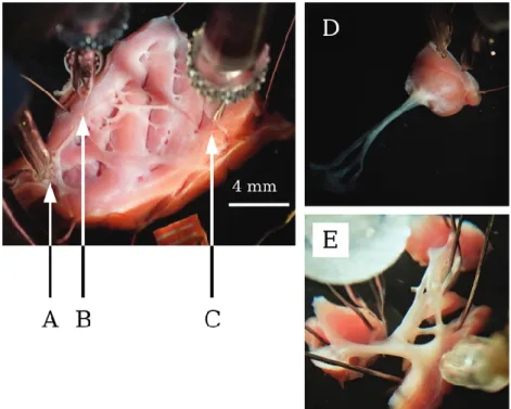
Preparations, containing free-running PFs and VM 25–35 mm in diameter and 2–4 mm in thickness (electrotonically coupled preparations from the left ventricle) (
F1 Figs. 1A–1C) and individual
PFs (Fig. 1E) with small muscle endings and individual papillary VM (Figs. 1D, electrotonically uncoupled preparations from both of the left and right ventricle) were obtained and individually mounted in a tissue chamber with a volume of 50 mL. Electro- tonically coupled preparations were paced from a PF, mimicking physiological cardiac conduction. Stimulation was executed using a pair of platinum electrodes in contact with the prepara- tion using rectangular current pulses of 0.5–2 ms duration. These stimuli were delivered at a constant cycle length of 1000 ms for at
least 60 min allowing the preparation to equilibrate before the measurements were initiated. Transmembrane potentials were simultaneously recorded from PF and subendocardial VM using conventional glass microelectrodes (Fig. 1)filled with 3 mol/L KCl and having tip resistances of 5–20 MXconnected to the input of a high-impedance electrometer (Experimetria, type 309, Budapest, Hungary), which was coupled to a dual-beam oscilloscope. The resting potential (RP), action potential amplitude (APA), maxi- mum upstroke velocity (Vmax), and APD measured at 50% and 90%
of repolarization (APD50 and APD90, respectively) were online monitored and offline recorded using a home-made software (APES) running on a computer equipped with an ADA 3300 ana- log-to-digital data acquisition board (Real Time Devices, Inc., State College, Pennsylvania) having a maximum sampling fre- quency of 40 kHz. Dispersion of repolarization was inferred from the difference of APD90values of PFs and VM, referred to as DAPD90. Stimulation with a constant cycle length of 1000 ms was applied in the course of all experiments. Attempts were made to maintain the same impalement throughout each experiment. In case an impalement became dislodged, adjustment was attempted, and if the action potential characteristics of the reestablished impalement deviated by less than 5% from the previous measure- ment, the experiment continued (Lengyel et al. 2001;Jost et al.
2005; Orvos et al. 2015,2019). All measurements were carried out at
AQ3
37 °C.
Statistical analysis
All data are expressed as means6SEM. The“n”number refers to the number of experiments. Depending on the type of compar- ison, Student’sttest was used either for independent samples (amiodarone) or for paired samples (dofetilide). The results were considered statistically significant whenpwas<0.05.
Results
Comparison of baseline electrophysiology of electrotonically coupled and uncoupled preparations
In electrotonically coupled control preparations (Tables 1and2; T1,T2 F2-F3 Figs. 2Aand3A) most action potential characteristics both of PF
and VM were comparable to those of individual PF and VM
(uncoupled) preparations (n= 21) (Tables 3and4;Figs. 2Cand3C). T3,T4 VM APD was slightly longer in coupled preparations compared to
the uncoupled preparations (Tables 3and4;Figs. 2Cand3C), while PF APD was slightly shorter in coupled preparations. APA, Vmax, and RP were similar to those of uncoupled (individual) prepara- tions. Baseline dispersion (DAPD90) was 39.664.0 ms (pooled con- trols, n = 21) in coupled control groups of the drug studies.
Conduction time (CT) to PFs was shorter in all preparations than that of VM, confirming an anterograde wave of depolarization.
Since individual, electrotonically uncoupled PF and VM prepara- tions were not necessarily taken from the same heart and were not in connection, differences in their APD cannot be directly meas- ured, but average values showed an APD difference of 72.8 ms between the two groups of preparations under control circumstan- ces (pooled controls,n= 13 and 16).
Effects of amiodarone
Electrotonically coupled preparations obtained from animals after chronic amiodarone treatment (n= 11) did not show statisti- cally significant changes in the RP, APA, andVmax. Amiodarone treatment increased APD90and APD75values of PF potentials (p<
0.01) (Table 1;Fig. 2B) while eliciting no effect on the early phases of repolarization. In VM, prolongation was measured in all stages of repolarization, from APD10to APD90(p<0.01). The prolonga- tion of AP duration in VM was more pronounced than in PFs;
thus,DAPD90decreased substantially (18.065.0 ms vs. 45.765.7 ms,p<0.01). APA andVmaxof VM remained unchanged compared to the control.
In uncoupled PF preparations, APD90and APD75were increased (p < 0.05); the prolongation was more pronounced than in coupled preparations (Table 3;Fig. 2D). In uncoupled VM prepara- tions, APD90and APD50were also increased (p<0.01), although this change was less pronounced compared to the VM measure- ments from coupled preparations. These effects reflected an im- portant difference between coupled and uncoupled preparations in response to chronic amiodarone treatment. Accordingly, in uncoupled preparations, amiodarone increased APD without sig- nificantly changing dispersion of repolarization between PF and VM, measured asDAPD90. On the contrary, in coupled preparations,
amiodarone increased APD in such a manner to result in a signifi- cant decrease of dispersion of repolarization, measured asDAPD90. Effects of dofetilide
In coupled preparations, acutely administered dofetilide (n= 8, 50 nM) induced a marked increase in APD90, APD75 and APD50 (p<0.001) values in PF compared to control measurements (Table 2, Fig. 3B). In VM, APD90 and APD75 values were also prolonged (p<0.01 andp<0.05 respectively), and APD50 was markedly increased. The more pronounced prolongation of AP duration in PFs led to an increase inDAPD90 to 75.2612.6 ms from 47.0611.1 ms Table 1. The electrophysiological effects of 50 mg·kg–1·day–1amiodarone in electrotonically coupled (“C”) Purkinjefiber (PF) and ventriculat muscle (VM) preparations at a basic cycle length of 1000 ms.
Sample CT (ms) RP (mV) APA (mV) Vmax(V/s) APD90(ms) APD50(ms) APD90difference (ms) Control PF-C (14) 5.260.7 –88.463.2 119.262.6 515.6643.2 259.865.5 170.965.7 41.065.6
Amiodarone (50 mg·kg–1·day–1) PF-C (11) 4.060.6 –84.662 120.762.8 516.2667.2 305.965.8### 184.8610.9 18.065##
Control VM-C (14) 13.561.2 –85.861.6 110.162.9 201.3627.9 218.867.6 165.065.5 Amiodarone (50 mg·kg–1·day–1) VM-C (11) 12.761.6 –87.862.7 106.863.8 150.4646.1 288.065.9### 218.267.6###
Note:CT, conduction time; RP, resting potential; APA, action potential amplitude;Vmax, maximum rate of depolarization; APD90and APD50, action potential durations at 90% and 50% of repolarization. Results are means6SEM.##p<0.01,###p<0.001, Student’sttest for unpaired data.
Table 2.The electrophysiological effects of 50 nmol/L dofetilide in electrotonically coupled (“C”) Purkinjefiber (PF) and ventriculat muscle (VM) preparations at a basic cycle length of 1000 ms.
Sample CT (ms) RP (mV) APA (mV) Vmax(V/s) APD90(ms) APD50(ms) APD90difference (ms)
Control PF-C (7) 6.560.9 –90.563.8 123.164.7 369.2655 265.064.4 203.464.4 37.064.3
Dofetilide (50 nmol/L) PF-C (7) 7.161.2 –87.463.3 122.265.5 350.4651.1 333.968.8*** 244.668.7*** 67.2610.3**
Control VM-C (7) 15.460.8 –82.863.7 110.664.5 169.9618.5 228.665.7 175.965.6 Dofetilide (50 nmol/L) VM-C (7) 16.161.6 –80.963.2 108.662.8 178.4633 261.3611.5* 188.9611
Note:CT, conduction time; RP, resting potential; APA, action potential amplitude;Vmax, maximum rate of depolarization; APD90and APD50, action potential durations at 90% and 50% of repolarization. Results are means6SEM. *p<0.05, **p<0.01, ***p<0.001, Student’sttest for paired data.
Fig. 1. Photographs of an electrotonically coupled ventricular preparation. (A) Pacing microelectrode; (B) microelectrode impaled in a Purkinjefiber; (C) microelectrode impaled in a ventricular muscle); (D) an uncoupled papillary muscle; (E) an uncoupled Purkinjefiber inside the tissue bath. [Colour online.]
(p<0.01). APA andVmaxdid not change. Conduction times (CTs) slightly increased after dofetilide treatment.
In uncoupled PF preparations (n = 6–6),
AQ4
APD90, APD75, andAPD50were also prolonged after dofetilide treatment (p<0.001), but prolongation was more pronounced compared to the change in coupled preparations (Table 4;Fig. 3D). In uncoupled VM prep- arations, APD90to APD25were all prolonged after dofetilide treat- ment (p<0.01). Even though the change in uncoupled VM APD90
is comparable to that of the coupled preparations, the AP of PFs showed a more pronounced prolongation, unlike with that of amiodarone treatment, indicated by the greatly increased differ- ence in average APD90values.
Discussion
Under control conditions, PF APDs were shorter in coupled preparations compared to uncoupled preparations, while VM APDs were longer when coupled with PF, as indicated in the con- trol values ofTables 1and2, when compared to those ofTables 3
and4. APs recorded from canine transitional cells in the PMJs have been previously described to have longer APD than VM and slower maximum rate of depolarization than PF but limited to the immediate surroundings of the PMJ (Martinez-Palomo et al.
1970), suggesting that the slight prolongation we observed in VM and abbreviation in PFs when measured in electrotonically coupled preparations may be attributable to the electrotonic interaction between VM and PFs. This is not the case in dissected ventricular and Purkinje preparations (i.e., individual PFs, papil- lary muscle, or trabecule). Therefore, these latter preparations are not affected by electrotonic coupling, which also leads to an exaggerated difference in APD; thus, when calculating dispersion from individual, uncoupled preparations, dispersion is likely to be overestimated.
Prolongation of APs in individual VM preparations has been previously reported after chronic amiodarone treatment, while PF has been not changed or shortened. Thus, the differences in APD have been decreased between PF and VM in uncoupled Fig. 2. The effect of chronic amiodarone (50 mg·kg–1·day–1) in (A and B) coupled and (C and D) uncoupled action potentials. Solid lines
represent Purkinjefiber potentials and dotted lines represent ventricular action potentials; stimulation frequency was 1 Hz. [Colour online.]
preparations (Papp et al. 1996). In our experiments, amiodarone was found to prolong APD of PF significantly in electrotonically coupled, but not in uncoupled, preparations, as seen inFig. 2, and in uncoupled preparations resulted
AQ5
in no difference betweenthe APD90 of PFs and VM. However, after chronic amiodarone treatment in electrotonically coupled preparations, we observed slight to moderate prolongation of PF repolarization accompa- nied by a much more pronounced prolongation of VM, leading to a decrease of dispersion of repolarization, reflected as lower DAPD90(
F4 Fig. 4).
The effect of dofetilide in uncoupled cardiac PF and VM prepa- rations is well documented (Gwilt et al. 1991;Knilans et al. 1991;
Bányász et al. 2009), but such measurements have not been reported in ex vivo coupled preparations. Therefore, in this study, direct comparison between coupled and uncoupled prepa- rations was possible after dofetilide administration. In coupled preparations, dofetilide increased DAPD90 by causing a much greater prolongation in PFs than in VM (Table 2). This difference
in mean APD90values was further increased in uncoupled prepa- rations, as seen inTable 4.
The different changes in dispersion may be partially explained by the different effects of each drug on ion channels: dofetilide is considered as a selective inhibitor of the delayed rectifier out- ward potassium current (IKr) (Carmeliet 1992;Kiehn et al. 1994;
Mounsey and DiMarco 2000), while amiodarone also inhibits inward currents, such asINaLandICaL(Follmer et al. 1987;Kodama et al. 1996;Nishimura et al. 1989), and outward currents, such as IKs, apart fromIKr(Balser et al. 1991;Bertran et al. 1998;Kodama et al. 1996;Sato et al. 1994;Varró et al. 1996). SinceINaLis consid- ered more prominent in PFs than in VM (Baláti et al. 1998;Haufe et al. 2005), blockingINaLby amiodarone would limit APD length- ening in PFs more than in VM, resulting in less dispersion of repolarization.
In this work, we did not study the possible role of calcium sig- naling in either tissue type. Nevertheless, it is worth mentioning that in previous studies performed in uncoupled PF and VM Fig. 3. The effect of dofetilide (50 nmol/L) in (A and B) coupled and (C and D) uncoupled action potentials. Solid lines represent Purkinje fiber potentials and dotted lines represent ventricular action potentials; stimulation frequency was 1 Hz. [Colour online.]
preparations, amiodarone abolished EADs and delayed afterde- polarizations (Varró et al. 2001). On the contrary, several studies have shown that dofetilide evoked EADs in various cardiac prepa- rations (Horváth et al. 2015;Nalos et al. 2012;Fedida et al. 2006).
AQ6
In some preliminary, additional experiments, we found that in coupled preparations, dofetilide evoked EADs only when admin- istered in combination with CsCl and Bay K8644, i.e., a situation where repolarization reserve had been previously attenuated and calcium current had been activated. Therefore, understand- ing calcium signaling in electrotonically coupled preparations should be an aim of further studies.
The extent of AP prolongation elicited by each drug also varies between the electronically coupled and uncoupled preparations.
Dofetilide elicited a more pronounced prolongation in uncoupled PFs compared to changes measured when coupled with VM. This can be witnessed when comparingFigs. 3Band3D. PF prolongation caused by chronic amiodarone treatment was similar in coupled and uncoupled conditions, but VM prolongation was more pro- nounced in coupled preparations (Fig. 4). These differences may be explained by the undisturbed electrotonic coupling between the PFs and the subendocardial VM, since this interaction may lead to a slight decrease in the measured APD of PFs and slight increase in that of VM, partially evening out the distinct difference in APDs measured when the conduction system and the myocardium are dissected. Accordingly, the extent of PF prolongation caused by dofetilide may partially be modulated by the neighboring ventricu- lar muscle, and the prolongation of VM caused by amiodarone may be, in a similar manner, potentiated by the interaction with PFs.
Thus, our data from coupled and uncoupled preparations suggest that the electrotonic interaction between PFs and subendocardial myocardium affects not only the baseline electrophysiology of tis- sues studied using the conventional microelectrode technique but also the measured effects elicited by antiarrhythmic agents.
Therefore, amiodarone ex vivo decreased not only transmural dispersion, previously demonstrated on canine (Sicouri et al.
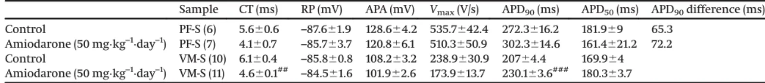
1997) and human (Drouin et al. 1998) preparations, but also between PFs and VM. In the clinical setting, the QT/QTc prolonga- tion increase is associated with an increased risk of VT/VF. How- ever, drugs that not only increase QT/QTc but also decrease the dispersion of QT/QTc or JT are accompanied by a lower proar- rhythmic risk (e.g., amiodarone), while agents that prolong QT/QTc without a decrease in dispersion (e.g., quinidine) have a higher risk of arrhythmic events (Cui et al. 1994;Antzelevitch et al. 1998). It has also been reported that dofetilide increases Table 3. The electrophysiological effects of 50 mg·kg–1·day–1amiodarone in uncoupled (“S”) Purkinjefiber (PF) and ventricular muscle (VM) preparations at a basic cycle length of 1000 ms.
Sample CT (ms) RP (mV) APA (mV) Vmax(V/s) APD90(ms) APD50(ms) APD90difference (ms)
Control PF-S (6) 5.660.6 –87.661.9 128.664.2 535.7642.4 272.3616.2 181.969 65.3
Amiodarone (50 mg·kg–1·day–1) PF-S (7) 4.160.7 –85.763.7 120.866.1 510.3650.9 302.3614.6 161.4621.2 72.2
Control VM-S (10) 6.160.4 –85.860.8 108.263.2 238.9630.9 20764.4 169.964
Amiodarone (50 mg·kg–1·day–1) VM-S (11) 4.660.1## –84.561.6 101.962.6 173.9613.7 230.163.6### 180.363.7
Note:CT, conduction time; RP, resting potential; APA, action potential amplitude;Vmax, maximum rate of depolarization; APD90and APD50, action potential durations at 90% and 50% of repolarization. Results are means6SEM.##p<0.01,
AQ9
###p<0.001, Student’sttest for unpaired data.Table 4.The electrophysiological effects of 50 nM dofetilide in uncoupled (“S”) Purkinjefiber (PF) and ventricular muscle (VM) preparations at a basic cycle length of 1000 ms.
Sample CT (ms) RP (mV) APA (mV) Vmax(V/s) APD90(ms) APD50(ms) APD90difference (ms) Control PF-S (7) 5.560.6 –87.261.8 133.562.6 487.3642.3 280623.5 181.9618.5 81.3
Dofetilide (50 nmol/L) PF-S (7) 6.160.8 –88.861.6 134.662.1 446.9640.5 409.9612*** 252.769.6*** 168.1
Control VM-S (6) 6.260.9 –86.262.4 116.763.8 192620.3 198.763.1 163.863
Dofetilide (50 nmol/L) VM-S (6) 6.360.76 –84.662.5 120.663 195.7622.8 241.866.6** 196.666.3**
Note:CT, conduction time; RP, resting potential; APA, action potential amplitude;Vmax, maximum rate of depolarization; APD90and APD50, action potential durations at 90% and 50% of repolarization. Results are means6SEM. **p<0.01, ***p<0.001, Student’sttest for unpaired
AQ10
data.Fig. 4. The action potential differences between Purkinjefibers (PF) and ventricular muscle (VM) in electrotonically coupled and uncoupled preparations during control conditions after chronic amiodarone treatment (50 mg·kg–1·day–1) and in the presence of dofetilide (50 nmol/L). Bars represent means6SEM. The white areas in the PF bars represent the difference in action potential duration between PF and VM; stimulation frequency was 1 Hz. [Colour online.]
electrical heterogeneity in the human heart (Stabenau et al.
2020). Dofetilide is associated with a higher risk of causing tor- sades than amiodarone (Brendorp et al. 2002). Amiodarone has also been shown to decrease dispersion of monophasic APD90 (Osaka et al. 2011). The decrease in dispersion between the car- diac conductive system and myocardium may also be a benefi- cial action of antiarrhythmic agents, similar to the reduction of transmural dispersion, by decreasing the diversity in refractori- ness between adjacent cardiac regions, consequently decreasing the risk of extra beats propagating by unidirectional block.
Conclusion
This study demonstrated that amiodarone, like dofetilide, lengthened cardiac repolarization but unlike dofetilide, it decreased dispersion of repolarization in a preparation that preserves elec- trotonic coupling between PFs and subendocardial VM. Also, car- diac electrophysiological drug effects can be better established in preparations with preserved electrotonic coupling than in uncoupled tissues. The observed marked differences between the effects of amiodarone and dofetilide on dispersion of repola- rization in both well-coupled and uncoupled PFs and VMfibers provide a further explanation why amiodarone has a signifi- cantly less proarrhythmic risk than dofetilide, in spite of both drugs exerting a similar degree of QT lengthening in patients.
This effect of amiodarone, unlike that of dofetilide, suggests an antiarrhythmic effect without a significant proarrhythmic risk.
In addition, this study highlights the importance of studying dis- persion of repolarization between PFs and VM in well-coupled preparations, since drug effects can be over- and underestimated in uncoupled preparations.
Acknowledgements
This work was funded by the National Research Development and Innovation Office (NKFIH K-119992 and GINOP-2.3.2-15-2016- 00048-STAY ALIVE), the Ministry of Human Capacities Hungary (20391-3/2018/FEKUSTRAT, EFOP-3.6.2-16-2017-00006-LIVE LONGER, and EFOP 3.6.3-VEKOP-16-2017-00009), and the Hungarian Academy of Sciences. The GINOP and EFOP projects are cofinanced by the European Union and the European Regional Development Fund.
References
Antzelevitch, C., Shimizu, W., Yan, G.X., and Sicouri, S. 1998. Cellular basis for QT dispersion. J. Electrocardiol.30(Suppl): 168–175. doi:10.1016/s0022- 0736(98)80070-8. PMID:9535495.
Baláti, B., Varró, A., and Papp, J.G. 1998. Comparison of the cellular electro- physiological characteristics of canine left ventricular epicardium, M cells, endocardium and Purkinje fibres. Acta Physiol. Scand. 164(2): 181–190.
doi:10.1046/j.1365-201X.1998.00416.x. PMID:9805105.
Balser, J.R., Bennett, P.B., Hondeghem, L.M., and Roden, D.M. 1991. Suppres- sion of time-dependent outward current in guinea pig ventricular myo- cytes. Actions of quinidine and amiodarone. Circ. Res.69(2): 519–529.
doi:10.1161/01.res.69.2.519. PMID:1860189.
Bányász, T., Horváth, B., Virág, L., Bárándi, L., Szentandrássy, N., Harmati, G., et al. 2009. Reverse rate dependency is an intrinsic property of canine car- diac preparations. Cardiovasc. Res.84(2): 237–244. doi:10.1093/cvr/cvp213.
PMID:19556280.
Bertran, G.C., Biagetti, M.O., Valverde, E.R., and Quinteiro, R.A. 1998. Effects of amiodarone and desethylamiodarone on the inward rectifying potas- sium current (IK1) in rabbit ventricular myocytes. J. Cardiovasc. Pharma- col.31(6): 914–920. doi:10.1097/00005344-199806000-00016. PMID:9641477.
Brendorp, B., Pedersen, O., Torp-Pedersen, C., Sahebzadah, N., and Køber, L.
2002. A benefit-risk assessment of class III antiarrhythmic agents. Drug Saf.
25(12): 847–865. doi:10.2165/00002018-200225120-00003. PMID:12241126.
Carmeliet, E. 1992. Voltage- and time-dependent block of the delayed K+ cur- rent in cardiac myocytes by dofetilide. J. Pharmacol. Exp. Ther.262(2):
809–817. PMID:1501123.
Cui, N., Sager, N., Singh, N., and Sen, N. 1998. Different effects of amioda- rone and quinidine on the homogeneity of myocardial refractoriness in patients with intraventricular conduction delay. J. Cardiovasc. Pharma- col. Ther.3(3): 201–208. doi:10.1177/107424849800300301. PMID:10684498.
Cui, G., Sen, L., Sager, P., Uppal, P., and Singh, B.N. 1994. Effects of amioda- rone, sematilide, and sotalol on QT dispersion. Am. J. Cardiol.74(9): 896–
900. doi:10.1016/0002-9149(94)90582-7. PMID:7526675.
Drouin, E., Lande, G., and Charpentier, F. 1998. Amiodarone reduces transmural heterogeneity of repolarization in the human heart. J. Am. Coll. Cardiol.
32(4): 1063–1067. doi:10.1016/s0735-1097(98)00330-1. PMID:9768733.
Fedida, D., Orth, P.M.R., Hesketh, J.C., and Ezrin, A.M. 2006. The role of late I and antiarrhythmic drugs in EAD formation and termination in Purkinjefibers.
J. Cardiovasc. Electrophysiol.17(s1): S71–S78. doi:10.1111/j.1540-8167.2006.00386.x.
PMID:16686685.
Follmer, C.H., Aomine, M., Yeh, J.Z., and Singer, D.H. 1987. Amiodarone- induced block of sodium current in isolated cardiac cells. J. Pharmacol.
Exp. Ther.243(1): 187–194. PMID:2444698.
Gilmour, R.F., and Watanabe, M. 1994. Dynamics of circus movement re- entry across canine Purkinjefibre-muscle junctions. J. Physiol.476(3):
473–485.doi:10.1113/jphysiol.1994.sp020148. PMID:8057255.
AQ7
Glukhov, A.V., Fedorov, V.V., Lou, Q., Ravikumar, V.K., Kalish, P.W., Schuessler, R.B., et al. 2010. Transmural dispersion of repolarization in failing and nonfailing human ventricle. Circ. Res. 106(5): 981–991.
AQ8
doi:10.1161/CIRCRESAHA.109.204891. PMID:20093630.
Gwilt, M., Arrowsmith, J.E., Blackburn, K.J., Burges, R.A., Cross, P.E., Dalrymple, H.W., and Higgins, A.J. 1991. UK-68,798: a novel, potent and highly selective class III antiarrhythmic agent which blocks potassium channels in cardiac cells. J. Pharmacol. Exp. Ther.256(1): 318–324. PMID:
1988662.
Haufe, V., Cordeiro, J.M., Zimmer, T., Wu, Y.S., Schiccitano, S., Benndorf, K., and Dumaine, R. 2005. Contribution of neuronal sodium channels to the car- diac fast sodium current INa is greater in dog heart Purkinjefibers than in ventricles. Cardiovasc. Res. 65(1): 117–127. doi:10.1016/j.cardiores.2004.08.017.
PMID:15621039.
Horváth, B., Hegyi, B., Kistamás, K., Váczi, K., Bányász, T., Magyar, J., et al.
2015. Cytosolic calcium changes affect the incidence of early afterdepola- rizations in canine ventricular myocytes. Can. J. Physiol. Pharmacol.
93(7): 527–534. doi:10.1139/cjpp-2014-0511. PMID:25928391.
Jost, N., Virág, L., Bitay, M., Takács, J., Lengyel, C., Biliczki, P., et al. 2005. Restrict- ing Excessive Cardiac Action Potential and QT Prolongation. Circulation, 112(10): 1392–1399. doi:10.1161/CIRCULATIONAHA.105.550111. PMID:16129791.
Kiehn, J., Villena, P., Beyer, T., and Brachmann, J. 1994. Differential effects of the new class III agent dofetilide on potassium currents in guinea pig cardiomyocytes. J. Cardiovasc. Pharmacol. 24(4): 566–572. doi:10.1097/
00005344-199410000-00007. PMID:7528839.
Knilans, T.K., Lathrop, D.A., Nánási, P.P., Schwartz, A., and Varró, A. 1991.
Rate and concentration-dependent effects of UK-68,798, a potent new class III antiarrhythmic, on canine Purkinjefibre action potential dura- tion andVmax. Br. J. Pharmacol.103(2): 1568–1572. doi:10.1111/j.1476-5381.
1991.tb09828.x. PMID:1884111.
Kodama, I., Kamiya, K., Honjo, H., and Toyama, J. 1996. Acute and chronic effects of amiodarone on mammalian ventricular cells. Jpn. Heart J.37(5): 719–730.
doi:10.1536/ihj.37.719. PMID:8973384.
Lengyel, C., Iost, N., Virág, L., Varró, A., Lathrop, D.A., and Papp, J.G. 2001.
Pharmacological block of the slow component of the outward delayed rectifier current (IKs) fails to lengthen rabbit ventricular muscle QTc and action potential duration. Br. J. Pharmacol.132(1): 101–110. doi:10.1038/sj.
bjp.0703777. PMID:11156566.
Martinez, M.E., Walton, R.D., Bayer, J.D., Haïssaguerre, M., Vigmond, E.J., Hocini, M., and Bernus, O. 2018. Role of the purkinje-muscle junction on the ventricular repolarization heterogeneity in the healthy and ischemic ovine ventricular myocardium. Front. Physiol. 9: 718. doi:10.3389/fphys.2018.00718.
PMID:29962961.
Martinez-Palomo, A., Alanis, J., and Benitez, D. 1970. Transitional cardiac cells of the conductive system of the dog heart. Distinguishing morphological and electrophysiological features. J. Cell Biol.47(1): 1–17. doi:10.1083/jcb.47.1.1.
PMID:5513552.
Mounsey, J.P., and DiMarco, J.P. 2000. Dofetilide. Circulation,102(21): 2665–
2670. doi:10.1161/01.CIR.102.21.2665. PMID:11085972.
Nalos, L., Varkevisser, R., Jonsson, M.K.B., Houtman, M.J.C., Beekman, J.D., van der Nagel, R., et al. 2012. Comparison of the IKr blockers moxifloxa- cin, dofetilide and E-4031 in five screening models of pro-arrhythmia reveals lack of specificity of isolated cardiomyocytes. Br. J. Pharmacol.
165(2): 467–478. doi:10.1111/j.1476-5381.2011.01558.x. PMID:21718297.
Nattel, S., and Quantz, M.A. 1988. Pharmacological response of quinidine induced early afterdepolarisations in canine cardiac Purkinje fibres:
insights into underlying ionic mechanisms. Cardiovasc. Res.22(11): 808–
817. doi:10.1093/cvr/22.11.808. PMID:3256422.
Nishimura, M., Follmer, C.H., and Singer, D.H. 1989. Amiodarone blocks cal- cium current in single guinea pig ventricular myocytes. J. Pharmacol.
Exp. Ther.251(2): 650–659. PMID:2553932.
Nogami, A. 2011a. Purkinje-related arrhythmias Part I: Monomorphic ventricu- lar tachycardias. Pacing Clin. Electrophysiol.34(5): 624–650. doi:10.1111/j.1540- 8159.2011.03044.x. PMID:21410719.
Nogami, A. 2011b. Purkinje-related arrhythmias Part II: Polymorphic ventric- ular tachycardia and ventricularfibrillation. Pacing Clin. Electrophysiol.
34(8): 1034–1049. doi:10.1111/j.1540-8159.2011.03145.x. PMID:21671950.
Orvos, P., Kohajda, Z., Szlovák, J., Gazdag, P., Árpádffy-Lovas, T., Tóth, D., et al. 2019. Evaluation of possible proarrhythmic potency: comparison of
the effect of dofetilide, cisapride, sotalol, terfenadine, and verapamil on hERG and native IKr currents and on cardiac action potential. Toxicol.
Sci.168(2): 365–380. doi:10.1093/toxsci/kfy299. PMID:30561737.
Osaka, T., Yokoyama, E., Hasebe, H., and Kodama, I. 2011. Effects of chronic amiodarone on the electrical restitution in the human ventricle with ref- erence to its antiarrhythmic efficacy. J. Cardiovasc. Electrophysiol.22(6):
669–676. doi:10.1111/j.1540-8167.2010.01990.x. PMID:21235669.
Papp, J.G., Németh, M., Krassói, I.I., Mester, L., Hála, O., and Varró, A. 1996.
Differential electrophysiologic effects of chronically administered amio- darone on canine purkinjefibers versus ventricular muscle. J. Cardiovasc.
Pharmacol. Ther. 1(4): 287–296. doi:10.1177/107424849600100404. PMID:
10684429.
Sato, R., Koumi, S., Singer, D.H., Hisatome, I., Jia, H., Eager, S., and Wasserstrom, J.A. 1994. Amiodarone blocks the inward rectifier potas- sium channel in isolated guinea pig ventricular cells. J. Pharmacol. Exp.
Ther.269(3): 1213–1219. PMID:8014865.
Sicouri, S., Moro, S., Litovsky, S., Elizari, M.V., and Antzelevitch, C. 1997.
Chronic amiodarone reduces transmural dispersion of repolarization in the canine heart. J. Cardiovasc. Electrophysiol.8(11): 1269–1279. doi:10.1111/j.1540- 8167.1997.tb01018.x. PMID:9395170.
Stabenau, H.F., Shen, C., Tereshchenko, L.G., and Waks, J.W. 2020. Changes in global electrical heterogeneity associated with dofetilide, quinidine, ranolazine, and verapamil. Heart Rhythm,17(3): 460–467. doi:10.1016/j.
hrthm.2019.09.017. PMID:31539628.
Varró, A., Lathrop, D.A., and Surawicz, B. 1990. Effects of propranolol on premature action potentials in canine Purkinje and ventricular muscle.
J. Cardiovasc. Pharmacol.16(5): 757–763. doi:10.1097/00005344-199011000- 00010. PMID:1703597.
Varró, A., Takács, J., Németh, M., Hála, O., Virág, L., Iost, N., et al. 2001. Elec- trophysiological effects of dronedarone (SR 33589), a noniodinated amio- darone derivative in the canine heart: comparison with amiodarone. Br. J.
Pharmacol.133(5): 625–634. doi:10.1038/sj.bjp.0704106. PMID:11429385.
Varró, A., Virág, L., and Papp, J.G. 1996. Comparison of the chronic and acute effects of amiodarone on the calcium and potassium currents in rabbit isolated cardiac myocytes. Br. J. Pharmacol. 117(6): 1181–1186.
doi:10.1111/j.1476-5381.1996.tb16713.x. PMID:8882613.
Walton, R.D., Martinez, M.E., Bishop, M.J., Hocini, M., Haïssaguerre, M., Plank, G., et al. 2014. Influence of the Purkinje-muscle junction on trans- mural repolarization heterogeneity. Cardiovasc. Res. 103(4): 629–640.
doi:10.1093/cvr/cvu165. PMID:24997066.