.. ..
.. ..
.. ..
.. ..
.. ..
.. ..
.. ..
.. ..
.. ..
Cardiac innervation in acute myocardial
ischaemia/reperfusion injury and cardioprotection
Derek J. Hausenloy
1,2,3,4,5,6*, Hans Erik Bøtker
7, Peter Ferdinandy
8,9, Gerd Heusch
10, G. Andre´ Ng
11, Andrew Redington
12,13, and David Garcia-Dorado
14,15*; on behalf of the EU-CARDIOPROTECTION COST Action (CA16225)
1Cardiovascular & Metabolic Disorders Program, Duke-National University of Singapore Medical School, Singapore;2National Heart Research Institute Singapore, National Heart Centre, Singapore;3Yong Loo Lin School of Medicine, National University Singapore, Singapore;4The Hatter Cardiovascular Institute, University College London, London, UK;5The National Institute of Health Research University College London Hospitals Biomedical Research Centre, Research & Development, London, UK;6Tecnologico de Monterrey, Centro de Biotecnologia-FEMSA, Nuevo Leon, Mexico;7Department of Cardiology, Aarhus University Hospital, Aarhus N, Denmark;8Department of Pharmacology and Pharmacotherapy, Semmelweis University, Budapest, Hungary;9Pharmahungary Group, Szeged, Hungary;10Institute for Pathophysiology, West German Heart and Vascular Center, University of Essen Medical School, Essen, Germany;11Department of Cardiovascular Sciences, University of Leicester, NIHR Leicester Biomedical Research Centre, Glenfield Hospital, LE3 9QP, UK;
12Cincinnati Children’s Hospital Medical Center, Heart Institute, Cincinnati, OH, USA;13Department of Pediatrics, University of Cincinnati College of Medicine, Cincinnati, OH, USA;
14Department of Cardiology, Vascular Biology and Metabolism Area, Vall d’Hebron University Hospital and Research Institute (VHIR), Universitat Auto´noma de Barcelona, Spain;
15Instituto CIBER de Enfermedades Cardiovasculares (CIBERCV): Instituto de Salud Carlos III, Madrid, Spain
Received 20 November 2018; revised 21 December 2018; editorial decision 15 January 2019; accepted 21 February 2019; online publish-ahead-of-print 23 February 2019
Abstract Acute myocardial infarction (AMI) and the heart failure (HF) that often complicates this condition, are among the leading causes of death and disability worldwide. To reduce myocardial infarct (MI) size and prevent heart failure, novel therapies are required to protect the heart against the detrimental effects of acute ischaemia/reperfusion in- jury (IRI). In this regard, targeting cardiac innervation may provide a novel therapeutic strategy for cardioprotection.
A number of cardiac neural pathways mediate the beneficial effects of cardioprotective strategies such as ischaemic preconditioning and remote ischaemic conditioning, and nerve stimulation may therefore provide a novel therapeu- tic strategy for cardioprotection. In this article, we provide an overview of cardiac innervation and its impact on acute myocardial IRI, the role of extrinsic and intrinsic cardiac neural pathways in cardioprotection, and highlight pe- ripheral and central nerve stimulation as a cardioprotective strategy with therapeutic potential for reducing MI size and preventing HF following AMI. This article is part of a Cardiovascular Research Spotlight Issue entitled
‘Cardioprotection Beyond the Cardiomyocyte’, and emerged as part of the discussions of the European Union (EU)-CARDIOPROTECTION Cooperation in Science and Technology (COST) Action, CA16225.
䊏 䊏 䊏 䊏 䊏 䊏 䊏 䊏 䊏 䊏 䊏 䊏 䊏 䊏 䊏 䊏 䊏 䊏 䊏 䊏 䊏 䊏 䊏 䊏 䊏 䊏 䊏 䊏 䊏 䊏 䊏 䊏 䊏 䊏 䊏 䊏 䊏 䊏 䊏 䊏 䊏 䊏 䊏 䊏 䊏 䊏 䊏 䊏 䊏 䊏 䊏 䊏 䊏 䊏 䊏 䊏 䊏 䊏 䊏 䊏 䊏 䊏 䊏 䊏 䊏 䊏 䊏 䊏 䊏 䊏 䊏 䊏 䊏 䊏 䊏 䊏 䊏 䊏 䊏 䊏 䊏 䊏 䊏 䊏 䊏 䊏 䊏 䊏 䊏 䊏 䊏 䊏 䊏 䊏 䊏 䊏 䊏 䊏 䊏 䊏 䊏 䊏 䊏 䊏 䊏 䊏 䊏 䊏 䊏 䊏 䊏 䊏 䊏 䊏 䊏 䊏 䊏 䊏 䊏 䊏 䊏 䊏 䊏 䊏 䊏 䊏 䊏 䊏 䊏 䊏 䊏 䊏 䊏 䊏 䊏 䊏 䊏 䊏 䊏 䊏 䊏 䊏 䊏 䊏 䊏 䊏 䊏 䊏 䊏 䊏 䊏 䊏 䊏 䊏 䊏 䊏 䊏 䊏 䊏 䊏 䊏 䊏 䊏 䊏 䊏 䊏 䊏 䊏 䊏 䊏 䊏 䊏 䊏 䊏 䊏 䊏 䊏 䊏 䊏 䊏 䊏 䊏 䊏 䊏 䊏 䊏 䊏 䊏 䊏 䊏 䊏 䊏 䊏 䊏 䊏 䊏 䊏 䊏 䊏 䊏 䊏 䊏 䊏 䊏 䊏 䊏 䊏 䊏 䊏 䊏 䊏 䊏
Keywords Ischaemia/reperfusion injury
•
Myocardial infarction•
Nervous system•
Cardioprotection䊏 䊏 䊏 䊏 䊏 䊏 䊏 䊏 䊏 䊏 䊏 䊏 䊏 䊏 䊏 䊏 䊏 䊏 䊏 䊏 䊏 䊏 䊏 䊏 䊏 䊏 䊏 䊏 䊏 䊏 䊏 䊏 䊏 䊏 䊏 䊏 䊏 䊏 䊏 䊏 䊏 䊏 䊏 䊏 䊏 䊏 䊏 䊏 䊏 䊏 䊏 䊏 䊏 䊏 䊏 䊏 䊏 䊏 䊏 䊏 䊏 䊏 䊏 䊏 䊏 䊏 䊏 䊏 䊏 䊏 䊏 䊏 䊏 䊏 䊏 䊏 䊏 䊏 䊏 䊏 䊏 䊏 䊏 䊏 䊏 䊏 䊏 䊏 䊏 䊏 䊏 䊏 䊏 䊏 䊏 䊏 䊏 䊏 䊏 䊏 䊏 䊏 䊏 䊏 䊏 䊏 䊏 䊏 䊏 䊏 䊏 䊏 䊏 䊏 䊏 䊏 䊏 䊏 䊏 䊏 䊏 䊏 䊏 䊏 䊏 䊏 䊏 䊏 䊏 䊏 䊏 䊏 䊏 䊏 䊏 䊏 䊏 䊏 䊏 䊏 䊏 䊏 䊏 䊏 䊏 䊏 䊏 䊏 䊏 䊏 䊏 䊏 䊏 䊏 䊏 䊏 䊏 䊏 䊏 䊏 䊏 䊏 䊏 䊏 䊏 䊏 䊏 䊏 䊏 䊏 䊏 䊏 䊏 䊏 䊏 䊏 䊏 䊏 䊏 䊏 䊏 䊏 䊏 䊏 䊏 䊏 䊏 䊏 䊏 䊏 䊏 䊏 䊏 䊏 䊏 䊏 䊏 䊏 䊏 䊏 䊏 䊏 䊏 䊏 䊏 䊏 䊏 䊏 䊏 䊏 䊏 䊏
This article is part of the Spotlight Issue on Cardioprotection Beyond the Cardiomyocyte.
1. Introduction
Acute myocardial infarction (AMI) and the heart failure that often compli- cates this condition, are among the leading causes of death and disability worldwide. To reduce myocardial infarct (MI) size and prevent heart fail- ure, novel therapies are required to protect the heart against acute ischae- mia/reperfusion injury (IRI). In this regard, the dense cardiac network of parasympathetic and sympathetic nerves and their interactions with the in- trinsic cardiac nerve system (ICNS), may provide novel targets for cardio- protection. This cardiac neural network influences myocardial rhythm and contractile function, and the susceptibility to acute IRI. It also contributes to cardioprotective strategies such as ischaemic preconditioning (IPC) and
remote ischaemic conditioning (RIC). In this article, we provide an over- view of cardiac innervation with a focus on acute myocardial IRI, the role of extrinsic and intrinsic cardiac innervation in cardioprotection, and highlight peripheral and central nerve stimulation as a cardioprotective strategy with therapeutic potential for improving clinical outcomes in AMI patients.
2. An overview of the cardiac neural network
The heart is innervated by a complex interacting hierarchal network of neural pathways within the central nervous system (CNS), intrathoracic
* Corresponding authors. E-mail: derek.hausenloy@duke-nus.edu.sg (D.J.H.); E-mail: dgdorado@vhebron.net (D.G.-D.)
Published on behalf of the European Society of Cardiology. All rights reserved.VCThe Author(s) 2019. For permissions, please email: journals.permissions@oup.com.
Downloaded from https://academic.oup.com/cardiovascres/article-abstract/115/7/1167/5364019 by Hungary EISZ Consortium user on 04 December 2019
.. ..
.. ..
.. ..
.. ..
.. ..
.. ..
.. ..
.. ..
.. ..
.. ..
.. ..
.. ..
.. ..
.. ..
.. ..
.. ..
.. ..
.. ..
.. ..
.. ..
.. ..
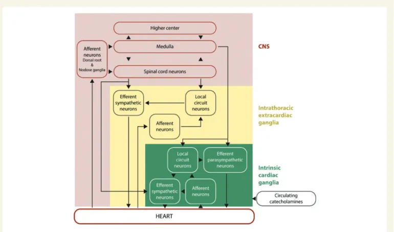
extracardiac ganglia, and intrinsic cardiac ganglia of the ICNS (seeFigure1).1
.
The heart is supplied and controlled by sympathetic and parasympa- thetic nerves, which receive sensory inputs from heart, blood vessels, and other organs. The parasympathetic nerves interface with ganglionic neurons of the ICNS, whereas the sympathetic nerves traverse ganglia without synapsing on ganglionic neurons, and together they provide beat-to-beat regulation of heart rhythm and contractile function.
Sympathetic stimulation increases heart rate and cardiac contractile function through activation of beta-adrenoceptors, and vagal activation reduces heart rate and in some species, cardiac contractile function, through activation of muscarinic receptors (reviewed in references2 and3).
Pre-ganglionic fibres of the parasympathetic nervous system (SNS) arise from the medulla oblongata, and via the vagal nerves, secrete ace- tylcholine (Ach) which binds to the nicotinic Ach receptors on the plasma membrane of post-ganglionic fibres. These in turn secrete Ach, which binds to the type 2 muscarinic Ach receptors present on the plasma membrane of cardiac cells in the sinoatrial (SA) node, atrioven- tricular (AV) node, left ventricle, and to some extent also other parts of the heart, resulting in a reduction in contraction rate of cardiac muscle by shortening its action potential duration (APD) and conduction veloc- ity, by hyperpolarizing SA nodal cells that reduce heart rate.
Within the myocardium there exists an ICNS, comprising cardiac gan- glia and interconnecting neurons (known as ganglionic plexuses), which process sensory information and modulate efferent post-ganglionic para- sympathetic and sympathetic activity within the heart, in the absence of any central modulation (reviewed in reference3). The extrinsic para- sympathetic and sympathetic nerves access the ICNS arterially, around
the roots of the pulmonary artery and aortic root, and interface with the venous portion of the heart around the roots of the pulmonary veins and superior vena cava. The number of cardiac ganglia varies between species from 19 in mice to over 800 in humans, and they are mainly lo- cated on the dorsal atrial surface, around the base of the aorta and pul- monary artery, dorsal and ventral to the pulmonary veins, and on the anterior ventricular surface. From these cardiac ganglia, intrinsic cardiac nerves extend epicardially from ganglionic plexuses to innervate the atria, interatrial septum and the ventricles. A number of neurochemicals shave been found within the ICNS, the presence of which highlight the existence of both parasympathetic and sympathetic nervous compo- nents within the atria and ventricles. The majority of the cardiac ganglia are cholinergic (containing choline acetyltransferase, responsible for the synthesis of acetylcholine) which innervate supraventricular myocardium in and around the sinoatrial and atrioventricular nodes as well as the left ventricle, and these co-exist with both neuronal nitric oxide synthase (nNOS) [responsible for producing nitric oxide (NO)], and vasoactive intestinal peptide. The cardiac ganglia also include adrenergic nerve fibres (containing tyrosine hydroxylase, for the production of noradrenaline) within the left and right coronary subplexuses that innervate the ven- tricles, with which neuropeptide Y (NPY) is co-released. Within the ICNS there are also neuronal subpopulations that are non-adrenergic and non-cholinergic.4Activation of the ICNS can result in local and/or remote cardiac changes with effects on cardiac function and rhythm that are dependent on location. The ganglionic plexuses can modulate post- ganglionic parasympathetic nerve activity and selectively modulate vagal control of heart rate, atrio-ventricular conduction and left ventricular inotropy.5–7 The ganglionic plexuses may also help mediate the
Figure 1Hierarchy of cardiac innervation to the heart. This figure shows the complex and hierarchal interactions between the different components of the neural pathways of the CNS, intrathoracic extracardiac ganglia, and intrinsic cardiac ganglia of the intrinsic cardiac nervous system. This figure has been modified from Armour with permission.1
Downloaded from https://academic.oup.com/cardiovascres/article-abstract/115/7/1167/5364019 by Hungary EISZ Consortium user on 04 December 2019
.. ..
.. ..
.. ..
.. ..
.. ..
.. ..
.. ..
.. ..
.. ..
.. ..
.. ..
.. ..
.. ..
.. ..
.. ..
.. ..
.. ..
.. ..
.. ..
.. ..
.. ..
.. ..
.. ..
.. ..
.. ..
.. ..
.. ..
.. ..
.. ..
.. ..
.. ..
.. ..
.. ..
.. ..
.. ..
.. ..
.. ..
.. ..
.. ..
.. ..
.. ..
.. ..
.. ..
.. ..
.. .
differential effects of sympathetic nerves stimulation of the heart, with nerves arising from the left sympathetic chain influencing LV contractile function and electrical conduction via the AVN to a greater degree than the right, whilst the nerves arising from the right sympathetic chain have a more significant modulator effect on sinus rate via the sinoatrial node.8 The ICNS can respond to a variety of stimuli including acute IRI and influ- ence cardiac function on a beat-to-beat basis and have been implicated in both acute IRI and cardioprotection.9
3. Cardiac innervation and acute myocardial IRA
Sensory nerve endings may detect consequences of acute ischaemia, such as hypoxia, lactate, Kþand low pH, which stimulate cardiac sensory nerves to release their neuropeptide transmitters.3,10 Local afferent function of these sensory nerves may have a strong influence on cardiac function through cardio-cardiac reflexes and initiate adaptive responses due to their NO and vasoactive neuropeptides, such as calcitonin gene-related peptide (CGRP), substance P, somatostatin.10–12Indeed, in selective sensory desensitization by capsaicin, a ligand for TRPV channels, cardiac sensory nerves were demonstrated to strongly influence gene expression patterns in rat hearts,13regulation of the cardiac NO-cGMP system,11SERCA function,14with potential effects on cardiac function.
Moreover, cardiac sensory nerves play a role in acute myocardial injury and adaptation to ischaemic stress,12,15 and in the mechanism of doxorubicin-induced heart failure.16Intact cardiac sensory nerves have been shown to protect against acute IRI-induced cell death via the local release of NO and cytoprotective neuropeptides.12Sympathetic effer- ent nerve terminals release norepinephrine (NE) and exacerbate IRI- induced cardiac cell death directly and indirectly by deterioration of oxy- gen supply and by increasing oxygen demand.2
Cardiac sympathetic afferent denervation attenuates cardiac remodel- ling and improves cardiovascular function in rats with heart failure.17 Modulation of neural networks outside the heart can also impact on post-AMI remodelling with renal nerve denervation preventing adverse post-AMI LV remodelling and preserving vascular function in both spon- taneously hypertensive rats and normotensive rats, effects which were mediated by reduced neprilysin activity and preservation of circulating natriuretic peptide levels.18 Interestingly, blockade of beta- adrenoceptors directly in the brain via chronic intracerebroventricular administration of metoprolol attenuated post-AMI LV remodelling in a rat model of myocardial infarction-induced heart failure, suggesting that the action of certain beta-blockers in the brain could contribute to the beneficial effect of beta-blockers in the failing heart.19
Cardiac sympathetic neurons in the stellate ganglia co-express NPY and the neurotransmitter NE. Following acute myocardial IRI, axonal damage and the inflammatory response to injury, result in suppression of NPY and NE expression, and enhanced expression of neuropeptides such as vasoactive intestinal peptide, substance P, and galanin. Habecker et al.20observed extensive axon damage after AMI, and this was associ- ated with a significant increase in galanin (a peptide which promotes re- generation of sensory neurons21) in cardiac sympathetic neurons in the left ventricle, suggesting the existence of an endogenous protective strat- egy based on neuropeptides in cardiac sympathetic neurons. The suscep- tibility to acute myocardial IRI differs between cardiomyocytes and neurons, and found that cardiac sympathetic neurons are more suscepti- ble to acute myocardial IRI than cardiomyocytes.22
Most cardiac neurons of the ICNS are perivascular, making them sus- ceptible to acute myocardial IRI, thereby setting an environment for neu- ronal remodelling following AMI. In response to acute myocardial IRI, pathological and degenerative changes to the cardiac ganglia occur with the appearance of cytoplasmic inclusions, a feature in common with neu- ronal degeneration disorders. Acute myocardial IRI induces reorganiza- tion and remodelling within ganglionic plexuses of the ICNS in the first 7 days post-AMI, resulting in increased adrenergic sensitivity and en- hanced nNOS expression within parasympathetic post-ganglionic neu- rons within the ICNS.23Pathological features of damaged cardiac nerves include enlargement, and degenerative changes to dendrites and axons, and the appearance of cytoplasmic inclusions.24,25Neuronal remodelling also occurs within regions of non-infarcted myocardium, presumably en- abling a compensatory response in remote myocardium and impacting on post-AMI cardiac remodelling. In this regard, it has been proposed that enhanced nNOS expression plays a protective role, attenuating the initial increase in centrally derived sympathetic activity and facilitating parasympathetic neuronal inputs.23However, the actual interplay be- tween the ICNS and extrinsic vagal or sympathetic nerves in the setting of AMI needs to be further elucidated.
3.1 Protection of cardiac neurons against acute myocardial IRI
The majority of studies investigating cardioprotective strategies for pro- tecting the heart against the detrimental effects of acute IRI have focused on preventing cell death of cardiomyocytes and only few studies have ex- plored the beneficial effects on cardiac neurons. During acute myocardial ischaemia, damage to cardiac sympathetic neurons results in the release of NE into the myocardial interstitial space. IPC can reduce myocardial NE levels following acute myocardial IRI in rat and rabbit hearts.26–28Miura et al.28demonstrated that the detrimental effects of acute myocardial IRI on cardiac sympathetic nerves were reduced by IPC, and the mechanism underlying this neuroprotective effect was attributed to KATP channel opening. The neurotrophin, nerve growth factor (NGF) is known to sup- port survival and differentiation of sympathetic neurons, and is elevated following AMI in a spinal nerve-dependent manner (thoracic epidural an- aesthesia prevented the increase in NGF following AMI).29Strandeet al.30 have shown that NGF administered prior to ischaemia reduced MI size in anin vivorat AMI model, and this effect was mediated through PI3K and NOS. A recent clinical study has shown that limb RIC can reduce the mus- cle sympathetic nerve activity in the forearm induced by acute ischaemia, and this was associated with decreased production of an erythrocyte marker of oxidative stress and the reduction of NO availability, and ame- liorated ischaemic reactive hyperaemia.31Further studies are required to investigate whether cardioprotective strategies can protect cardiac para- sympathetic neurons and the ICNS against the detrimental effects of acute myocardial IRI.
3.2 Cardiac innervation and ventricular arrhythmias following acute myocardial IRA
Ischaemia can directly provoke cardiomyocyte electrical instability, APD heterogeneity, and arrhythmias as a result of ATP depletion, lactate pro- duction, reactive oxygen species, Kþaccumulation, and other substances e.g. endothelin, which enhances the response of perivascular afferent nerves to autonomic reflexes. Some myocardial ischaemic events can be triggered and enhanced by abnormal central autonomic activity such as emotional stress leading to an imbalance in cardiac sympathovagal tone,
Downloaded from https://academic.oup.com/cardiovascres/article-abstract/115/7/1167/5364019 by Hungary EISZ Consortium user on 04 December 2019
.. ..
.. ..
.. ..
.. ..
.. ..
.. ..
.. ..
.. ..
.. ..
.. ..
.. ..
.. ..
.. ..
.. ..
.. ..
.. ..
.. ..
.. ..
.. ..
.. ..
.. ..
.. ..
.. ..
.. ..
.. ..
.. ..
.. ..
.. ..
.. ..
.. ..
.. ..
.. ..
.. ..
.. ..
.. ..
.. ..
.. ..
.. ..
.. ..
.. ..
.. ..
.. ..
.. ..
.. ..
.. .
reflexly increasing cardiac sympathetic activity leading to further coro- nary vasoconstriction.32,33This is often accompanied by vagal withdrawal perpetuating the clinical scenario. Since the ICNS has an integrative role and can exert considerable influence on cardiodynamics, it is possible that significant interaction occurs between the heart’s ‘little brain’3locally and with peripheral nerves that mediate important mechanisms underly- ing arrhythmogenesis during acute myocardial IRI. Vagal control of heart rate and release of Ach in non-ischaemic ventricular regions are both blunted following a developed myocardial infarct,34 showing that re- gional ischaemia can affect non-ischaemic sites both proximal and distal to the insult site.
Preclinical studies support the notion that SNS activity is pro-arrhyth- mic,35,36whilst vagal nerve stimulation is anti-arrhythmic.37High levels of vagal activity exert powerful anti-arrhythmic effects, which can counter the effects of acute ischaemia and sympathetic activation. Mechanisms are complex and include indirect effects of accentuated antagonism against sympathetic activity and direct protective effects on electrophysi- ological parameters, Ca2þ-handling and other important factors such as inflammation and gap junctions.38Increased dispersion of repolarization is an important mechanism in ventricular arrhythmogenesis, and sympa- thetic stimulation increases dispersion of repolarizationin vivo,39espe- cially in the ischaemic border which increases propensity to arrhythmias.40
In an innervated isolated heart preparation,41the kinetics of the APD restitution relationship appear to be a key mechanism by which sympa- thetic stimulation precipitates ventricular fibrillation (VF), resulting in a steepening of APD-restitution slope—this facilitates alternans and hence wave-breaks to generate VF.42Vagal nerve stimulation, on the other hand, protects against VF initiation by flattening the slope, an effect which is mediated via NO from neuronal NO synthase,43a property which appears to be independent of muscarinic activation.44 This NO-mediated protection has been shown to be important with intra- pericardial perfusion of L-arginine increasing NO synthase activity, and protecting against VF in open chest dogs during acute coronary artery occlusion.45Recent proof-of-concept evidence suggests that vagal stimu- lation via low-level tragus stimulation can reduce arrhythmias related to acute myocardial IRI in patients with STEMI, a finding which needs to be confirmed in larger studies.46
Cardiac remodelling as a result of AMI exaggerates influences on dis- persion and APD kinetics which results in increased arrhythmogeneity coupled with cardiac fibrosis, regional denervation,47and adaptive nerve sprouting and heterogeneous hyperinnervation.48 Neural remodelling also occurs in the stellate ganglion,49and ICNS which further promotes instability in the already arrhythmogenic environment.25Preclinical stud- ies have shown a beneficial effect of reducing sympathetic tone through renal artery denervation on ventricular arrhythmias associated to post- AMI LV remodelling.50,51
Clinically, left cardiac sympathetic denervation is effective in reducing arrhythmia burden in otherwise refractory ventricular arrhythmias,52but with accompanying side effects. Recent evidence supports a cardiotopic arrangement whereby functionally distinct neurons arise from discrete regions of the sympathetic chain,53which should be targeted for more focused therapy. On the other hand, vagal protection against VF initia- tion appears to be mediated through a specific population of anti- fibrillatory nitrergic neurons,54 although other indirect and non- arrhythmic mechanisms may also be at work including anti-inflammatory actions and effects on gap junctions.38Clinical studies using implanted va- gal nerve stimulators in patients with heart failure have not produced positive outcomes to date.55Much work is needed to understand the
mechanisms underlying the autonomic modulation of lethal arrhythmias especially following AMI, in order to develop effective therapeutic options.
3.3 Coronary vascular effects of cardiac sympathetic and vagal innervation in myocardial IRI
Both, cardiac sympathetic and vagal nerve activation impact on coronary blood flow through changes in heart rate with secondary effects on MI size.56Their direct effect on the coronary circulation is more immediate and short-lasting such that it is of greater importance in acute episodes of reversible ischaemic injury than in AMI. Sympathetic activation during exercise, excitement, or pain not only increases heart rate and cardiac contractile function through activation of b-adrenoceptors but also coronary vasoconstriction of epicardial and resistive vessels through activation ofa-adrenoceptors.57In the presence of coronary stenosis, a-adrenergic coronary vasoconstriction is powerful enough to induce lactate production and ischaemic contractile dysfunction.58,59 Acute myocardial ischaemia then elicits a further positive-feedback activation of cardiac sympathetic nerves which then results in progressive a- adrenergic coronary vasoconstriction but can be eliminated by spinal an- aesthesia.33 Coronary collateral vessels in dogs have no functional a-adrenoceptors. Hence, the blood flow into collateral-dependent myo- cardium is not reduced by sympathetic activation.60Accordingly, chronic sympathetic denervation does not increase collateral blood flow or re- duce infarct size after 3 h coronary occlusion in conscious dogs,61and the same is true in anaesthetized rabbits.62,63However, chronic sympa- thetic denervation in mice attenuates post-infarct inflammation and ad- verse remodelling.64 In anaesthetized pigs, carvedilol but not propranolol improved coronary blood flow after 3 h coronary occlu- sion/reperfusion and reduced coronary no-reflow, suggesting an action througha- rather than ß-adrenoceptor blockade.65a-Adrenergic coro- nary vasoconstriction contributes to acute myocardial ischaemia also in humans.66In particular, the cardiac sympatho-excitatory reflex elicitsa- adrenergic coronary vasoconstriction during stenting in patients with stable angina and with AMI, and a-blockade may therefore improve blood flow during reperfusion following AMI.67
Activation of cardiac vagal nerves reduces MI size, not only through HR reduction, but through a number of mechanisms, including improved mitochondrial function, attenuated formation of reactive oxygen species, and inflammation.68There is no evidence that cardiac vagal nerve activa- tion improves collateral blood flow during coronary occlusion.
However, cardiac vagal nerve activation just prior to reperfusion not only reduces MI size69,70but also decreases areas of no-reflow after re- perfusion.70Vagal activation by electrical stimulation of the auricular tra- gus also reduced MI size in patients with AMI.46
3.4 Cardiac innervation and inflammation
The sympathetic control of the immune cell system has been investi- gated in conditions such as rheumatoid arthritis, asthma, sepsis, and coli- tis. Peripheral effects of SNS activation have been linked to the release of monocytes from the bone marrow,71macrophage programming,72cyto- kine expression of various immune cells,73and B cell antibody produc- tion.74More recently, the SNS has been suggested to play a role in the immune response to cardiovascular disease.75
Following AMI, the inflammatory response to acute myocardial IRI plays a critical role in determining MI size and subsequent LV remodelling (reviewed in references76and77). Recent studies have investigated the
Downloaded from https://academic.oup.com/cardiovascres/article-abstract/115/7/1167/5364019 by Hungary EISZ Consortium user on 04 December 2019
.. ..
.. ..
.. ..
.. ..
.. ..
.. ..
.. ..
.. ..
.. ..
.. ..
.. ..
.. ..
.. ..
.. ..
.. ..
.. ..
.. ..
.. ..
.. ..
.. ..
.. ..
.. ..
.. ..
.. ..
.. ..
.. ..
.. ..
.. ..
.. ..
.. ..
.. ..
.. ..
.. ..
.. ..
.. ..
.. ..
.. ..
.. ..
.. ..
.. ..
.. ..
.. ..
.. ..
.. ..
.. .
role of the cardiac SNS in the regulation of the inflammatory response to AMI. In a murine model of AMI, Ziegleret al.64surgically removed the right and the left superior cervical ganglia, which resulted in near com- plete loss of myocardial sympathetic innervation in the LV anterior wall.
Although this method of cardiac sympathetic denervation did not affect acute myocardial injury and MI size, it did attenuate myocardial inflamma- tion (with less infiltration of macrophages, neutrophils and T cells), and prevent subsequent adverse LV remodelling in terms of less cardiomyo- cyte hypertrophy, and preserved cardiac function. These findings confirm the importance of chronic SNS activation in post-AMI heart failure as a contributor to adverse LV remodelling. However, the mechanisms through which the cardiac SNS modulates the inflammatory response post-AMI is not known and requires further study. Interestingly, the in- teraction between the SNS and the immune cell system appears to be mutual, with a study showing less sympathetic hyperinnervation of re- mote myocardium post-AMI following chemical depleting macrophages with systemic clodronate.78
4. Cardiac innervation and cardioprotection
A number of experimental studies have investigated the role of cardiac innervation, and more recently the ICNS in endogenous cardioprotec- tive strategies.79Pacing-induced preconditioning requires intact cardiac capsaicin-sensitive sensory innervation, and the release of NO and CGRP from capsaicin-sensitive nerves may be involved in the mechanism of pacing-induced preconditioning.
Cardiomyocytesper seare capable of synthesizing and releasing ACh, an intrinsic cholinergic system which is known as the non-neuronal cholinergic system within the heart.80Ach is also produced in the myocardium during acute myocardial ischaemia, and exogenous acetylcholine can be a trigger of IPC cardioprotection.81Bilateral vagotomy did not inhibit ischaemia- induced Ach release in the myocardium.82,83The role of Ach in the ICNS as a mediator of IPC has recently demonstrated the involvement of intrinsic cardiac ganglia. In an isolated perfused rat heart subjected to acute IRI, the ganglion blocker, hexamethonium, and the muscarinic receptor antagonist, atropine, abrogated IPC cardioprotection. Interestingly, IPC increased ace- tylcholine in the perfusate, and the cardioprotection induced by this perfus- ate in a naı¨ve rat heart was also blocked by hexamethonium.84However, the mechanism through which IPC stimulates the intrinsic cardiac ganglion is not clear, and whether this pathway is operativein vivois not known. In contrast to these findings, an earlier study by Kudejet al.85had found that in- tact cardiac nerves were not required for classical IPC in a porcine acute myocardial IRI model but were required for the second window of protec- tion through the activation ofa1-adrenergic receptor and increased ex- pression of iNOS and COX-2. Atropine and bilateral vagotomy did not abolish the infarct-limiting effects of classical IPC in rats.86,87
In the field of cardioprotection, most studies have focused on the role of peripheral and cardiac innervation in RIC, the phenomenon by which brief cycles of non-lethal ischaemia and reperfusion to an organ or tissue away from the heart is able to protect the heart against AMI.88–94
4.1 Cardiac innervation and cardioprotection by RIC
The actual mechanisms underlying cardioprotection induced by RIC remain unclear, although a neuro-hormonal pathway has been implicated (Figure 2).95 With respect to the neural component of RIC
cardioprotection, an intact neural pathway is required for application of RIC to the remote organ or tissue. The neural element to the stimulus was demonstrated in some of the earliest experimental studies of RIC, which showed that the cardioprotection induced for example by transient mesen- teric ischaemia was completely abrogated when animals were pre-treated with the ganglion blocker hexamethonium.96Resection of the neural path- way to the lower limb abolished RIC-induced cardioprotection by transient limb ischaemia,87,97,98showing the dependency of the RIC stimulus upon neural connections between the remote organ and the heart.
However, these observations predated the finding that a key component of RIC is the release of cardioprotective substances into the blood, the plasma, and plasma dialysate from animals and humans subjected to tran- sient limb ischaemia being highly cardioprotective when used to perfuse naı¨ve hearts subjected to prolonged ischaemia, or when used to pre-treat isolated cardiomyocytes subjected to simulated IRI.99,100While the identifi- cation of the substance (or substances) released by RIC remain to be deter- mined completely, there is little doubt that their release is dependent upon intact neural pathways to the triggering organ. For example, the aforemen- tioned abrogation of RIC by femoral nerve transection prior to limb RIC was associated with failure to release humoral factor(s), the plasma dialy- sate from such animals having no cardioprotective activity when tested in Langendorff preparation.101The testing of ‘cardioprotectivity’ of plasma in this way has proven to be a useful biomarker for dissecting the neuro-hu- moral pathways potentially involved in other conditioning stimuli.102It is perhaps unsurprising, given the earlier discussion, that direct stimulation of the femoral nerve leads to release of humoral factor(s) and recapitulates the cardioprotectivity associated with transient limb ischaemia.103 However other, less direct, neural stimuli appear also to invoke this neuro- humoral response. For example, it has long been known that local IPC of the heart and other organs involves the stimulation of capsaicin-sensitive sensory nerves (C-sensory fibres),104and more recently both surgical inci- sion (presumably via stimulation of sensory fibres) and direct stimulation of sensory nerves in the skin (using topical capsaicin) was shown to induce po- tent ‘remote’ cardioprotection.105 In subsequent studies, topical capsai- cin87,103 and stimulation of sensory nerves via transcutaneous nerve stimulation106were both shown to release cardioprotective humoral fac- tor(s) into the blood. Interestingly, this humoral response was abolished by pre-treatment with topical DMSO (a sensory nerve blocker) and intra-arterial injection of the NO donor SNAP, presumably via the neuro- inhibitory effects of NO on unmyelinated sensory nerves. Similarly, although conceivably working via other signalling pathways, the ‘precondi- tioning’ effect of targeted electro-acupuncture (EA) was associated with re- lease of humoral factor(s) and can provide equally potent cardioprotection to that of RIC induced by transient limb ischaemia in experimental ani- mals.107Interestingly, EA has been shown to be cardioprotective in the clin- ical setting, where it reduces peri-operative myocardial injury in patients undergoing cardiac surgery.108,109
Activation of the somatosensory system, the spinal cord, and the auto- nomic nervous system have been shown to mediate the release of yet unidentified humoral factor(s) that elicit the response in the target organ in the setting of RIC. The sensory afferent nerve appears to be the pivotal com- munication from the conditioned limb or organ as release of the humoral mediator following RIC depends on an intact sensory pathway.101,110,111
The stimulus may not only originate from local IRI in the conditioned organ or limb, but may also be initiated by local surgical trauma, which appears to re- cruit similar signalling pathways within the heart as RIC.105,111,112
Spinal cord involvement in RIC has been supported by loss of RIC cardi- oprotection with spinal cord transection at T7-T10, or intrathecal spinal opioid receptor blockade with naloxone, and MI size reduction can be
Downloaded from https://academic.oup.com/cardiovascres/article-abstract/115/7/1167/5364019 by Hungary EISZ Consortium user on 04 December 2019
.. ..
.. ..
.. ..
.. ..
.. ..
.. ..
.. ..
.. ..
.. ..
.. ..
.. ..
.. ..
recapitulated via spinal cord stimulation at C8-T2.113,114It appears that car- diac sympathetic nerves are involved in the observed MI size reduction upon spinal cord stimulation, and this cardioprotective effect is attenuated by the a1-blocker, prazosin, and the beta-blocker, timolol.114 It is well- established that systemic administration of morphine is cardioprotective, but it has recently shown that lower doses of morphine can be adminis- tered intrathecally into the cerebrospinal fluid to induce cardioprotec- tion.115–117This protective effect appears to be mediated by spinalm-opioid receptors and signals through the spinal NOS-NO-cGMP pathway.116,117
The efferent cardioprotective efficacy of the humoral mediator on the myocardium is dependent on functioning intrinsic neural loops and recruitment of intrinsic cardiac ganglia, which regulate cardiac neural activity in the absence of any extracardiac neural input. Transmission via intrinsic cardiac ganglia is dependent on acetylcholine release to activate nicotinic acetylcholine receptors (nAchR) on the post-ganglionic nerve and initiate a nerve impulse. The ganglionic blocker, hexamethonium,
which prevents transmission of information at the ganglia by antagonizing nAchR, abrogates protection by local bradykinin administration or RIC in most,96,105,118but not all studies.119Another ganglionic blocker, tri- metaphan, also abrogates the protection by RIC from ischaemia–reper- fusion induced endothelial dysfunction in humans.120
Further studies are clearly required, but the potential role of direct or indirect neural stimulation as a cardioprotective strategy is compelling, and further understanding of the neural component of the neuro- humoral pathways of RIC may be important in understanding the vaga- ries of response when RIC is used clinically.
4.2 Vagal nerve stimulation and cardioprotection
The role of vagal stimulation, either as part of a remote stimulus or via di- rect stimulation is an emerging area of interest in the field of Figure 2Cardiac innervation and cardioprotection. The heart is innervated by the cardiac sympathetic and parasympathetic afferent and efferent neu- ral pathways which interact with intrinsic cardiac nerves within the heart to modulate myocardial function, susceptibility to acute IRI, and cardiac arrhyth- mias. Cardioprotection induced by endogenous strategies such as IPC and RIC can modulate the intrinsic cardiac nerves and peripheral sensory afferent nerves in the limb and the vagus nerve, respectively. IPC cardioprotection in the isolated perfused has shown to be dependent on the function of intrinsic cardiac nerves within the heart. RIC which comprises brief non-lethal cycles of ischaemia and reperfusion to the limb, via cuff inflation/deflation, causes lo- cal autacoid release. This in turn activates sensory afferent neurons which relay, via the spinal cord, to the dorsal nucleus of vagal nerve (DMVN) in the CNS. Activation of nuclei within the DMVN results in increased vagal nerve firing to the heart which, via release of Ach and subsequent activation of muscarinic Ach receptors induces the cardioprotective phenotype. In addition, following activation of afferent sensory neurons in the conditioned limb, there is release of a dialysable cardioprotective factor into the systemic circulation. The source of this factor remains unknown, although possibilities include: (i) from the conditioned limb itself, (ii) from the central nervous system, (iii) from pre-/post-ganglionic parasympathetic nerve endings within the heart, and (iv) from a non-conditioned remote organ/tissue such as the gut or spleen. Neural stimulation of sensory afferent nerves [by RIC, transcutane- ous nerve stimulation (TENS), trauma, EA, or topical capsaicin] or of the vagus nerve can induce cardioprotection. This figure has been modified from Sivaramanet al.91
Downloaded from https://academic.oup.com/cardiovascres/article-abstract/115/7/1167/5364019 by Hungary EISZ Consortium user on 04 December 2019
.. ..
.. ..
.. ..
.. ..
.. ..
.. ..
.. ..
.. ..
.. ..
.. ..
.. ..
.. ..
.. ..
.. ..
.. ..
.. ..
.. ..
.. ..
.. ..
.. ..
.. ..
.. ..
.. ..
.. ..
.. ..
.. ..
.. ..
.. ..
.. ..
.. ..
.. ..
.. ..
.. ..
.. ..
.. ..
.. ..
.. ..
.. ..
.. ..
.. ..
.. ..
.. ..
.. ..
.. ..
.. .
cardioprotection. Electrical stimulation of the vagal nerve is cardioprotec- tive. Vagal stimulation reduced MI size in anin vivorat AMI model when per- formed either prior to ischaemia or a the onset of reperfusion, with preconditioning vagal stimulation activating the Akt/GSK-3b muscarinic pathway, whereas post-conditioning vagal stimulation activateda7-nico- tinic acetylcholine receptors and JAK2, independently of the cholinergic anti-inflammatory pathway.121In a comprehensive study, Donatoet al.122 demonstrated the involvement of these neural pathways in RIC induced cardioprotection via limb ischaemia. The need for afferent innervation to the limb was confirmed, since cardioprotection was abrogated in animals undergoing femoral and sciatic nerve transection. More importantly, prior transection of the spinal cord, or the left and right vagus nerves at the mid- cervical level, or pre-treatment with atropine, also abolished the cardiopro- tective effect of remote preconditioning by transient limb ischaemia.
Mastitskayaet al.123,124further dissected the role of vagal innervation in experiments using highly selective sectioning of different branches of the vagus nerve. The authors concluded that the posterior gastric branch of the vagus alone was pivotal in signal transduction of the preconditioning stimulus from limb to heart. Although they did not prepare plasma dialy- sate for confirmation of a coincident humoral signal, Mastitskaya concluded that their results suggest ‘that the circulating factor (or factors) of RPc are produced and released into the systemic circulation by the visceral organ(s) innervated by the posterior gastric branch of the vagus nerve’. In a different study that vagal stimulation induced the release of glucagon-like peptide 1 (GLP1),125and GLP-1 signalling has been shown to limit MI size in isolated hearts and intact pigs,126as well as in proof-of-concept clinical trials.127 Although the signal transduction involved is not clear, there is increasing evidence that GLP-1 signalling induces a metabolic shift towards glycolysis in cardiomyocytes which is independent of insulin.128,129
Interestingly, it has recently been shown in pigs and rats that the vago- splenic axis is required for RIC cardioprotection.130Splenic denervation or splenectomy abolished protection and muscarinergic stimulation of an isolated perfused spleen released a substance which reduced infarct size in an isolated perfused heart, indicating that the integrity of the vago- splenic axis is essential for RIC cardioprotection. However, the nature of the spleen-derived cardioprotective substance was not identified. Also, the role of the vago-splenic axis in the more clinically relevant remote ischaemic per-conditioning or post-conditioning, was not investigated, but this limitation applies also to all other of the above studies. Although the underlying mechanisms are not known, it is proposed that the spleen acts as a source of neuroprotective,131and cardioprotective substan- ces.130Most recently, acute cardioprotection via vagal nerve stimulation has been tested in the clinical setting of AMI with the demonstration that transcutaneous vagal activation by low-level electrical stimulation at the right tragus reducing MI size.46,68
Chronic neuropathic pain impacts on the susceptibility to acute myo- cardial IRI,132and MI size was reduced in a murine model of chronic neu- ropathic pain. This cardioprotective effect could be recapitulated via activation of anterior nucleus of paraventricular thalamus (PVA)- dependent parasympathetic pathway, as evidenced by the fact that phar- macological inhibition of Erk activation in the PVA abolished neuropathic pain-induced cardioprotection, whereas activation of PVA neurons phar- macologically, or by optogenetic stimulation, induced cardioprotection.
4.3 Anaesthesia and cardioprotection by neural stimulation
Any anaesthesia impacts on the autonomic nervous system and its bal- ance. Of particular concern with respect to cardioprotection is the use
of pentobarbital anaesthesia in experimental studies since pentobarbital augments sympathetic activity and its impact on ischaemic/reperfused myocardium. Accordingly, sympathetic denervation augments ischaemic myocardial blood flow and reduces MI size in pentobarbital-anaesthe- tized dogs,133,134and this effect is not seen in conscious, chronically instrumented dogs.61Of even greater concern is the use of propofol in experimental and clinical studies on cardioprotection.135Propofol inter- feres withc-aminobutyrate-mediated central nervous control of cardiac vagal nerves.136,137Propofol, in contrast to volatile anaesthesia, inter- feres with the cardioprotection by RIC in rats138and in patients under- going cardiovascular surgery,139–141 and this interference may have accounted for the apparent lack of cardioprotection in two large ran- domized clinical trials.142–144
4.4 Diabetic neuropathy as modulator of cardioprotection by RIC
The efficacy of IPC is decreased in animal and human models of diabetes mellitus,145–151while the responses to RIC in humans with diabetes have been varying.110,152,153
Depending on the presence of peripheral neu- ropathy, dialysed plasma from diabetic patients subjected to RIC has revealed differential responses. Plasma from diabetic patients without neuropathy was cardioprotective in naı¨ve recipient rabbit hearts, while plasma from patients with peripheral neuropathy failed to provide cardi- oprotective plasma.110The findings confirm the interaction between the neural and humoral components of RIC and that release of the humoral mediator following RIC is dependent on an intact sensory innervation in the conditioned limb.98
As described above, the vagal nerve is an essential neural mediator for limb RIC cardioprotection and facilitates the release of the blood-borne mediator.100,122,123
However, studies exploring the impact of autonomic neuropathy upon the efficacy of RIC in a human context have not been identified. Despite deprivation of extracardiac innervation, experimental studies using isolated hearts have demonstrated consistent attenuation of the efficacy of RIC by diabetes. Acute myocardial IRI appears to be de- pendent on diabetes duration, but the efficacy of RIC is not.150,154,155
Moreover, the majority of studies have been conducted in young experi- mental animals with a low likelihood of diabetic complications. Although extracardiac autonomic neuropathy may be involved, it does not seem to be a leading mechanism behind the impaired response to RIC in dia- betic individuals.
Degenerative changes and reduced numbers of nerve fibres and intra- cardiac ganglia have been demonstrated in patients and animal models of type 1 and 2 diabetes,156–158and the density of cholinergic nerves may be changed in diabetic rats.159The functional impact of disarrays in intrin- sic neural cardiac loops and cardiac ganglia, which regulate cardiac neural activity and intracellular signalling pathways involved in cytoprotection, and interference with RIC is currently unknown.
5. Clinical implications and future perspectives
Limb RIC appears to the most promising strategy for limiting MI size in patients with AMI.160There is compelling evidence that neural stimula- tion is a key element in triggering and coordinating RIC cardioprotection, but the contribution of the neural network is complex and depends on the type of RIC intervention (pre-, per-, and post-conditioning), the ani- mal species and other factors, and that may be additive or redundant.
Downloaded from https://academic.oup.com/cardiovascres/article-abstract/115/7/1167/5364019 by Hungary EISZ Consortium user on 04 December 2019
.. ..
.. ..
.. ..
.. ..
.. ..
.. ..
.. ..
.. ..
.. ..
.. ..
.. ..
.. ..
.. ..
.. ..
.. ..
.. ..
.. ..
.. ..
.. ..
.. ..
.. ..
.. ..
.. ..
.. ..
.. ..
.. ..
.. ..
.. ..
.. ..
.. ..
.. ..
.. ..
.. ..
.. ..
.. ..
.. ..
.. ..
.. ..
.. ..
.. ..
.. ..
.. ..
.. ..
.. ..
.. .
Elucidating the exact role of the different neuronal pathways involved in each situation appears as an essential step to bring the maximal benefit of RIC strategies to patients. It should help optimize RIC protocols for different clinical contexts, as the type of ischaemic insult to the myocar- dium, age, sex, comorbidities, and co-medications, as well as to identify situations of resistance to RIC strategies and opportunities for combina- tion therapies. It should also help to develop new treatments that could reproduce the cardioprotection afforded by remote ischaemia with pharmacological or physical methods or combinations of both. Among these new treatments, different modalities of direct nerve stimulation and neuromodulation appear as a promising, safe, and effective strategy.
In this regard, transcutaneous vagal nerve stimulation has been shown to reduce MI size in AMI patients,46and EA has been reported to reduce peri-operative myocardial injury patients undergoing cardiac sur- gery.108,109Finally, limb RIC appears to be the most promising strategy for limiting MI size in patients with AMI,160and whether it can improve clinical outcomes is being tested in the CONDI2/ERIC-PPCI trial,161 which reports its result in Summer 2019.
Conflict of interest: H.E.B. is a shareholder in CellAegis Inc. P.F. is a founder and CEO of Pharmahungary, a Group of R&D companies. All other authors have no relevant disclosures.
Funding
This work was supported by the National Institute for Health Research University College London Hospitals Biomedical Research Centre to D.H.;
British Heart Foundation (FS/10/039/28270 to D.H.; FS/12/2/29300, PG/13/
57/30385, and RG/17/3/32774 to G.A.N.); Duke-National University Singapore Medical School to D.H.]; Singapore Ministry of Health’s National Medical Research Council under its Clinician Scientist-Senior Investigator scheme (NMRC/CSA-SI/0011/2017 to D.H.) and Collaborative Centre Grant scheme (NMRC/CGAug16C006 to D.H.); Singapore Ministry of Education Academic Research Fund Tier 2 (MOE2016-T2-2-021 to D.H.);
the Instituto de Salud Carlos III, CIBERCV-Instituto de Salud Carlos III, Spain [grant CB16/11/00479, co-funded with European Regional Development Fund-FEDER contribution to (DGD)]; and grants (PIE/2013-00047 and PI 17/
1397 to D.G.D.); the German Research Foundation (SFB 1116, B08 to G.H.);
The Danish Council for Strategic Research (11-108354 to H.E.B.); Novo Nordisk Foundation (Conditioning Based Intervention Strategies – ConBis to H.E.B.), Trygfonden; the National Research, Development and Innovation Office of Hungary (NVKP_16-1-2016-0017; OTKA KH 125570; OTKA 115378 to P.F.); the Higher Education Institutional Excellence Programme of the Ministry of Human Capacities in Hungary, within the framework of the Therapeutic Development thematic programme of the Semmelweis University to P.F. This article is based upon work from COST Action EU- CARDIOPROTECTION CA16225 supported by COST (European Cooperation in Science and Technology).
References
1. Armour JA. Potential clinical relevance of the ‘little brain’ on the mammalian heart.
Exp Physiol2008;93:165–176.
2. Kingma JG, Simard D, Rouleau JR. Influence of cardiac nerve status on cardiovascu- lar regulation and cardioprotection.World J Cardiol2017;9:508–520.
3. Wake E, Brack K. Characterization of the intrinsic cardiac nervous system.Auton Neurosci2016;199:3–16.
4. Allen E, Coote JH, Grubb BD, Batten TF, Pauza DH, Ng GA, Brack KE. The electro- physiological effects of nicotinic and electrical stimulation of intrinsic cardiac ganglia in the absence of extrinsic autonomic nerves in the rabbit heart.Heart Rhythm2018;15:
1698–1707.
5. Gatti PJ, Johnson TA, Phan P, Jordan IK III, Coleman W, Massari VJ. The physiologi- cal and anatomical demonstration of functionally selective parasympathetic ganglia located in discrete fat pads on the feline myocardium.J Auton Nerv Syst1995;51:
255–259.
6. Gray AL, Johnson TA, Ardell JL, Massari VJ. Parasympathetic control of the heart. II.
A novel interganglionic intrinsic cardiac circuit mediates neural control of heart rate.J Appl Physiol (1985)2004;96:2273–2278.
7. Dickerson LW, Rodak DJ, Fleming TJ, Gatti PJ, Massari VJ, McKenzie JC, Gillis RA.
Parasympathetic neurons in the cranial medial ventricular fat pad on the dog heart selectively decrease ventricular contractility.J Auton Nerv Syst1998;70:129–141.
8. Winter J, Tanko AS, Brack KE, Coote JH, Ng GA. Differential cardiac responses to unilateral sympathetic nerve stimulation in the isolated innervated rabbit heart.
Auton Neurosci2012;166:4–14.
9. Beaumont E, Salavatian S, Southerland EM, Vinet A, Jacquemet V, Armour JA, Ardell JL. Network interactions within the canine intrinsic cardiac nervous system: implica- tions for reflex control of regional cardiac function.J Physiol2013;591:4515–4533.
10. Franco-Cereceda A, Lundberg JM. Actions of calcitonin gene-related peptide and tachykinins in relation to the contractile effects of capsaicin in the guinea-pig and rat heart in vitro.Naunyn Schmiedebergs Arch Pharmacol1988;337:649–655.
11. Csont T, Csonka C, Kovacs P, Jancso G, Ferdinandy P. Capsaicin-sensitive sensory neurons regulate myocardial nitric oxide and cGMP signaling.Eur J Pharmacol2003;
476:107–113.
12. Ferdinandy P, Csont T, Csonka C, Torok M, Dux M, Nemeth J, Horvath LI, Dux L, Szilvassy Z, Jancso G. Capsaicin-sensitive local sensory innervation is involved in pacing-induced preconditioning in rat hearts: role of nitric oxide and CGRP?Naunyn Schmiedebergs Arch Pharmacol1997;356:356–363.
13. Zvara A, Bencsik P, Fodor G, Csont T, Hackler L, Jr., Dux M, Furst S, Jancso G, Puskas LG, Ferdinandy P. Capsaicin-sensitive sensory neurons regulate myocardial function and gene expression pattern of rat hearts: a DNA microarray study.FASEB J2006;20:160–162.
14. Bencsik P, Kupai K, Giricz Z, Gorbe A, Huliak I, Furst S, Dux L, Csont T, Jancso G, Ferdinandy P. Cardiac capsaicin-sensitive sensory nerves regulate myocardial relaxation via S-nitrosylation of SERCA: role of peroxynitrite.Br J Pharmacol2008;153:488–496.
15. Wang L, Wang DH. TRPV1 gene knockout impairs postischemic recovery in iso- lated perfused heart in mice.Circulation2005;112:3617–3623.
16. Katona M, Boros K, Santha P, Ferdinandy P, Dux M, Jancso G. Selective sensory de- nervation by capsaicin aggravates adriamycin-induced cardiomyopathy in rats.
Naunyn Schmiedebergs Arch Pharmacol2004;370:436–443.
17. Wang HJ, Wang W, Cornish KG, Rozanski GJ, Zucker IH. Cardiac sympathetic af- ferent denervation attenuates cardiac remodeling and improves cardiovascular dys- function in rats with heart failure.Hypertension2014;64:745–755.
18. Polhemus DJ, Trivedi RK, Gao J, Li Z, Scarborough AL, Goodchild TT, Varner KJ, Xia H, Smart FW, Kapusta DR, Lefer DJ. Renal sympathetic denervation protects the failing heart via inhibition of neprilysin activity in the kidney.J Am Coll Cardiol 2017;70:2139–2153.
19. Gourine A, Bondar SI, Spyer KM, Gourine AV. Beneficial effect of the central ner- vous system beta-adrenoceptor blockade on the failing heart.Circ Res2008;102:
633–636.
20. Habecker BA, Anderson ME, Birren SJ, Fukuda K, Herring N, Hoover DB, Kanazawa H, Paterson DJ, Ripplinger CM. Molecular and cellular neurocardiology: develop- ment, and cellular and molecular adaptations to heart disease.J Physiol (Lond)2016;
594:3853–3875.
21. Mahoney SA, Hosking R, Farrant S, Holmes FE, Jacoby AS, Shine J, Iismaa TP, Scott MK, Schmidt R, Wynick D. The second galanin receptor GalR2 plays a key role in neurite outgrowth from adult sensory neurons.J Neurosci2003;23:416–421.
22. Werner RA, Maya Y, Rischpler C, Javadi MS, Fukushima K, Lapa C, Herrmann K, Higuchi T. Sympathetic nerve damage and restoration after ischemia-reperfusion injury as assessed by (11)C-hydroxyephedrine.Eur J Nucl Med Mol Imaging2016;43:312–318.
23. Hardwick JC, Ryan SE, Beaumont E, Ardell JL, Southerland EM. Dynamic remodeling of the guinea pig intrinsic cardiac plexus induced by chronic myocardial infarction.
Auton Neurosci2014;181:4–12.
24. Hopkins DA, Macdonald SE, Murphy DA, Armour JA. Pathology of intrinsic cardiac neurons from ischemic human hearts.Anat Rec2000;259:424–436.
25. Rajendran PS, Nakamura K, Ajijola OA, Vaseghi M, Armour JA, Ardell JL, Shivkumar K. Myocardial infarction induces structural and functional remodelling of the intrin- sic cardiac nervous system.J Physiol (Lond)2016;594:321–341.
26. Seyfarth M, Richardt G, Mizsnyak A, Kurz T, Schomig A. Transient ischemia reduces norepinephrine release during sustained ischemia. Neural preconditioning in iso- lated rat heart.Circ Res1996;78:573–580.
27. de Jong JW, Cargnoni A, Bradamante S, Curello S, Janssen M, Pasini E, Ceconi C, Bunger R, Ferrari R. Intermittent v continuous ischemia decelerates adenylate breakdown and prevents norepinephrine release in reperfused rabbit heart.J Mol Cell Cardiol1995;27:659–671.
28. Miura T, Kawamura S, Tatsuno H, Ikeda Y, Mikami S, Iwamoto H, Okamura T, Iwatate M, Kimura M, Dairaku Y, Maekawa T, Matsuzaki M. Ischemic preconditioning attenuates cardiac sympathetic nerve injury via ATP-sensitive potassium channels during myocardial ischemia.Circulation2001;104:1053–1058.
29. Yue W, Guo Z. Blockade of spinal nerves inhibits expression of neural growth fac- tor in the myocardium at an early stage of acute myocardial infarction in rats.Br J Anaesth2012;109:345–351.
30. Strande JL, Routhu KV, Lecht S, Lazarovici P. Nerve growth factor reduces myocar- dial ischemia/reperfusion injury in rat hearts.J Basic Clin Physiol Pharmacol2013;24:
81–84.
Downloaded from https://academic.oup.com/cardiovascres/article-abstract/115/7/1167/5364019 by Hungary EISZ Consortium user on 04 December 2019