ORIGINAL INVESTIGATION
Oral treatment with a zinc complex
of acetylsalicylic acid prevents diabetic cardiomyopathy in a rat model of type‑2 diabetes: activation of the Akt pathway
Sevil Korkmaz‑Icöz1*, Samer Al Said1, Tamás Radovits2, Shiliang Li1, Maik Brune3, Péter Hegedűs1,
Ayhan Atmanli1, Mihály Ruppert1,2, Paige Brlecic1, Lorenz Heyne Lehmann4, Bernd Lahrmann5,6, Niels Grabe5,6,7, Yutaka Yoshikawa8, Hiroyuki Yasui8, Patrick Most9, Matthias Karck1 and Gábor Szabó1
Abstract
Background: Type‑2 diabetics have an increased risk of cardiomyopathy, and heart failure is a major cause of death among these patients. Growing evidence indicates that proinflammatory cytokines may induce the development of insulin resistance, and that anti‑inflammatory medications may reverse this process. We investigated the effects of the oral administration of zinc and acetylsalicylic acid, in the form of bis(aspirinato)zinc(II)‑complex Zn(ASA)2, on different aspects of cardiac damage in Zucker diabetic fatty (ZDF) rats, an experimental model of type‑2 diabetic cardiomyopathy.
Methods: Nondiabetic control (ZL) and ZDF rats were treated orally with vehicle or Zn(ASA)2 for 24 days. At the age of 29–30 weeks, the electrical activities, left‑ventricular functional parameters and left‑ventricular wall thicknesses were assessed. Nitrotyrosine immunohistochemistry, TUNEL‑assay, and hematoxylin‑eosin staining were performed.
The protein expression of the insulin‑receptor and PI3K/AKT pathway were quantified by Western blot.
Results: Zn(ASA)2‑treatment significantly decreased plasma glucose concentration in ZDF rats (39.0 ± 3.6 vs 49.4 ± 2.8 mM, P < 0.05) while serum insulin‑levels were similar among the groups. Data from cardiac catheterization showed that Zn(ASA)2 normalized the increased left‑ventricular diastolic stiffness (end‑diastolic pressure–volume relationship: 0.064 ± 0.008 vs 0.084 ± 0.014 mmHg/µl; end‑diastolic pressure: 6.5 ± 0.6 vs 7.9 ± 0.7 mmHg, P < 0.05).
Furthermore, ECG‑recordings revealed a restoration of prolonged QT‑intervals (63 ± 3 vs 83 ± 4 ms, P < 0.05) with Zn(ASA)2. Left‑ventricular wall thickness, assessed by echocardiography, did not differ among the groups. However histological examination revealed an increase in the cardiomyocytes’ transverse cross‑section area in ZDF compared to the ZL rats, which was significantly decreased after Zn(ASA)2‑treatment. Additionally, a significant fibrotic remod‑
eling was observed in the diabetic rats compared to ZL rats, and Zn(ASA)2‑administered ZDF rats showed a similar collagen content as ZL animals. In diabetic hearts Zn(ASA)2 significantly decreased DNA‑fragmentation, and nitro‑
oxidative stress, and up‑regulated myocardial phosphorylated‑AKT/AKT protein expression. Zn(ASA)2 reduced cardio‑
myocyte death in a cellular model of oxidative stress. Zn(ASA)2 had no effects on altered myocardial CD36, GLUT‑4, and PI3K protein expression.
© 2016 Korkmaz‑Icöz et al. This article is distributed under the terms of the Creative Commons Attribution 4.0 International License (http://creativecommons.org/licenses/by/4.0/), which permits unrestricted use, distribution, and reproduction in any medium, provided you give appropriate credit to the original author(s) and the source, provide a link to the Creative Commons license, and indicate if changes were made. The Creative Commons Public Domain Dedication waiver (http://creativecommons.
org/publicdomain/zero/1.0/) applies to the data made available in this article, unless otherwise stated.
Open Access
*Correspondence: korkmaz@uni‑heidelberg.de
1 Laboratory of Cardiac Surgery, Department of Cardiac Surgery, University Hospital Heidelberg, Im Neuenheimer Feld 326, 69120 Heidelberg, Germany
Full list of author information is available at the end of the article
Background
Diabetes mellitus (DM) is a metabolic disorder charac- terized by chronic hyperglycemia due to an impairment of insulin secretion, defects of insulin action, or both. The increasing prevalence of DM, and its association with cardiovascular disease have become serious public health issues. Primarily, the complications of diabetes have been attributed to increased atherosclerosis. However, DM is also able to alter the cardiac structure and function in the absence of coronary atherosclerosis, hypertension and significant valvular disease [1], a condition called diabetic cardiomyopathy (DCM) [2, 3]. Depending on its cause, diabetes can be classified into two main catego- ries, type-1 and type-2 DM. More than 90 % of diabetic patients are diagnosed with type-2 diabetes dominated by hyperinsulinemia, hyperglycemia and dyslipidemia.
Additionally, about 80 % of type-2 diabetic patients are overweight or obese. Importantly, obesity is not only associated with lipid accumulation in adipose tissue, but also in non-adipose tissues such as the myocardium. This lipid overload in the cytoplasm of the cells may cause cel- lular dysfunction, cell death, eventual cardiac dysfunc- tion and heart failure, and also may be an explanation for DCM [4–6]. Additionally, the direct effects of hypergly- cemia on cardiomyocytes and oxidative stress due to the production of reactive oxygen species and nitrogen spe- cies, have also been proposed to be the root cause under- lying the development of DCM [7]. Furthermore, recent studies have revealed that proinflammatory cytokines can cause the sustained development of insulin resistance [8], and that anti-inflammatory medications may reverse this process [9, 10]. Thus therapeutic interventions tar- geting free radical generation, inflammation, and/or endogenous antioxidant enzymes enhancement, and gly- cemic metabolism by using insulin-enhancing or insulin- mimetic agents would be useful in patients with type-2 DM at risk for developing DCM.
In recent years some metal ions and their complexes have been proposed as candidates for treating DM. Zinc is an essential trace element for the synthesis, storage, and release of insulin [11]. Orally active zinc(II)-com- plexes have been found to have in vitro insulinomimetic activity and an in vivo blood glucose lowering effect [12].
In addition to glycemic control, zinc(II) ions are known to possess anti-ulcer activities as well as anti-inflammatory
properties [13]. Therefore, the dietary supplementation of zinc has been recommended as a novel strategy to manage diabetes-associated cardiac complications [14].
The proposed molecular mechanism of zinc consists of the activation of the entire insulin signalling pathway, including the activation of protein kinase B (also known as AKT), a serine/threonine-specific protein kinase.
Thus, lipogenesis is increased and the glucose trans- port (GLUT)-4 protein is translocated to the cell mem- brane, facilitating the uptake of glucose into the cell [11]. Moreover, antioxidant activities of zinc complexes have been described [15–17], implying the activation of antioxidant enzymes, the attenuation of oxygen-derived free radicals, and the generation of free radicals scaven- gers. A high dose of salicylates has been found to reverse hyperglycemia and hyperinsulinemia in type-2 diabetic patients [10]. Therefore, the zinc complex of acetylsali- cylic acid (aspirin) (Zn(ASA)2) was synthesized [18]. The anti-inflammatory effect of aspirin can be increased, the gastrointestinal absorption of aspirin may be enhanced, the ulcergenicity of aspirin may be reduced, and zinc can be presented in a better tolerated dosage form. Recently, it has been shown that the synergic effect of zinc and aspirin, in the form of bis(aspirinato)zinc(II) complex improved not only hyperglycemia, insulin resistance, leptin resistance, and hypoadiponectinemia, but also improved hypertension in type-2 diabetic KK-Ay mice [19]. However, experimental investigations of the effects of the zinc complex of acetylsalicylic acid on diabetic hearts using in vivo animal models are yet lacking.
The Zucker diabetic fatty (ZDF) rat is the most widely used type-2 diabetic obese rodent model. At the age of 25 weeks ZDF rats develop a severe metabolic syndrome associated with profound modifications of the cardiac transcriptome, which may be involved in the develop- ment of cardiac pathology [20]. Additionally, old ZDF rats show gender-related differences, such as female rodents are less susceptible to high-fat diet-induced insu- lin resistance, that might be relevant to humans [21].
One of the therapeutic options concerning diabetic patients without cardiovascular disease or with ischemic heart disease, includes aspirin treatment. We hypoth- esized that the use of zinc and aspirin in the form of bis(aspirinato)Zinc(II) complex, may be a new treat- ment concept for these patients due to its blood glucose Conclusions: We demonstrated that treatment of type‑2 diabetic rats with Zn(ASA)2 reduced plasma glucose‑levels and prevented diabetic cardiomyopathy. The increased myocardial AKT activation could, in part, help to explain the cardioprotective effects of Zn(ASA)2. The oral administration of Zn(ASA)2 may have therapeutic potential, aiming to prevent/treat cardiac complications in type‑2 diabetic patients.
Keywords: Cardiac function, Diabetic cardiomyopathy, Type‑2 diabetes mellitus, Zinc‑aspirin complex
lowering function [19], cardioprotective actions [22], and preventive effects against restenosis [23]. In light of these findings, we investigated the effects of an oral administra- tion of the zinc complex of acetylsalicylic acid upon dia- betes-induced changes in cardiac structure, function, and electrical activity. As the AKT-GLUT-4 signalling path- way has been found to be activated by zinc complexes, we also investigated whether their protein expressions play a crucial role in our experimental setting.
Methods Animals
Male Zucker diabetic fatty (ZDF) rats and Zucker lean (ZL) control rats were obtained at an age of 12 weeks (Charles River, Saint Germain-Nuelles, France). The Animals were kept in local facilities, housed in standard cages with 12 h light/dark cycles at a constant tempera- ture of 22 ± 2 °C, a Purina 5008 diet, as recommended by the supplier, was fed with water ad libitum. Both the care of the animals and the experimental procedures were conducted according to the ‘Principles of Laboratory Animal Care’ drafted by the National Society for Medical Research and the ‘Guide for the Care and Use of Labo- ratory Animals’, prepared by the Institute of Laboratory Animal Resources and published by the National Insti- tute of Health (NIH Publication No. 86-23, revised 1996).
The experiments were approved by the Ethical Commit- tee of the Land Baden-Württemberg for Animal Experi- mentation (G-56/13).
Rat model of type‑2 DM
The ZDF male rat (fa/fa) is genetically inbred and devel- ops obesity, fasting hyperglycemia, and type-2 diabe- tes (ZDF group, n = 13) while fed a special diet. The homozygous (+/+) and heterozygous (fa/+) Zucker gen- otypes remain lean (ZL group, n = 20). Based on litera- ture data and the results of our previous studies [24, 25], final experiments on the rats were performed at an age of 29–30 weeks.
Experimental groups
At the age of 25–26 week-old the rats were randomly divided into four groups: (1) ZL group (n = 10): ZL rats received a polyethylene glycol vehicle, (2) ZDF group (n = 7): ZDF rats received a polyethylene glycol vehi- cle, (3) ZDF + Zn(ASA)2 group (n = 6): ZDF rats were treated with Zn(ASA)2, and (4) ZL + Zn(ASA)2 group (n = 10): ZL rats received Zn(ASA)2. Zn(ASA)2 was pre- pared as a suspension in a polyethylene glycol vehicle at a volume of 2 ml/kg. It was administered once daily, by oral gavage, for 24 days at the dose of 15 mg/kg. The applica- tion and dosage of Zn(ASA)2 were determined according
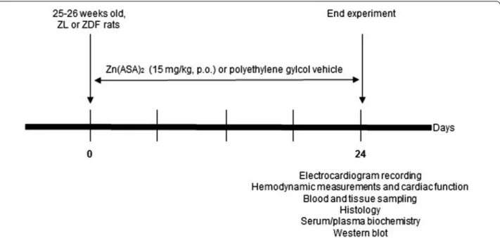
to the results of previous rodent experiments [19]. The experimental protocol is shown in Fig. 1.
Echocardiography
One day before the final experiments, the rats were slightly anesthetized with 1.5–2.0 % isoflurane by mask, the left side of the chest was shaved to obtain a clear image, and the animals were situated in the supine posi- tion on a warming pad. Two-dimensional (long- and short-axis) and motion-mode (M-mode) images of the left ventricle were obtained from a parasternal view, car- ried out with vevo 2100 ultrasound system, and using standard echocardiography. All data was analysed off-line at the end of the study, with the software resident on the ultrasound system. Interventricular diameters includ- ing left ventricular internal end-diastolic dimensions (LVIDd) and left ventricular internal end-systolic dimen- sions (LVIDs), were measured. All measurements were based on the mean of at least three consecutive cardiac cycles on the M-mode tracing at the level of the papillary muscle, and the mean values were used in our analyses.
Electrocardiogram (ECG) recording
The rats were anesthetized with sodium pentobarbi- tal (60 mg/kg) intraperitoneally, and they were kept in a supine position. Their body temperature (measured via rectal probe) was maintained at 37 °C. The stand- ard 12-lead electrocardiogram (leads I, II, III, aVR, aVL, aVF, V1, V2, V3, V4, V5, and V6) was recorded using subcutaneously placed needle electrodes. All leads were connected to a standard direct-writing recorder (Mor- tara Instrument, WI, USA). The paper speed was set at 50 mm/s, and the sensitivity at 10 mm/mV. The ECG analysis was evaluated by lead II including the following measurements: ST-segment elevation, QRS complex, and QT-intervals. The QT-interval, measured from the onset of the QRS complex to the end of the T wave, was cor- rected using the normalized Bazett’s formula adjusted for rats (nQTc = QT/(RR/f)1/2) (RR corresponding to cardiac cycle length and f to frequency) [26]. The electrocardiog- raphy was analyzed by an examiner blinded to the experi- mental groups.
In vivo hemodynamic measurements and cardiac function After the ECG recording, the animals were tracheot- omised, intubated, and artificially ventilated. A polyeth- ylene catheter was inserted into the left external jugular vein for fluid administration purposes. A 2F microtip pressure–volume catheter (SPR-838, Millar Instruments, Houston, TX, USA) was inserted into the right carotid artery and advanced into the ascending aorta. After allowing an initial stability period of 5 min, the systolic
blood pressure (SBP), diastolic blood pressure (DBP), mean arterial pressure (MAP), and heart rate were recorded. After that, the catheter was advanced into the left ventricle under pressure control. With the use of a special pressure–volume analysis program (PVAN, Millar Instruments, Houston, TX, USA), stroke volume, cardiac output (CO), stroke work, left-ventricular end-systolic pressure (LVESP), left-ventricular end-diastolic pressure (LVEDP), and time constant of left-ventricular (LV) pres- sure decay (Τau) were calculated. The pressure–volume relations of the LV cavity were assessed by briefly com- pressing the inferior vena cava. The slope [end-systolic elastance (Ees)] of the LV end-systolic pressure–volume relationship (ESPVR) according to the parabolic curvi- linear model [27] was calculated as a load-independent index of LV contractility. The slope of the LV end-dias- tolic pressure–volume relationship (EDPVR) was calcu- lated as a reliable index of LV stiffness.
Biochemical estimation
After the heart function was measured the blood was collected from the abdominal aorta in Lithium-Heparin- Gel Monovette® and serum tubes. Then, after centrifu- gation, plasma and serum samples were obtained. The levels of plasma glucose, high-density lipoprotein (HDL) cholesterol, total cholesterol, triglyceride and aspartate aminotransferase (AST) were determined in the cen- tral laboratory of the Heidelberg University clinic on an ADVIA 2400 chemistry analyser (Siemens, Germany).
The levels of serum insulin were measured via ELISA kit (Mercodia, Uppsala, Sweden) and non-esterified fatty acids were determined with a commercially available enzymatic and colorimetric assay kit (Wako Chemicals, Germany). Fast protein liquid chromatography (FPLC) was used for the analysis of lipoprotein profiles. Serum samples from each experimental group were pooled and separated by chromatography on an ÄKTApurifier FPLC system (GE Healthcare, Sweden). Briefly, 400 μl of pooled serum were separated using a Superose-6 size exclusion column, and the eluate was fractionated. Total choles- terol and triglyceride contents of the individual fractions were determined via commercially available kits (Randox Laboratories, UK and Human Diagnostica, Germany, respectively).
Histopathological process
After the blood samples were collected pieces of myo- cardial tissue were fixed in a buffered paraformaldehyde solution (4 %), embedded in paraffin, and cut to 5-μm.
All of the histological evaluations were conducted by an examiner blinded to the experimental groups.
Hematoxylin and eosin staining
Paraffin-embedded sections were placed on adhesive slides and stained with hematoxylin and eosin. Cardio- myocyte cross-sectional areas were calculated under a microscope using the Cell^A software (Olympus Soft Imaging Solutions GmbH, Germany).
Fig. 1 Experimental protocol. At the age of 25–26 week‑old, ZL or ZDF rats were treated with Zn(ASA)2 or a polyethylene glycol vehicle (15 mg/kg) orally for 24 consecutive days. Zn(ASA)2 indicates a zinc complex of acetylsalicylic acid, ZDF Zucker diabetic fatty rats, ZL Zucker lean
Terminal deoxynucleotidyl transferase‑mediated dUTP nick end‑labeling (TUNEL)
TUNEL assay was performed according to the manufac- turer’s instructions (Chemicon International, Temecula, CA, USA), to detect DNA strand breakages (free 3′-OH DNA ends). Briefly, the rehydrated sections were digested with 20 μg/ml DNase-free Proteinase K (Sigma-Aldrich, Germany) to retrieve antigenic epitopes, and endogenous peroxidases were blocked with 3 % hydrogen peroxide (H2O2). The free 3ʹ-OH termini of the DNA ends were labeled with a reaction mixture of terminal deoxynucle- otidyl transferase (TdT) and digoxigenin-dUTP at 37 °C for 1 h (Chemicon International, Temecula, CA, USA).
Incorporated digoxigenin-conjugated nucleotides were detected using a horseradish peroxidase conjugated anti- digoxigenin antibody and 3,3ʹ-diaminobenzidine. The sections were counterstained with Gill’s hematoxylin.
Dehydrated sections were cleared in xylene, mounted with Permount (Fischer Scientific, Germany), and cover- slips were applied. Based on the intensity and distribution of the labelling, a semi-quantitative histomorphological assessment was performed using conventional micros- copy. For the assessment of TUNEL-labelled cells, the number of positive cell nuclei per microscopic examina- tion field (with 200× magnification) was counted in four fields for each sample, averaged, and the mean was calcu- lated for each experimental group.
Acid fuchsin orange G (AFOG)
An AFOG-stain was used to detect collagen fibers in the heart tissue. Histological sections were automatically imaged, using 20× magnification (resolution: 0.46 μm/
pixel), with the Hamamatsu NanoZoomer 2.0-HT Scan System (Hamamatsu Photonics, Hamamatsu Japan). The slide scanner automatically detects the region of interest containing the tissue, and also automatically determines a valid focal plane for scanning. Image processing algo- rithms have been developed using VisiomorphDP version 4.5.1.324 (Visiopharm, Hoersholm, Denmark). Image pro- cessing was performed in several distinguished steps: (a) Automatic region of interest detection using thresholding methods in the RGB-Colorspace. As a result of this step, the tissue regions are separated from the background for further analysis; (b) Pixelwise classification of the whole slide by using a linear Bayesian Classifier [28] in RGB col- orspace. After training the classifier by manually marking background areas within the tissue regions, the normal muscle tissue, as well as the fibrotic areas, the classifier was able to separate the processed area into one of these three subcategories. A second subsequent classifier was trained to detect folded tissue and artifacts; (c) As a post- processing step, areas that were too small (small tissue fragments, staining artifacts, dust particles) were removed
by using morphological operations like opening and clos- ing. The ratio of the fibrotic area in relation to the total tissue can be calculated using these output variables.
Immunohistochemical detection of nitrotyrosine
According to previously described methods [29] we per- formed immunohistochemical staining on heart tissue to detect nitrotyrosine, a marker for nitro-oxidative stress.
Positive immunohistological staining was scored using semiquantitative analysis. The total score was then given based on the sum of the intensity and the area scores.
The intensity was evaluated as follows: no staining (score 0), weak staining (score 1), moderate staining (score 2), and severe staining (score 3). The area score was evalu- ated (from 0 to 4) within score 0 representing no stain- ing, score 1 (positive cells <10 %), score 2 (positive cells 11–50 %), score 3 (positive cells 51–80 %) and score 4 (positive cells >80 %).
Western blotting
Proteins were extracted from the heart tissue in a 1x RIPA buffer (Melford, Ipswich, UK). The protein concentra- tion was determined with Bradford assay. Total protein homogenates 20 μg/30 μl were denatured, separated on sodium dodecyl sulfate polyacrylamide electrophoresis gels, and transferred to a polyvinylidene fluoride mem- brane (Millipore, Darmstadt, Germany). The membrane was blocked with 2.5 % BSA in Tris- Buffered Saline Tween 20 for 1 h, before being incubated overnight at 4 °C with CD36 (1:1000, Cell Signalling Technology, Cam- bridge, UK), GLUT-4 (1:100, Santa Cruz, Biotechnology, Heidelberg, Germany), insulin receptor-β (1:1000, Cell Signalling Technology, Cambridge, UK), phosphorylated- PI3K (1:1000, Cell Signalling Technology, Cambridge, UK), PI3K (1:1000, Cell Signalling Technology, Cambridge, UK), AKT, or phosphorylated-AKT (1:500, Abcam, Cambridge, UK). After washing the blots to remove excessive primary antibody binding, the blots were incubated for 1 h with a horseradish peroxydase conjugated secondary antibody at room temperature. Glyceraldehyde-3-phosphate dehy- drogenase (GAPDH), housekeeping protein, was used for loading control and protein normalization. The immuno- reactive protein bands were developed using the Enhanced Chemiluminescence system (PerkinElmer, Rodgau-Jue- gesheim, Germany). The intensity of the immunoblot bands was detected with Chemismart 5100.
Cellular model of hydrogen peroxide‑induced cardiomyocytes oxidative stress
Isolation and culture of neonatal rat cardiomyocytes, measurement protocol
Neonatal cardiomyocytes were isolated from 1- to 2-day- old newborn rats (Charles River, Sulzfeld, Germany).
Briefly, the hearts were excised and rapidly placed into an ice-cold buffer (in mmol/L: NaCl 116.4, HEPES 20, NaH2PO4 1, glucose 5.5, KCl 5.4, MgSO4 0.8; pH 7.4).
The atria and great vessels were trimmed and discarded.
The ventricles were cut into small pieces and incubated (37 °C, 20 min) repeatedly (5 to 6 times) in a buffer sup- plemented with collagenase type II (Worthington Bio- chemical Corporation, New Jersey, USA) and pancreatin (0.6 mg/mL; Gibco BRL). After each round of digestion, the supernatant was centrifuged (1200 rpm, 10 min), and the resulting cell pellet was resuspended in DMEM/
M199 (4:1) supplemented, with 5 % horse serum (Bio- chrom, Berlin, Germany), 10 % fetal bovine serum (Gibco by Thermo Fischer Scientific, Karlsruhe, Germany), penicillin G (100 U/mL; Gibco by Thermo Fischer Sci- entific, Karlsruhe, Germany), and streptomycin (100 μg/
mL; Gibco by Thermo Fischer Scientific, Karlsruhe, Ger- many). Then, cells were pooled and separated on a dis- continuous Percoll (GE Healthcare Bio-Sciences AB, Uppsala, Sweden) gradient (top density = 1.059 g/ml, bottom density = 1.082 g/ml) by centrifugation. The middle band at the interface of the two Percoll layers was collected. Cell damage was estimated based on lac- tate dehydrogenase activity released from damaged cells into the cell culture, using a colorimetric lactate dehy- drogenase-Cytotoxicity Assay kit (BioVision, CA, USA).
The absorbance was measured at 490–500 nm using a plate reader (Thermo Labsystems Multiskan Ascent, MD, USA), and the percentage of cytotoxicity was then calculated.
Experimental groups
To investigate the effects of Zn(ASA)2 upon H2O2- induced oxidative stress in cardiomyocytes, the cul- tured neonatal cardiomyocytes were divided into two groups: (1) vehicle + H2O2 group (n = 25 wells): car- diac cells were pre-treated with the vehicle (DMSO, 2 h),
then incubated with H2O2; (2) Zn(ASA)2 + H2O2 group (n = 25 wells): cells were pre-treated with Zn(ASA)2 (10−3 M; 2 h), then incubated with H2O2. The concentra- tion of H2O2 was 500 µM, the incubation time was 2 h.
Statistics
All data is expressed as mean ± standard error of the mean (SEM). Intergroup comparisons were performed by using one-way analysis of variance, followed by a Stu- dent’s unpaired t test with Bonferroni’s correction for multiple comparisons. A value of P <0.05 was considered statistically significant.
Results
Biochemical parameters
The plasma glucose levels were significantly higher in ZDF rats compared to ZL animals (49.4 ± 2.8 mM vs.
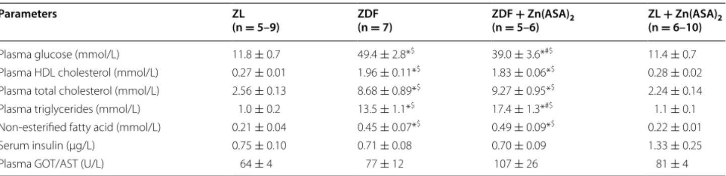
11.8 ± 0.7 mM, P < 0.05), indicating the manifestation of an overt diabetes. When ZDF rats received daily oral administration of Zn(ASA)2 for 24 days, the plasma glu- cose levels were significantly reduced in comparison to ZDF rats (Table 1). Total cholesterol, HDL cholesterol, and free non-esterified fatty acid levels were significantly higher in ZDF rats than in ZL rats, and the Zn(ASA)2 treatment provided no changes. Additionally, triglycer- ide levels in the diabetic group were significantly elevated compared to both ZL groups. Treatment with Zn(ASA)2 further significantly increased plasma triglyceride con- centrations in the diabetic rats, while having no effect upon the lean group. As the triglycerides were abnor- mally high, the LDL cholesterol calculation could not be performed. Therefore, the serum lipoproteins were sepa- rated by fast performance liquid chromatography (FPLC).
Three major peaks were detected, representing the lipoprotein classes VLDL, LDL, and HDL, respectively (Fig. 2). Peak size analysis confirmed increased levels of HDL (Table 1; Fig. 2) and showed high VLDL and LDL
Table 1 Biochemical parameters
Biochemical profile of ZDF (Zucker diabetic fatty) and Zucker Lean (nondiabetic) rats after 24 days of treatment with either vehicle or zinc complex of acetylsalicylic acid (Zn(ASA)2). Values are mean ± SEM. * P < 0.05 vs ZL, # P < 0.05 vs ZDF, $ P < 0.05 vs ZL + Zn(ASA)2
GPT indicates glutamate–oxaloacetate transaminase, AST aspartate aminotransferase, n number of animals analysed
Parameters ZL
(n = 5–9) ZDF
(n = 7) ZDF + Zn(ASA)2
(n = 5–6) ZL + Zn(ASA)2 (n = 6–10)
Plasma glucose (mmol/L) 11.8 ± 0.7 49.4 ± 2.8*$ 39.0 ± 3.6*#$ 11.4 ± 0.7
Plasma HDL cholesterol (mmol/L) 0.27 ± 0.01 1.96 ± 0.11*$ 1.83 ± 0.06*$ 0.28 ± 0.02
Plasma total cholesterol (mmol/L) 2.56 ± 0.13 8.68 ± 0.89*$ 9.27 ± 0.95*$ 2.24 ± 0.14
Plasma triglycerides (mmol/L) 1.0 ± 0.2 13.5 ± 1.1*$ 17.4 ± 1.3*#$ 1.1 ± 0.1
Non‑esterified fatty acid (mmol/L) 0.21 ± 0.04 0.45 ± 0.07*$ 0.49 ± 0.09*$ 0.22 ± 0.01
Serum insulin (μg/L) 0.75 ± 0.10 0.71 ± 0.08 0.70 ± 0.09 1.33 ± 0.25
Plasma GOT/AST (U/L) 64 ± 4 77 ± 12 107 ± 26 81 ± 4
(Fig. 2) in diabetic rats. The administration of Zn(ASA)2 had further increased VLDL levels, while there was no effect on LDL cholesterol. Furthermore, serum insulin levels were similar among the groups with a tendency toward increase in the nondiabetic control group treated with Zn(ASA)2. Plasma GOT/AST concentrations, which show the degree of hepatic disturbance, were also identi- cal to those of the untreated rats.
General characteristics and LV wall thickness
During 24 days of treatment, the ZL rats gained (+0.20 ± 2.8 g), whereas the ZDF rats lost weight (−75 ± 38 g), without reaching statistical significance (P > 0.05). Treatment with Zn(ASA)2 had no effect on body weight loss (ZDF + Zn(ASA)2: −17 ± 6 g;
ZL + Zn(ASA)2: −7 ± 2 g). Furthermore, the body weight, heart weight, heart-to-body weight ratio, and
heart weight-to-tibia length ratio were similar among the groups (Table 2). LV wall thickness, assessed by echocar- diography, did not differ among the groups (Table 2).
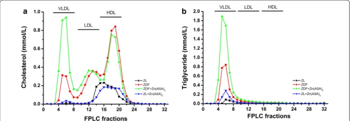
Zn(ASA)2 improves ECG patterns
In ECG recordings, type-2 DM was associated with an elongated corrected QT-interval for heart rate, reduced PQ-interval, and moderate ST-segment elevation com- pared to ZL rats. Treatment with Zn(ASA)2 significantly reduced the prolonged corrected QT-interval and ST- segment elevation (Fig. 3).
Zn (ASA)2 improves LV cardiac function Basic hemodynamic data
The heart rate, systolic and diastolic blood pressures, mean arterial pressure, and the cardiac output were simi- lar among all the experimental groups (Table 3).
Fig. 2 Total cholesterol and triglyceride levels of fast performance liquid chromatography (FPLC) fractions. The graph shows a cholesterol and b triglyceride concentrations of FPLC fractions from pooled serum. Zn(ASA)2 indicates a zinc complex of acetylsalicylic acid, ZDF Zucker diabetic fatty rats, ZL Zucker lean, VLDL very low density lipoprotein, LDL low density lipoprotein, HDL high density lipoprotein
Table 2 Body weight, heart weight, heart-to-body weight ratio, heart weight-to-tibia length ratio and left-ventricular wall thickness assessed by echocardiography
Values are mean ± SEM. ZL indicates Zucker lean, ZDF Zucker diabetic fatty acid, Zn(ASA)2 zinc complex of acetylsalicylic acid, LVPW, d, thickness of the left ventricular posterior wall in diastole, LVPW, s thickness of the left ventricular posterior wall in systole, n number of animals analysed
ZL(n = 7–10) ZDF
(n = 7–8) ZDF + Zn(ASA)2
(n = 5–6) ZL + Zn(ASA)2
(n = 8–10) Weights
Body weight (g) 419 ± 9 376 ± 18 394 ± 27 416 ± 6
Heart weight (g) 1.36 ± 0.03 1.22 ± 0.04 1.35 ± 0.14 1.32 ± 0.03
Heart‑to‑body weight (g/kg) 3.26 ± 0.05 3.26 ± 0.11 3.39 ± 0.10 3.19 ± 0.07
Heart weight‑to‑tibia length (g/cm) 0.32± 0.01 0.30 ± 0.01 0.33 ± 0.03 0.30 ± 0.01
Left‑ventricular wall thickness
LVPW, d (mm) 2.49 ± 0.31 2.95 ± 0.42 1.95 ± 0.17 1.94 ± 0.20
LVPW, s (mm) 3.20 ± 0.27 4.24 ± 0.40 3.16 ± 0.19 2.69 ± 0.25
Systolic function
The stroke volume, stroke work, and LVESP did not differ among the groups (Table 3). Figure 4 shows representa- tive original pressure–volume loops registered during transient occlusion of the inferior vena cava. The end- systolic pressure–volume relationship of the diabetic rats demonstrated only slightly decreased slope (Ees) in com- parison to that of the ZL animals (Table 3).
Diastolic function
Increased slope of EDPVR and the LVEDP in ZDF ani- mals suggested a markedly increased LV diastolic stiff- ness (Fig. 4). Moreover, these passive diastolic function parameters were similar in ZL and ZDF + Zn(ASA)2 ani- mals. The LV active relaxation as assessed by Tau-w did not differ among the groups (Table 3).
Zn(ASA)2 decreases cardiomyocyte hypertrophy
Histological examination revealed that the cardiomyo- cyte transverse cross-section area in the ZDF rats was significantly increased in the H&E stained sections com- pared to the ZL animals (Fig. 5a, b). This enlargement was significantly decreased after the Zn(ASA)2 treatment.
Zn(ASA)2 decreases nitro‑oxidative stress
A significant increase in nitrotyrosine production could be observed in ZDF rats when compared with ZL ani- mals. Treatment with Zn(ASA)2 significantly decreased nitrotyrosine production, as testified by decreased brown staining (Fig. 5a, c).
Effect of Zn(ASA)2 on myocardial fibrosis
The histopathologic examination of myocardial tissue, using acid fuchsin orange G-stain (AFOG), revealed a significant increase in fibrotic formation in ZDF rats compared to ZL animals. ZDF rats treated with Zn (ASA)2 showed a collagen content comparable to the ZL groups (Fig. 6a–c).
Zn(ASA)2 decreases DNA strand breaks
A significant increase in the density of TUNEL-positive cell nuclei was observed in the myocardium of ZDF rats compared to ZL animals, indicating DNA-fragmenta- tion. Treatment with Zn(ASA)2 significantly decreased the percentage of myocardial cells with DNA damage (Fig. 6d–g).
Fig. 3 Zn(ASA)2 improves electrocardiographic pattern. a Representative surface 12‑lead ECG tracing; b corrected QT interval; c PQ‑interval, and d ST‑segment elevation. Values are mean ± SEM, n = 6‑10. *P < 0.05 vs ZL, #P < 0.05 vs ZDF, $P < 0.05 vs Zn(ASA)2, nQTc = QT/(RR/f )1/2. Zn(ASA)2
indicates a zinc complex of acetylsalicylic acid, ZDF Zucker diabetic fatty rats, ZL Zucker lean, RR corresponding to cardiac cycle length and f to frequency, nQTc QT intervals corrected with RR intervals
Zn(ASA)2 regulates myocardial protein expression
Western blot analysis (Fig. 7) revealed that the expres- sion of CD36 was significantly increased, and GLUT-4 was decreased in ZDF rats compared to the nondia- betic group. The insulin receptor β expression was simi- lar among the groups. Zn(ASA)2 had no effect on these proteins. Even though there was no significant change in phosphorylated-PI3K, type-2 DM promotes the expres- sion of total PI3K compared to the control, but the ratio remained unchanged. Additionally, despite no signifi- cant alteration in phosphorylated and total AKT, ZDF rats showed a significant decrease in phosphorylated- AKT/AKT ratio compared to the ZL group. Treatment with Zn(ASA)2 significantly decreased total AKT and considerably increased the phosphorylated-AKT/AKT expression.
Zn(ASA)2 reduces the cytotoxicity induced by H2O2 in cardiomyocytes
H2O2, at a concentration of 500 μM, induced 73 ± 2 % cytotoxicity in cultured neonatal cardiomyocytes, which was significantly decreased to 26 ± 1 % after a pre-treat- ment of cardiac cells with Zn(ASA)2.
Discussion
The aim of this study was to investigate the effects of the oral administration of zinc and aspirin, in the form of bis(aspirinato)zinc(II) complex on diabetes-induced cardiac damage. Our results suggest that its blood glu- cose lowering effect with increased activation of the
serine-threonine kinase AKT in the myocardium could, in part, explain the cardioprotective effects of Zn(ASA)2 upon DCM.
DCM has been defined as ventricular dysfunction along with abnormal myocardial structure in the absence of changes in blood pressure and coronary artery dis- ease. Multiple pathophysiological mechanisms have been proposed to explain this phenomenon, such as myocar- dial fibrosis, myocyte hypertrophy, contractile dysfunc- tion, changes in calcium handling and in mitochondrial function, abnormal cardiomyocyte loss by apoptosis and unsuccessful metabolic adaptation [30]. Despite all these mechanisms, hyperglycemia is considered to be one of the main pathogenic mechanisms of DCM. The potential role of a zinc complex of acetylsalicylic acid in treating DM has recently been demonstrated. An orally administered complex of zinc and aspirin ameliorated hyperglycemia and metabolic syndrome-like disorders in an animal model of type-2 DM [19]. The anti-diabetic effect of Zn(ASA)2 is also supported by our findings in ZDF rats, which showed that a treatment with Zn(ASA)2 caused a significant reduction of high plasma glucose lev- els. However, its administration did not have a significant impact on the lipid profile, except that the VLDL-triglyc- eride levels were increased further by the treatment. It might be that the Zn(ASA)2 lowers blood glucose levels by enhancing the conversion of excess glucose to triglyc- erides in the liver. However, we do not have unequivo- cal evidence at this point, and further investigations are needed to clarify these metabolic aspects of Zn(ASA)2 Table 3 Hemodynamic parameters
In vivo cardiac catheterization findings of ZDF (Zucker diabetic fatty) and Zucker lean (nondiabetic) rats after 24 days of treatment with either vehicle or Zn(ASA)2. Values are mean ± SEM, * P < 0.05 vs. ZL, $ P < 0.05 vs. ZL + Zn(ASA)2
Zn(ASA)2 indicates a zinc complex of acetylsalicylic acid, SBP systolic blood pressure, DBP diastolic blood pressure, MAP mean blood pressure, CO cardiac output, LVESP left ventricular end systolic pressure, Ees the slope Ees of the left ventricular end‑systolic pressure–volume relationship, Tau time constant of left ventricular decay, n number of animals analysed
Parameters ZL
(n = 7–10) ZDF
(n = 7) ZDF + Zn(ASA)2
(n = 6) ZL + Zn(ASA)2
(n = 9) Basic hemodynamic data
Heart rate (beats/min) 366 ± 12 346 ± 14 355 ± 12 373 ± 13
SBP (mmHg) 138 ± 5 151 ± 6 153 ± 6 141 ± 7
DBP (mmHg) 106 ± 3 108 ± 7 110 ± 5 107 ± 6
MAP (mmHg) 112 ± 4 122 ± 7 124 ± 5 118 ± 6
CO (ml/min) 31.0 ± 2.8 35.6 ± 5.3 35.6 ± 2.3 30.2 ± 4.1
Systolic function
Stroke volume (µl) 85 ± 7 93 ± 13 100 ± 5 81 ± 11
Stroke work (mmHg*ml) 8.56 ± 0.88 10.71 ± 1.28 11.25 ± 0.78 7.86 ± 1.09
LVESP (mmHg) 131 ± 5 131 ± 6 128 ± 7 133 ± 7
Ees (mmHg/µl) 1.13 ± 0.13 0.75 ± 0.11 0.85 ± 0.06 1.18 ± 0.24
Diastolic function
Tau (Weiss) (ms) 9.8 ± 0.3 10.3 ± 0.7 10.2 ± 0.3 9.9 ± 0.4
treatment. Its underlying mechanisms involve glucose uptake into the adipocytes by facilitating insulin’s binding to its receptor, leading to the activation of insulin recep- tor β-subunit, insulin receptor substrates, PI3K which in turn activates protein kinase B/AKT. Thus, the transloca- tion of GLUT-4 to the adipocytes membrane stimulated by insulin, normalizes blood glucose levels in the experi- mental diabetic animals [31]. In the present study set up, Zn(ASA)2 does not influence insulin production how- ever, the small amounts of insulin released by the pan- creas can be more effective after a Zn(ASA)2 treatment.
Furthermore, the insulin-mimetic activity of zinc com- plexes was previously examined in terms of free fatty acid release, and was found to inhibit free fatty acid release from isolated rat adipocytes treated with epinephrine (adrenaline) [31], also indirectly indicating their in vitro glucose-uptake ability into the cells.
Metabolic abnormalities present in diabetes (hyperin- sulinemia, hyperlipidemia, and hyperglycemia) might be the initial event which causes endothelial dysfunction, inflammation, apoptosis, and oxidative stress [32]. In the present study, we demonstrated in an in vitro model, Fig. 4 Zn(ASA)2 improves left‑ventricular cardiac function. a Representative original pressure–volume loops registered during transient occlusion of the inferior vena cava. The slope of end‑systolic pressure–volume relationship (red line) and end‑diastolic pressure–volume relationship (EDPVR, green line); b the slope of end‑diastolic pressure–volume relationship (EDPVR); and c left‑ventricular end‑diastolic pressure (LVEDP). Values are mean ± SEM, n = 6–10. *P < 0.05 vs ZL, #P < 0.05 vs ZDF, $P < 0.05 vs Zn(ASA)2. Zn(ASA)2 indicates a zinc complex of acetylsalicylic acid, ZDF Zucker diabetic fatty rats, ZL Zucker lean
that Zn(ASA)2 protects cardiomyocytes from oxidative stress-induced damage. Furthermore, the presence of nitrotyrosine on proteins in the diabetic myocardium, as shown by our immunohistochemical staining, as a marker for nitro-oxidative stress in vivo, is in agreement with previous data [33, 34]. According to the semi-quan- titative scoring of the staining, pronounced nitro-oxida- tive stress can be prevented by a Zn(ASA)2 treatment.
Other studies supporting our observations have shown that zinc-induced overexpression of the antioxidant protein metallothionein protects the heart against dia- betes-induced cardiac protein nitration in the develop- ment of DCM [35]. Zinc supplementation was sufficient to prevent DCM at least partially by reducing oxidative stress [36]. Molecular events of oxidative stress are lipid peroxydation [37], protein oxidation [38], and oxidative damage of DNA [39]. One of the most widely used meth- ods for detecting DNA breaks and DNA fragmentation in
tissue sections is TUNEL, which was described to detect cells undergoing apoptosis [40]. Oxidative stress-induced cell deaths, such as apoptosis, have important roles in the pathogenesis of DCM. In the present study, upon treatment of diabetic rats with Zn(ASA)2, the number of cardiac apoptotic nuclei was significantly decreased.
In addition, apoptosis contributes to the loss of cardiac myocytes leading to adverse cardiac remodeling, includ- ing fibrosis and hypertrophy, impaired cardiac function, and eventually heart failure. The accumulation of extra- cellular matrix proteins, in particular collagens is one of the key processes in DCM. Sárközy et al. have shown an altered expression of several genes related to cardiac hypertrophy and remodeling [41]. Minerals, vitamins, and trace elements treatment in diabetic rats resulted in opposite gene expression changes, showing benefi- cial effects in DCM [41]. In the present study enhanced nitro-oxidative stress and apoptosis in the diabetic Fig. 5 Zn(ASA)2 decreases cardiomyocyte hypertrophy and nitro‑oxidative stress. a Hematoxylin and eosin staining micrographs of transverse sec‑
tions of myocardium (magnification ×400; scale bar: 50 μm) and representative photomicrographs of nitrotyrosine immunohistochemistry staining;
magnification ×200, scale bar: 20 µm. b Quantitative analysis of cardiomyocyte cross‑sectional area using of ~20 cardiomyocytes in each group and c immunohistochemical scores for nitrotyrosine in the myocardium. Values are mean ± SEM, n = 6–9. *P < 0.05 vs ZL, #P < 0.05 vs ZDF, $P < 0.05 vs Zn(ASA)2. Zn(ASA)2 indicates a zinc complex of acetylsalicylic acid, ZDF Zucker diabetic fatty rats, ZL Zucker lean
myocardium, and a high histopathological cardiac fibro- sis score were shown in accordance with the literature, and were proven to be normalized by a Zn(ASA)2 treat- ment. Despite the ability of the antihyperglycemic agents, such as metformin and sulfonylureas, to maintain nor- moglycemia, many patients still develop DCM or are at risk for cardiovascular events [30]. To overcome these limitations and drawbacks for the use of conventional metabolic treatments, the findings of the present study may open novel therapeutic strategies, using Zn(ASA)2 directed against DCM.
Structural changes of the myocardium are often referred to as remodeling [42]. The main feature of this process is an increase in ventricular mass and at cellular level the hypertrophy of individual cardiomyocytes. An increase in myocyte profile surface area, as evident in the present work through enlarged cardiomyocytes in dia- betic rats, has been observed. Potential causes of cardio- myocyte hypertrophy have been accredited to a number
of factors, including mechanical stress [43], the sympa- thetic nervous system [44], the renin-angiotensin-aldos- terone system [43], growth factors, and inflammatory cytokines [45]. Oral administration of Zn(ASA)2 protects the heart from pronounced hypertrophy of single cardio- myocytes. However, the LV wall thickness, assessed by echocardiography, did not significantly differ among the groups.
Metabolic disturbance in diabetes contribute to the development of DCM. Impaired cardiac function, due to cardiomyocyte hypertrophy, apoptosis, and fibrosis has been well documented in the setting of DCM [46].
Additionally, high glucose has been shown to exert det- rimental effects on the myocardial contractile function in ZDF rats [47]. In the present study, with the use of in vivo cardiac catheterization and LV pressure–volume analysis, we showed that the slope Ees of ESPVR, as a load-insen- sitive index of contractility, was only slightly decreased in the diabetic rats. In consonance with our observations, Fig. 6 Effect of Zn(ASA)2 on myocardial fibrosis and DNA‑strand breaks in cardiomyocytes. Representative photomicrographs showing the whole‑
slide after a the region of interest detection and b pixelwise classification of the tissue; blue fibrotic areas, green normal tissue, orange excluded areas in the acid fuchsin orange G (AFOG) stained sections. c Quantitative analysis of interstitial fibrosis in the myocardium. Representative photomicro‑
graphs of d nuclei with 4′,6‑diamidino‑2phenylindole (DAPI‑stained nuclei, blue); e nuclei with fragmented DNA visualized by Terminal Deoxynucle‑
otidyl Transferase‑Mediated dUTP Nick End‑Labeling (TUNEL) staining (TUNEL‑positive nuclei, red), and f merged image (red/blue double stained) (magnification ×400, scale bar: 50 µm) from the same sample which belong to the ZDF group. g Quantification of TUNEL‑positive cells for each group. Values are mean ± SEM, n = 6–10. *P < 0.05 vs ZL, #P < 0.05 vs ZDF, $P < 0.05 vs Zn(ASA)2. Zn(ASA)2 indicates a zinc complex of acetylsalicylic acid, ZDF Zucker diabetic fatty rats, ZL Zucker lean
our previous studies showed that LV contractility, with the use of load-independent variables, was not severely impaired in type-2 diabetic animals [24, 25]. Additionally, it has been shown that ZDF rats exhibited LV diastolic dysfunction [24], and that subclinical LV diastolic dys- function is already present in the early phase of disturbed glucose metabolism, before the onset of type-2 DM,
being mainly associated with the state of insulin resist- ance and not only with sustained hyperglycemia [48]. The administration of drugs that may reduce insulin resist- ance and related cardiovascular risk, would be useful in patients with type-2 DM at risk for developing DCM.
In the present work, diabetes associated changes to the indexes of LV diastolic stiffness (LVEDP and slope of Fig. 7 Effects of Zn(ASA)2 on myocardial protein expression. Immunoblot analysis for a CD36, b insulin receptor (IR)‑β, c glucose transporter (GLUT)‑
4, d phosphorylated PI3K, e PI3K, f phosphorylated phosphatidylinositol 3‑kinase‑(PI3K)/PI3K ratio, g phosphorylated, h total AKT and i phosphoryl‑
ated AKT/total Akt ratio protein band densities in the myocardium. Glyceraldehyde‑3‑phosphate dehydrogenase (GAPDH), housekeeping protein was used as reference. Values are mean ± SEM, n = 6–7. *P < 0.05 vs ZL, #P < 0.05 vs ZDF, $P < 0.05 vs Zn(ASA)2. Zn(ASA)2 indicates a zinc complex of acetylsalicylic acid, ZDF Zucker diabetic fatty rats, ZL Zucker lean
EDPVR) were significantly higher in type-2 diabetic rats, reflecting the impairment of passive, rather than active, LV diastolic function. The LV diastolic dysfunction was improved by Zn(ASA)2 treatment. Even though Xie and al. have shown only minor electrophysiological changes (in action potential duration and conduction velocity) during normoxic perfusion in 13–16 weeks old Zucker obese diabetic fatty rats [49], in the present study rats of the diabetic group presented a delayed repolarization of the ventricular myocardium manifested in prolonged QT intervals on the surface ECG, which was attenu- ated by a Zn(ASA)2 treatment. Our results showed that Zn(ASA)2 administration reduces the risk for ventricular tachyarrhythmia.
Being a key component of the insulin signaling pathway and its downstream targets, including serine/threonine protein kinase AKT, mammalian target of rapamycin (mTOR), and glycogen synthase kinase (GSK), PI3K plays important roles in cardiac adaptation including protein synthesis, inhibition of apoptosis, and fatty acid and glucose metabolism, as well as in gene expression and cell survival. In DM advanced glycation endproducts (AGEs)/receptor for AGEs interaction (RAGE) has been shown to contribute to the development of DCM [50]. A recent study indicates that AGEs trigger cardiomyocyte autophagy (a type II programmed cell death) by at least in part inhibiting the PI3K/AKT/mTOR pathway via RAGE [51]. The activation of the PI3K signaling increases ser- ine phosphorylation of protein kinase B, and this results in the uptake of glucose by inducing the translocation of the GLUT-4 protein to the cell membrane. In the present study, the ratios of expression of phosphorylated AKT to total AKT, as measured by densitometry of Western blot, were significantly lower in heart samples from dia- betic animals. The oral treatment of rats with Zn(ASA)2 reversed this alteration to the levels observed in ZL rats.
It has been recently shown that zinc supplementation can rescue the detrimental effects of Akt2 gene deletion on the heart, probably associated with its insulin mimetic effect on the cardiac glucose-metabolism [52]. In the pre- sent study even though diabetes resulted in a reduction in myocardial GLUT-4 protein expression, a Zn(ASA)2 treatment had no effect upon this factor. The down-regu- lation of GLUT4 expression in the heart has been postu- lated to contribute to the altered substrate utilization and impaired contractility observed in diabetic cardiomyo- pathy [53]. Additionally, observations in GLUT4-defi- cient mice indirectly suggest that a down-regulation of GLUT4 may be involved in the pathogenesis of hypertro- phy [54]. As our results showed that Zn(ASA)2 improves LV diabetic heart function not by decreasing myocardial hypertrophy but likely by its action on fibrosis, these observations may in part, explain why the Zn(ASA)2
treatment failed to regulate the expression of myocardial GLUT4 protein. All together, AKT activation seems to play a key role in the cardioprotective effects of Zn(ASA)2 against the DCM.
However some limitations of this study need to be addressed. ZDF rats represent a progressive and severe type-2 DM with diabetic complications that were left untreated until 25–26 weeks of age. We did not investi- gate whether improving metabolic control with a tra- ditional oral antidiabetic treatment could also prevent diabetic cardiomyopathy. Additionally, the ZDF strain is an inbred rat model produced by a mutation in the leptin-receptor gene, whereas leptin deficiencies are not common in humans. However, ZDF rats were selected as an appropriate diabetic experimental model to verify the present study’s hypothesis.
Conclusions
In summary, the oral administration of Zn(ASA)2 not only decreases blood glucose levels, but also alleviates type-2 diabetes-induced damage in rat cardiac tissue, in part via the activation of the myocardial AKT signal- ling pathway. We recently showed that Zn(ASA)2 pro- tects the heart against myocardial ischemia in rats [22].
As a whole, our results suggest that Zn(ASA)2 might be an ideal therapeutic strategy for diabetic patients at risk for ischemic heart disease. Further detailed studies are required to reveal the exact mechanisms underlying Zn(ASA)2′s protective effects.
Abbreviations
AFOG: acid fuchsin orange G; AST: aspartate aminotransferase; CO: cardiac out‑
put; DBP: diastolic blood pressure; DM: diabetes mellitus; EDPVR: slope of the end‑diastolic pressure–volume relationship; Ees: slope of the LV end‑systolic pressure–volume relationship; f: frequency; ECG: electrocardiogram; FPLC: fast performance liquid chromatography; GAPDH: glyceraldehyde‑3‑phosphate dehydrogenase; GOT: glutamate‑oxaloacetate transaminase; HDL: high den‑
sity lipoprotein; H2O2: hydrogen peroxide; LDL: low density lipoprotein; LV: left‑
ventricular; LVEDP: left‑ventricular end‑diastolic pressure; LVESP: left‑ventricular end‑systolic pressure; MAP: mean arterial blood pressure; mTOR: mammalian target of rapamycin; RR: cardiac cycle length; SBP: systolic blood pressure; Tau:
time constant of left ventricular pressure decay; TdT: terminal deoxynucleoti‑
dyl transferase; TUNEL: Terminal Deoxynucleotidyl Transferase‑Mediated dUTP Nick End‑Labeling; VLDL: very low density lipoprotein; ZDF: Zucker diabetic fatty; Zn(ASA)2: zinc complex of acetylsalicylic acid.
Authors’ contributions
Conceived and designed the experiments: SKI, TR, GS, MK. Performed the experiments: SKI, SAS, SL, MB, PH, AA, MR. Analyzed the data: SKI, SAS, TR, SL, MB, PH, AA, MR, PB, LL, BL, YY, HY, GS. Wrote the manuscript: SKI, SAS, TR, MB, MR, PB, YY, HY, PM, GS. All authors read and approved the final manuscript.
Author details
1 Laboratory of Cardiac Surgery, Department of Cardiac Surgery, University Hospital Heidelberg, Im Neuenheimer Feld 326, 69120 Heidelberg, Ger‑
many. 2 Heart and Vascular Center, Semmelweis University, Városmajor u.
68, Budapest 1122, Hungary. 3 Department of Internal Medicine I and Clini‑
cal Chemistry, University Hospital Heidelberg, Im Neuenheimer Feld 671, 69120 Heidelberg, Germany. 4 Department of Cardiology, Angiology and Pulmonology, University Hospital Heidelberg, Im Neuenheimer Feld
410, 69120 Heidelberg, Germany. 5 Hamamatsu Tissue Imaging and Analysis Center (TIGA), Bioquant, University of Heidelberg, 69120 Heidelberg, Germany.
6 Steinbeis Transfer Center for Medical Systems Biology, 69124 Heidelberg, Germany. 7 Department of Medical Oncology, National Center for Tumor Diseases, University of Heidelberg, 69120 Heidelberg, Germany. 8 Department of Analytical and Bioinorganic Chemistry, Kyoto Pharmaceutical University, Kyoto 607‑8414, Japan. 9 Molecular and Translational Cardiology, Department of Internal Medicine III, University Hospital Heidelberg, Im Neuenheimer Feld 410, 69120 Heidelberg, Germany.
Acknowledgements
This study was supported by the Deutsche Stiftung für Herzforschung (German Heart Research Foundation) (to S. Korkmaz‑Icöz and T. Radovits), by the Medical Faculty of the University of Heidelberg (to S. Korkmaz‑Icöz), by the Hungarian Scientific Research Fund (OTKA PD100245 to T. Radovits), and by the János Bolyai Research Scholarship of the Hungarian Academy of Sciences (to T. Radovits). The excellent technical assistance of Patricia Kraft, Tobias Mayer, Karin Sonnenberg, Ulrike Vogt, and Lutz Hoffmann is greatly acknowledged.
Competing interests
The authors declare that they have no competing interests.
Received: 30 October 2015 Accepted: 5 April 2016
References
1. Aneja A, Tang WH, Bansilal S, Garcia MJ, Farkouh ME. Diabetic cardiomyo‑
pathy: insights into pathogenesis, diagnostic challenges, and therapeutic options. Am J Med. 2008;121:748–57.
2. Regan TJ, Lyons MM, Ahmed SS, Levinson GE, Oldewurtel HA, Ahmad MR, et al. Evidence for cardiomyopathy in familial diabetes mellitus. J Clin Invest. 1977;60:884–99.
3. Fein FS. Diabetic cardiomyopathy. Diabetes Care. 1990;13:1169–79.
4. Schaffer JE. Lipotoxicity: when tissues overeat. Curr Opin Lipidol.
2003;14:281–7.
5. Unger RH, Orci L. Diseases of liporegulation: new perspective on obesity and related disorders. Faseb J. 2001;15:312–21.
6. van Herpen NA, Schrauwen‑Hinderling VB. Lipid accumulation in non‑
adipose tissue and lipotoxicity. Physiol Behav. 2008;94:231–41.
7. Maharjan BR, Jha JC, Adhikari D, Vishwanath P, Baxi J, Alurkar VM, et al. A study of oxidative stress, antioxidant status and lipid profile in dia‑
betic patient in the western region of Nepal. Kathmandu Univ Med J.
2008;6:16–22.
8. Hotamisligil GS. Inflammatory pathways and insulin action. Int J Obes Relat Metab Disord. 2003;27(Suppl 3):S53–5.
9. Hundal RS, Petersen KF, Mayerson AB, Randhawa PS, Inzucchi S, Shoelson SE, et al. Mechanism by which high‑dose aspirin improves glucose metabolism in type 2 diabetes. J Clin Invest. 2002;109:1321–6.
10. Yuan M, Konstantopoulos N, Lee J, Hansen L, Li ZW, Karin M, et al. Reversal of obesity‑ and diet‑induced insulin resistance with salicylates or targeted disruption of Ikkbeta. Science. 2001;293:1673–7.
11. Chimienti F. Zinc, pancreatic islet cell function and diabetes: new insights into an old story. Nutr Res Rev. 2013;26:1–11.
12. Yoshikawa Y, Ueda E, Sakurai H, Kojima Y. Anti‑diabetes effect of Zn(II)/car‑
nitine complex by oral administration. Chem Pharm Bull. 2003;51:230–1.
13. Frommer DJ. The healing of gastric ulcers by zinc sulphate. Med J Aust.
1975;2:793–6.
14. Ozturk N, Olgar Y, Ozdemir S. Trace elements in diabetic cardiomyopathy:
an electrophysiological overview. World J Diabetes. 2013;4:92–100.
15. Atahan E, Ergun Y, Belge Kurutas E, Cetinus E, Guney Ergun U. Ischemia‑
reperfusion injury in rat skeletal muscle is attenuated by zinc aspartate. J Surg Res. 2007;137:109–16.
16. Ozkan KU, Boran C, Kilinc M, Garipardic M, Kurutas EB. The effect of zinc aspartate pretreatment on ischemia‑reperfusion injury and early changes of blood and tissue antioxidant enzyme activities after unilateral testicu‑
lar torsion‑detorsion. J Pediatr Surg. 2004;39:91–5.
17. Turut H, Kurutas EB, Bulbuloglu E, Yasim A, Ozkaya M, Onder A, et al. Zinc aspartate alleviates lung injury induced by intestinal ischemia‑reperfu‑
sion in rats. J Surg Res. 2009;151:62–7.
18. Singla AK, Wadhwa H. zinc‑aspirin complex—synthesis, physicochemical and biological evaluation. Int J Pharm. 1994;108:173–85.
19. Yoshikawa Y, Adachi Y, Yasui H, Hattori M, Sakurai H. Oral administration of Bis(aspirinato)zinc(II) complex ameliorates hyperglycemia and meta‑
bolic syndrome‑like disorders in spontaneously diabetic KK‑A(y) mice:
structure‑activity relationship on zinc‑salicylate complexes. Chem Pharm Bull. 2011;59:972–7.
20. Sarkozy M, Zvara A, Gyemant N, Fekete V, Kocsis GF, Pipis J, et al.
Metabolic syndrome influences cardiac gene expression pattern at the transcript level in male ZDF rats. Cardiovasc Diabetol. 2013;12:16.
21. Corsetti JP, Sparks JD, Peterson RG, Smith RL, Sparks CE. Effect of dietary fat on the development of non‑insulin dependent diabetes mellitus in obese Zucker diabetic fatty male and female rats. Atherosclerosis.
2000;148:231–41.
22. Korkmaz S, Atmanli A, Li S, Radovits T, Hegedus P, Barnucz E, et al.
Superiority of zinc complex of acetylsalicylic acid to acetylsalicylic acid in preventing postischemic myocardial dysfunction. Exp Biol Med.
2015;2:113–25.
23. Hegedus P, Korkmaz S, Radovits T, Schmidt H, Li S, Yoshikawa Y, et al.
Bis (Aspirinato) zinc (II) complex successfully inhibits carotid arterial neointima formation after balloon‑injury in rats. Cardiovasc Drugs Ther.
2014;28:533–9.
24. Radovits T, Korkmaz S, Loganathan S, Barnucz E, Bomicke T, Arif R, et al.
Comparative investigation of the left ventricular pressure–volume relationship in rat models of type 1 and type 2 diabetes mellitus. Am J Physiol Heart Circ Physiol. 2009;297:H125–33.
25. Radovits T, Korkmaz S, Matyas C, Olah A, Nemeth BT, Pali S, et al. An altered pattern of myocardial histopathological and molecular changes underlies the different characteristics of type‑1 and type‑2 diabetic cardiac dysfunction. J Diabetes Res. 2015;2015:728741.
26. Kmecova J, Klimas J. Heart rate correction of the QT duration in rats. Eur J Pharmacol. 2010;641:187–92.
27. Kass DA, Beyar R, Lankford E, Heard M, Maughan WL, Sagawa K. Influence of contractile state on curvilinearity of in situ end‑systolic pressure–vol‑
ume relations. Circulation. 1989;79:167–78.
28. Wang CW, Fennell D, Paul I, Savage K, Hamilton P. Robust automated tumour segmentation on histological and immunohistochemical tissue images. PLoS One. 2011;6:e15818.
29. Liaudet L, Soriano FG, Szabo E, Virag L, Mabley JG, Salzman AL, et al.
Protection against hemorrhagic shock in mice genetically deficient in poly(ADP‑ribose)polymerase. Proc Natl Acad Sci USA. 2000;97:10203–8.
30. Fuentes‑Antras J, Picatoste B, Ramirez E, Egido J, Tunon J, Lorenzo O. Targeting metabolic disturbance in the diabetic heart. Cardiovasc Diabetol. 2015;14:17.
31. Yoshikawa Y, Ueda E, Kojima Y, Sakurai H. The action mechanism of zinc(II) complexes with insulinomimetic activity in rat adipocytes. Life Sci.
2004;75:741–51.
32. Tarquini R, Lazzeri C, Pala L, Rotella CM, Gensini GF. The diabetic cardio‑
myopathy. Acta Diabetol. 2011;48:173–81.
33. Kajstura J, Fiordaliso F, Andreoli AM, Li B, Chimenti S, Medow MS, et al.
IGF‑1 overexpression inhibits the development of diabetic cardio‑
myopathy and angiotensin II‑mediated oxidative stress. Diabetes.
2001;50:1414–24.
34. Song D, Kuo KH, Yao R, Hutchings SR, Pang CC. Inducible nitric oxide synthase depresses cardiac contractile function in Zucker diabetic fatty rats. Eur J Pharmacol. 2008;579:253–9.
35. Cong W, Zhao T, Zhu Z, Huang B, Ma W, Wang Y, et al. Metallothionein prevents cardiac pathological changes in diabetes by modulating nitration and inactivation of cardiac ATP synthase. J Nutr Biochem.
2014;25:463–74.
36. Lu Y, Liu Y, Li H, Wang X, Wu W, Gao L. Effect and mechanisms of zinc supplementation in protecting against diabetic cardiomyopathy in a rat model of type 2 diabetes. Bosn J Basic Med Sci. 2015;15:14–20.
37. Esterbauer H, Gebicki J, Puhl H, Jurgens G. The role of lipid peroxidation and antioxidants in oxidative modification of LDL. Free Radic Biol Med.
1992;13:341–90.
38. Sohal RS, Agarwal S, Dubey A, Orr WC. Protein oxidative damage is associated with life expectancy of houseflies. Proc Natl Acad Sci USA.
1993;90:7255–9.