REMOTE ISCHEMIC CONDITIONING AND ITS MOLECULAR MECHANISM IN THE
HEART
Ph.D thesis
Tamás Baranyai, MD
Pharmaceutical Sciences Doctoral School Semmelweis University
Supervisor: Péter Ferdinandy, MD, D.Sc Official reviewers:
Attila Szijártó, MD, Ph.D István Baczkó, MD, Ph.D
Head of the Final Examination Committee:
Attila Tordai, MD, Ph.D
Members of the Final Examination Committee:
Tamás Radovits, MD, Ph.D István Koncz, MD, Ph.D
Budapest
2017
2
1. Introduction
Despite the wide availability of advanced revascularisation techniques, acute myocardial infarction (AMI) is one of the leading causes of mortality and morbidity in developed countries. Early restoration of the coronary circulation is obligatory in order to prevent the myocardium from the ischemia-triggered myocardial damage. Paradoxically, reperfusion per se exacerbates further the myocardial injury. Thus, both ischemia and reperfusion are responsible for the overall myocardial damage. This is termed ischemia/reperfusion injury.
Ischemic conditioning is a promising protective strategy against ischemia/reperfusion injury. Ischemic preconditioning (i.e., brief cycles of ischemia and reperfusion of the affected coronary artery before sustained ischemia; IPreC) was shown first to attenuate subsequent myocardial damage.
Since then, it has been demonstrated that ischemic conditioning leads to cardioprotection if applied after cardiac ischemia (ischemic postconditioning) which is more feasible for clinical application. Moreover, cardioprotection could be elicited by applying ischemic stimuli on a distant area of the heart or on a remote organ (e.g., kidney, skeletal muscle). This is termed remote ischemic conditioning (RIC) which is also reported to be cardioprotective in preclinical context. Although it has been proposed that humoral and neuronal factors are involved in the signal transport of RIC, it is hardly understood.
The translation of cardioprotective conditioning stimuli into the clinical practice has proven difficult and disappointing despite numerous positive proof-of-concept clinical trials. Thus far, a completed small-scale clinical trial involving ST-elevation myocardial infarction patients demonstrated long term efficacy of RIC as assessed by significant improvement of major adverse cardiac and cerebrovascular events. Nevertheless, two large clinical trials
3
reported that RIC did not improve the long-term outcome after cardiac surgery (ERICCA and RIPHEART trials).
The neutral results have been attributed to many factors, such as recruitment of inadequate patient population, type of revascularisation, inclusion of late revascularisations, comorbidities, comedications, and most importantly the unidentified mechanism of RIC. Moreover, the strict adherence to certain endpoints, such as myocardial necrosis and the neglect of the microvasculature might also hinder the successful translation of RIC.
2. Aims
• To test the cardioprotective effect of RIC in rat and porcine model of AMI.
• To investigate whether acute hyperglycemia interferes with the cardioprotective effect of RIC.
• To investigate whether RIC signal is transported via extracellular vesicles.
3. Materials and Methods
This investigation conforms to the Guide for the Care and Use of Laboratory Animals published by the US National Institutes of Health, to the EU Directive and was approved by the animal ethics committee of the Semmelweis University and by the animal ethics committee of Hungarian National Food Chain Safety Office.
3.1. Experimental protocol to test remote ischemic conditioning in vivo in a rat model of cardiac ischemia/reperfusion injury
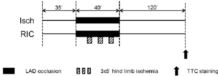
Male Wistar rats were anaesthetized with pentobarbital. Rats were randomized into two groups (Figure 1): (1) control ischemic and (2) RIC. At 35 min of the study protocol, LAD was occluded for 40 min with a suture. RIC was induced by three cycles of 5 min occlusion and 5 min reperfusion of the
4
right femoral vessels starting after 10 min of left anterior descending coronary artery (LAD) occlusion. Both the femoral artery and vein were occluded with a metal vessel clamp after isolation of the vessels from the surrounding connective tissue and femoral nerve. At the end of the 40 min ischemia, reperfusion was induced by loosening the suture. At the end of the 120 min reperfusion, hearts were harvested for the evaluation of the extent of the myocardial necrosis.
Figure 1. Experimental protocol to test remote ischemic conditioning in vivo in a rat model of cardiac ischemia/reperfusion injury. Isch – control ischemia only group, RIC – remote ischemic conditioning, LAD – left anterior descending coronary artery, TTC – triphenyltetrazolium chloride.
3.2. Experimental protocol to test remote ischemic conditioning in vivo in a clinically relevant porcine model of cardiac ischemia/reperfusion injury
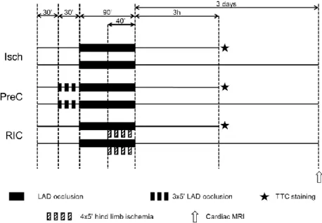
Domestic female pigs were block randomized into three groups (Figure 2):
(1) ischemia only (Isch), (2) IPreC and (3) RIC. One day prior to the experiments, pigs were given acetyl salicylic acid and clopidogrel. Animals were sedated with ketamine/xylazine/atropine mixture. Anesthesia was induced by inhalation of isoflurane. Selective angiography of left coronary artery was performed. After the analysis of the baseline angiogram, balloon catheter was placed in the mid part of the LAD after the origin of the second diagonal branch. For induction of AMI, the intracoronary balloon was inflated, for the induction of reperfusion (3 h or 3 days), the balloon was deflated. In IPreC group, LAD was occluded by the inflation of the balloon three times for 5 min
5
followed by 5 min of reperfusion, while in other animals the balloon was left deflated for 30 min. Then LAD was occluded by inflating coronary balloon for 90 min.
Figure 2. Experimental protocol to test remote ischemic conditioning in an in vivo porcine model of cardiac ischemia/reperfusion injury. Isch – ischemia only, IPreC – ischemic preconditioning, RIC – remote ischemic conditioning, LAD – left anterior descending coronary artery, TTC – triphenyltetrazolium chloride, MRI – magnetic resonance imaging.
RIC was performed by four cycles of 5 min occlusion and 5 min reperfusion of the femoral vessels by tightening and releasing of a snare around the right hindlimb starting at the 50th min of LAD occlusion. Final reperfusion was confirmed with coronarography. Anesthesia was either maintained for 3 h or in case of 3 days reperfusion, wounds were closed and anaesthesia was terminated by the withdrawal of isoflurane. After three hours of reperfusion, hearts were harvested for histological measurement of myocardial necrosis. After 3 days of
6
reperfusion, cardiac MRI was performed to determine myocardial function, necrosis, edema and microvascular obstruction.
3.3. Experimental protocol to test the effect of hyperglycemia on remote ischemic conditioning in an in vivo rat model of ischemia/reperfusion injury
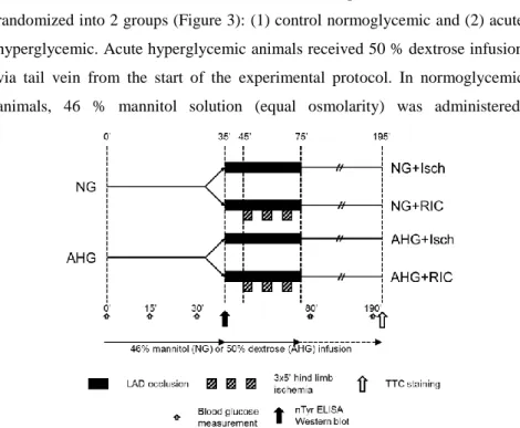
Male Wistar rats were anaesthetized with pentobarbital. Rats were randomized into 2 groups (Figure 3): (1) control normoglycemic and (2) acute hyperglycemic. Acute hyperglycemic animals received 50 % dextrose infusion via tail vein from the start of the experimental protocol. In normoglycemic animals, 46 % mannitol solution (equal osmolarity) was administered.
11111111 111111 111111 111111 111111 111111 111111 111111 111111 111111 111111 111111 111111 111111 111111 11111 Qq qqqqqq 11111 111111 111111 111111 111111 11ewe3232e3 323 2323 11 111111 111111 11 111 111
Figure 3. Experimental protocol to test the effect of hyperglycemia on remote ischemic conditioning in an in vivo rat model of acute myocardial infarction. NG – normoglycemia, AHG – acute hyperglycemia, nTyr – 3 nitrotyrosine, ELISA - enzyme-linked immunosorbent assay, TTC – triphenyltetrazolium chloride.
After 35 min in vivo perfusion, half of the animals from normoglycemic and acute hyperglycemic groups were sacrificed, and hearts were excised, washed
7
in Krebs-Henseleit solution, and were snap-frozen until further experiments.
The other half of the animals were further randomized into four groups: (1) control ischemic with normoglycemia (NG + Isch), (2) RIC with normoglycemia (NG + RIC), (3) ischemic with acute hyperglycemia (AHG + Isch), and (4) RIC with acute hyperglycemia (AHG + RIC). At 35 min of the study protocol, LAD was occluded for 40 min. RIC was induced by three cycles of 5 min occlusion and 5 min reperfusion of the right femoral vessels starting after 10 min of the LAD occlusion. Both the femoral artery and vein were occluded with a metal vessel clamp after isolation of the vessels from the surrounding connective tissue and femoral nerve. At the end of the 40 min ischemia, reperfusion was induced by loosening the suture. At the end of the 120 min reperfusion, hearts were harvested for the evaluation of the extent of the myocardial necrosis.
3.4. Experimental protocol to test the vesicular nature of remote ischemic conditioning ex vivo
Male Wistar rats were anesthetized with ketamine/xylazine and heparinized.
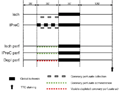
Hearts were isolated and perfused in Langendorff mode with 37 °C Krebs-Henseleit solution for 20 min for stabilization. Hearts were randomized into five groups (Figure 4): (1) 30 min global ischemia (Isch); (2) additional IPreC with 3×5 min global ischemia/reperfusion; (3) treated with perfusate collected from Isch hearts prior to the 30 min global ischemia starting from the 20th min (Isch perf); (4) treated with perfusate collected from IPreC hearts prior to the 30 min global ischemia starting from the 20th min (IPreC perf); (5) treated with vesicle-depleted perfusate collected from IPreC hearts prior to the 30 min global ischemia starting from the 20th min (Depl perf). After 120 min of reperfusion, hearts were harvested for histological staining of myocardial necrosis. Extracellular vesicles were isolated from collected coronary perfusates by filtration and differential centrifugation. Pellets were saved as
8
vesicle-rich pellet and the supernatant was saved as vesicle-depleted perfusate.
Vesicle-depleted perfusates were reconstituted with Krebs Henseleit solution to their original volume measured at the time of coronary perfusate collection.
Reconstituted perfusates were used in heart perfusion experiments same day.
Figure 4. Experimental protocol to test vesicular nature of remote ischemic conditioning ex vivo.
Isch – ischemia only, IPreC – ischemic preconditioning, perf – perfused with perfusate collected from Isch or IPreC hearts, Depl perf - perfused with perfusate collected from IPreC hearts and depleted from extracellular vesicles, TTC - triphenyltetrazolium chloride.
3.5. Measurement of myocardial damage
Measurement of area at risk (AAR): After various periods of reperfusion, LAD was reoccluded, and the AAR was negatively stained with Evans blue retrogradely. AAR was expressed as the proportion of the left ventricular mass. AAR in experiments, in which cardiac MRI was performed, was established by using the modified APPROACH score based on coronary angiography.
9
Histological staining of myocardial necrosis: In order to determine the myocardial necrosis, isolated rat or porcine heart slices were incubated in triphenyltetrazolium chloride to stain viable areas. By using planimetric analysis myocardial necrosis was delineated.
Cardiac MRI to determine myocardial necrosis, edema and microvascular obstruction: For the assessment of myocardial necrosis, edema and microvascular obstruction in porcine hearts, cardiac MRI was performed using a 1.5 T clinical scanner after three days of reperfusion. To determine myocardial necrosis and microvascular obstruction, T1-weighed, late gadolinium enhanced images were recorded 12-15 min after administration of a gadolinium-based contrast agent. To determine myocardial edema, T2-weighed imaging was performed with dark blood sequence. Myocardial necrosis, edema and microvascular obstruction were quantified after manual planimetry.
3.6. Characterisation of extracellular vesicles
To detect extracellular vesicles, transmission electron microscopy was performed. Size distribution of extracellular vesicles was measured by dynamic light scattering measurements by using a goniometer. Particle size distributions were determined by the maximum entropy method. The diameter of the particles was calculated using the sphere approximation.
3.7. Arrhythmia analysis in rats
An electrocardiogram was recorded throughout the entire experiment.
Arrhythmia analysis was performed according to the Lambeth conventions, and arrhythmia incidence and duration scores were calculated.
3.8. Protein detection with Western blot in various samples
Extracellular vesicle sample preparation for Western blot: In order to demonstrate the presence or the absence of extracellular vesicles in the perfusate of ex vivo perfused hearts, extracellular vesicles were pelleted from coronary perfusates which were collected from Isch and IPreC hearts.
10
Supernatants of the centrifugation were kept as extracellular vesicle-depleted perfusates. Pellets were resuspended in homogenation buffer containing protease inhibitor.
Heart sample preparation for Western blot: Left ventricular tissue was homogenized in homogenation buffer supplemented with protease inhibitor, sodium fluoride and phenylmethylsulfonyl fluoride.
Western blot: Equal volume of homogenates (extracellular vesicles) or equal amount of protein (heart samples) was loaded and separated in a Tris- glycine SDS polyacrilamide gel. Proteins were transferred onto a polyvinylidene difluoride membrane. Membranes were blocked with bovine serum albumin or non-fat dry milk. Membranes were probed with primary antibodies, and with corresponding horseradish peroxidase-conjugated secondary antibodies. Signals were detected with an enhanced chemiluminescence kit. Antibodies detecting phosphorylated epitopes were removed with a stripping buffer before incubation with antibodies detecting the total protein.
3.9. Myocardial 3-nitrotyrosine measurement
In order to determine the nitrative stress, free myocardial 3-nitrotyrosine was measured from left ventricular samples with 3-nitrotyrosine ELISA according to the manufacturer’s protocol.
3.10. Statistics
Student’s t-test was performed where two groups including contionuous variables were compared. One-way ANOVA was performed in case of the comparison of multiple groups including contionuous variables. Repeated measures ANOVA was used if multiple groups with contionuous variables were compared at different time points. To test the effect and interaction of two variables (i.e., AHG and RIC), two-way ANOVA was performed. After ANOVA studies, either LSD or Dunnett’s post hoc test was applied. To limit
11
the case numbers in the cardiac MRI study, one-way ANOVA was performed by using bootstrapping with 1000-sample replacement. Groups with discrete variables (i.e., scores) were compared with Kruskal–Wallis analysis. Statistical significance was accepted if p<0.05. Statistical tests were performed with IBM SPSS Statistics, Version 19.
4. Results
4.1. The cardioprotective effect of remote ischemic conditioning in rat and porcine hearts
Remote ischemic conditioning in an in vivo rat model of ischemia/reperfusion injury (Figure 1): RIC significantly attenuated the myocardial necrosis compared to the Control group (n=5-6) in rats. AAR was not different between groups (n=5-6).
Remote ischemic conditioning in an in vivo porcine model of ischemia/reperfusion injury (Figure 2): To quantify myocardial necrosis, edema and MVO in vivo, cardiac MRI was performed 3 days after coronary occlusion and reperfusion. Myocardial necrosis was not affected by RIC (n=8), however, IPreC (n=4) attenuated it significantly as compared to the Isch group (n=7). Myocardial edema was significantly decreased by RIC (n=7) as compared to the Isch group (n=7), however, only a tendency of decrease was observed by IPreC (n=4; p=0.06). AAR based on angiographic score was not different among groups. MVO volume was significantly decreased by IPreC (n=4), but not by RIC (n=8) as compared to the Isch group (n=7). To assess myocardial necrosis and AAR by an ex vivo histological method, the gold standard triphenyltetrazolium chloride- and Evans blue staining were performed after 3 h of reperfusion. RIC did not decrease myocardial necrosis (n=5), however, IPreC (n=6), significantly decreased it as compared to the Isch
12
group (n=5) as measured by triphenyltetrazolium chloride. AAR was not different among groups as evaluated by Evans blue staining.
4.2. The cardioprotective effect of remote ischemic conditioning is abolished by acute hyperglycemia
Acute hyperglycemia was induced with a dextrose infusion during in vivo ischemia/reperfusion rat experiments as described in Figure 3. Blood glucose level was significantly elevated due to dextrose perfusion in both AHG+Isch and AHG+RIC groups compared to the corresponding normoglycemic groups.
Elevated blood glucose levels did not influence the heart rate and blood pressure of the rats.
Myocardial necrosis was significantly smaller in NG+RIC group (n=10) compared to NG+Isch (n=7), while RIC failed to decrease it in AHG+RIC group (n=6) when compared to AHG+Isch (n=5). Furthermore, acute hyperglycemia per se did not aggravate myocardial necrosis. AAR was not different among groups.
Arrhythmia analysis revealed that acute hyperglycemia compared to normoglycemia significantly increased the incidence and the duration of arrhythmias. RIC did not alter arrhythmia scores from the time point it was applied, as compared either to NG+Isch or AHG+Isch groups.
Increased nitrative stress is often implicated in the disruption of cardioprotective interventions. Therefore, 3-nitrotyrosine content, a marker of nitrative stress, was measured in rat hearts. Cardiac 3-nitrotyrosine was significantly elevated due to acute hyperglycemia (n=8).
Since the nitrative stress has been previously shown to interact with mTOR pathway, we evaluated the expression and/or phosphorylation of mTOR pathway associated proteins. The phosphorylations of mTOR (Ser2448) and S6 (Ser235/236) were significantly elevated (n=7-9) which indicates that the activity of mTOR complex I was increased in acute hyperglycemia.
13
Phosphorylation of AKT (Ser473) was also significantly elevated in acute hyperglycemia (n=7-9), however, other mTOR regulators (p-AMPKα [Thr172]
and p-Erk1/2 [Thr202/Tyr204]) were unchanged in acute hyperglycemia as compared to normoglycemia (n=7-9).
Since nitrative stress and mTOR pathway have been shown to interact with autophagy, expression and/or phosphorylation of autophagy-related proteins were also assessed. LC3II/LC3I ratio was significantly decreased due to acute hyperglycemia (n=7-9), however, other autophagy-related proteins (Beclin-1, p62, p-ULK1 [Ser555], ATG7 and BNIP3) were unchanged in acute hyperglycemia (n=7-9).
4.3. Remote ischemic conditioning is mediated by extracellular vesicles Vesicular transfer of RIC mediators was investigated in an ex vivo setup of cardiac ischemia/reperfusion injury as described in Figure 4.
Coronary perfusates from IPreC hearts contained more extracellular vesicles than perfusates isolated from Isch hearts as evidenced by Western blot against HSP60, a well-accepted marker of extracellular vesicles. Electron micrographs revealed that these extracellular vesicles can be classified as microvesicle and exosomes. Dynamic light scattering revealed three populations of particles: (1) ~10 nm, (2) size range of exosomes (<100 nm), and (3) microvesicles (100–1000 nm).
IPreC significantly reduced myocardial necrosis as compared to Isch hearts. In hearts treated with coronary perfusates collected from IPreC hearts (IPreC perf) myocardial necrosis was significantly lower than in hearts treated with coronary perfusate from Isch hearts (Isch perf). Perfusates of IPreC hearts which had been depleted of extracellular vesicles were also given to recipient hearts (Depl perf). Myocardial necrosis in Depl perf hearts (i.e., where extracellular vesicle-depleted perfusate was applied) did not differ significantly from myocardial necrosis observed in Isch perf hearts (n = 5-8).
14
5. Discussion
5.1. The promise of remote ischemic conditioning: the role of microvasculature
Translational models of AMI play important roles in the development of interventions for the clinical practice. For this purpose, pigs are excellent model animals, since their cardiac anatomy and cardiovascular physiology exhibit similarities to the human heart.
According to previous studies, we demonstrated that RIC reduced myocardial necrosis in an in vivo rat model of AMI. However, in our porcine model of AMI, RIC did not reduce myocardial necrosis, but the positive control, IPreC, did. Previously, RIC has been shown to reduce myocardial necrosis in closed-chest swine models of AMI. The discrepancy between our results and those of others might be explained by the significantly different perioperative medication and experimental design. Previous studies applied medications required only for the perioperative procedures (e.g., anesthesia, pain control), however, the therapeutical management of AMI consists of other drugs as well. We treated animals with acetylsalicylic acid and clopidogrel according to the clinical guidelines. However, it has been shown that cyclooxygenase-2 is an essential mediator of ischemic conditioning.
Clopidogrel has been retrospectively shown to reduce cardiac necrosis and to decrease cardiovascular events after AMI in clinical trials, which might be attributed to its antiplatelet activity and a direct cardioprotective effect.
Therefore, one may conclude that the myocardium is already in a protective state, and further protection could not be elicited. Anesthesia was maintained by isoflurane. It is well-documented that certain anaesthetics, inclucing the fluranes, induce cardioprotection and/or interfere with cardioprotective
15
interventions. These data indicate that to ensure translational value of animal studies, it is essential to apply perioperative medication according to clinical guidelines.
Here we demonstrated with ex vivo Evans blue staining and in vivo angiography scoring that AAR was not affected by conditioning stimuli, while myocardial edema was significantly decreased by RIC indicating that the extensive damage of the cardiac microvasculature was attenuated. These results indicate that edema might be independent from AAR in cardioprotection studies, and for this reason, applying myocardial edema as AAR may introduce bias and lead to false conclusions in clinical trials.
5.2. Acute hyperglycemia abolishes the cardioprotective effect of remote ischemic conditioning
We have demonstrated that acute hyperglycemia with no preceding diabetes mellitus abolished the myocardial necrosis limiting effect of RIC in an in vivo rat model of ischemia/reperfusion injury. Furthermore, we have shown here that acute hyperglycemia did not influence autophagy, but increased nitrative stress in the heart plausibly through the activation of the AKT-mTOR pathway. Our finding that experimentally induced acute hyperglycemia with no preceding diabetes diminishes cardioprotective effect of RIC, supports previous observations showing that other forms of cardioprotection (e.g., IPreC) may be affected by acute hyperglycemia. Although we showed here that acute hyperglycemia did not influence the extent of myocardial necrosis, a few studies have reported that acute hyperglycemia without any pre-exisisting pathophysiological conditions aggravate myocardial necrosis.
The underlying mechanism of the loss of RIC-induced cardioprotection by acute hyperglycemia is not fully understood. Increased oxidative and nitrative stresses are implicated in the disruption of cardioprotective interventions by metabolic comorbidities, and conditioning stimuli, such as RIC, alleviate
16
nitrative stress. It was shown here that nitrative stress was also increased in acute hyperglycemia in rat heart in vivo, and similar results have been shown in isolated rat hearts perfused with hyperglycemic solution. These findings clearly signal the pivotal role of excessive nitrative stress in the loss of cardioprotection in disturbed glucose homeostasis.
Nitrative stress has also been shown to directly disrupt autophagy.
Therefore, we assessed cardiac autophagy and its regulatory pathways in acute hyperglycemia. However, we found that autophagy was unlikely to be disrupted, as only LC3II/LC3I ratio was significantly reduced, but other autophagy-related parameters were not. Although cardiac autophagy was not modulated, its most important regulator, the mTOR pathway, was largely activated by acute hyperglycemia. Since it has been shown that the inhibition of mTOR by rapamycine elicits cardioprotective effect in vivo, and that RIC, while protecting the myocardium against ischemia, downregulates mTOR, we hypothesize that the upregulated mTOR pathway might be responsible for this loss of cardioprotection by RIC in acute hyperglycemia.
5.3. Remote ischemic conditioning is mediated by extracellular vesicles We have shown that the release of extracellular vesicles from the heart after IPreC stimuli is increased and that extracellular vesicles are responsible for the transmission of RIC signals for cardioprotection.
Previously several humoral and neuronal transmitter mechanisms have been hypothesized to play a role in the propagation of RIC, however, to date none of them is generally accepted. First, the involvement of humoral transmission pathways have been proposed as transfusion of blood from preconditioned rabbits confers cardioprotection in a naïve non-preconditioned animal against ischemia/reperfusion injury. The role of neuronal pathways has also been studied, but results are also still controversial.
17
Here we evidence a novel, vesicular mechanism for the transmission of cardioprotective signals from a preconditioned heart to another heart subjected to coronary occlusion and reperfusion, which might explain how the suspected humoral and/or released neuronal factors of RIC are transmitted. Ischemia- induced release of extracellular vesicles from cultured cardiomyocytes was reported recently, which is in agreement with our current findings. Elsewhere, exosomes derived from mesenchymal stem cell cultures have been shown to exert cardioprotection in mice, and microvesicles isolated from cell culture medium of bone marrow stem cells protected neonatal cardiomyocytes from ischemic injuries. Since in the latter two reports extracellular vesicles from untreated cells induced pro-survival signals, based on our current findings, we cannot exclude the possibility that extracellular vesicles released from the heart under basal conditions might be also cardioprotective, would their amount be as high as after preconditioning stimuli.
6. Conclusion
Remote ischemic conditioning attenuates myocardial necrosis in rat, while it protects the microvasculature in porcine model of acute myocardial infarction.
Acute hyperglycemia abolishes the cardioprotection conferred by remote ischemic conditioning.
Extracellular vesicles mediates remote ischemic conditioning.
7. Own publications
Own publications involved in the current thesis:
Baranyai T, Giricz Z, Varga ZV, Koncsos G, Lukovic D, Makkos A, Sárközy M, Pávó N, Jakab A, Czimbalmos C, Vágó H, Ruzsa Z, Tóth L,
18
Garamvölgyi R, Merkely B, Schulz R, Gyöngyösi G, Ferdinandy P. In vivo MRI and ex vivo histological assessment of the cardioprotection induced by ischemic preconditioning, postconditioning and remote conditioning in a closed-chest porcine model of reperfused acute myocardial infarction:
importance of microvasculature. J Transl Med. 2017 Apr 1;15(1):67.
[IF: 3.694]
Baranyai T, Nagy CT, Koncsos G, Onódi Z, Károlyi-Szabó M, Makkos A, Varga ZV, Ferdinandy P, Giricz Z. Acute hyperglycemia abolishes cardioprotection by remote ischemic perconditioning. Cardiovasc Diabetol.
2015 Nov 18;14(1):151. [IF: 4.534]
Giricz Z, Varga ZV, Baranyai T, Sipos P, Paloczi K, Kittel A, Buzas EI, Ferdinandy P. Cardioprotection by remote ischemic preconditioning of the rat heart is mediated by extracellular vesicles. J Mol Cell Cardiol. 2014 Mar;68:75-8. [IF: 4.655]
Own publications not involved in the current thesis:
Giricz Z, Koncsos G, Rajtík T, Varga ZV, Baranyai T, Csonka C, Szobi A, Adameová A, Gottlieb RA, Ferdinandy P. Hypercholesterolemia downregulates autophagy in the rat heart. Lipids Health Dis. 2017 Mar 23;16(1):60. [IF: 2.137]
Pavo N, Lukovic D, Zlabinger K, Zimba A, Lorant D, Goliasch G, Winkler J, Pils D, Auer K, Jan Ankersmit H, Giricz Z, Baranyai T, Sárközy M, Jakab A, Garamvölgyi R, Emmert MY, Hoerstrup SP, Hausenloy DJ, Ferdinandy P, Maurer G, Gyöngyösi M. Sequential activation of different pathway networks in ischemia-affected and non-affected myocardium, inducing intrinsic remote conditioning to prevent left ventricular remodeling.
Sci Rep. 2017 Mar 7;7:43958. [IF: 5.228]
19
Koncsos G, Varga ZV, Baranyai T, Boengler K, Rohrbach S, Li L, Schlüter KD, Schreckenberg R, Radovits T, Oláh A, Mátyás C, Lux Á, Al- Khrasani M, Komlódi T, Bukosza N, Máthé D, Deres L, Barteková M, Rajtík T, Adameová A, Szigeti K, Hamar P, Helyes Z, Tretter L, Pacher P, Merkely B, Giricz Z, Schulz R, Ferdinandy P. Diastolic dysfunction in prediabetic male rats: Role of mitochondrial oxidative stress. Am J Physiol Heart Circ Physiol.
2016 Oct 1;311(4):H927-H943. [IF: 3.324]
Sódar BW, Kittel Á, Pálóczi K, Vukman KV, Osteikoetxea X, Szabó- Taylor K, Németh A, Sperlágh B, Baranyai T, Giricz Z, Wiener Z, Turiák L, Drahos L, Pállinger É, Vékey K, Ferdinandy P, Falus A, Buzás EI. Low- density lipoprotein mimics blood plasma-derived exosomes and microvesicles during isolation and detection. Sci Rep. 2016 Apr 18;6:24316. [IF: 5.228]
Baranyai T, Herczeg K, Onódi Z, Voszka I, Módos K, Marton N, Nagy G, Mäger I, Wood MJ, El Andaloussi S, Pálinkás Z, Kumar V, Nagy P, Kittel Á, Buzás EI, Ferdinandy P, Giricz Z. Isolation of exosomes from blood plasma:
qualitative and quantitative comparison of ultracentrifugation and size exclusion chromatography methods. PLoS One. 2015 Dec 21;10(12):e0145686. [IF: 3.057]
Baán J, Varga ZV, Leszek P, Ku Mierczyk M, Baranyai T, Dux L, Ferdinandy P, Braun T, Mendler L. Myostatin and IGF-I signaling in end-stage human heart failure: a qRT-PCR study. J Transl Med. 2015 Jan 16;13(1):1. [IF:
3.694]
Baranyai T, Terzin V, Vajda A, Wittmann T, Czako L. Acute pancreatitis caused by hypertriglyceridemia. Orv Hetil. 2010 Nov 7;151(45):1869-74.