Neuronal Nitric Oxide Mediates the Anti-Inflammatory Effects of Intestinal Ischemic Preconditioning
Sa´ndor Varga, MD,
aLa´szlo´ Juha´sz, PhD,
bPe´ter Ga´l, MD,
cGa´bor Boga´ts, MD, PhD,
aMiha´ly Boros, MD, DSc,
bZsolt Pala´sthy, MD, PhD,
dAndrea Szabo´, MD, PhD,
b,* and Jo´zsef Kaszaki, PhD
baDepartment of Cardiac Surgery, Faculty of Medicine, University of Szeged, Szeged, Hungary
bInstitute of Surgical Research, Faculty of Medicine, University of Szeged, Szeged, Hungary
cDepartment of Pediatrics, Faculty of Medicine, University of Szeged, Szeged, Hungary
dDepartment of Surgery, Faculty of Medicine, University of Szeged, Szeged, Hungary
a r t i c l e i n f o
Article history:
Received 14 March 2019 Received in revised form 15 May 2019
Accepted 14 June 2019 Available online xxx
Keywords:
Ischemiaereperfusion Intestinal motility Neutrophil accumulation Mast cell degranulation Mucosal injury
a b s t r a c t
Background:Ischemic preconditioning (IPC) can provide a defense against ischemiaereperfusion (IR)-induced acute inflammation and barrier dysfunction in many organs. Because nitric oxide (NO) has been implicated as a trigger or mediator in the IPC mechanism and because neuronal NO synthase (nNOS) is a dominant isoform of NOS in the gastrointestinal tract, our aim was to investigate the role of nNOS in IPC-induced protection after mesenteric IR.
Materials and methods: Intestinal IR was induced in sodium pentobarbitaleanesthetized dogs by clamping the superior mesenteric artery for 60 min followed by 2 h of reperfu- sion (IR group;n¼7). In further groups, IPC was used (three cycles of 5-min ischemia/5-min reperfusion periods) before IR in the presence or absence of selective inhibition of nNOS with 7-nitroindazole (5 mg/kg, intravenously, in a bolus 15 min before IPC,n ¼6 each).
Changes in mesenteric vascular resistance, intramucosal pH (pHi), and small bowel motility were monitored. Plasma nitrite/nitrate levels, intestinal NO synthase activity, leukocyte accumulation, mast cell degranulation, and histologic injury were also determined.
Results:Ischemia significantly decreased mesenteric vascular resistance and pHi, whereas IR induced a temporary bowel hypermotility and acute inflammatory reaction. IPC facili- tated pHi recovery, attenuated motility dysfunction, elevated NOS-dependent NO pro- duction, and reduced leukocyte accumulation, mast cell degranulation, and mucosal injury. Pretreatment with 7-nitroindazole halted the IPC-induced increase in NO avail- ability, pHi recovery, and the anti-inflammatory and morphologic effects.
Conclusions:Our data demonstrate that NO generated by intestinal nNOS plays a pivotal role in IPC-linked tissue protection by inhibiting an IR-related acute inflammatory response.
ª2019 Published by Elsevier Inc.
* Corresponding author.Institute of Surgical Research, University of Szeged, Pulz u. 1, H-6724 Szeged, Hungary. Tel.:þ36-62 545-103; fax:
þ36-62 545-743.
E-mail address:szabo.andrea.exp@med.u-szeged.hu(A. Szabo´).
Available online atwww.sciencedirect.com
ScienceDirect
journal home page: www.Journa lofSurgicalResea rch.com
0022-4804/$esee front matterª2019 Published by Elsevier Inc.
https://doi.org/10.1016/j.jss.2019.06.053
tion. This hypothesis is supported by the immediate increase in tissue nitrite and nitrate (NOx) levels after IPC in the rat intestine.2It has also been shown that endogenous NO pro- duction by NO synthases (NOSs) plays a regulatory role in intestinal motility after reperfusion8and the preservation of mucosal barrier function on IR injury.9 Furthermore, NO- releasing compounds or NO donors (e.g., SIN-1, sodium nitroprusside, CAS 754, and FK409) exerted similar protection against mucosal injury in rats,10cats,11and dogs12as well.
Among the two calcium-dependent constitutive isoforms, neuronal NO synthase (nNOS or type 1 NOS) is a dominant isoenzyme in the small intestine of both rodents10and large animals.13 Nevertheless, the role of nNOS in intestinal IR injuryeinduced acute inflammatory reactions is still debated.
A number of studies have demonstrated a proinflammatory role for nNOS,14-16but anti-inflammatory properties have also been described.17Furthermore, treatment with nNOS inhibi- tor 7-nitroindazole (7-NI) in healthy rats for 4 d exhibited higher intestinal myeloperoxidase (MPO), inducible NOS, and nuclear factor-kappa binding activity with concomitant lower IkBaexpression.18The paradoxical role of nNOS-mediated NO signaling during inflammation may be due to the differences in the severity and time course of model diseases (i.e., colitis, sepsis, or cerebrovascular inflammation) and species differ- ences (i.e., rodentsversuslarger animals) as well.
In the present study, we aimed to investigate the possible role of nNOS in the local IPC-induced protection in the gastrointestinal tract of a large animal model, where inner- vation is similar to that of humans.19To this end, 7-NI, a specific nNOS inhibitor with a high neuronal uptake,20was administered before IPC in anesthetized dogs. Particular emphasis was placed on intestinal motility function and in- flammatory reactions in the early reperfusion phase when the parallel involvement of NOS activation was also determined.
Materials and methods
The experiments complied with the ARRIVE guidelines and the project were approved by the National Scientific Ethics Committee on Animal Experimentation (National Competent Authority) in Hungary under license number V./148/2013. The study was performed in compliance with EU Directive 2010/
63/EU on the protection of animals used for experimental and other scientific purposes and the National Institutes of Health
the root of the superior mesenteric artery (SMA) was dissected free, and an ultrasonic flow probe (Transonic Systems Inc, Ithaca, NY) was placed around the exposed SMA to measure mesenteric blood flow. A branch of a tributary of the ileal vein supplying the terminal part of the ileum was cannulated with a 2-F polyethylene catheter to measure mesenteric venous pressure so as to obtain blood samples (see later).
Experimental protocol
Ten male and nine female outbred dogs were randomly allo- cated to IR (n¼7; male¼4 and female¼3) and IPC (n¼12;
male ¼ 6 and female¼ 6) groups (Fig. 1). In the IR group, mesenteric ischemia was elicited by occluding the SMA for 60 min with a miniclip, which was followed by 2 h of reper- fusion. In the IPC group, animals were assigned into 7- NIetreated or vehicle groups and were treated with the se- lective nNOS inhibitor 7-NI (5 mg/kg; n ¼6; male ¼3 and female¼3) or saline vehicle (n¼6; male¼3 and female¼3) in an intravenous bolus 15 min before IPC. IPC was induced in two groups by clamping the SMA using three cycles of 5-min ischemia/5-min reperfusion periods starting 60 min before IR. Stock solution of 7-NI (SigmaeAldrich Co, St. Louis, MO) was dissolved in a mixture of 75% dimethyl sulfoxide/saline and stored at20C; the aliquots were diluted in 3 mL of 0.9%
saline before administration. In the IR group, animals were treated with saline vehicle at a matching time point.
Blood samples were collected from the mesenteric vein for later determination of plasma NOx levels at different time points (Fig. 1). Tissue biopsies were also harvested from the intestine for further biochemical/histologic (e.g., MPO, NOS activity, and mast cell (MC) degranulation) analyses (at base- line as well as at the 15thand 120thmin of reperfusion).
Hemodynamic and blood gas measurements; calculation of mucosal pH
The peripheral arterial and mesenteric venous pressures (using Statham P23 Db transducers) were registered with a computerized data acquisition system (Haemosys 1.17 Experimetria Ltd, Budapest, Hungary). Mesenteric vascular resistance was calculated with a standard formula. A silastic balloon catheter (TGS Tonomitor; Tonometrics Inc, Worcester, MA) was introduced through a small enterotomy into the in- testinal lumen. Arterial blood gases and intramucosal pCO2
were measured with a blood gas analyzer (AVL, Graz, Austria).
Intestinal pH (pHi) was calculated using the modified HendersoneHasselbach formula with a correction factor for 30 min of equilibration.
Intestinal motility measurement
To monitor the small bowel motility, a modified strain gauge transducer technique was applied,13 the transducers being sutured to the circular muscle layer of a terminal ileal segment. Motility index was calculated to estimate the neurogenic integrity of the intestine.21
Plasma nitrate/nitrite level measurements
Plasma NOx concentrations were determined from samples taken at baseline (t¼ 30 min), before ischemia as well as the 15th, 60th, and 120th min of reperfusion by means of the Griess reaction.13
Measurement of NOS enzyme activity
NOS activity was determined in intestinal tissue biopsies based on the enzymatic conversion of3[H]-L-arginine to3[H]- L-citrulline using the Lowry method; the Ca2þ-dependent and Ca2þ-independent NOS activities were assessed in the pres- ence and absence of Ca-calmodulin, respectively.13
Tissue MPO activity measurements
MPO, a marker of neutrophil granulocyte infiltration, was measured from mucosal biopsies using a standard photo- metric method.22
Determination of MC degranulation
Ileal biopsies were placed into an ice-cold Carnoy’s fixative, embedded in paraffin, sectioned (6 mm), and stained with Alcian blue and Safranin O. Histologic analysis was performed in coded sections by one investigator (S.V.) at 40 optical magnification in a blinded fashion. MCs were differentiated based on the location of granules, injury to the cell membrane, and volume of the cytoplasm. Intact cells were characterized by (1) intact membrane and (2) cytoplasmic granules, while criteria for degranulated cells were specified by (1) cell mem- brane injury, (2) reduced cytoplasmic volume and (3) loss of cytoplasmic granules. The percentage of degranulation (MC
%) was calculated based on the number of intact MC (iMC) and degranulated MC (dMC) mast cells: MC% ¼ dMC/
(dMCþiMC).23
Determination of morphologic changes in the structure of the small intestine
Analysis of morphologic injury to the same ileal biopsies was performed on slides stained with hematoxylin-eosin in coded slides in a blinded fashion. Measurements of the height of different portions of the mucosa were conducted with a computer-assisted image analysis system (IVM Pictron, Budapest, Hungary). The extent of mucosal damage was determined on the basis of (1) villus heightdtotal mucosal height ratio (%) and (2) villus heightdcrypt height ratio (%);
these were determined for each villus of the current slide (at 3- 5 high power fields at 4magnification). Villus tip injury was also assessed using a modified version of Chiu’s method for each villus of the current slide (at 4-7 high power fields at 10 magnification).24
Fig. 1eExperimental protocol. In the first group, 60 min of ischemia was induced by occluding the SMA, which was followed by 120 min of reperfusion (IR). In the two other groups, IR was preceded by IPC, which was elicited by inducing three cycles of 5-min ischemia/5-min reperfusion periods in the presence and absence of a bolus injection of 7-NI (5 mg/kg) 15 min before IPC (IPCDIRD7-NI and IPCDIR groups, respectively). IR and IPCDIR groups were treated with a vehicle of 7- NI (a mixture of 75% DMSO/saline) at corresponding time points. As indicated, blood samples from the mesenteric vein (BS) and tissue biopsies from the affected bowel area (marked by an arrowhead) were also taken for later assessments of plasma nitrate/nitrite levels and NOS and MPO activities, as well as to assess MC degranulation and mucosal injury.
Results
Hemodynamic changes
Apart from an abrupt decrease during ischemia (because zero flow and pressure during occlusion in the affected intestinal area), no statistically significant differences between the groups or in comparison with baseline values could be detected in mesenteric vascular resistance (Fig. 2).
Changes in intramucosal pH
No difference in pHi values could be detected at baseline;
neither 7-NI nor IPC influenced this parameter (Fig. 3). In response to 60 min of ischemia, however, a steep decrease in pHi could be detected (P<0.05) in all of the groups, which was followed by a gradual recovery. Restoration in this parameter appeared to be more rapid in both IPC groups as pHi values were significantly higher than in the IR group during the 30th- 90th min of reperfusion. At the end of reperfusion (reperfusion 120 min), pHi values were significantly higher only in the IPCþIR group than those in the test ischemia (IR) group.
Small bowel motility changes
Small bowel motility was not influenced by 7-NI alone or IPC at baseline, but a transiently reduced motility was observed in the 7-NI-treated group immediately after the IPC procedure (Fig. 4); this vanished in 30 min (just before the beginning of ischemia). Ischemia alone did not induce any significant changes in this parameter, but the early reperfusion phase (30 min of reperfusion) was associated with an increased in- testinal motility. This intestinal hypermotility was prevented completely by IPC (to a similar extent in both the vehicle- treated and 7-NIetreated groups), and at the 60th min of reperfusion, even lower motility values could be detected than those seen at baseline. By the end of 2 h of reperfusion, the motility index was normalized in all the groups.
Changes in plasma NOxlevels
In the IR group, no significant changes in NOxvalues could be detected at any phase of the experiments (Fig. 5). A temporary increase was observed in the vehicle-treated IPC group in
blood samples taken before the beginning of ischemia and in both IPC groups at the beginning (at 15 min) of reperfusion.
This IPC-induced increase in NOx levels, however, was missing in the 7-NIetreated IPC þIR group; furthermore, a marked increase in this parameter was seen at the last time point (120th min) of reperfusion.
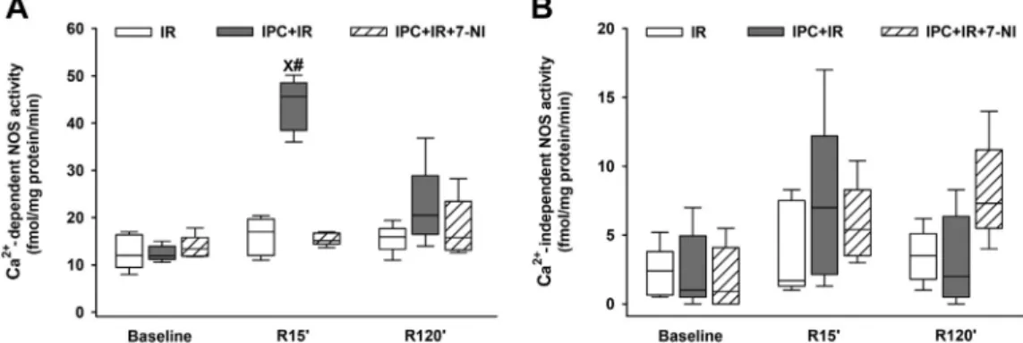
Changes in ileal NOS activities
Ca2þ-independent and Ca2þ-dependent NOS activities were not significantly different between examined groups at base- line (Fig. 6A and B). As for Ca2þ-independent NOS (iNOS) ac- tivity, no statistically significant changes could be detected throughout the entire experimental period in any of the groups (Fig. 6B). In the IR group, Ca2þ-dependent NOS activity showed no changes during any phase of the entire examina- tion period either. In the vehicle-treated IPC group, however, a marked increase in Ca2þ-dependent NOS activity was found at the 15th min of reperfusion, but this increase was not present in the 7-NIetreated group.
Changes in ileal MPO activity
The baseline MPO activities were similar in the different groups (Fig. 7), but significant increases were seen at the 15th and 120th min of reperfusion in the IR group and at the 15th min of reperfusion in the 7-NIþIPCþIR groups. As compared with the IR groups, a significant reduction in MPO activity was evidenced in the IPCþIR group at both examined time points of reperfusion.
Fig. 2eTime course of changes in mesenteric vascular resistance during baseline and during 60 min of SMA occlusion followed by 120 min of reperfusion (IR group).
Further groups of animals were also subjected to IPC in the presence and absence of 7-NI treatment (IPCDIRD7-NI and IPCDIR groups, respectively). Here, values are indicated as mediansD25th and 75th percentiles. Time- dependent differences from the baseline were assessed with Dunn’s method. Differences between groups were analyzed with the KruskaleWallis (one-way analysis of variance on ranks) test, followed by Dunn’s method for pairwise multiple comparison.
Mucosal injury and mast cell degranulation
Mast cell degranulation or mucosal injury was not present at baseline in any of the groups (Fig. 8A-D,Fig. 9A and B,Fig. 10A).
Fifteen minutes of reperfusion after a 60-min SMA occlu- sion caused a significant increase in mast cell degranulation (Fig. 8A,Fig. 9C and D) and mucosal injury (as evidenced by loss of enterocyte integrity and subepithelial changes; see details in legend forFigs. 9 and 10) and a reduction in mucosal thickness (Fig. 8C and D) in the IR and IPCþIRþ7-NI groups.
These changes were exacerbated only moderately by the end of the observation period (120 min of reperfusion). In the IPCþIR group, however, a significantly lower extent of both mast cell degranulation and histologic injury were observed than in the other groups (Figs. 8 and 10C).
Discussion
These data confirm the results of previous studies2,5,6,25,26on the protective effect of IPC in the intestine with the involve- ment of nNOS in the IPC-induced intestinal protection demonstrated here for the first time. The alleviating effects of IPC were marked by a more rapid recovery of intramucosal pH, restored IR-induced motility changes, and reduced local in- flammatory reaction, including leukocyte accumulation and mast cell degranulation. As a result, IPC preserved mucosal integrity. Compared with rodents, pigs and dogs have similar innervation of the circular muscle layer to that of humans,19 Fig. 3eTime course of changes in intramucosal pH during
baseline and 60 min of SMA occlusion followed by 120 min of reperfusion (IR group). Further groups of animals were also subjected to IPC in the presence and absence of 7-NI treatment (IPCDIRD7-NI and IPCDIR groups,
respectively). Here, values are indicated as mediansD25th and 75thpercentiles. Time-dependent differences from the baseline were assessed with Dunn’s method. Differences between groups were analyzed with the KruskaleWallis (one-way analysis of variance on ranks) test, followed by Dunn’s method for pairwise multiple comparison. X representsP<0.05versusbaseline, and #P<0.05versusIR.
Fig. 4eTime course of changes in motility index during baseline and during 60 min of SMA occlusion followed by 120 min of reperfusion (IR group). Further groups of animals were also subjected to IPC in the presence and absence of 7-NI treatment (IPCDIRD7-NI and IPCDIR groups, respectively). Here, values are indicated as mediansD25thand 75thpercentiles. Time-dependent differences from the baseline were assessed with Dunn’s method. Differences between groups were analyzed with the KruskaleWallis (one-way analysis of variance on ranks) test, followed by Dunn’s method for pairwise multiple comparison. X representsP<0.05versus baseline, and #P<0.05versusIR.
Fig. 5eTime course of changes in plasma NOxduring baseline and during 120 min of reperfusion after a 60-min of SMA occlusion (IR group). Further groups of animals were also subjected to IPC in the presence and absence of 7-NI treatment (IPCDIRD7-NI and IPCDIR groups,
respectively). Here, values are indicated as mediansD25th and 75thpercentiles. Time-dependent differences from the baseline were assessed with Dunn’s method. Differences between groups were analyzed with the KruskaleWallis (one-way analysis of variance on ranks) test, followed by Dunn’s method for pairwise multiple comparison. X representsP<0.05versusbaseline, and #P<0.05versusIR.
and it was possible to investigate IR- and IPC-induced changes in a complex and detailed fashion in this large animal model.
Reperfusion injury of the bowel affects different structural elements of the bowel in a particular sequence. The early reperfusion phase affects the integrity of the enterocyte lining and the rest of the mucosa relatively early, followed by injury of the muscle and neuronal structures.27In our study, the IPC- induced protection of the intestinal mucosal barrier (in
particular the villus tips) developed as early as the 15th min of reperfusion, which persisted over the examined 120-min reperfusion period. A similarly early (<120 min) protective effect was also demonstrated by others in rats as well.5,6,25 The mechanisms of protection provided by IPC in the intes- tine were strongly linked to oxidative stress4,5,28but were also attributed to NO-dependent mechanisms.2,29A transient peak in NO release was detected during the early postischemic phase in the rat intestine,2and this phenomenon was also observed if IR was preceded by IPC.30IR has been shown to trigger nNOS expression at the early stages of reperfusion31 even as early as 5 min of reperfusion in vitro (in Guinea pigs)32or after 3 h of reperfusionin vivoin the rat jejunum.31 The effect of IPC on the expression and function of nNOS was examined here for the first time (at least to our knowl- edge). Specifically, an nNOS-dependent NO release can be presumed in response to IPC as levels of NO metabolites in the venous effluent of the intestine were effectively reduced when the specific NOS inhibitor 7-NI was administered before IPC.
At the beginning (15th min) of reperfusion, the IPC-induced increase in Ca-dependent NOS activity was also prevented by 7-NI. We also saw a marked increase in NOxmetabolites occurring in the late phase of reperfusion (which is rather unexpected); this can probably be explained by nonenzymatic production of NO33resulting from hypoxia via impairment of the microcirculation in the postischemic bowel tissue.
Despite the previously observed debates,14-17 the contri- bution of nNOS to IPC-induced postischemic reactions is obviously positive in our study. Although the role of nNOS in the mechanisms of IPC was not examined elsewhere in the intestine, nNOS was also found to be protective in other or- gans, such as in heart IPC (bothin vitro andin vivo).34This protection in the heart involves reduced oxidative/nitrosative stress, and the positive effects of IPC are lost after nonspecific NOS inhibition and in nNOS knockout animals.35Here, we could not examine the effect of nNOS inhibition on IR-induced changes (in adherence to the 3Rs approaches in animal research), but others also demonstrated a predominantly animals were also subjected to IPC in the presence and absence of 7-NI treatment (IPCDIRD7-NI and IPCDIR groups, respectively). Here, plots demonstrate the median (horizontal line in the box) and the 25th (lower whisker) and 75th (upper whisker) percentiles. Time-dependent differences from the baseline were assessed with Dunn’s method. Differences between groups were analyzed with the KruskaleWallis (one-way analysis of variance on ranks) test, followed by Dunn’s method for a pairwise multiple comparison. X representsP<0.05versusbaseline, and #P<0.05versusIR.
Fig. 7eChanges in leukocyte accumulation (MPO activity) in the affected ileal segment during baseline and at 15 and 120 min of reperfusion (IR). Further groups of animals were also subjected to IPC in the presence and absence of 7-NI treatment (IPCDIRD7-NI and IPCDIR groups,
respectively). Here, plots demonstrate the median (horizontal line in the box) and the 25th (lower whisker) and 75th (upper whisker) percentiles. Time-dependent differences from the baseline were assessed with Dunn’s method. Differences between groups were analyzed with the KruskaleWallis (one-way analysis of variance on ranks) test, followed by Dunn’s method for pairwise multiple comparison. X representsP<0.05versus baseline, and #P<0.05versusIR.
protective role of nNOS against intestinal reperfusion injury (with respect to bowel muscle contractility and poly- morphonuclear leukocyte accumulation).36
It appears that bowel IR may cause blood supply- dependent and nitrosative stressemediated neuronal injury of the bowel, but this usually occurs after relatively long ischemic challenges (also depending on the species) and typically develops at later stages of reperfusion.7,8,37,38Based on examinations of bowel motility in the present study, we found no major sign of neurogenic dysfunction of the bowel within the observed reperfusion period; furthermore, we noted a temporary postischemic enhancement in this regard at an early stage of reperfusion. On the other hand, because 7- NI was given before IPC, we had a chance to assess its direct effects (independently of IR). Interestingly, this manifested in an immediate, but temporary decrease in bowel motility.
Because nNOS immunoreactive myenteric neurons are inhibitory motoneurons and descending interneurons, the above effect of 7-NI on bowel motility is surprising. It is known that NO, synthesized at a peripheral level by nNOS, reduces intestinal motility (as was demonstrated in the sheep, for instance)39via sustained smooth muscle hyperpolarization;
this causes inhibition of spontaneous motility.40On the other hand, nonspecific inhibition of nNOS reduced gastrointestinal motility not only in our study but also elsewhere,8,41and we
found a similar reducing effect of 7-NI on the colon motility in a subacute bowel obstruction model as well.13 In nNOS knockout animals, bowel motility (after 2 h of ischemia) did not decrease at 3 h but did so at 48 h of reperfusion,36whereas reduced intestinal transit time (i.e., increased motility) was observed under similar circumstances after nNOS inhibi- tion.41 The effects of IPC- on IR-induced bowel motility changes were only examined in the long term, showing that IPC restores the reduced motility caused by 30 min of ischemia followed 6 h of reperfusion in rats.42In our study, IPC pre- vented the IR-induced temporary increase in bowel motility at the early reperfusion phase, and 7-NI had no specific effect on this reaction within the examined time frame.
Although postischemic mucosa/enterocyte injury is believed to be linked mostly to intracellular free radical- mediated processes, bowel IR also induces marked inflam- matory reactions (e.g., polymorphonuclear leukocyte accu- mulation and MC degranulation); these can also be inhibited by IPC.43,44We saw simultaneous early increases in both tis- sue MPO and MC degranulation as early as 15 min after ischemia, which were both ameliorated by IPC. Similar, rela- tively early MPO changes were also demonstrated in rats (examined 30-60 min after ischemia).41,43 The nNOS de- pendency of the latter reaction was also proven as inhibition of nNOS (or KO)36reversed this protection. Interestingly, the Fig. 8eChanges in (A) MC degranulation, (B) mucosal histologic injury according to Chiu’s grade, and mucosal height ([C] as expressed as villus/crypt height ratio and [D] villus/total mucosa height ratio) in samples taken from the affected ileal segment during baseline and at 15 and 120 min of reperfusion (IR). Further groups of animals were also subjected to IPC in the presence and absence of 7-NI treatment (IPCDIRD7-NI and IPCDIR groups, respectively). Here, plots demonstrate the median (horizontal line in the box) and the 25th (lower whisker) and 75th (upper whisker) percentiles. Time-dependent differences from the baseline were assessed with Dunn’s method. Differences between groups were analyzed with the KruskaleWallis (one-way analysis of variance on ranks) test, followed by Dunn’s method for pairwise multiple comparison.
X representsP<0.05versusbaseline, and #P<0.05versusIR.
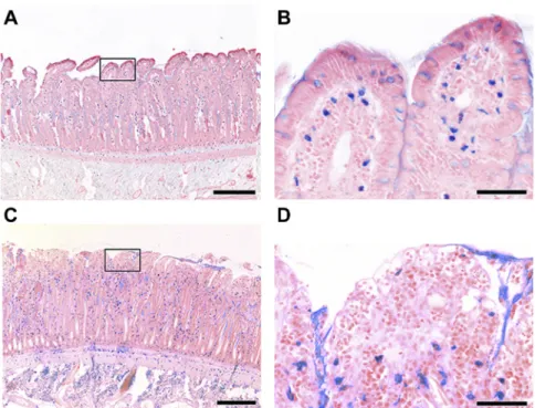
Fig. 9eRepresentative micrographs showing mucosal MCs stained with Alcian blue and Safranin O at (A and C) lower (bar denotes 200mm) and (B and D) higher magnifications (bar denotes 50mm). In Panel B, please note intact intracellular granules, whereas Panel D shows a loss of intracellular granules and stained material dispersed diffusely and extracellularly as signs of MC degranulation at the 120thmin of reperfusion in the IR group. (Color version of figure is available online.)
Fig. 10eRepresentative micrographs of the ileal mucosa stained with hematoxylin and eosin staining at (A) baseline and (B) 120 min of reperfusion in the IR group, (C) the IPCDIR group, and (D) the IPCDIRD7-NI group. Panels B and D demonstrate massive epithelial lifting down the side of villi, denuded villi, increased cellularity of the lamina propria, hemorrhage, and the end of the finger-like appearance of villous structures. Panel C displays the development of subepithelial Gruenhagen’s space at the apex of the villus, capillary congestion, and extension of the subepithelial space with a lifting of the epithelial layer from the lamina propria. The bar denotes 200mm in all figures.
positive effect of IPC was also dependent on mast cell- dependent mediator release in the small bowel.44 In our study, however, the IR-induced degranulation of MCs was not increased, but rather greatly prevented by IPC, which was reversed by 7-NI. It is therefore reasonable to assume that altered MC degranulation is a manifestation of reduced in- flammatory reactions caused by nNOS in this IPC model, and this process may also be involved in mediating the structural injury of the intestinal mucosa.
Apart from oxidative injury, the integrity of the enterocyte lining is also highly dependent on adequate oxygen delivery.
Similar to others (although working with rats),45 our study found evidence of relatively minor IR-induced macro- hemodynamic changes, but microvascular perfusion (as esti- mated indirectly by tonometry) underwent a marked and lasting deterioration. The positive effects of IPC on IR-induced deterioration of microvascular perfusion, tissue oxygenation, and leukocyte-endothelial interaction within the microvas- culature of the small intestine are in evidence here and in a number of other studies.3,46,47 These reactions were also explained with an IPC-induced reduction of intestinal oxida- tive stress.28It appears that our study is the first to show that nNOS also plays a role in a more rapid microcirculatory re- covery after bowel IPC. Because there was also remarkably early evidence of an increase in MPO and the appearance of morphologic injury in the reperfusion phase in our study, the importance of leukocyte-mediated reactions in preserving morphologic integrity (as another manifestation of nNOS- dependent IPC effects) cannot be ruled out either.
Conclusions
In the present study, the beneficial effects of intestinal IPC were examined in a relatively detailed fashion (with respect to pHi, motility dysfunction, leukocyte accumulation, mast cell degranulation, and mucosal injury). Changes in some of the examined parameters (in particular, in IPC-induced marked increase in NO availability: e.g., NOx and constitutive NOS activity, pHi, inflammatory, and morphologic changes) could be influenced/reversed by pretreatment with the nNOS in- hibitor 7-NI. Hence, our data strongly support the contribution of intestinal nNOS enzyme in the protective effect of IPC against the IR-related acute inflammatory response and morphologic injury in the small intestine.
Acknowledgment
This study was funded by research grants from the Hungarian National Research, Development and Innovation Office (NKFIH K116689 to J.K. and K120232 to M.B.), GINOP-2.3.2-15- 2016-00034 (J.K.), and EFOP-3.6.2-16-2017-00006 (A.S.). The authors are grateful to Ms A´ gnes Fekete and Ms Anna Nagyi- va´n for their skillful assistance.
Authors’ contributions: S.V., P.G., and Z.P. contributed to acquisition of data or analysis and interpretation of data. G.B.
and J.K. contributed to conception and design of the study. L.J.
drafted the article. M.B. and A.S. contributed to correction and
proofreading the article. All the authors have read and approved the final article. A.S. and J.K. contributed equally to the study.
Disclosure
The authors reported no proprietary or commercial interest in any product mentioned or concept discussed in this article.
r e f e r e n c e s
1. Murry CE, Jennings RB, Reimer KA. Preconditioning with ischemia: a delay of lethal cell injury in ischemic myocardium.Circulation. 1986;74:1124e1136.
2. Hotter G, Closa D, Prados M, et al. Intestinal preconditioning is mediated by a transient increase in nitric oxide.Biochem Biophys Res Commun. 1996;222:27e32.
3. Wolfa´rd A, Kaszaki J, Varga S, La´za´r G, Boros M. Early microcirculatory changes after ischemic preconditioning and small bowel autotransplantation.Eur Surg Res.
2007;39:284e290.
4. Camprodon RA, Bowles MJ, Pockley AG, de Oca J. Anti- inflammatory effects of ischemic preconditioning on rat small bowel allografts.Transplant Proc. 2014;46:2146e2149.
5. Ferencz A, Sza´nto´ Z, Borsiczky B, et al. The effects of preconditioning on the oxidative stress in small-bowel autotransplantation.Surgery. 2002;132:877e884.
6. Sileri P, Sica G, Gentileschi P, et al. Ischemic preconditioning protects intestine from prolonged ischemia.Transplant Proc.
2004;36:283e285.
7. Taha MO, Miranda-Ferreira R, Chang AC, et al. Effect of ischemic preconditioning on injuries caused by ischemia and reperfusion in rat intestine.Transplant Proc.
2012;44:2304e2308.
8. Calcina F, Barocelli E, Bertoni S, et al. Effect of N-methyl-d- aspartate receptor blockade on neuronal plasticity and gastrointestinal transit delay induced by ischemia/
reperfusion in rats.Neuroscience. 2005;134:39e49.
9. Kubes P. Ischemia-reperfusion in feline small intestine: a role for nitric oxide.Am J Physiol. 1993;264:G143eG149.
10.Qu XW, Wang H, Rozenfeld RA, Huang W, Hsueh W. Type I nitric oxide synthase (NOS) is the predominant NOS in rat small intestine. Regulation by platelet-activating factor.
Biochim Biophys Acta. 1999;1451:211e217.
11.Kanwar S, Tepperman BL, Payne D, Sutherland LR, Kubes P.
Time course of nitric oxide production and epithelial dysfunction during ischemia/reperfusion of the feline small intestine.Circ Shock. 1994;42:135e140.
12.Kawata K, Takeyoshi I, Iwanami K, et al. A spontaneous nitric oxide donor ameliorates small bowel ischemia-reperfusion injury in dogs.Dig Dis Sci. 2001;46:1748e1756.
13.Pala´sthy Z, Kaszaki J, La´za´r G, Nagy S, Boros M. Intestinal nitric oxide synthase activity changes during experimental colon obstruction.Scand J Gastroenterol. 2006;41:910e918.
14.Qu XW, Thaete LG, Rozenfeld RA, et al. Tetrahydrobiopterin prevents platelet-activating factor-induced intestinal hypoperfusion and necrosis: role of neuronal nitric oxide synthase.Crit Care Med. 2005;33:1050e1056.
15.Iijima H, Tulic MK, Duguet A, et al. NOS 1 is required for allergen-induced expression of NOS 2 in mice.Int Arch Allergy Immunol. 2005;138:40e50.
16.Altay T, Gonzales ER, Park TS, Gidday JM. Cerebrovascular inflammation after brief episodic hypoxia: modulation by neuronal and endothelial nitric oxide synthase.J Appl Physiol (1985). 2004;96:1223e1230.
neutrophil content in brain and lung tissue by a modified myeloperoxidase assay.Int J Microcirc Clin Exp. 1996;16:89e97.
23.Gera L, Varga R, To¨ro¨k L, et al. Beneficial effects of
phosphatidylcholine during hindlimb reperfusion.J Surg Res.
2007;139:45e50.
24.Chiu CJ, McArdle AH, Brown R, Scott HJ, Gurd FN. Intestinal mucosal lesion in low-flow states. I. A morphological, hemodynamic, and metabolic reappraisal.Arch Surg.
1970;101:478e483.
25.McCallion K, Wattanasirichaigoon S, Gardiner KR, Fink MP.
Ischemic preconditioning ameliorates ischemia- and reperfusion-induced intestinal epithelial hyperpermeability in rats.Shock. 2000;14:429e434.
26.Yang B, Chen Y, Long YH, et al. Intestinal and limb ischemic preconditioning provides a combined protective effect in the late phase, but not in the early phase, against intestinal injury induced by intestinal ischemia-reperfusion in rats.Shock.
2018;49:596e603.
27.Pontell L, Sharma P, Rivera LR, et al. Damaging effects of ischemia/reperfusion on intestinal muscle.Cell Tissue Res.
2011;343:411e419.
28.Ji YY, Wang ZD, Wang SF, et al. Ischemic preconditioning ameliorates intestinal injury induced by ischemia- reperfusion in rats.World J Gastroenterol. 2015;21:8081e8088.
29.Sola A, De Oca J, Gonza´lez R, et al. Protective effect of ischemic preconditioning on cold preservation and reperfusion injury associated with rat intestinal transplantation.Ann Surg. 2001;234:98e106.
30.Watanabe T, Owada S, Kobayashi H, et al. Real-time monitoring of nitric oxide (NO) and pO2 levels under ischemic conditions associated with small bowel ischemia/reperfusion injury using selective electrodes for NO and oxygen molecules.Transplant Proc. 2007;39:3007e3009.
31.Lai CH, Lee CH, Hung CY, Lo HC. Oral citrulline mitigates inflammation and jejunal damage via the inactivation of neuronal nitric oxide synthase and nuclear factor-kB in intestinal ischemia and reperfusion.JPEN J Parenter Enteral Nutr. 2017;41:422e435.
32.Giaroni C, Marchet S, Carpanese E, et al. Role of neuronal and inducible nitric oxide synthases in the Guinea pig ileum
receptor.Dig Dis Sci. 2013;58:3429e3439.
38. Rivera LR, Thacker M, Pontell L, Cho HJ, Furness JB.
Deleterious effects of intestinal ischemia/reperfusion injury in the mouse enteric nervous system are associated with protein nitrosylation.Cell Tissue Res. 2011;344:111e123.
39. Castro M, Mun˜oz JM, Arruebo MP, et al. Involvement of neuronal nitric oxide synthase (nNOS) in the regulation of migrating motor complex (MMC) in sheep.Vet J.
2012;192:352e358.
40. Gil V, Gallego D, Grasa L, Martı´n MT, Jime´nez M. Purinergic and nitrergic neuromuscular transmission mediates spontaneous neuronal activity in the rat colon.Am J Physiol Gastrointest Liver Physiol. 2010;299:G158eG169.
41. Filpa V, Carpanese E, Marchet S, et al. Nitric oxide regulates homeoprotein OTX1 and OTX2 expression in the rat myenteric plexus after intestinal ischemia-reperfusion injury.Am J Physiol Gastrointest Liver Physiol.
2017;312:G374eG389.
42. Moore-Olufemi SD, Kozar RA, Moore FA, et al. Ischemic preconditioning protects against gut dysfunction and mucosal injury after ischemia/reperfusion injury.Shock.
2005;23:258e263.
43. de Oca J, Hotter G, Sola A, et al. Role of nitric oxide in preconditioning for intestinal transplantation.Transplant Proc. 1999;31:2573.
44. Xing D, Zhang R, Li S, et al. Pivotal role of mast cell carboxypeptidase A in mediating protection against small intestinal ischemia-reperfusion injury in rats after ischemic preconditioning.J Surg Res. 2014;192:177e186.
45. Jansson L, Carlsson PO, Bodin B, Andersson A, Ka¨llskog O.
Neuronal nitric oxide synthase and splanchnic blood flow in anaesthetized rats.Acta Physiol Scand. 2005;183:257e262.
46. Mallick IH, Yang W, Winslet MC, Seifalian AM. Ischaemic preconditioning improves microvascular perfusion and oxygenation following reperfusion injury of the intestine.Br J Surg. 2005;92:1169e1176.
47. Mallick IH, Yang W, Winslet MC, Seifalian AM. Protective effects of ischemic preconditioning on the intestinal mucosal microcirculation following ischemia-reperfusion of the intestine.Microcirculation. 2005;12:615e625.