SEMMELWEIS EGYETEM DOKTORI ISKOLA
Ph.D. értekezések
2082.
BARANYAI TAMÁS
Experimentális és klinikai farmakológia című program
Programvezető: Dr. Magyar Kálmán, professor emeritus Témavezető: Dr. Ferdinandy Péter, egyetemi tanár
REMOTE ISCHEMIC CONDITIONING AND ITS MOLECULAR MECHANISM IN THE
HEART
Ph.D thesis
Tamás Baranyai, MD
Semmelweis University
Doctoral School of Pharmaceutical Sciences
Supervisor: Péter Ferdinandy, MD, D.Sc Official reviewers: Attila Szijártó, MD, Ph.D
István Baczkó, MD, Ph.D
Head of the Final Examination Committee: Attila Tordai, MD, Ph.D Members of the Final Examination Committee: Tamás Radovits, MD, Ph.D
István Koncz, MD, Ph.D
Budapest
2017
1. Table of contents
1. Table of contents ... 2
2. List of abbreviations... 6
3. Introduction ... 8
3.1. Ischemia/reperfusion injury ... 8
3.2. Ischemic conditioning... 10
3.2.1. Ischemic preconditioning ...11
3.2.2. Ischemic postconditioning...12
3.2.3. Remote ischemic conditioning ...13
3.3. Mechanism of ischemic conditioning ... 13
3.3.1. Stimuli/triggers ...14
3.3.2. Signal transport from remote tissue...14
3.3.3. Intracellular mediators ...15
3.4. Interaction between comorbidities and ischemic conditioning... 17
3.4.1. Interaction with chronic comorbidities...17
3.4.2. Interaction with acute comorbidities ...17
4. Aims ... 19
5. Materials and methods ... 20
5.1. Experimental protocol to test remote ischemic conditioningin vivoin a rat model of cardiac ischemia/reperfusion injury ... 20
5.2. Experimental protocol to test remote ischemic conditioningin vivoin a clinically relevant porcine model of cardiac ischemia/reperfusion injury ... 21
5.3. Experimental protocol to test the effect of hyperglycemia on remote ischemic conditioning in anin vivorat model of ischemia/reperfusion injury... 23
5.4. Experimental protocol to test the vesicular nature of remote ischemic conditioningex vivo... 25
5.5. Measurement of area at risk, myocardial function, myocardial necrosis, edema
and microvascular obstruction... 27
5.5.1. Assessment of area at risk with coronary angiography analysis ...27
5.5.2. Assessment of myocardial necrosis by triphenyltetrazolium chloride staining ...27
5.5.3. Assessment of myocardial function, necrosis, edema and microvascular obstruction with porcine cardiac magnetic resonance imaging...28
5.6. Characterisation of extracellular vesicles ... 29
5.6.1. Detection of extracellular vesicles with transmission electron microscopy ...29
5.6.2. Size distribution of extracellular vesicles with dynamic light scattering ..30
5.7. Arrhythmia analysis in rat model of ischemia/reperfusion injury ... 30
5.8. Protein detection with Western blot in various samples ... 30
5.8.1. Sample preparation for Western blot...30
5.8.2. General description of Western blot ...31
5.8.3. Special Western blot description of Triton X-100-insoluble SQSTM1/p62 detection ...32
5.9. Myocardial 3-nitrotyrosine measurement for the assessment of the nitrative stress ... 32
5.10. Statistics ... 33
6. Results ... 34
6.1. The cardioprotective effect of remote ischemic conditioning in rat and porcine hearts ... 34
6.1.1. The cardioprotective effect of remote ischemic conditioning in an in vivo rat model of ischemia/reperfusion injury ...34
6.1.2. The cardioprotective effect of remote ischemic conditioning in an in vivo porcine model of ischemia/reperfusion injury...35
6.1.2.1. Myocardial necrosis, edema and microvascular obstruction by cardiac
magnetic resonance imaging ...37
6.1.2.2. Myocardial necrosis and area at risk evaluated by ex vivo staining ...37
6.1.2.3. Myocardial function by magnetic resonance imaging and echocardiography ...37
6.2. The cardioprotective effect of remote ischemic conditioning is abolished by acute hyperglycemia ... 38
6.2.1. Myocardial necrosis is not reduced by remote ischemic conditioning during acute hyperglycemia ...39
6.2.2. Nitrative stress is increased by acute hyperglycemia ...41
6.2.3. mTOR pathway is overactivated by acute hyperglycemia ...41
6.2.4. Autophagy is not affected by acute hyperglycemia...43
6.3. Remote ischemic conditioning is mediated by extracellular vesicles... 43
6.3.1. Ischemic preconditioning increases the release of cardiac extracellular vesicles ...43
6.3.2. Coronary perfusate depleted from extracellular vesicles did not decrease myocardial necrosis ...45
7. Discussion ... 49
7.1. The promise of remote ischemic conditioning: the role of microvasculature . 49 7.1.1. Remote ischemic conditioning did not reduce myocardial necrosis ...49
7.1.2. Remote ischemic conditioning reduced myocardial edema ...50
7.1.3. Remote ischemic conditioning did not reduce microvascular obstruction 51 7.1.4. Remote ischemic conditioning did not influence myocardial function ...51
7.2. Acute hyperglycemia abolishes the cardioprotective effect of remote ischemic conditioning ... 52
7.2.1. Remote ischemic conditioning is influenced by acute hyperglycemia...52
7.2.2. mTOR is overactivated due to acute hyperglycemia...53
7.3. Remote ischemic conditioning is mediated by extracellular vesicles... 54
7.4. Limitations ... 55
8. Conclusions ... 57
9. Summary ... 58
10. Összefoglalás... 59
11. References ... 60
12. List of own publications ... 84
12.1. Own publications involved in the current thesis ... 84
12.2. Own publications not involved in the current thesis ... 84
13. Acknowledgements ... 86
2. List of abbreviations
AAR - area at risk
AMI - acute myocardial infarction
AMPK - 5' adenosine monophosphate-activated protein kinase ATG7 - autophagy-related gene 7
BNIP3 - Bcl-2/E1B - interacting protein 3 cGMP - cyclic guanosine monophosphate eNOS - endothelial nitric oxide synthase
Erk1,2 - extracellular signal-regulated kinase 1,2 GAPDH - glyceraldehyde-3-phosphate dehydrogenase GSK3β - glycogen synthase kinase 3 beta
IL-6 - interleukin 6
IPostC - ischemic postconditioning IPreC - ischemic preconditioning
JAK - Janus kinase
LAD - left anterior descending coronary artery LC3 - microtubule-associated protein 1 light chain 3
LV - left ventricle
MEK - mitogen-activated protein kinase/ extracellular signal-regulated kinase kinase
mPTP - mitochondrial permeability transition pore
MRI - magnetic resonance imaging
mTOR - mechanistic target of rapamycin MVO - microvascular obstruction
NO - nitric oxide
PI3K - phosphoinositide 3-kinase
PKG - protein kinase G
PTEN - phosphatase and tensin homolog RIC - remote ischemic conditioning RISK - reperfusion injury salvage kinase SAFE - survivor activating factor enhancement
SR - sarcoplasmic reticulum
STAT3 - signal transducer and activator of transcription 3 TNF-α - tumor necrosis factor alpha
ULK1 - UNC-51-like kinase 1
3. Introduction
Ischemic heart disease, characterized by limited oxygen and nutrient supply to myocardial cells, and particularly its subgroup of acute coronary syndrome – a clinical syndrome defined as the relatively rapid oxygen and nutrient depletion of the myocardium – has been a leading cause of mortality and morbidity in the developed countries ever since the 1950s [1]. Only in 2012, 7.4 million people died of ischemic heart disease. Although ischemic heart disease mortality is the highest in the developed countries, it will soon become the number one cause of mortality in low-income countries as well [2]. Advanced treatment of ischemic heart disease and acute coronary syndrome has significantly decreased the mortality rates, especially in the high-income countries.
The most pioneering improvements to be mentioned involve the establishment of coronary care units with full access to monitors [3], the development of percutaneous coronary catheterization and balloon angioplasty [4], and of the surgical revascularisation techniques [5]. Nevertheless, the effective treatment of established cardiovascular risk factors is also responsible for the reduction in morbidity and mortality [6].
3.1. Ischemia/reperfusion injury
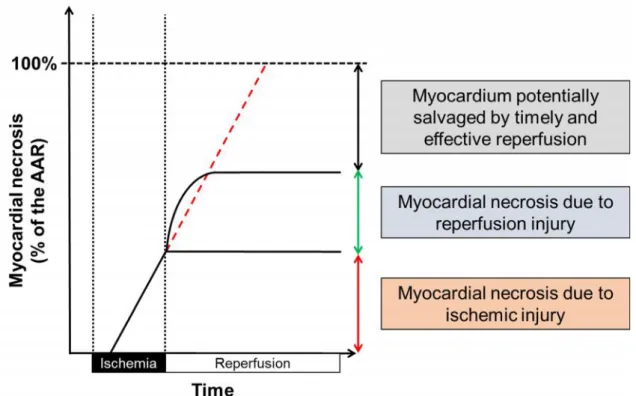
Early restoration of coronary circulation is obligatory in order to prevent the ischemia-triggered myocardial damage. Paradoxically, reperfusion per se further exacerbates myocardial injury. Thus, both ischemia and reperfusion are responsible for the overall myocardial damage seen following acute myocardial ischemia and subsequent reperfusion therapy. This is termed ischemia/reperfusion injury (Figure 1) [7].
During ischemia, the nutrient deprivation and decrease in oxygen supply lead to a disturbance of the myocardial homeostasis [8]. After 8-10 seconds hypoxia, all oxygen offered by haemoglobin, myoglobin and creatine-phosphate is extracted and utilized from the cellular proximity [9], cellular oxidative phosphorylation shuts down, and aerob metabolism shifts to anaerobic one [10]. Once the anaerobic reserve burns out, myocardial cells start to decay. Many factors, such as mitocondrial depolarization and dysfunction, sodium- and calcium overload, intra- and extracellular acidosis and reactive oxygen species production all contribute to the myocardial cell death as coordinated by cardiac myocytes, endothelial and inflammatory cells,etc.[11].
Figure 1.The contribution of ischemia and reperfusion to the final myocardial necrosis.
Reperfusion may responsible for 50 % of the total necrosis [7].
The reperfusion injury occurs in the first few minutes of the reperfusion, and similarly to the ischemic injury, is a complex phenomenon [11]. As the myocardial perfusion is restored, tissue oxygen level increases rapidly, metabolites are washed out and different units of the respiratory chain are recruited rapidly. However, the rapid restoration of the downregulated respiratory chain elements results in an increased formation of reactive oxygen species, leading to protein-, lipid- and nucleic acid oxidation [12].
Simultaneously, the already increased cellular calcium concentration [13] further increases via sarcoplasmic calcium release, thereby activating apoptotic pathways [14- 16]. Nevertheless, it seems the primary mediator of reperfusion injury is the opening of the mitochondrial permeability transition pore (mPTP) [17].
The (pre)clinical measurement of ischemia/reperfusion injury may be analyzed from three major aspects, which are closely linked together and define the most important endpoint, the mortality: myocardial contractility, electrophysiological function, and irreversible and reversible cellular damage.
Myocardial contractile dysfunction may occur as an acute heart failure during ischemia or reperfusion. Chronic heart failure may develop due to ischemia/reperfusion
injury over years. Importantly, reversibly injured regions of the myocardium may recover completely in a delayed manner termed myocardial stunning, or remain hypoperfused and hibernated [18-20]. The onset of arrhythmias (ranging from isolated premature beats to ventricular fibrillation) during ischemia or upon reperfusion is quite frequent complication of acute myocardial infarction (AMI) [21, 22]. Irreversible and reversible cellular damage can be measured in many ways. It is well-established that the final myocardial necrosis correlates significantly with the mortality [23]. However, the development of the final necrosis is an extremely complex chain of events with many cellular components and players [7]. For instance, it seems that microvascular damage is one of the most important factors contributing to the ultimate damage [7, 24]. Myocardial edema [25], microvascular obstruction (MVO; ischemic area where myocardial perfusion is not restored despite successful revascularization) [26], endothelial dysfunction and microembolisation are major signs of microvascular damage [27].
Many pharmaceutical agents have been proposed to attenuate myocardial ischemia/reperfusion injury. The common feature of these compounds are that they either interfere with well-established detrimental molecular pathways or that they facilitate/mimic the protective endogenous mechanisms activated upon reperfusion (see Section 3.3). The most promising agents identified in preclinical studies have already undergone clinical trials after encouraging preclinical results: adenosine [28], cyclosporin-A [29], erythropoietin [30], statins [31], insulin [32, 33], nitrite/nitrates and soluble guanylate cyclase activators [34, 35], exenatide [36], metoprolol [37] and nicorandil [35]. However, the majority of the studies aiming to modify a single target failed to improve the overall outcome after AMI. Nevertheless, ischemic conditioning enables a more comprehensive way to target multiple pathways orchestrating the effects of the ischemia/reperfusion injury.
3.2. Ischemic conditioning
During ischemic conditioning of the heart, short cycles of ischemia/reperfusion achieve a more resistant state of the myocardium against myocardial ischemia/reperfusion injury due to sustained injury,i.e., index myocardial ischemia (Figure 2). Classification of the ischemic conditioning strategies is based on several parameters including the site of the applied ischemia/reperfusion cycles or based on the timing referenced to the index
ischemia. Ischemic conditioning stimuli can be elicited on the affected coronary artery of the heart or on a coronary artery distant to the index ischemia or even on a remote organ (e.g., kidney, skeletal muscle) termed remote ischemic conditioning (RIC). Temporal variants of ischemic conditioning are ischemic pre-, per- and postconditioning [38], in which the conditioning is applied before, during and after the ischemic event, respectively.
Figure 2.The concept of ischemic pre-, post- and remote conditioning. Black bars represent ischemic periods over time. Figure is adapted from Ferdinandyet al.2014
[39].
3.2.1. Ischemic preconditioning
In 1986, Murryet al.showed for the first time that ischemic preconditioning (IPreC;
i.e., brief cycles of ischemia and reperfusion of the involved coronary artery before index ischemia) is able to attenuate subsequent myocardial damage [40]. Since then, the cardioprotective effect of IPreC has been confirmed numerous times both in preclinical
models and in clinical trials [41]. It has been demonstrated that IPreC significantly reduces myocardial necrosis [40], enhances postischemic cardiac function [42], improves endothelial dysfunction [43], prevents reperfusion-induced cardiac arrhythmias [44] and increases the resistance against hypoxia [45]. Specially, IPreC might be extremely useful in elective cardiac interventions. It has been reported that IPreC of the human heart results in reduced lactate and/or creatine kinase MB release, attenuated ECG alterations and/or reduced postinterventional complaints of patients after a secondary, elective percutaneous coronary intervention compared to the first intervention [46-48]. Moreover, numerous proof-of-concept studies on coronary artery bypass graft surgery have demonstrated an IPreC-induced cardioprotection proven by a reduction in cardiac biomarkers, such as creatine kinase MB and troponin T or I [41]. It is believed that pre-infarction angina endogenously mimics IPreC with better clinical outcome, nevertheless, this has been challenged by others [41].
3.2.2. Ischemic postconditioning
The clinical application of IPreC is limited to elective interventions accompanying myocardial ischemia, as the conditioning stimuli should precede the prolonged ischemia.
However, primary physicians, clinicians, etc., diagnose and treat myocardial ischemia after its onset, and IPreC is no longer a therapeutic option. Nonetheless, it has been demonstrated that ischemic conditioning reduces myocardial ischemia/reperfusion injury if appliedaftercardiac ischemia (termed as ischemic postconditioning; IPostC) [49, 50], which has already been postulated in earlier works [51, 52]. Similarly to IPreC, IPostC protects the myocardium from ischemia/reperfusion injury by improving myocardial function, attenuating myocardial oxidative damage, inflammation, edema, etc., after myocardial ischemia, however, the cardioprotection is less pronounced than IPreC [53].
Theoretically, IPostC could be applied either after elective or acute cardiac interventions/cardiothoracic surgeries. Indeed, IPostC has been reported to be cardioprotective both in preclinical [38] and clinical [54, 55] settings. However, to date, the largest clinical trials investigating the efficacy of IPostC did not show any benefit of IPostC on ST-elevation myocardial infarction patients after primary percutaneous intervention (POST and DANAMI 3-iPOST trial) in terms of long-term outcome [56, 57].
3.2.3. Remote ischemic conditioning
IPreC is impossible to apply in acute situations, and IPostC is by definition limited to be effective against the reperfusion-induced myocardial damage only. The discovery of RIC initiated an extensive research activity in the cardiovascular field. Originally, RIC demonstrated that short episodes of ischemia and reperfusion to a canine circumflex artery are able to alleviate the deteriorating effects of prolonged ischemia of the left anterior descending coronary artery (LAD) [58]. Since then, the cardioprotective effect of RIC was demonstrated that not only a distant part of the same organ but applying short ischemia/reperfusion cycles on a distant organ (brain, skin, intestine, kidney,etc.) triggers cardioprotection [59]. More importantly, Birnbaumet al.discovered that restricted blood flow and electrical pacing of rabbit gastrocnemius muscle protects the myocardium from the subsequent ischemia/reperfusion injury [60]. Noninvasively, Oxmanet al.described that RIC stimuli could be elicited with a simple tourniquet in rats [61], and thereafter, Günaydinet al.[62] and Kharbandaet al.[63] showed that this can be demonstrated in humans. This noninvasive approach facilitated the rapid translation into the clinical setting, and it has been justified by many proof-of-concept clinical trials [64-66].
However, the long-term outcome of such interventions is still controversial. Thus far, a completed small-scale clinical trial involving ST-elevation myocardial infarction patients demonstrated long-term efficacy of RIC as assessed by significant improvement of major adverse cardiac and cerebrovascular events [67]. Nevertheless, two large clinical trials reported that RIC did not improve the long-term outcome after cardiac surgery (ERICCA [68] and RIPHEART [69] trials).
3.3. Mechanism of ischemic conditioning
There are thousands of studies investigating the major mechanism of ischemic conditioning. It is clear that ischemic conditioning seems to be a well-conserved, endogenous program of the heart that shares a highly orchestrated spatiotemporal signal transduction with similar elements [53]. Theoretically, the major parts of the signal transduction are the stimulus or trigger, and the intracellular mediators. Since RIC stimulus occurs in a distant organ, it involves a third element as well, namely the signal transport to the target organ.
3.3.1. Stimuli/triggers
Cardioprotective triggers are by nature chemical or biological compounds, and many of which are part of the myocardial endogenous adaptation ischemia/reperfusion injury.
Ischemia and reperfusion are very complex pathophysiological phenomena. Ischemia and reperfusion induce the release of a mixture of chemical and biological components, and simultaneously trigger endogenous alterations of the cellular homeostasis.
The major chemical stimuli released upon short ischemia/reperfusion are reactive oxygen species, such as nitric oxide. Although reactive oxygen species have a pivotal role in cardioprotection, they behave as a double-edged sword [70]: a small amount of reactive oxygen species recruits protective intracellular signals (e.g., oxidize possibly protective cytosolic kinases) [71-73], while a more pronounced reactive oxygen species formation induces irreversible injury [70]. It has been also indicated that calcium ions [74] and hydrogen sulfide [75] may exert similar cardioprotection.
A significant amount of cardioprotective triggers belongs to the biologically active molecules, which act on specific receptors and throughout regulating intracellular pathways modifies the defense of the myocytes: autocoids (adenosine and bradykinin [76]), neurotransmitters (acetylcholine, catecholamines, endothelin, opiods [77]), peptide hormons (natriuretic peptides [78], adrenomedullin [79]) and cytokines (tumor necrosis factor-α and interleukins [80, 81]).
3.3.2. Signal transport from remote tissue
RIC is a unique conditioning phenomenon, since the stimulus is applied apart from the site of protection, necessitating a mechanistic pathway conveying the protective information. Two major form of signal transfer have been proposed: neuronal and humoral [38].
It is well-established that an intact afferent neuronal pathway is required for the proper signal transmission [82]. Furthermore, stimulation of the afferent neuronal fibres can mimic RIC-induced cardioprotection [83]. The mechanism of the efferent neuronal pathway is less clear, however, the vagal nerve may have a pivotal role [84, 85].
Humoral signal transfer is based on an early observation where the coronary effluent of a preconditioned heart was transferred to a naïve acceptor heart and exerted a similar
degree of cardioprotection [86]. Since then, many protective factors have been identified, such as stromal-derived factor-1α [87], nitric oxide [88], microRNA-144 [89], hypoxia-inducible factor-1α [90] and apolipoprotein a-I [91].
Nevertheless, the true nature of the protection may be a well-coordinated interplay between neuronal and humoral factors [38, 92].
3.3.3. Intracellular mediators
Chemical and biological triggers initiate the temporal and spatial orchestration of intracellular mediators of cardioprotection (Figure 3). The intracellular protective pathway identified was the protein kinase C pathway [93]. Initially, Ytrehuset al., showed that protein kinase C inhibitors abrogated the cardioprotective effect of IPreC subjected to rabbit hearts [93]. Later on, many other pathways have been identified, such as the nitric oxide/cyclic guanosine monophosphate/protein kinase G- [94], the Reperfusion Injury Salvage Kinase (RISK)- [95], the Survivor Activating Factor Enhancement (SAFE)- [96], the hypoxia-induced factor 1α- [97], the mitogen-activated protein kinase- [98] and a microRNA-regulated [99] pathways.
A number of investigations has been conducted to reveal the primary role of the mitochondria and sarcoplasmic reticulum (SR) in the cardioprotection. During short ischemic periods, mPTP opens transiently, preparing or recapitulating the mitochondrial reactive oxygen species- and calcium-homeostasis for the subsequent or elapsed, prolonged ischemia [100, 101]. Conversely, during reperfusion, the mPTP stays closed by the abovementioned mediators, preventing the mitochondrial depolarization [102, 103]. In addition, the activation of the mitochondrial ATP-dependent potassium channels rigorously regulates the calcium-flow and metabolic enzymes [104, 105]. Ion homeostasis is also maintained by the RISK- and SAFE-dependent phosphorylation of connexin-43, a gap junction-related poreforming protein [106]. To preserve the calcium homeostasis, a close functional relationship between mPTP and SR is required [107, 108]. Furthermore, SR facilitates the unfolded, damaged protein degradation [109] and activates autophagy to eliminate injured organelles [110, 111].
Figure 3.Major intracellular pathways of ischemic/pharmacological conditionings.
(Own graph based on Heusch, 2015 [53]). RISK – Reperfusion Injury Salvage Kinase;
SAFE – survivor activating factor enhancement; PTEN – phosphatase and tensin homolog; PI3K – phosphoinositide 3-kinase; eNOS – endothelial nitric oxide synthase;
NO – nitric oxide; cGMP – cyclic guanosine monophosphate; PKG – protein kinase G;
GSK3β – glycogen synthase kinase 3 beta; AMPK – 5' adenosine monophosphate- activated protein kinase; mTOR – mechanistic target of rapamycin;
MEK – mitogen-activated protein kinase/ extracellular signal-regulated kinase kinase;
Erk1,2 – extracellular signal-regulated kinase 1,2; IL-6 – interleukin 6; TNF-α – tumor necrosis factor alpha; JAK – Janus kinase; STAT3 - signal transducer and activator of transcription 3; mPTP – mitochondrial permeability transition pore; SR – sarcoplasmic
reticulum
3.4. Interaction between comorbidities and ischemic conditioning
3.4.1. Interaction with chronic comorbidities
In the clinical routine, the majority of ischemic heart diseases has been preceded by risk factors, such as hyperlipidemia, hypertension, diabetes mellitus, chronic renal failure, ageing,etc.[39, 112]. Moreover, to prevent the development or manifestation of ischemic heart diseases, patients are treated with a great amount of medications [39].
Comorbidities, aging and medications significantly influence and alter the structure and intracellular homeostasis of the heart. However, during preclinical research, healthy, juvenile and drug-naïve animals are used to test the efficacy of ischemic conditioning phenomena. Hypercholesterolemia significantly deteriorates the myocardial homeostasis and function without the development of atherosclerosis [113]. It has been shown that hyperlipidemia abolishes the cardioprotective effect of IPreC and IPostC [114-116].
Interestingly, IPreC, but not IPostC, could be elicited in hypertensive preclinical models [117, 118]. Similarly, IPreC [119], IPostC [120] and RIC [121] are ineffective in the presence of diabetes mellitus (type 1 or 2). Mechanisms contributing to the abolished cardioprotective strategies have been attributed to the impaired RISK/SAFE and nitric oxide/cyclic guanosine monophosphate/protein kinase G pathways, to the increased oxidative-nitrative stress, to the altered 5' AMP-activated protein kinase function, etc.
(see reviews for details [39, 112]). Nevertheless, the cardioprotective effect of IPreC was preserved in chronic renal failure [122].
3.4.2. Interaction with acute comorbidities
It is well-established that chronic comorbidities significantly alter the cardiac homeostasis and metabolism, and therefore the response to the same stimuli, such as ischemic conditioning [39]. However, it is not well-studied how acute pathologies without any chronic comorbidities (e.g., hypertensive crisis, hyperglycemia and acute renal failure) influence IPreC, IPostC or RIC.
Apart from being a major component of chronic metabolic diseases, hyperglycemia may also occur in acute situations for example as seen in sympathetic overactivation
during acute coronary syndrome [123]. Hyperglycemia in non-diabetic patients – a group of patients in which hyperglycemia is unlikely to persist – is generally associated with adverse outcomes after an AMI [124]. It is not known whether this acute hyperglycemia is the cause of adverse outcomes or it only reflects the severity of the AMI [123].
Furthermore, it has been shown that hyperglycemia inhibits cardioprotection conferred by IPreC and cardioprotection by various pharmacological agents [125-127].
4. Aims
To test the cardioprotective effect of RIC in rat and porcine model of AMI.
To investigate whether acute hyperglycemia interferes with the cardioprotective effect of RIC.
To investigate whether RIC signal is transported via extracellular vesicles.
5. Materials and methods
These investigations conform to the Guide for the Care and Use of Laboratory Animals published by the US National Institutes of Health (NIH publication No. 85–23, revised 1996), to the EU Directive (2010/63/EU) and was approved by the animal ethics committee of the Semmelweis University and by the animal ethics committee of Hungarian National Food Chain Safety Office (SOI/31/26-11/2014).
5.1. Experimental protocol to test remote ischemic conditioning in vivo in a rat model of cardiac ischemia/reperfusion injury
Unfasted, 220–280 g male Wistar rats were anesthetized with 60 mg/kg pentobarbital.
The absence of pedal reflex was considered as a sign of deep surgical anesthesia. Electric activity of the heart was monitored (AD Instruments, Bella Vista, Australia). Blood pressure was measured in the carotid artery (AD Instruments, Bella Vista, Australia).
Body temperature was maintained with a heat pad at physiological temperature (35.8–
38.3 °C). Rats were ventilated with 10 mL/kg stroke volume at rate of 80 strokes/min (Ugo-Basile, Gemonio, Italy).
Figure 4.Experimental protocol to test remote ischemic conditioningin vivoin a rat model of cardiac ischemia/reperfusion injury. Isch – ischemia only group, RIC – remote
ischemic conditioning, LAD – left anterior descending coronary artery, TTC – triphenyltetrazolium chloride.
Rats were randomized into two groups (Figure 4): (1) control ischemic and (2) RIC.
At 35 min of the study protocol, LAD was occluded with a 6–0 polypropylene suture via
median thoracotomy for 40 min. Occlusion was confirmed by ST-segment elevation, arrhythmias and paling of the occluded area. RIC was induced by three cycles of 5 min occlusion and 5 min reperfusion of the right femoral vessels starting after 10 min of the LAD occlusion. Both the femoral artery and vein were occluded with a metal vessel clamp after isolation of the vessels from the surrounding connective tissue and femoral nerve.
At the end of the 40 min index ischemia, reperfusion was induced by loosening the suture.
At the end of the 120 min reperfusion, hearts were harvested in order to evaluate the extent of myocardial necrosis.
5.2. Experimental protocol to test remote ischemic conditioning in vivo in a clinically relevant porcine model of cardiac ischemia/reperfusion injury
Domestic female pigs (25-35 kg; genotype: DanBred hybrid) were purchased from the Product Development and Monitoring Research Center of University of Kaposvár, and were kept according to the Big Dutchman principles. Animals were fed with pregnant sow diet with low energy and balanced protein level produced by Dalmand co. Ltd.
Pigs were block randomized [128] into three groups (Figure 5): ischemia only (Isch;
n=17), IPreC (n=12) and RIC (n=17). Three animals died during myocardial ischemia (Isch: 1; IPreC: 1; RIC: 1) and 3 during reperfusion due to therapy resistant malignant ventricular rhythm disturbances (Isch: 2; IPreC: 1; RIC: 0). Furthermore, 3 animals were excluded due to procedural technical reasons. The final case numbers were 14, 9 and 14 in Isch, IPreC, and RIC groups, respectively.
One day prior to the experiments, pigs were given 500 mg acetyl salicylic acid and 300 mg clopidogrel. Animals were sedated with 12 mg/kg ketamine hydrochloride, 1 mg/kg xylazine, and 0.04 mg/kg atropine intramuscularly after overnight fasting.
Anesthesia was induced by inhalation of isoflurane (2-2.5 vol%). Animals were intubated endotracheally and anesthesia was maintained by isoflurane oxygen mix inhalation (2-2.5 vol% and 3 L/min). Magnesium sulphate (4.06 mEq diluted in 10 mL, in every 60 min) and continuous amiodarone infusion (300 mg diluted in 500 mL saline) were being administered throughout the procedure via an ear vein. Sheaths were inserted into femoral artery and femoral vein to have entry routes for the catheterization, and
5000-5000 IU heparin was administered via each sheath. During preparation, cardiac function was assessed with echocardiography. Selective angiography of the left coronary artery was performed. After the analysis of the baseline angiogram, balloon catheter (2.75 mm diameter, 8 mm length) (Abbott, Abbott Park, IL, US) was placed in the mid part of the LAD after the origin of the second diagonal branch. For induction of AMI, the intracoronary balloon was inflated with 5 atm for 90 min, followed by deflation of the balloon, resulting in reperfusion (3 h or 3 days) confirmed by coronarography.
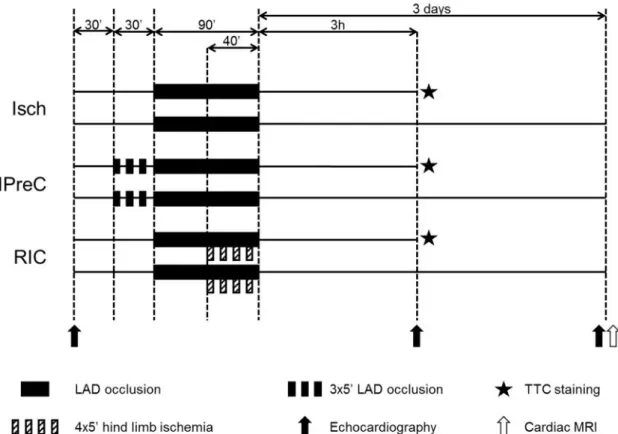
Figure 5.Experimental protocol to test remote ischemic conditioning in anin vivo porcine model of acute myocardial infarction. Isch – ischemia only, IPreC – ischemic preconditioning, RIC – remote ischemic conditioning, LAD – left anterior descending
coronary artery, TTC – triphenyltetrazolium chloride, MRI – magnetic resonance imaging.
In IPreC group, LAD was occluded by the inflation of the balloon at 5 atm 3 times for 5 min followed by 5 min of reperfusion, while in other animals the balloon was left deflated for 30 min. Then LAD was occluded by inflating coronary balloon, which was confirmed by coronarography. RIC was performed by four cycles of 5 min occlusion and 5 min reperfusion of the femoral vessels by tightening and releasing a snare around the
right hind limb starting at the 50th min of LAD occlusion. We documented the hind limb ischemia in three ways: 1) apparent lividity during ischemia and pronounced hyperemia during reperfusion were observed, 2) in each animal, a superficial femoral artery was cannulated distal to the occlusion, and blood pressure was measured during the intervention. The minimum of 30/30 mmHg blood pressure with 0 mmHg pulse pressure was achieved while the wire was tightened around the hind limb, and 3) in one particular animal, we performed femoral angiography before and during hind limb ischemia as well.
The interventional cardiologist was not blinded during the investigation due to the nature of the procedure: IPreC was achieved by inflating a balloon in the LAD, which require an unblinded interventional cardiologist. Nonetheless, the interventional cardiologist was not aware of the allocation until the initiation of the experimental intervention.
After reperfusion, 5000 IU heparin was initiated by intracoronary administration.
Final reperfusion was confirmed with coronarography and the catheters were subsequently removed. Anesthesia was either maintained for 3 h or in the case of 3 days reperfusion, wounds were closed and anesthesia was terminated by the withdrawal of isoflurane. Analgesia was applied by intramuscular injections of 1 g metamizole. An antibiotic cocktail (100 mg benzathine benzylpenicillin, 100 mg procaine benzylpenicillin, 200 mg dihydrostreptomycin-sulphate) was given im. before recovery.
5.3. Experimental protocol to test the effect of hyperglycemia on remote ischemic conditioning in an in vivo rat model of ischemia/reperfusion injury
In vivo experiments of RIC in rats has been performed as described in Section 5.1.
Rats were randomized into 2 groups (Figure 6): control normoglycemic, and acute hyperglycemic. Acute hyperglycemic animals received 50 % dextrose (vWR, Radnor, PA, US) infusion via the tail vein from the start of the experimental protocol. A blood glucose level of 15–20 mM was reached within 5 min by an infusion rate of 150 µL/min.
Then infusion rate was adjusted to maintain blood glucose levels between 15 and 20 mM throughout the entire protocol (0–60 µL/min, with an average of 50 µL/min), which was measured every 15 min (Accu-Check, Roche, Basel, Switzerland). In normoglycemic animals, an equal osmolarity, 46 % mannitol solution was administered (vWR, Radnor,
PA, US, induction rate: 150 µL/min for 5 min, then 50 µL/min).
After 35 min ofin vivoperfusion, half of the animals from normoglycemic and acute hyperglycemic groups were sacrificed, and hearts were excised, immersed readily in ice-cold Krebs-Henseleit solution until they were mounted. They were then perfused in Langendorff mode for 1 min with oxygenated (95 % oxygen/5 % CO2 gas mixture) Krebs-Henseleit solution (118 mM NaCl, 4.7 mM KCl, 1.2 mM MgSO4, 1.25 mM CaCl2, 1.2 mM KH2PO4, 25 mM NaHCO3and 11 mM glucose) at 37 °C to wash out blood, as described earlier [129]. The hearts were then snap-frozen in liquid nitrogen and stored at
−80 °C until further experiments. The other half of the animals were further randomized into four groups: (1) control ischemic with normoglycemia (NG + Isch), (2) RIC with normoglycemia (NG + RIC), (3) ischemic with acute hyperglycemia (AHG + Isch) and (4) RIC with acute hyperglycemia (AHG + RIC). At 35 min of the study protocol, LAD
Figure 6.Experimental protocol to test the effect of hyperglycemia on remote ischemic conditioning in anin vivorat model of acute myocardial infarction.
NG – normoglycemia, AHG – acute hyperglycemia, Isch – coronary ischemia, RIC – coronary ischemia and remote ischemic conditioning, nTyr – 3-nitrotyrosine, ELISA – enzyme-linked immunosorbent assay, TTC – triphenyltetrazolium chloride.
was occluded with a 6–0 polypropylene suture via median thoracotomy for 40 min.
Occlusion was confirmed by ST-segment elevation, arrhythmias and paling of the occluded area. RIC was induced by three cycles of 5 min occlusion and 5 min reperfusion of the right femoral vessels starting 10 min after the LAD occlusion. Both the femoral artery and vein were occluded with a metal vessel clamp after isolation of the vessels from the surrounding connective tissue and femoral nerve. At the end of the 40 min index ischemia, reperfusion was induced by loosening the suture. At the end of the 120 min reperfusion, hearts were harvested for evaluation of the degree of myocardial necrosis.
5.4. Experimental protocol to test the vesicular nature of remote ischemic conditioning ex vivo
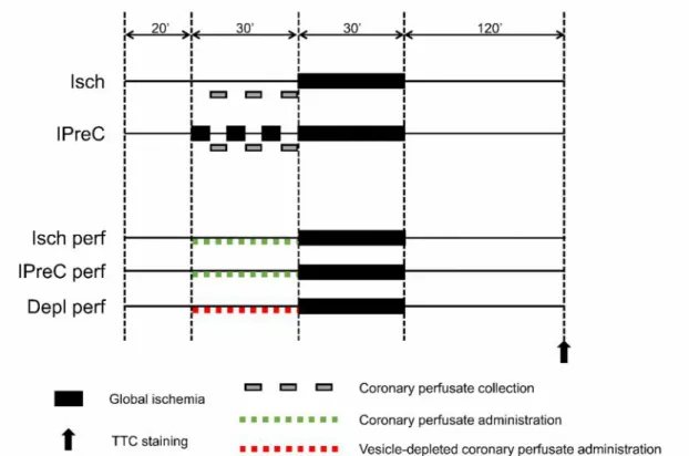
Male Wistar rats (250–350 g) were anesthetized by 85 mg/kg ketamine and 10 mg/kg xylazine and heparinized (500 U/kg). Hearts were isolated and perfused in Langendorff mode with 37 °C Krebs–Henseleit solution (118 mM NaCl, 4.7 mM KCl, 1.2 mM MgSO4, 1.25 mM CaCl2, 1.2 mM KH2PO4, 25 mM NaHCO3and 11 mM glucose) for 20 min for stabilization. Hearts were randomized into five groups (Figure 7): 1) 30 min global ischemia (Isch); 2) additional IPreC with 3×5 min global ischemia/reperfusion;
3) perfused with perfusate collected from Isch hearts prior to the 30 min global ischemia starting from the 20th min (Isch perf); 4) perfused with perfusate collected from IPreC hearts prior to the 30 min global ischemia starting from the 20th min (IPreC perf);
5) perfused with vesicle-depleted perfusate collected from IPreC hearts prior to the 30-min global ischemia starting from the 20th min (Depl perf). After 120 min of reperfusion, hearts were sliced into 2 mm slices, and stained with triphenyltetrazolium chloride as in Section 5.5.2.
Extracellular vesicles were isolated from collected coronary perfusates by filtration and differential centrifugation. Freshly prepared coronary perfusates were dialyzed in cellulose dialysis tube (Sigma, St. Louis, MO, US) against 0.45 % saline containing 5 mM EDTA for 4 h at room temperature. CFs were then vacuum-distilled to 40 mL (for approximately 40 min) at room temperature. Concentrated coronary perfusates were filtered through a 800 nm filter (Merck, Darmstadt, Germany) by gravity, and filtrates were centrifuged at 12,200×g for 20 min at 4 °C. Pellets were saved as
microvesicle/microparticle fraction. Supernatants of the previous centrifugation were filtered through a 200 nm filter (Merck, Darmstadt, Germany) by gravity and centrifuged at 100,000×g for 90 min at 4 °C in Beckman MLA-55 rotors. Pellets were saved as exosome-rich pellet and the supernatant was saved as vesicle-depleted perfusate. Vesicle- depleted perfusates were then reconstituted to their original volume measured at the time of coronary perfusate collection with Krebs-Henseleit solution (e.g., in case 200 mL coronary perfusate was collected, 40 mL of depleted perfusate was combined with 160 mL Krebs-Henseleit solution). Reconstituted perfusates were used in heart perfusion experiments on the same day.
Figure 7.Experimental protocol to test vesicular nature of remote ischemic conditioningex vivo. Isch – ischemia only, IPreC – ischemic preconditioning, perf – perfused with perfusate collected from Isch or IPreC hearts, Depl perf – perfused
with perfusate collected from IPreC hearts and depleted from extracellular vesicles, TTC – triphenyltetrazolium chloride.
5.5. Measurement of area at risk, myocardial function, myocardial necrosis, edema and microvascular obstruction
5.5.1. Assessment of area at risk with coronary angiography analysis
All animals underwent coronary angiography according to the protocol established by the catheterization laboratory. Anterograde flow in the artery before and after balloon inflation was characterized using the TIMI (Thrombolysis in Myocardial Infarction) system [130]. TIMI myocardial perfusion grade and myocardial blush grade were assessed visually on the angiogram and made by expert interventional cardiologist, and all data were entered prospectively into a database. Myocardial blush grade has been defined as follows: 0, no myocardial blush or contrast density; 1, minimal myocardial blush or contrast density; 2, moderate myocardial blush or contrast density but less than that obtained during angiography of a contralateral or ipsilateral non-infarct-related coronary artery; and 3, normal myocardial blush or contrast density, comparable with that obtained during angiography of a contralateral or ipsilateral non-infarct-related coronary artery. When myocardial blush persisted (“staining”), this phenomenon suggested leakage of contrast medium into the extravascular space and was graded 0 [131, 132]. No digital techniques were used. The area at risk (AAR) was established by using the modified APPROACH score [133].
5.5.2. Assessment of myocardial necrosis by triphenyltetrazolium chloride staining
Hearts were excised at the end of the indicated reperfusion periods. Rat hearts from in vivoexperiments were perfused with oxygenated (95 % oxygen/5 % CO2gas mixture) 37 °C Krebs-Henseleit solution (118 mM NaCl, 4.7 mM KCl, 1.2 mM MgSO4, 1.25 mM CaCl2, 1.2 mM KH2PO4, 25 mM NaHCO3and 11 mM glucose) in Langendorff mode for 1 min. LAD was reoccluded, and the AAR was negatively stained with Evans blue (Sigma, St. Louis, MO, US) retrogradely. Then 2 mm slices were cut. Rat hearts fromex vivoexperiments were cut into 2 mm slices without Evans blue (Sigma, St. Louis, MO, US) staining at the end of reperfusion.
Porcine hearts were placed immediately in ice-cold saline. Coronary angiography of
the pig was reviewed, and the occlusion site was identified. LAD was reoccludedex vivo at the same place asin vivowith a pean, and Evans blue (Sigma, St. Louis, MO, US) was injected into the coronary arteries through their orifices to negatively stain the AAR. Then 10 mm slices were cut. Heart slices were incubated in 1 % triphenyltetrazolium chloride (Sigma, St. Louis, MO, US) at 37 °C for 15 min to stain viable areas. After overnight fixation with 4 % formalin, slices were weighed and scanned for blind planimetric analysis (InfarctSize 2.4b software [Pharmahungary Group, Szeged, Hungary]). AAR was expressed as the proportion of the left ventricular (LV) mass, and necrosis as the proportion of the AAR mass. In case of hearts receivingex vivo global ischemia, AAR was equal to the LV.
5.5.3. Assessment of myocardial function, necrosis, edema and microvascular obstruction with porcine cardiac magnetic resonance imaging
Three days (70-78 h) after the deflation of the balloon in LAD of pigs, anesthesia was induced by inhalation of isoflurane-oxygen mix. Prior to cardiac magnetic resonance imaging (MRI), atracurium was administered and ventilation was maintained with mechanical ventilation. For the assessment of myocardial necrosis and edema, cardiac MRI was performed using a 1.5 T clinical scanner (Avanto, Siemens, Erlangen, Germany) using a phased array coil and a vector electrocardiogram system. Cine MRI images were acquired using a retrospectively electrocardiography-gated, steady-state free precession cine MRI technique (Cinetruefisp sequence) in short-axis and long-axis views of the heart using 1.2 ms echo time, 40 ms repetition time, 65 degree flip angle, 15 segments, 360 mm field-of-view, 8 mm slice thickness, and 256×256 image matrix. The image resolution was 1.4×1.4*8 mm. Dark blood prepared IR-TSE sequence single slice breath-hold acquisition T2w protocol with IR preparation (technical detail: TI=170 ms) 15 segments using every second trigger pulse was used to detect edema (technical details: TE 74 ms, flip angle 180 degree). The slice position and resolution was identical as cine images. The late gadolinium-enhanced images were acquired to determine the amount of myocardial necrosis and MVO. A 2-dimensional single shot Truefisp sequence with non-selective IR pulse shift acquisition to a diastolic phase of the cardiac cycle by adjusting the TR was
used 12 to 15 min after administration of a gadolinium-based contrast agent (0.13 mmol/kg gadobutrol [Bayer, Whippany, NJ, US]), with slice positions identical to the cine images. Typical in-plane resolution was 1.4×1.4×8 mm (Technical details: echo time 1.2 ms, flip angle 50°, triggering to every other heart beat). The inversion time was set to null the signal of viable myocardium and ranged from 280 to 320 ms. Left and right ventricular end-diastolic and end-systolic volumes, stroke volumes, ejection fractions and masses were quantified using manual planimetry of end-diastolic and end-systolic short-axis SSFP cine images with MASS 7.6 analysis software (Magnetic Resonance Analytical Software System, Medis Medical Imaging Software, Leiden, The Netherlands).
Myocardial necrosis and edema were quantified after manual planimetry both on the delayed contrast enhancement and T2-weighted images by delineation of myocardium with signal intensity 4 SDs above the mean signal obtained in the remote non-infarcted myocardium using MASS 7.6 analysis software. If present, the hypointense zone in the center of the hyperenhancement (MVO) was quantified and added to the infarct volume as previously described [134]. Values were expressed relative to the LV mass.
5.6. Characterisation of extracellular vesicles
5.6.1. Detection of extracellular vesicles with transmission electron microscopy
Extracellular vesicle pellet was prepared by centrifuging samples at 100,000×g for 90 min at 4 °C in Beckman MLA-55 rotors. Formalin (4 %) was layered carefully onto the pellets. After formalin was washed out, pellets were post-fixed with 1 % osmium tetraoxide (OsO4) for 20-30 min, and block stained with 1 % uranyl acetate (in 50 % ethanol) for 20-30 min. Then, pellets were dehydrated by graded ethanol for 5 min in 70 %, 90 %, 96 %, and 3×5 min in 100 % ethanol. Pelleted samples were embedded in Taab 812 (Taab Laboratories, Aldermaston, UK). Then ultrathin sections were prepared.
Prepared samples were analyzed under Hitachi 7100 electron microscope equipped by Veleta, a 2k×2k MegaPixel side-mounted TEM CCD camera (Olympus, Tokyo, Japan).
Contrast and brightness of electron micrographs were edited in Adobe Photoshop CS3 (Adobe Systems Incorporated, San Jose, CA, US).
5.6.2. Size distribution of extracellular vesicles with dynamic light scattering
Size distribution of extracellular vesicles was measured by dynamic light scattering measurements by using an ALV goniometer with a Melles Griot diode-pumped solid-state laser at 457.5 nm wavelength (type: 58 BLD 301). The intensity of the scattered light was measured at 90° and the autocorrelation function was calculated using an IBM PC-based data acquisition system. Particle size distributions were determined by the maximum entropy method. The diameter of the particles was calculated using sphere approximation.
5.7. Arrhythmia analysis in rat model of ischemia/reperfusion injury
An electrocardiogram was recorded throughout the entire experiment. Arrhythmia analysis was performed according to the Lambeth conventions, and arrhythmia incidence and duration scores were calculated [135].
5.8. Protein detection with Western blot in various samples 5.8.1. Sample preparation for Western blot
In order to demonstrate presence or absence of extracellular vesicles in the perfusate of Langendorff perfused hearts, coronary perfusate collected from Isch and IPreC hearts (n=3) were dialyzed and concentrated as described above. After passing concentrated samples through 800 nm filters by gravity, a total extracellular vesicle pellet was prepared by centrifuging samples at 100,000×g for 90 min at 4 °C in Beckman MLA-55 rotors.
Supernatants of the centrifugation were kept as extracellular vesicle-depleted perfusates.
Total extracellular vesicle pellets were resuspended in 35 µL radioimmunoprecipitation assay buffer (Cell Signaling, Danvers, MA, US) containing protease inhibitor cocktail (Roche, Basel, Switzerland). Radioimmunoprecipitation assay buffer (Cell Signaling, Danvers, MA, US) containing protease inhibitor was added to extracellular vesicle-depleted perfusates as well in 1/10 ratio. Twenty µL of each sample was mixed
with 5× Laemmli-buffer (Bio-Rad, Hercules, CA) and loaded on 4-20 % Tris-glycine SDS-polyacrylamide gels (Bio-Rad, Hercules, CA).
LV tissue was homogenized in radioimmunoprecipitation assay buffer (Cell Signaling, Danvers, MA, US) supplemented with protease inhibitor (Roche, Basel, Switzerland), sodium fluoride (Sigma, St. Louis, MO, US) and phenylmethylsulfonyl fluoride (Sigma, St. Louis, MO, US). Protein concentration of the homogenates was measured by Bicinchoninic Acid Assay kit (Thermo Fisher Scientific, Waltham, MA, US).
5.8.2. General description of Western blot
Twenty µL of each sample (samples containing extracellular vesicles) or equal amount of protein was mixed with reducing 5× Laemmli buffer, loaded and separated in a 4-20 % precast Tris-glycine SDS polyacrilamide gel (Bio-Rad, Hercules, CA, US) with 90-110 V. Proteins were transferred onto a polyvinylidene difluoride membrane (Bio-Rad, Hercules, CA, US) at 350 mA for 2 h or 200 mA overnight. Proper transfer was visualized with Ponceau staining (Sigma, St. Louis, MO, US). Membranes were blocked with 5 % bovine serum albumin (Santa Cruz Biotechnology, Dallas, TX, US) or non-fat dry milk (Bio-Rad, Hercules, CA, US) in Tris-buffered saline containing 0.05 % Tween-20 (Sigma, St. Louis, MO, US) at room temperature for 2 h. Membranes were probed with primary antibodies purchased from Cell Signaling (Danvers, MA, US) overnight at 4 °C (HSP60 - #4870; phospho-mTOR [Ser2448] - #2971; mTOR - #2972;
phospho - S6 [Ser235/236] - #2211; ribosomal S6 - #2317; phospho-AKT [Ser473] - #9271; AKT - #9272; phospho-AMP-activated protein kinase α [AMPKα;
Thr172] - #2535; AMPKα - #5831; phospho-extracellular signal-regulated kinase 1/2 [Erk1/2; Thr202/Tyr204] - #9106; Erk1/2 - #9107; microtubule-associated protein 1 light chain 3 A/B [LC3A/B] - #4108; beclin-1 - #3495; SQSTM1/p62 - #5114;
phospho-UNC-51-like kinase 1 [p-ULK1; Ser555] - #5869; ULK1 - #4773;
autophagy-related gene 7 [ATG7] - #8558; Bcl-2/E1B - interacting protein 3 [BNIP3] - #3769; loading control: glyceraldehyde-3-phosphate dehydrogenase [GAPDH] - #5174), and with corresponding horseradish peroxidase-conjugated secondary antibodies (Cell Signaling, Danvers, MA, US) for 2 h at room temperature.
Signals were detected with an enhanced chemiluminescence kit (Bio-Rad, Hercules, CA, US) by Chemidoc XRS+ (Bio-Rad, Hercules, CA, US). Antibodies detecting phosphorylated epitopes were removed with Pierce Stripping Buffer (Thermo Fisher Scientific, Waltham, MA, US) before incubation with antibodies detecting the total protein.
5.8.3. Special Western blot description of Triton X-100-insoluble SQSTM1/p62 detection
LV tissue was homogenized with TissueLyser (Qiagen, Venlo, The Netherlands) in a homogenation buffer containing 50 mM Tris, 150 mM NaCl, 1 mM EDTA, 10 % glycerol and 2 % Triton X-100 (pH 8.0) supplemented with protease inhibitor (Roche, Basel, Switzerland), sodium fluoride (Sigma, St. Louis, MO, US) and phenylmethylsulfonyl fluoride (Sigma, St. Louis, MO, US). Homogenates were centrifuged (10,000×g, 10 min, 4 °C) supernatant was carefully removed and discarded.
Pellet was washed with the above mentioned homogenation buffer once again (10,000×g, 10 min, 4 °C). Pellets were then resuspended in sample buffer containing 62.5 mM Tris-HCl, 5 % glycerol and 1.3 % SDS. Protein concentration of the homogenates was measured by Bicinchoninic Acid Assay kit (Thermo Fisher Scientific, Waltham, MA, US). Equal amount of protein (20 µg) was loaded, separated and processed under reducing conditions as described above.
5.9. Myocardial 3-nitrotyrosine measurement for the assessment of the nitrative stress
Free myocardial 3-nitrotyrosine was measured from LV samples harvested at 35 min with 3-nitrotyrosine ELISA (Cayman, Ann Arbor, MI, US) according to the manufacturer’s protocol.
5.10. Statistics
Data was expressed asmean±standard error of mean.Student’s t-testwas performed where two groups including contionuous variables were compared.One-way ANOVA was performed in case of the comparison of multiple groups including contionuous variables. Repeated measures ANOVA was used if multiple groups with continuous variables were compared at different time points. To test the effect and interaction of two variables (i.e., AHG and RIC),two-way ANOVAwas performed. After ANOVA studies, either LSD or Dunnett’s post hoc test was applied. To limit the case numbers in the cardiac MRI study, one-way ANOVA was performed by using bootstrapping with 1000-sample replacement [136], as frequently used in clinical studies [137-139] and recommended for preclinical studies as well [140]. Groups with discrete variables (i.e., scores) were compared withKruskal–Wallis analysis. Mortality statistics has been developed by using theKaplan–Meier estimation. Statistical significance was accepted if p<0.05. Statistical tests were performed with IBM SPSS Statistics, Version 19.
6. Results
6.1. The cardioprotective effect of remote ischemic conditioning in rat and porcine hearts
6.1.1. The cardioprotective effect of remote ischemic conditioning in an in vivo rat model of ischemia/reperfusion injury
To investigate whether RIC attenuates myocardial necrosis, RIC was tested in an in vivo rat model of AMI as described in Section 5.1 and Figure 4. RIC significantly attenuated the myocardial necrosis compared to the Control group (p<0.05; n=5-6; Figure 8a). There was no difference between the AAR of groups (n=5-6).
Figure 8. aRIC attenuated myocardial necrosis.bAAR was not different between groups.cRepresentative images of Evans blue/triphenyltetrazolium chloride-stained heart sections indicating myocardial necrosis and AAR. Orange: slice outline, green:
ventricular chamber, purple: AAR, yellow: myocardial necrosis. *p<0.05 vs. Control.
RIC – remote ischemic conditioning, AAR – area at risk, LV – left ventricle.
6.1.2. The cardioprotective effect of remote ischemic conditioning in an in vivo porcine model of ischemia/reperfusion injury
To investigate whether RIC attenuates myocardial necrosis, RIC was tested in an in vivo porcine model of ischemia/reperfusion injury as described in Section 5.2 and Figure5.
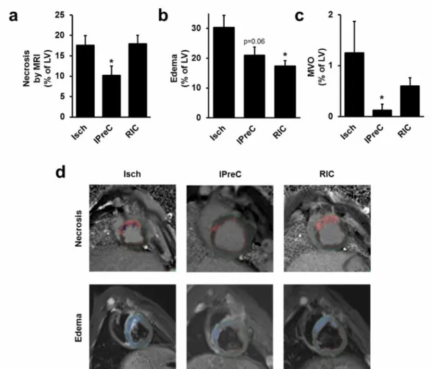
Figure 9. aIPreC, but not RIC, reduced myocardial necrosis.bIPreC and RIC decreased myocardial edema.cIPreC reduced MVO.dRepresentative MRI images of
myocardial necrosis and edema. Green line: epicardial outline, red line: endocardial outline, red area: myocardial necrosis, dark blue area: MVO, light blue area: myocardial
edema. *p<0.05 vs. Isch. Isch – ischemia only, IPreC – ischemic preconditioning, RIC – remote ischemic conditioning, MRI–magnetic resonance imaging, LV – left
ventricle, MVO – microvascular obstruction.
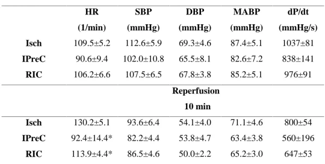
Table 1.At baseline, hemodynamic parameters were not significantly different between groups, whereas the heart rate was significantly lower in IPreC and RIC groups as compared to the Isch group. *p<0.05 vs. Isch. Isch – ischemia only, IPreC – ischemic preconditioning, RIC – remote ischemic conditioning, HR – heart rate, SBP – systolic blood pressure, DBP – diastolic blood pressure, MABP – mean arterial blood pressure.
Table 2.There was no difference in various angiographic parameters between various groups. Isch – ischemia only, IPreC – ischemic preconditioning, RIC – remote ischemic
conditioning, LV – left ventricle.
APPROACH score (% of LV)
TIMI score Myocardial blush grade
TIMI myocardial
perfusion grade
Isch 19.2±2.2 2.4±0.3 2.5±0.3 2.3±0.3
IPreC 18.6±2.4 2.2±0.3 2.4±0.3 2.3±0.3
RIC 19.7±2.5 2.3±0.3 2.5±0.3 2.5±0.3
Baseline HR
(1/min)
SBP (mmHg)
DBP (mmHg)
MABP (mmHg)
dP/dt (mmHg/s)
Isch 109.5±5.2 112.6±5.9 69.3±4.6 87.4±5.1 1037±81
IPreC 90.6±9.4 102.0±10.8 65.5±8.1 82.6±7.2 838±141
RIC 106.2±6.6 107.5±6.5 67.8±3.8 85.2±5.1 976±91
Reperfusion 10 min
Isch 130.2±5.1 93.6±6.4 54.1±4.0 71.1±4.6 800±54
IPreC 92.4±14.4* 82.2±4.4 53.8±4.7 63.4±3.8 560±196
RIC 113.9±4.4* 86.5±4.6 50.0±2.2 65.2±3.0 647±53
6.1.2.1. Myocardial necrosis, edema and microvascular obstruction by cardiac magnetic resonance imaging
To quantify myocardial necrosis, edema and MVO, cardiac MRI was performed 3 days after coronary occlusion and reperfusion. Myocardial necrosis (% of LV) was not affected by RIC, however, IPreC attenuated it significantly as compared to the Isch group (p<0.05; n=4-8), although only 4 IPreC cases were studied with MRI (Figure 9a).
Myocardial edema (% of LV) was significantly decreased by RIC as compared to the Isch group (p<0.05; n=7-7), however, only a tendency of decrease was observed by IPreC (n=4; p=0.06; Figure 9b). At baseline, hemodynamic parameters were not significantly different between groups, whereas the heart rate was significantly lower in IPreC and RIC groups as compared to the Isch group (Table 1). AAR based on angiographic score was not different between groups (Table 2). Furthermore, angiographic measures of reperfusion, i.e., TIMI score, myocardial blush grade, and TIMI myocardial perfusion grade were not affected by conditioning stimuli (Table 2). MVO volume (% of LV) was significantly decreased by IPreC (p<0.05), but not by RIC as compared to the Isch group (n=4-8; Figure 9c).
6.1.2.2. Myocardial necrosis and area at risk evaluated by ex vivo staining
To assess myocardial necrosis and AAR by an ex vivohistological method, the gold standard triphenyltetrazolium chloride- and Evans blue staining were applied after 3 h of reperfusion. RIC did not decrease myocardial necrosis (% of AAR), however, IPreC significantly decreased it as compared to the Isch group (n=5-6; Figure 10a). There was no difference in AARs between the experimental groups (% of LV) as evaluated by Evans blue staining (Figure 10b).
6.1.2.3. Myocardial function by magnetic resonance imaging and echocardiography
Myocardial function was analyzed by cardiac MRI. Myocardial function was not different between groups after 3 days of reperfusion (Table 3). Similar data was obtained by echocardiographic analysis (Data not shown).
Table 3.There was no difference in myocardial function between groups as assessed by cardiac MRI. Isch – ischemia only, IPreC – ischemic preconditioning, RIC – remote ischemic conditioning, HR – heart rate, LV – left ventricle, RV – right ventricle, EDVI – end-diastolic volume index, ESVI – end-systolic volume index, SVI – stroke volume
index, EF – ejection fraction, COI – cardiac output index.
Isch IPreC RIC
HR (1/min) 96.26±7.76 100.50±5.06 100.13±5.07 LV EDVI (mL/kg) 3.63±0.25 3.10±0.02 3.55±0.09 LV ESVI (mL/kg) 2.07±0.15 1.81±0.14 2.09±0.17 LV SVI (mL/kg) 1.49±0.11 1.29±0.15 1.40±0.10 LV EF (%) 43.47±1.67 41.64±4.75 41.55±2.77 LV COI (L/min/kg) 0.15±0.02 0.11±0.01 0.14±0.02 RV EDVI (mL/kg) 2.86±0.21 2.80±0.18 2.59±0.10 RV ESVI (mL/kg) 1.40±0.09 1.65±0.05 1.32±0.08 RV SVI (mL/kg) 1.46±0.15 1.15±0.18 1.28±0.08 RV EF (%) 43.95±2.43 40.63±3.66 47.42±2.61 RV COI (L/min/kg) 0.14±0.02 0.09±0.01 0.12±0.01
6.2. The cardioprotective effect of remote ischemic conditioning is abolished by acute hyperglycemia
To investigate the effect of acute hyperglycemia on the efficacy of RIC, acute hyperglycemia was induced with a 50 % dextrose infusion during in vivo ischemia/reperfusion experiments as described in Section 5.3 and Figure 6. Blood glucose levels were significantly elevated due to dextrose perfusion in both AHG+Isch and AHG+RIC groups compared to the corresponding normoglycemic groups from at least the 15th min of perfusion (Table 4, preliminary results showed that acute hyperglycemia developed in 5 min [17.2±1.8 mM, n=4]). Elevated blood glucose levels did not influence the heart rate and blood pressure of the rats (Table 5).
Figure 10. aIPreC, but not IPostC and RIC, reduced myocardial necrosis.bThere was no difference between AARs.cRepresentative images of Evans
blue/triphenyltetrazolium chloride-stained heart sections indicating myocardial necrosis and AAR. Orange: slice outline, green: ventricular chamber, purple: AAR, yellow:
myocardial necrosis. *p<0.05 vs. Isch. Isch–ischemia only, IPreC – ischemic preconditioning, RIC – remote ischemic conditioning, AAR–area at risk, LV – left
ventricle.
6.2.1. Myocardial necrosis is not reduced by remote ischemic conditioning during acute hyperglycemia
Myocardial necrosis was significantly smaller in NG+RIC group compared to NG+Isch (p<0.05; n=7-10), while RIC failed to decrease myocardial necrosis in
AHG+RIC group when compared to AHG+Isch (n=5-6; Figure 11a). Furthermore, acute hyperglycemia per se did not aggravate cardiac necrosis (Figure 11a). There was no difference between the AAR of various groups (n=5-10; Figure 11b).
Arrhythmia analyses revealed that acute hyperglycemia compared to normoglycemia significantly increased the incidence and duration of arrhythmias during the whole myocardial ischemic period, but arrhythmia incidence and duration were not different during reperfusion (Table 6). RIC per sedid not alter arrhythmia scores from the time point it was applied, as compared to Isch group (Table 6). Furthermore, RIC did not decrease arrhythmia scores during acute hyperglycemia compared to AHG+RIC (Table 6).
Figure 11. aAcute hyperglycemia abolished cardioprotective effect of RIC.bThere was no difference between AARs.cRepresentative images of Evans
blue/triphenyltetrazolium chloride-stained heart sections indicating myocardial necrosis and AAR. Orange: slice outline, green: ventricular chamber, purple: AAR, yellow:
myocardial necrosis. *p<0.05 vs. NG + Isch. #p<0.05 vs. NG + RIC.
NG – normoglycemia, AHG – acute hyperglycemia, Isch – ischemia only group, RIC – remote ischemic perconditioning, AAR – area at risk, LV – left ventricle.
*
Myocardial necrosis
6.2.2. Nitrative stress is increased by acute hyperglycemia
Increased oxidative and/or nitrative stress is often implicated in the disruption of cardioprotective interventions. The 3-nitrotyrosine content, a marker of nitrative stress, was therefore measured in the hearts of normoglycemic and acute hyperglycemic rats at 35 min. Cardiac 3-nitrotyrosine was significantly elevated due to acute hyperglycemia (p<0.05; n=8-8; Figure 12).
Table 4.Blood glucose (mM) is elevated in acute hyperglycemia. *p<0.05 vs.
corresponding time point of NG+Isch group. #p<0.05 vs. corresponding time point of NG+RIC group. NG – normoglycemia, AHG – acute hyperglycemia, Isch – ischemia
only group, RIC – remote ischemic perconditioning.
6.2.3. mTOR pathway is overactivated by acute hyperglycemia
Because the oxidative and nitrative stress have been previously shown to interact with mTOR pathway [141], we evaluated the expression and/or phosphorylation of mTOR pathway associated proteins. Phosphorylation levels of mTOR (Ser2448) and S6 (Ser235/236) were significantly elevated (p<0.05; n = 7-9; Figure 13a-b), which indicates that the activity of mTOR complex I was increased in acute hyperglycemia.
Phosphorylation of AKT at site Ser473 was also significantly elevated in acute hyperglycemia (p<0.05; n=7-9; Figure 13c), however, other mTOR regulators, such as phosphorylated AMPKα (Thr172) and Erk1/2 (Thr202/Tyr204) were unchanged in acute hyperglycemia as compared to normoglycemia (n=7-9; Figure 13d-e).
Table 5.Acute hyperglycemia does not influence heart rate (HR) and mean arterial blood pressure (MABP). NG – normoglycemia, AHG – acute hyperglycemia,
Isch – ischemia only group, RIC – remote ischemic conditioning.
Figure 12.Acute hyperglycemia increased cardiac nitrative stress. 3-nitrotyrosine content of hearts of NG or AHG rats. *p<0.05 vs. NG. NG – normoglycemia,
AHG – acute hyperglycemia.
6.2.4. Autophagy is not affected by acute hyperglycemia
Since oxidative/nitrative stress and mTOR pathway have been shown to interact with autophagy, expression and/or phosphorylation of autophagy-related proteins were assessed in normoglycemia and acute hyperglycemia. LC3II/LC3I ratio was significantly decreased due to acute hyperglycemia (p<0.05; n=7-9; Figure 14a), however, other autophagy-related proteins such as Beclin-1, total and Triton X-100-insoluble SQSTM1/p62, phospho-ULK1 (Ser555), ATG7 and BNIP3 were unchanged in acute hyperglycemia (n=7-9; Figure 14b-g).
6.3. Remote ischemic conditioning is mediated by extracellular vesicles
Vesicular transfer of RIC mediators was investigated in an ex vivo setup of cardiac ischemia/reperfusion injury as described in Section 5.4 and Figure 7.
6.3.1. Ischemic preconditioning increases the release of cardiac extracellular vesicles
Coronary perfusates from IPreC hearts contained more extracellular vesicles than perfusates isolated from control (Isch) hearts as evidenced by immunoblotting against HSP60, a well-accepted marker of extracellular vesicles (Figure 15a). Electron micrographs revealed that these extracellular vesicles can be classified as
microvesicle/microparticles as defined by a diameter of >100 nm and light vesicular structure (Figure 15b), and exosomes of <100 nm in diameter (Figure 15b). To further demonstrate that size of the particulate matter in coronary perfusate conforms to the range of exosomes and microvesicles, we assessed their size distribution by dynamic light scattering in perfusates from IPreC hearts. As the representative diagram (Figure 15c) shows, three populations of particles could be distinguished in our samples. Besides a small fraction of particles with hypothetical diameter of approximately 10 nm, the main particulate constituents fell into the size range of exosomes (<100 nm) and microvesicles (100–1000 nm).
Table 6.Acute hyperglycemia exacerbates the incidence and duration of arrhythmias during ischemia. *p<0.05 vs. corresponding time point of NG+Isch group. #p<0.05 vs.
corresponding time point of NG+RIC group. NG – normoglycemia, AHG – acute hyperglycemia, Isch – ischemia only group, RIC – remote ischemic conditioning.