MicroRNAs associated with ischemia-reperfusion injury and cardioprotection by ischemic pre- and postconditioning: protectomiRs
Zoltán V. Varga,1,2,6Ágnes Zvara,3Nóra Faragó,3Gabriella F. Kocsis,1Márton Pipicz,1Renáta Gáspár,1 Péter Bencsik,1,6Anikó Görbe,1,6Csaba Csonka,1,6László G. Puskás,3Thomas Thum,4,5Tamás Csont,1,6* and Péter Ferdinandy1,2,6*
1Cardiovascular Research Group, Department of Biochemistry, University of Szeged, Szeged, Hungary;2Department of Pharmacology and Pharmacotherapy, Semmelweis University, Budapest, Hungary;3Institute of Genetics, Biological Research Center, Hungarian Academy of Sciences, Szeged, Hungary;4Institue of Molecular and Translational Therapeutic Strategies, Hannover Medical School, Hannover, Germany;5National Heart and Lung Institute, Imperial College London, London, United Kingdom; and6Pharmahungary Group, Szeged, Hungary
Submitted 17 October 2013; accepted in final form 20 May 2014
Varga ZV, Zvara Á, Faragó N, Kocsis GF, Pipicz M, Gáspár R, Bencsik P, Görbe A, Csonka C, Puskás LG, Thum T, Csont T, Ferdi- nandy P. MicroRNAs associated with ischemia-reperfusion injury and cardioprotection by ischemic pre- and postconditioning: protectomiRs.Am J Physiol Heart Circ Physiol307: H216–H227, 2014. First published May 23, 2014; doi:10.1152/ajpheart.00812.2013.—We aimed to characterize early changes in microRNA expression in acute cardioprotection by isch- emic pre- and postconditioning in rat hearts. Hearts isolated from male Wistar rats were subjected to1) time-matched nonischemic perfusion, 2) ischemia-reperfusion (30 min of coronary occlusion and 120 min of reperfusion), 3) preconditioning (3⫻ 5 min of coronary occlusion) followed by ischemia-reperfusion, or 4) ischemia-reperfusion with postconditioning (6⫻10 s of global ischemia-reperfusion at the onset of reperfusion). Infarct size was significantly reduced by both inter- ventions. Of 350 different microRNAs assessed by microarray anal- ysis, 147–160 microRNAs showed detectable expression levels. Com- pared with microRNA alterations induced by ischemia-reperfusion versus time-matched nonischemic controls, five microRNAs were significantly affected by both pre- and postconditioning (microRNA- 125b*, microRNA-139-3p, microRNA-320, microRNA-532-3p, and microRNA-188), four microRNAs were significantly affected by preconditioning (microRNA-487b, microRNA-139-5p, microRNA- 192, and microRNA-212), and nine microRNAs were significantly affected by postconditioning (microRNA-1, microRNA let-7i, microRNA let-7e, microRNA let-7b, microRNA-181a, microRNA-208, mi- croRNA-328, microRNA-335, and microRNA-503). Expression of randomly selected microRNAs was validated by quantitative real-time PCR. By a systematic comparison of the direction of microRNA expression changes in all groups, we identified microRNAs, specific mimics, or antagomiRs that may have pre- and postconditioning-like cardioprotective effects (protectomiRs). Transfection of selected pro- tectomiRs (mimics of microRNA-139-5p, microRNA-125b*, microRNA let-7b, and inhibitor of microRNA-487b) into cardiac myocytes subjected to simulated ischemia-reperfusion showed a significant cytoprotective effect. This is the first demonstration that the ischemia- reperfusion-induced microRNA expression profile is significantly in- fluenced by both pre- and postconditioning, which shows the involve- ment of microRNAs in cardioprotective signaling. Moreover, by analysis of microRNA expression patterns in cardioprotection by pre- and postconditioning, specific protectomiRs can be revealed as po- tential therapeutic tools for the treatment of ischemia-reperfusion injury.
microRNA; miRNA; miR; reperfusion injury; cardioprotection
MICRORNAS ARE SMALL (19 –23 nucleotides) noncoding RNA molecules that are currently being recognized as endogenous physiological regulators of gene expression (1). It has been well established that microRNAs play an essential role in the posttranscriptional regulation of gene expression by repression or activation of translation/transcription (“RNA interference”) (5, 27). MicroRNAs are known to play important roles in many physiological and pathological processes in the heart, including cardiac fibrosis, the hypertrophic response, myocyte contrac- tility, cardiac development, and arrhythmogenesis (36). Re- cently, several research groups have reported the involvement of some microRNAs in the pathophysiology of myocardial infarction (11, 31). The importance of microRNAs in cardiac pathologies suggests their promising therapeutic potential (15, 19). Modulation of microRNA expression in vivo with specific synthetic oligonucleotides is a feasible approach, as appli- cation of both microRNA inhibitors (antagomiRs) and mi- croRNA mimics offer a potential therapeutic option for cardioprotection (35).
However, little is known on the role of microRNAs in the ischemic myocardium, particularly in the early phase of myocardial ischemia-reperfusion (I/R) injury and in endog- enous cardioprotective maneuvers such as ischemic pre- and postconditioning. These cardioprotective interventions, es- pecially ischemic postconditioning, has emerged as a clinically applicable approach for limiting myocardial injury in humans (30). However, despite of intensive research in the last de- cades, the exact biological mechanisms underlying the cardio- protective effect of pre- and postconditioning remain largely unclear due to the complexity of the cellular events and the interference of other comorbidities and risk factors of ischemic heart diseases (17, 30). Therefore, the use of systems biological tools seems necessary to reveal the complex molecular events.
Accordingly, we have previously shown by cDNA microarrays that ischemic preconditioning (29) and ischemic postcondition- ing (10) markedly influenced the gene expression pattern of rat hearts.
In the present study, we aimed to identify microRNAs involved in pre- and postconditioning-induced cardioprotec- tion by the application of a systematic comparison of the microRNA expression patterns of different myocardial pheno- types, including nonischemic, I/R, protected by preconditioning,
* T. Csont and P. Ferdinandy contributed equally to this work.
Address for reprint requests and other correspondence: P. Ferdinandy, Cardiovascular Research Group, Dept. of Pharmacology and Pharmacother- apy, Semmelweis Univ., Nagyvárad tér 4, Budapest 1089, Hungary (e-mail:
peter.ferdinandy@pharmahungary.com).
First published May 23, 2014; doi:10.1152/ajpheart.00812.2013.
and protected by postconditioning. In fact, preceding ischemic preconditioning and subsequent postconditioning stimuli can be considered as effective “inhibitors” of I/R injury. Therefore, by comparing microRNA expression changes and the direction of the changes (i.e., down- or upregulation) induced by I/R versus time-matched nonischemic controls and in I/R with pre- or postconditioning, respectively, we were able to reveal possible causative relationships between microRNA changes and en- dogenous cardioprotection. Using microRNA microarrays of 350 known rat microRNA genes, in the present study, we identified several microRNAs associated with cardioprotection by ischemic pre- and postconditioning. These microRNAs are potential therapeutic targets for cardioprotection, appropriate mimics, or antagomiRs, which were termed here as “protecto- miRs.” Validation of four of the potential protectomiRs was also performed in cardiomyocytes subjected to simulated I/R in vitro.
MATERIALS AND METHODS
This investigation conformed with the National Institutes of Health Guide for the Care and Use of Laboratory Animals(NIH Pub. No.
85-23, Revised 1996) and was approved by local animal ethics committee of the University of Szeged.
Experimental Protocol
Male Wistar rats (250 –300 g) were anesthetized with diethyl ether and given 500 U/kg heparin intravenously. Hearts were then isolated and perfused according to Langendorff with oxygenated, normother- mic Krebs-Henseleit buffer as previously described (9). Four different perfusion protocols were applied (Fig. 1). A time-matched nonisch- emic control group was aerobically perfused for 190 min. In the I/R group, regional ischemia was induced by 30 min of occlusion of the left anterior descending coronary artery (LAD) followed by 120 min of reperfusion. In the preconditioning group, hearts were subjected to a preconditioning protocol (induced by three intermittent cycles of 5 min of coronary occlusion/5 min of reperfusion) followed by I/R. In the postconditioning group, postconditioning was induced by six consecutive cycles of 10 s of global ischemia and 10 s of reperfusion applied immediately at the onset of the 120 min of reperfusion. Heart
rate and coronary flow were monitored throughout the perfusion protocol in all groups. To assess I/R injury and cardioprotection, infarct size was determined by planimetry (Infarctsize 2.4, Pharma- hungary, Szeged, Hungary) after standard triphenyltetrazolium chlo- ride staining at the end of reperfusion (n⫽8 –10/group). Infarct size was expressed as a percentage of the area at risk (8). To further assess tissue injury, lactate dehydrogenase (LDH) release of hearts was measured using a LDH-P kit (Diagnosticum, Budapest, Hungary) from coronary effluents collected for 5 min upon reperfusion after test ischemia (n⫽12/group). LDH release was expressed as milliunits per minute per gram wet heart weight. In separate experiments, at the end of the 120 min of reperfusion, we sampled the LAD-supplied risk zone of the left ventricle for further analysis of microRNA expression.
Sampling was performed by a single oblique cut with scissors from the origin of the LAD toward the right side of the apical area that involves the majority of the anterior wall of the left ventricle as well as the apex of the heart. Samples from all groups (n⫽6/group) were rapidly frozen in liquid nitrogen for microRNA isolation.
MicroRNA Isolation
MicroRNA isolation was performed according to the protocol for isolation of microRNA from tissues as supplied in the manufacturer’s kit (Roche) with some modifications (14). RNA quality and quantity were assessed spectrophotometrically (Nanodrop) with a 2100 Bio- analyzer (Agilent).
Microarray Analysis of MicroRNA Expression
MicroRNAs were labeled using the miRNA Complete Labeling and Hyb kit system (Agilent Technologies, Palo Alto, CA). Labeled samples were completely vacuum dried on the medium-high (45°C) heat setting and hybridized onto the surface of a 8⫻15k Rat miRNA Microarray (Agilent Technologies), which was done in microarray hybridization chambers (Agilent Technologies), as previously de- scribed (13). Each array was scanned as previously described (16).
Statistical analysis was performed on fluorescent signal intensity data representing microRNA expression, as described below.
Changes in gene expression were determined as ratios of signal intensity values. For visual comparison and representation of both down- and upregulation, data were shown as log2changes. Averages of log2changes were plotted with SD values.
30 min 40 min
Ischemia/reperfusion (IR)
120 min Time-matched, non-ischemic control (C)
190 min
30 min Ischemic preconditioning (PRE)
120 min
5’ 5’ 5’
30 min 40 min
Ischemic postconditioning (POST)
120 min
10’ 5’ 5’ 5’
6x10” I/R
aerobic perfusion regional ischemia
perfusate collection infarct size measurement tissue sampling for microRNA assays
Global ischemia cycles to induce POST
Fig. 1. Experimental protocol. Hearts isolated from male Wistar rats were subjected to either time- matched aerobic perfusion [i.e., time-matched non- ischemic control (C)], 30 min of coronary occlusion and 120 min of reperfusion [ischemia/reperfusion (I/R; IR)], 3⫻5 min of I/R followed by 30 min of coronary occlusion and 120 min of reperfusion [pre- conditioning (PRE)], or 6 ⫻ 10 s of global I/R applied immediately after 30 min of coronary oc- clusion and followed by 120 min of reperfusion [postconditioning (POST)] protocols. Coronary ef- fluent was collected upon the first 5 min of reper- fusion after coronary occlusion for lactate dehydro- genase release detection. Infarct size was measured at the 120th min of reperfusion. For microRNA isolation, in separate experiments, left ventricles were frozen in liquid nitrogen at the end of the perfusion protocol.
Validation of Microarray Data by Quantitative Real-Time PCR To confirm microarray results, quantitative real-time PCR was used on randomly selected microRNAs, as previously described (12).
Experiments were performed with the TaqMan MicroRNA Reverse Transcription Kit (Applied Biosystems) and with TaqMan MicroRNA Assays (Applied Biosystems).
Identification of ProtectomiRs
Ischemic preconditioning and postconditioning can be considered as effective “inhibitors” of I/R injury. Therefore, by comparing microRNA expression changes and the direction of the changes (i.e., down- or upregulation) induced by I/R versus time-matched nonisch- emic controls and in I/R with pre- or postconditioning, respectively, possible causative relationships between microRNA changes and endogenous cardioprotection can be revealed. Accordingly, we first selected microRNAs that were significantly affected by ischemic preconditioning and/or ischemic postconditioning compared with I/R versus nonischemic controls. These microRNAs were then divided into three groups according to the effect of I/R compared with time-matched nonischemic controls and further subdivided into two additional categories according to the direction of microRNA expres- sion changes, thereby yielding the following six categories.
Category 1.MicroRNAs that were downregulated by I/R compared with time-matched nonischemic controls but their downregulation was inhibited by ischemic preconditioning and/or ischemic postcon- ditioning were considered to be associated with the cardioprotective mechanism of ischemic preconditioning and/or ischemic postcondi- tioning. Therefore, these microRNAs or their mimics are considered as protectomiRs.
Category 2.MicroRNAs that were downregulated by I/R compared with time-matched nonischemic controls but their down-regulation was further enhanced by ischemic preconditioning and/or ischemic postconditioning were considered to be associated with the cardio- protective mechanism of ischemic preconditioning and/or ischemic postconditioning. Therefore, the antagomiRs of these microRNAs can also be considered as protectomiRs. However, no example of this microRNA group was found.
Category 3.MicroRNAs that were upregulated by I/R compared with time-matched nonischemic controls but their upregulation was inhibited by ischemic preconditioning and/or ischemic postcondition- ing were considered to be associated with the development of I/R injury. Therefore, the antagomiRs of these microRNAs can be also considered as protectomiRs.
Category 4.MicroRNAs that were upregulated by I/R compared with time-matched nonischemic controls but their up-regulation was further enhanced by ischemic preconditioning and/or ischemic post- conditioning were considered to be associated with the cardioprotec- tive mechanism of ischemic preconditioning and/or ischemic postcon- ditioning. Therefore, these microRNAs or their mimics are considered as protectomiRs.
Category 5. MicroRNAs that were not affected by I/R alone compared with time-matched nonischemic controls but were signifi- cantly upregulated by ischemic preconditioning and/or ischemic postconditioning were considered to be associated with the cardio- protective mechanism of ischemic preconditioning and/or ischemic postconditioning. Therefore, these microRNAs or their mimics are considered as protectomiRs.
Category 6. This group of microRNAs was not affected by I/R alone compared with time-matched nonischemic controls but was downregulated by ischemic preconditioning and/or ischemic postcon- ditioning. Inhibition of these microRNAs was considered to be asso- ciated with the cardioprotective mechanism of ischemic precondition- ing and/or ischemic postconditioning. Therefore, the antagomiRs of these microRNAs can be also considered as protectomiRs.
MicroRNA Transfection into Rat Cardiac Myocytes Subjected to Simulated I/R
To validate the cytoprotective effect of selected protectomiRs, neonatal cardiomyocyte cultures were prepared from newborn Wistar rats (40). Cells were kept in a standard CO2incubator (5% CO2) and supplied with proliferation medium for 1 day (DMEM⫹10% FBS).
Differentiation medium (DMEM⫹1% FBS) was then administered for 2 days.
Synthetized microRNAs (Dharmacon) were transfected into cardi- omyocytes using a previously described transfection protocol (40).
Miridian microRNA mimics were diluted in antibiotic-free medium and were incubated with cells for 10 h with DharmaFect 1 transfection reagent, which was followed by a 10-h-long recovery period. For negative control transfection, cel-miR-67 (a nontargetingCaenorhab- ditis elegansmicroRNA) was used, which shows no sequence identity to known rat microRNAs. After completion of the transfection pro- cedure, transfected cells were subjected to a simulated I/R protocol.
To simulate ischemic conditions, the culture medium was changed to hypoxic solution, plates were placed into a hypoxic chamber, and cells were exposed to a constant flow of a mixture of 95% N2and 5% CO2
at 37°C for 4 h. Simulated ischemia was followed by 2 h of simulated reperfusion using differentiation medium and normoxia. To assess cell viability, cardiomyocytes were incubated with 1 M calcein AM (Sigma, St. Louis, MO) at room temperature for 30 min. Fluorescence intensity was measured with a fluorescence plate reader (Fluostar Optima, BMG Labtech, Ortenberg, Germany) with 490-nm excitation and 520-nm emission filters. The cytoprotective effect of different microRNA modulators was compared with the negative control, i.e., cardiomyocytes transfected with a nontargeting microRNA and sub- jected to simulated I/R. We expressed viability as a percentage of nontargeting microRNA-transfected normoxic control groups.
Statistical Analysis
Microarray and quantitative real-time PCR data are presented as means ⫾ SD; all other data are means ⫾ SE. One-way ANOVA followed by Fisher’s least-significant-difference post hoc test was used to evaluate differences in mean values of infarct size, LDH release, and hemodynamic parameters between groups.
Statistical analysis of microRNA microarrays were done as previ- ously described using Feature Extraction software (Agilent Technol- ogies) (14). All individual microRNAs were represented by 20 dif- ferent probes on the array. A microRNA was considered as detected if at least 1 probe from all 20 probes was detected. The total gene signal is equal to the sum of all signals of the individual probes.
Expressions of all 350 microRNAs found in the Sanger miRBase (version 10.1) were checked. Using a two-tailed two-sample unequal variance Student’st-test, thePvalue was determined to find signifi- cant gene expression changes. A correctedPvalue was determined for each gene to control the false discovery rate using the Benjamini and Hochberg multiple testing correction protocol. MicroRNA expression ratios withPvalues of⬍0.05 and log2changes of less than or equal to⫺0.586 or log2changes ofⱖ0.586 were considered as repression or overexpression, respectively.
RESULTS
Evaluation of I/R Injury and Cardioprotection
To confirm the cardioprotective effect of preconditioning and postconditioning in the present study, infarct size and LDH release were measured after I/R. Both ischemic precondition- ing and ischemic postconditioning significantly decreased in- farct size as measured at the 120th min of reperfusion com- pared with the I/R group (Table 1). LDH release measured in the first 5 min of reperfusion after 30 min of ischemia was
significantly reduced in both ischemic pre- and postcondition- ing groups (Table 1). Neither heart rate nor coronary flow was affected significantly by any of the interventions (Table 1).
Global Expression Changes of MicroRNAs
To determine microRNAs in1) time-matched nonischemic control, 2) I/R, 3) preconditioning ⫹ I/R, and 4) I/R ⫹ postconditioning groups, microRNAs were isolated from the anterior wall of left ventricles after 2 h of reperfusion from all groups. Of the total 350 different rat microRNAs studied, 147 microRNAs in the time-matched nonischemic control group, 154 microRNAs in the I/R group, 160 microRNAs in the preconditioning group, and 156 microRNAs in the postcondi- tioning group showed expression, respectively.
Identification of MicroRNAs Associated With I/R Injury To determine microRNAs associated with I/R injury, microRNA expression induced in the I/R group was compared with the time-matched nonischemic control group as well as in ischemic preconditioning and postconditioning groups. Eight different microRNAs (microRNA-125a-5p, microRNA-322*, microRNA-331, microRNA-378*, microRNA-494, microRNA- 652, microRNA-877, and microRNA-92a) were significantly altered by I/R compared with the time-matched nonischemic control group, and their alterations were not affected signifi- cantly either by pre- or postconditioning, which represented microRNAs associated with I/R injury alone without being associated with cardioprotective signaling (Fig. 2.).
Identification of MicroRNAs Associated With
Cardioprotection by Ischemic Pre- and/or Postconditioning To assess cardioprotective microRNAs associated with isch- emic preconditioning, we analyzed expression alterations (in- cluding the direction of changes) induced by ischemic precon- ditioning followed by I/R compared with the I/R and time- matched nonischemic control groups, respectively. We found three different microRNAs that were significantly upregulated by preconditioning compared with both the time-matched non- ischemic control group and the I/R group (microRNA-139-5p, microRNA-192, and microRNA-212; Fig. 3A). There was also one microRNA that was significantly upregulated by I/R com- pared with time-matched nonischemic controls; however, pre- conditioning inhibited this alteration, i.e., downregulated this alteration compared with I/R (microRNA-487b; Fig. 3B). We considered these microRNAs to be associated with the cardio- protective mechanisms of preconditioning.
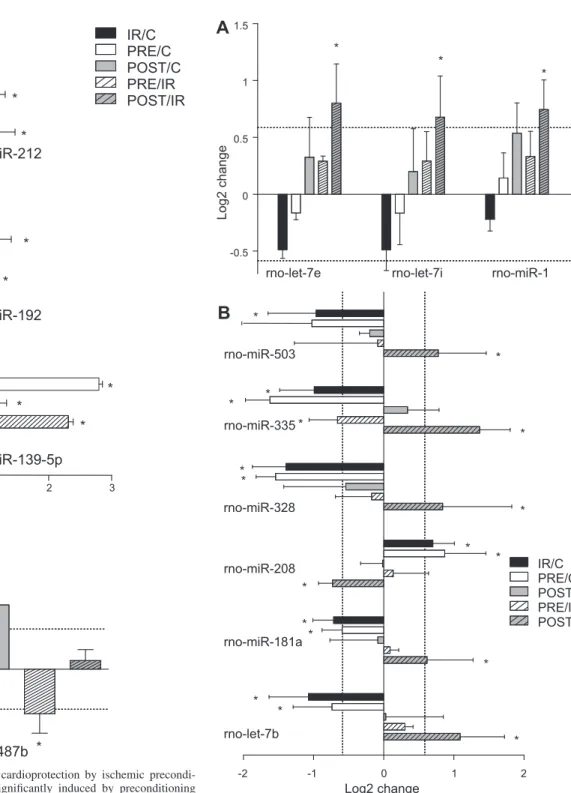
To further assess cardioprotective microRNAs, the effect of ischemic postconditioning on myocardial microRNA expres- sion was determined. We found three microRNAs that were not affected by I/R itself compared with nonischemic controls;
however, their expression was significantly upregulated by the subsequent postconditioning stimulus compared with the I/R group (microRNA-1, microRNA let-7i, and microRNA let-7e;
Fig. 4A). There were also six different microRNAs that were significantly altered (up- or downregulated) by I/R compared with time-matched nonischemic controls; however, postcondi- tioning significantly inhibited these alterations (microRNA let-7b, microRNA-181a, microRNA-208, microRNA-328, microRNA-335, and microRNA-503; Fig. 4B). We considered these nine microRNAs to be associated with the cardioprotec- tive mechanisms of postconditioning.
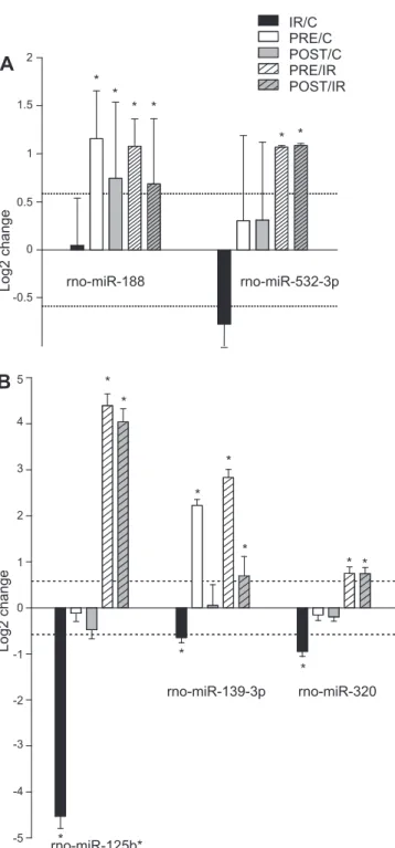
We found five different microRNAs (microRNA-188, mi- croRNA-532-3p, microRNA-125b*, microRNA139-3p, and microRNA-320) that showed similar expression patterns due to both ischemic preconditioning and postconditioning according to the following results. Both ischemic preconditioning and ischemic postconditioning significantly inhibited I/R-induced downregulation of microRNA-125b*, microRNA-139-3p, and microRNA-320 (Fig. 5B). In the case of microRNA-188 and microRNA-532-3p, both pre- and postconditioning in- duced significant upregulation compared with I/R; however, I/R alone did not affect the expression of these microRNAs (Fig. 5A). We considered these five microRNAs to play a pivotal role in endogenous cardioprotective signaling in both pre- and postconditioning.
Validation of Microarray Data by Quantitative Real-Time PCR
To confirm the microarray data, we measured the expression of 12 randomly selected microRNAs using quantitative real- time PCR. The expression changes of 10 microRNAs were confirmed by quantitative real-time PCR and showed good reliability of the microarray data (Table 2). In addition, quan- titative real-time PCR experiments were performed in the case of microRNA-125b*, which showed the most pronounced alterations due to I/R and pre- and postconditioning as assessed by microarray. The quantitative real-time PCR results con- firmed the microarray data (Table 2).
Identification of ProtectomiRs
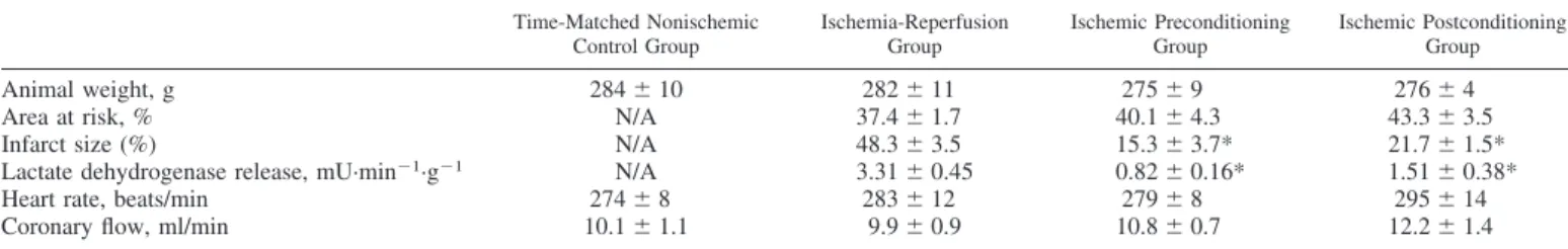
To identify protectomiRs, we systematically compared mi- croRNA changes and the direction of their changes in all Table 1. Markers of myocardial tissue injury
Time-Matched Nonischemic Control Group
Ischemia-Reperfusion Group
Ischemic Preconditioning Group
Ischemic Postconditioning Group
Animal weight, g 284⫾10 282⫾11 275⫾9 276⫾4
Area at risk, % N/A 37.4⫾1.7 40.1⫾4.3 43.3⫾3.5
Infarct size (%) N/A 48.3⫾3.5 15.3⫾3.7* 21.7⫾1.5*
Lactate dehydrogenase release, mU·min⫺1·g⫺1 N/A 3.31⫾0.45 0.82⫾0.16* 1.51⫾0.38*
Heart rate, beats/min 274⫾8 283⫾12 279⫾8 295⫾14
Coronary flow, ml/min 10.1⫾1.1 9.9⫾0.9 10.8⫾0.7 12.2⫾1.4
Values are means⫾SE. The area at risk and infarct size were measured after 120 min of reperfusion. Lactate dehydrogenase release was measured from coronary effluent collected during the first 5 min of reperfusion. The heart rate and coronary flow data presented here were measured at the end of the perfusion protocols. N/A, not applicable. *P⬍0.05 vs. the ischemia-reperfusion group.
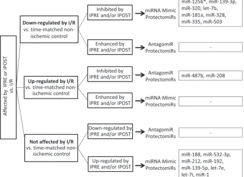
groups. Based on the comparative data, potential protectomiRs were classified into six theoretical categories, as described in
METHODS(Fig. 6). The protectomiRs identified here belonged to three of the six theoretical categories, as follows. Mimics of microRNA-125b*, microRNA-139-3p, microRNA-320, microRNA let-7b, microRNA-181a, microRNA-328, microRNA-335, and microRNA-503 were considered as protectomiRs as these
microRNAs were downregulated by I/R compared with time- matched nonischemic controls and their downregulation was inhibited by ischemic pre- and/or postconditioning. Further mimics of microRNA-188, microRNA-532-3p, microRNA- 212, microRNA-192, microRNA-139-5p, microRNA let-7e, microRNA let-7i, and microRNA-1 were considered as pro- tectomiRs as these microRNAs were not affected by I/R
1
IR/C PRE/C POST/C PRE/IR POST/IR
Log2 change
0 2 3 4
-1 -2
rno-miR-92a
rno-miR-877
rno-miR-652
rno-miR-494
rno-miR-378*
rno-miR-331
rno-miR-322*
rno-miR-125a-5p
*
*
*
*
*
*
*
*
*
*
*
*
*
*
* Fig. 2. Cardiac microRNAs (miR) associated with
I/R injury. This graph shows microRNAs that were significantly changed by I/R compared with time- matched nonischemic controls but neither pre- nor postconditioning influenced their changes due to I/R. This shows that these microRNAs were asso- ciated with I/R and unaffected by endogenous car- dioprotective mechanisms. Bars show alterations in microRNA expression in I/R versus time-matched nonischemic controls (IR/C), preconditioning ver- sus time-matched nonischemic controls (PRE/C), postconditioning versus time-matched nonischemic controls (POST/C), preconditioning versus I/R (PRE/IR), and postconditioning versus I/R (POST/
IR). Values are log2expression changes ⫾ SD.
*P ⬍0.05 and a log2change of greater than or equal to⫾0.585.
IR/C PRE/C POST/C PRE/IR POST/IR
Log2 change
Log2 change
0 0.5 1 1.5
-1 -0.5
rno-miR-212
rno-miR-192
rno-miR-139-5p
rno-miR-487b *
* *
*
*
*
*
*
*
*
A
B
Fig. 3. MicroRNAs associated with cardioprotection by ischemic precondi- tioning. A: microRNAs that were significantly induced by preconditioning compared with both I/R and time-matched nonischemic controls, demonstrat- ing that these microRNAs are specifically induced by preconditioning.
B: microRNA-487b, which was significantly upregulated by I/R compared with time-matched nonischemic controls; however, preconditioning significantly inhibited its expression change due to I/R compared with nonischemic con- trols. Bars show alterations in microRNA expression in I/R versus time- matched nonischemic controls, preconditioning versus time-matched nonisch- emic controls, postconditioning versus time-matched nonischemic controls, preconditioning versus I/R, and postconditioning versus I/R. Values are log2
expression changes⫾SD. *P⬍0.05 and a log2change of greater than or equal to⫾0.585.
Log2 change
0 1 1.5
-0.5 0.5
IR/C PRE/C POST/C PRE/IR POST/IR
2
0 1
1 - -2
Log2 change rno-miR-503
*
*
*
rno-miR-335
*
* *
rno-miR-328
**
*
* *
* rno-miR-208
rno-miR-181a
rno-let-7b
**
*
*
* *
rno-let-7e rno-let-7i rno-miR-1
*
*
*
A
B
Fig. 4. MicroRNAs associated with cardioprotection by ischemic postcondi- tioning.A: microRNAs that were significantly induced by postconditioning compared with I/R, demonstrating that these microRNAs are specifically induced by postconditioning.B: microRNAs that were significantly affected by I/R compared with time-matched nonischemic controls; however, their alter- ations were significantly inhibited postconditioning. Bars show alterations in microRNA expression in I/R versus time-matched nonischemic controls, preconditioning versus time-matched nonischemic controls, postconditioning versus time-matched nonischemic controls, preconditioning versus I/R, and postconditioning versus I/R. Values are log2expression changes⫾SD. *P⬍ 0.05 and a log2change of greater than or equal to⫾0.585.
compared with time-matched nonischemic controls; however, they were upregulated by ischemic pre- and/or postcondition- ing compared with I/R. AntagomiRs of microRNA-487b and microRNA-208 were considered as protectomiRs as these microRNAs were upregulated by I/R compared with time- matched nonischemic controls and their upregulation was in- hibited by ischemic pre- and/or postconditioning.
Validation of Cardiocytoprotection by ProtectomiRs in Cardiomyocytes Subjected to Simulated I/R
Based on the microRNA microarray results, several microRNAs were identified to be associated with cardioprotection by pre- and/or postconditioning. To further validate the possi- ble causative relationship between the affected microRNAs and cytoprotection, a postconditioning-associated mimick protectomiR (rno-let7b) and a preconditioning-associated mimick protectomiR (rno-miR-139-5p) as well as an inhib- itor protectomiR (rno-miR-487b) and a mimick protectomiR associated with both pre- and postconditioning-induced car- dioprotection (rno-miR-125b*) were selected for further study in cardiac myocytes subjected to simulated I/R. Trans- fection of cardiomyocytes with any of the four selected protectomiRs significantly decreased cardiomyocyte cell death due to simulated I/R (Fig. 7).
DISCUSSION
In the present study, we have shown using a microarray containing all 350 rat microRNAs available in Sanger miRBase (version 10.1) that in the early phase of I/R injury (i.e., at 2 h of reperfusion) the microRNA expression pattern is altered in the rat heart, which is significantly influenced by preceding preconditioning and/or subsequent postconditioning stimuli.
Moreover, by a systematic comparison of microRNA expres- sion changes and the direction of the changes induced by I/R with or without pre- or postconditioning, we revealed possible causative relationships between microRNA changes and en- dogenous cardioprotection. This is the first demonstration that ischemic pre- and postconditioning significantly affect the I/R-induced global microRNA expression pattern. Further- more, we validated the cardiocytoprotective effect of four of the potential cardioprotective microRNAs (i.e., the mimic of microRNA-125b*, microRNA let-7b, microRNA-139-5p, and inhibitor of microRNA-487b) in cardiomyocytes subjected to simulated I/R in vitro. Therefore, we identified specific cardio- protective microRNAs as potential therapeutic targets, the modula- tion of which with specific microRNA mimics or antagomiRs may provide cardioprotection. These microRNA mimics or antagomiRs are commonly termed here as protectomiRs.
MicroRNAs Associated With I/R Injury
Some studies have previously reported alterations of global microRNA expression after myocardial infarction in different animal models; nevertheless, the majority of these studies focused on late stages of infarction (days after coronary artery ligation), thereby showing the possible involvement of microRNAs in cardiac remodeling and heart failure (32, 34, 39).
However, there is limited information regarding microRNA alterations in the first few hours of I/R injury, when irreversible processes of cell injury are already triggered (11, 31, 41).
0 1 2 3
-2 -1
-3
-4
-5 4 5
IR/C PRE/C POST/C PRE/IR POST/IR
Log2 change
rno-miR-125b*
0 0.5 1
Log2 change
1.5 2
-0.5
A
B
rno-miR-139-3p rno-miR-320 rno-miR-188 rno-miR-532-3p
* *
*
*
*
*
*
*
*
*
*
* *
*
*
*
Fig. 5. MicroRNAs associated with cardioprotection by both preconditioning and postconditioning.A: microRNA changes induced by pre- and postcondi- tioning stimuli per se.B: microRNAs that were significantly changed by I/R compared with time-matched nonischemic controls; however, both precondi- tioning and postconditioning significantly inhibited their expression changes due to I/R. This shows that these microRNAs are associated with cardiopro- tection by both pre- and postconditioning via a reversal of the deleterious mechanisms of I/R. Bars show alterations in microRNA expression in I/R versus time-matched nonischemic controls, preconditioning versus time- matched nonischemic controls, postconditioning versus time-matched nonisch- emic controls, preconditioning versus I/R, and postconditioning versus I/R.
Values are log2expression changes⫾SD. *P⬍0.05 and a log2change of greater than or equal to⫾0.585.
In the present study, we considered microRNAs to be associ- ated with acute I/R injury (i.e., 2 h of reperfusion) when an alteration in the expression of a specific microRNA was signifi- cantly changed by I/R compared with time-matched nonischemic controls but when neither preceding preconditioning nor subse- quent postconditioning influenced the I/R-induced change of the particular microRNA. This shows that these microRNAs were associated with I/R but unaffected by endogenous cardioprotec- tive mechanisms. Such an expression pattern was characteristic in the case of eight different microRNAs (microRNA-125a-5p, mi- croRNA-322*, microRNA-331, microRNA-378*, microRNA- 494, microRNA-652, microRNA-877, and microRNA-92a). Ac- cordingly, microRNA-92a, microRNA-494, and microRNA-378*
have been previously described to be affected by ischemia in different tissues of rodents (2, 26, 41). Nevertheless, to date, little is known on the role of these microRNAs in acute I/R injury.
MicroRNAs Associated With Ischemic Preconditioning Our present study is the first in the literature focusing on the global microRNA expression pattern in the early phase of I/R injury preceded by ischemic preconditioning. Here, we found three different microRNAs (microRNA-139-5p, microRNA-192, and microRNA-212) that were significantly upregulated by pre- conditioning compared with both the I/R and time-matched non- ischemic control groups, suggesting that these microRNAs are specifically induced by preconditioning stimuli. Furthermore, we found a single microRNA (i.e., microRNA-487b) that was sig- nificantly upregulated by I/R; however, preconditioning inhib- ited its upregulation, thereby demonstrating a specific counter- regulation of this microRNA in I/R by preceding precondition- ing. By this approach, we revealed a possible causative relationship between these microRNAs and the cardioprotec- tive effect of preconditioning. To further validate the possible causative relationships, we selected the mimick of microRNA- 139-5p and inhibitor of microRNA-487b as potential precon- ditioning-related protectomiRs for transfection into primary neonatal cardiomyocytes subjected to simulated I/R and found that both the mimick of microRNA-139-5p and inhibitor of microRNA-487b were cardiocytoprotective. These results con-
firmed a causative relationship between microRNAs identified by the above-mentioned approach and cardioprotection.
Among the microRNAs affected by ischemic precondition- ing in the present study are several ones with unknown func- tion. For instance, the function of microRNA-139 has been demonstrated only in mouse hepatocytes so far (21), i.e., microRNA-139 decreased the amount of FoxO1 protein, a key factor in the regulation of cardiomyocyte autophagy (33).
Autophagy, on the other hand, has been shown to be involved in ischemic preconditioning-induced cardioprotection (25).
In a late preconditioning model, Yin et al. (44) investigated the involvement of microRNA-1, microRNA-21, and microRNA- 24 in mice; however, we could not demonstrate a role of these microRNAs in our classic preconditioning model. Wang et al.
(42) detected significant microRNA-144 and microRNA-451 upregulation immediately after preconditioning stimuli. More- over, they showed that infarct size in microRNA-144/451 knockout mice is higher and cannot be preconditioned (42). In our present data set, microRNA-451 also showed some upregu- lation due to preconditioning compared with I/R; however, the level of upregulation did not reach statistical significance.
These discrepancies could be due to differences in species and in the time of tissue sampling after ischemia.
MicroRNAs Associated With Ischemic Postconditioning The present study is the first in the literature focusing on global microRNA expression changes in the early phase of ischemic postconditioning-induced protection. Here, we iden- tified nine different microRNAs (microRNA-1, microRNA let-7i, microRNA let-7e, microRNA let-7b, microRNA-181a, microRNA-208, microRNA-328, microRNA-335, and microRNA-503) that were significantly affected by subsequent postconditioning compared with I/R, showing that these microRNAs are specifically regulated by the postconditioning stimuli. We found six different microRNAs (microRNA let-7b, microRNA-181a, microRNA-208, microRNA-328, mi- croRNA-335, and microRNA-503) that were significantly al- tered (up- or downregulated) by I/R compared with time- matched nonischemic controls; however, postconditioning sig- Table 2. Validation of the microarray data with quantitative real-time PCR
microRNA Name Chip Result Quantitative Real-Time PCR Result Confirmed
Ischemia-reperfusion group versus time-matched nonischemic control group
rno-let-7c ⫺0.92⫾0.38 ⫺1.25⫾0.20 Yes
rno-let-7d ⫺0.66⫾0.24 ⫺1.65⫾1.02 Yes
rno-miR-92a ⫺0.82⫾0.25 0.69⫾0.04 No
rno-miR-19a 0.60⫾0.11 1.14⫾0.72 Yes
rno-miR-19b 0.70⫾0.25 1.33⫾0.60 Yes
rno-miR-208 0.70⫾0.30 1.30⫾0.53 Yes
rno-miR-30c 0.32⫾0.14 ⫺0.07⫾0.54 Yes
rno-miR-335 ⫺0.98⫾0.49 ⫺1.91⫾0.83 Yes
rno-miR-125b* ⫺4.53⫾0.26 ⫺5.02⫾0.29 Yes
Ischemic preconditioning group versus ischemia-reperfusion group
rno-miR-320 0.74⫾0.13 1.75⫾0.93 Yes
rno-miR-335 ⫺0.65⫾0.4 0.19⫾0.32 No
rno-miR-125b* 4.39⫾0.25 4.66⫾0.36 Yes
Ischemic postconditioning group versus ischemia-reperfusion group
rno-miR-1 0.74⫾0.26 1.93⫾0.47 Yes
rno-miR-335 1.36⫾0.43 1.93⫾0.75 Yes
rno-miR-125b* 4.03⫾0.28 0.92⫾0.29 Yes
Values are means⫾SD of log2expression changes.
nificantly inhibited these alterations. These findings demonstrate that I/R-induced expression changes of these six different microRNAs are specifically counterregulated by sub- sequent postconditioning stimuli. In addition, we found three different microRNAs (microRNA-1, microRNA let-7i, and microRNA let-7e) that were not affected by I/R compared with nonischemic controls; however, these microRNAs were up- regulated due to postconditioning compared with I/R. By this approach, we revealed a possible causative relationship be- tween these microRNAs and the cardioprotective effect of postconditioning. To further validate this, we selected microRNA let-7b as a potential postconditioning-related protec- tomiR for transfection into primary neonatal cardiomyocytes subjected to simulated I/R and found that microRNA let-7b was cardiocytoprotective. This result confirmed a causative relationship between microRNAs identified by the above- mentioned approach and cardioprotection.
The exact role of most of the postconditioning-related pro- tectomiRs in cardioprotection is unclear, but some of these
microRNAs have been previously studied in the heart. He et al.
(24) showed that microRNA-1 and microRNA-133a were in- volved in the cardioprotective effect of ischemic postcondi- tioning in the rat and found that these microRNAs were capable of decreasing cardiomyocyte apoptosis in vitro. Here, we found a similar upregulation of microRNA-1 after subse- quent postconditioning stimuli. This microRNA has been shown to be involved in the regulation of apoptotic signaling by modulating the phosphatase and tensin homolog/Akt path- way (20), which is known to be a major participant of I/R injury and one of the intracellular mediator of cardioprotective signaling (18, 23). In the present study, microRNA-208 was significantly upregulated by I/R compared with nonischemic controls, which was inhibited by subsequent postconditioning stimuli. A similar upregulation of myocardial microRNA-208 has been previously described by Bostjancic et al. (3, 4) after myocardial infarction in humans. This microRNA is encoded in an intron of the ␣-myosin heavy chain 6 gene, and it is believed to be cardiac specific (38). MicroRNA-208 mediates
Fig. 6. Selection process of cardiac protectomiRs. MicroRNAs that were significantly affected by ischemic preconditioning (IPRE) and/or ischemic postconditioning (IPOST) compared with I/R alone were identified first. These microRNAs were then divided into three groups according to the effect of I/R compared with time-matched nonischemic controls and further subdivided into two additional categories according to the direction of microRNA expression changes, thereby yielding the following six categories. Category 1comprised microRNAs that were downregulated by I/R compared with time-matched nonischemic controls, but their down-regulation was inhibited by ischemic preconditioning and/or postconditioning. They were considered to be associated with the cardioprotective mechanism of ischemic preconditioning and/or postconditioning; therefore, these microRNAs or their mimics are considered as protectomiRs.Category 2comprised microRNAs that were downregulated by I/R compared with time-matched nonischemic controls, but their down-regulation was further enhanced by ischemic preconditioning and/or ischemic postconditioning. They were considered to be associated with the cardioprotective mechanism of ischemic preconditioning and/or postconditioning; therefore, the antagomiRs of these microRNAs can be also considered as protectomiRs. However, no example of this microRNA group was found.Category 3comprised microRNAs that were upregulated by I/R compared with time-matched nonischemic controls, but their upregulation was inhibited by ischemic preconditioning and/or postconditioning. They were considered to be involved in the development of I/R injury;
therefore, the antagomiRs of these microRNAs can be also considered as protectomiRs.Category 4comprised microRNAs that were upregulated by I/R compared with time-matched nonischemic controls, but their upregulation was further enhanced by ischemic preconditioning and/or postconditioning. They were considered to be associated with the cardioprotective mechanism of ischemic preconditioning and/or postconditioning; therefore, these microRNAs or their mimics are considered as protectomiRs.Category 5comprised microRNAs that were not affected by I/R alone compared with time-matched nonischemic controls but were significantly upregulated by ischemic preconditioning and/or postconditioning. They were considered to be associated with the cardioprotective mechanism of ischemic preconditioning and/or postconditioning; therefore, these microRNAs or their mimics are considered as protectomiRs. Category 6 comprised a group of microRNAs not affected by I/R alone compared with time-matched nonischemic controls, but their expression was downregulated by ischemic preconditioning and/or postconditioning. Inhibition of these microRNAs was considered to be involved in the cardioprotective mechanism of ischemic preconditioning and/or postconditioning; therefore, the antagomiRs of these microRNAs can be also considered as protectomiRs.
downregulation of the ␣-myosin heavy chain gene and up- regulation of the-myosin heavy chain (37), which are both involved in the development of hypertrophic response (43). In addition, some other targets of microRNA-208 have been determined by Callis et al. (7), including myostatin, a negative regulator of muscle growth and hypertrophy. These results, including those in the present study, suggest that an antagomiR of microRNA-208 could be a promising protectomiR, having additional therapeutic value in cardiac hypertrophy. The 15 different protectomiRs identified in the present study to be involved in the cardioprotective effect of postconditioning represent a therapeutic target group against reperfusion injury.
MicroRNAs Commonly Associated With Cardioprotection by Both Pre- and Postconditioning
Our present study is the first to identify microRNAs commonly associated with cardioprotection induced by both pre- and post- conditioning. These five different microRNAs (microRNA-188, microRNA-532-3p, microRNA-125b*, microRNA-139-3p, and microRNA-320) represent especially important therapeutic targets for ischemia- as well as reperfusion-induced pathologies. In case of microRNA-188 and microRNA-532-3p, both pre- and postcondition- ing significantly induced upregulation compared with I/R; however, I/R alone did not affect the expression of these microRNAs. This is the first demonstration that microRNA-188 and microRNA-532-3p are specifically associated with both pre- and postconditioning stimuli. However, there are no data in the literature on the mRNA targets of microRNA-188 and microRNA-532-3p in the heart. In the case of microRNA-125b*, microRNA-139-3p, and microRNA-320, this is the first demonstration that both preceding preconditioning and subsequent postconditioning stimuli resulted in a significant reversal of I/R-induced mi- croRNA expression changes, suggesting the prominent role of these microRNAs in cardioprotective signaling. However, there are no data on the role of microRNA-139-3p, whereas
there are limited data on microRNA-320 and microRNA- 125b* in cardiac pathologies (6, 31). Ren et al. (31) showed that microRNA-320 was significantly downregulated in an ex vivo mouse model of 45 min of global ischemia and 2 h of reperfusion as well as in an in vivo mouse model of 30 min of coronary occlusion and 24 h of reperfusion. In the present study, we detected a similar downregulation of microRNA-320 after acute I/R. However, we have also shown here that both preconditioning and postconditioning inhibited the I/R-induced downregulation of microRNA-320, indicating that this mi- croRNA plays an important role in cardioprotection. MicroRNA- 125b* showed the most significant changes in the present study, i.e., more than 20-fold down-regulation after I/R, while both preceding preconditioning and subsequent postconditioning stimuli markedly inhibited its downregulation. The exact role of microRNA-125b in cardioprotective cellular signaling re- mains to be investigated. Nevertheless, it should be mentioned that Busk et al. (6) reported a significant upregulation of microRNA-125b during the early hypertrophic growth in rat hearts after aortic banding.
According to the above-described approach, we revealed a causative relationship between microRNA-188, microRNA- 532-3p, microRNA-125b*, microRNA-139-3p, and microRNA- 320 and the cardioprotective effect of both pre- and postcon- ditioning. To further validate these causative relationships, we selected microRNA-125b* as a potential pre- and postcondi- tioning-related protectomiR for transfection into primary neo- natal cardiomyocytes subjected to simulated I/R and found that microRNA-125b* was cardiocytoprotective. This result further confirmed a causative relationship between microRNAs iden- tified by the above-mentioned approach and cardioprotection.
ProtectomiRs
In the present study, we report a novel approach to identify therapeutically applicable microRNAs with cardioprotective features. The analysis of the direction of microRNA changes due to pre- and/or postconditioning allowed the identification of mimics or antagomiRs of specific microRNAs, which were termed here as protectomiRs. Interestingly, all of the identified microRNA belonged to three major categories of the theoret- ically possible six categories, as described inMETHODS. More- over, the majority (18 of 20) of the identified protectomiRs were microRNA mimics, suggesting that the loss of specific microRNAs in the ischemic myocardium may play a role in I/R injury.
Limitations of the Study
In the present study, global microRNA expression was investigated in a single time point upon reperfusion; therefore, the time course of changes in microRNA expressions remained unknown. Thirty minutes of coronary occlusion followed by 120 min of reperfusion, when microRNA expression was measured, is considered an early phase of cell injury due to necrotic and/or apoptotic processes (28). However, it cannot be excluded that early cell death processes might affect mi- croRNA alterations induced by I/R injury and cytoprotection by pre- and postconditioning.
Although we identified several protectomiRs in the present study, investigation of the exact role of each protectomiR in cardioprotective signaling was outside of the scope of this
0 20 40 60 80 100
Percent survival(%)
*
# # #
NORMOXIA HYPOXIA
Non- targeting
Control
Non- targeting
Control miR- 139-5p
mimic miR- 125b*
mimic let-
7b mimic
#
miR- 487b inhibitor Fig. 7. Cardiocytoprotection by selected protectomiRs. Transfection of primary cardiomyocytes with the postconditioning-associated protectomiR mimic (rno- let7b), a preconditioning-associated protectomiR mimic (rno-miR-139-5p) and inhibitor (rno-miR-487b), and a protectomiR mimic associated with both pre- and postconditioning-induced cardioprotection (rno-miR-125b*) showed that these protectomiRs were cardiocytoprotective. Values are means⫾SE. *P⬍ 0.05 vs. normoxic control; #P⬍0.05 vs. hypoxic control.
![Fig. 1. Experimental protocol. Hearts isolated from male Wistar rats were subjected to either time-matched aerobic perfusion [i.e., time-time-matched non-ischemic control (C)], 30 min of coronary occlusion and 120 min of reperfusion [ischemia/reperfusion](https://thumb-eu.123doks.com/thumbv2/9dokorg/1373539.112708/2.904.67.571.784.1068/experimental-protocol-isolated-subjected-perfusion-occlusion-reperfusion-reperfusion.webp)