Ischemic preconditioning in liver surgery. From animal model to human use.
Ph.D. Theses
Oszkár Hahn M.D.
Semmelweis University
Doctoral School of Clinical Medicine
Tutor: Péter Kupcsulik M.D., Ph.D
Opponents: Irén Mikó M.D., Ph.D
Gabriella Lengyel M.D., Ph.D
Board of final examination: Ferenc Szalay M.D., D.Sc.
Pál Ondrejka M.D., Ph.D József Sándor M.D., Ph.D
Budapest
2014
INTRODUCTION
Resection is the most common curative treatment for liver diseases of malignant origin and is also sometimes necessary for treatment of tumors of benign origin. For reducing the potentially lethal risk of massive bleeding and blood transfusions with adverse
immunomodulatory adverse effects, major hepatic resection is frequently performed under clampage of the hepatic pedicle. As a consequence of vascular occlusion, in situ warm ischemia followed by reperfusion may result in severe organ dysfunction and organ failure, called by ischemia reperfusion injury (IRI).
The main components of IRI are cellular activation and cytokine release, expression of adhesion molecules and microcirculatory dysfunction.
There are two phases of hepatic IRI. The first, occurring in the first 4 h after reperfusion of the liver, is activation of Kupffer cells. This results in production and release of reactive oxygen species (ROS) and proinflammatory cytokines, particularly tumour necrosis factor (TNF) α and interleukin (IL) 1. Other interleukins, such as IL-6 and IL-12, are also
implicated. One of the results of this cytokine release is increased expression of intercellular adhesion molecules (ICAMs) by endothelial cells, leading to the second phase of IR injury.
This second phase, occurring after 6 h of reperfusion, mainly involves the activation of neutrophils, which accumulate within the sinusoids and postsinusoidal venules, and adhere to the endothelial cell lining. These neutrophils then extravasate, causing parenchymal injury by production and release of their own ROS and proteases, and phagocytosis.
1. Kupffer cells, citokines
IP reduces Kupfer cell activation and leucocyte-endothelial interaction.
Kupffer cell plays a central role in hepatic IRI and IP modulates its activity. IP reduces the ROS-mediated injury to parenchymal and non-parenchymal cells both by the suppression of ROS production by Kupffer cells and induction of cellular resistance to ROS. This effect reduces oxidative cellular damage by 50 per cent in warm ischaemia. The production of TNF- α by Kupffer cells is also inhibited by IP. This decrease in hepatic TNF-α release is thought to be responsible for the effect of remote preconditioning on other organs following hepatic IP.
2. Adhesion molecules
IP decreases endothelial P-selectin expression in warm IRI, an effect attributed to the absence of stimulation by TNF-α.The resultant decrease in neutrophil adhesion,
transmigration and activation protects the parenchyma. Reduced ‘stickiness’ of post-
ischaemic neutrophils in preconditioned tissue has been attributed to diminished I/R-induced P-selectin expression and downregulation of inter-cellular adhesion molecule(ICAM)-1 release.
3. Microcirculation
The microcirculatory dysfunction that occurs in hepatic IRI is multifactorial, involving direct cellular injury, leukocyte and platelet accumulation, and vasoconstriction. The decrease in ROS production and neutrophil-mediated injury by IP leads to preservation of the
architecture of the microcirculation. As a result, along with its influence on nitric oxide production, IP improves microcirculatory perfusion, an effect that can be demonstrated by intravital microscopy, leading to improved sinusoidal perfusion.
4. NO and adenosine
The protective effects of classical preconditioning depend on endogenous substances that act immediately. The main compounds implicated in hepatic IP before warm ischaemia are nitric oxide and adenosine, both produced by endothelial cells. Adenosine production and
release in response to IP in the rat liver have been demonstrated to protect SECs by activation of the A2 receptor, leading to increased production of nitric oxide. Nitric oxide appears to play a pivotal role in early IP, where it is thought to act as a trigger, and delayed IP, where it acts as a trigger and a mediator. Endothelial nitric oxide synthase (eNOS) is present in liver endothelial cells, and is responsible for the nitric oxide produced in early preconditioning.
Inducible nitric oxide synthase (iNOS) is present in all liver cell types and is the source of nitric oxide in late preconditioning. Serum nitric oxide levels are increased, and the transcription and expression of nitric oxide synthase is increased, in rats undergoing liver transplantation with IP. Nitric oxide is thought to mediate early IP by several mechanisms, including its influence (along with the activated kinases) on adenosine 5-triphosphate- sensitive potassium channels. Opening of these channels is a significant feature in early IP, but whether this is the endpoint or another step in the pathway is not known. Another
mechanism involves an increase in levels of guanosine 3, 5-cyclic monophosphate (cGMP) in hepatocytes. This increase in cGMP causes cytoprotection by reducing oxygen consumption and energy demand.
5. Energy preservation
IP reduces cellular energy demand during ischaemia. One possible mechanismby which metabolism can be downregulated is through adenosine 5-monophosphate-activated protein kinase (AMPK), which inhibits glycolysis. Degradation of ATP during IP leads to accumulation of adenosine 5-monophosphate (AMP), an allosteric activator of AMPK.
Inhibition of AMPK production abolished the protective effects of preconditioning on energy metabolism and hepatocyte injury. ATP preservation prevents cell necrosis, but appears to provide the necessary substrate for the cell to undergo apoptosis instead. Clearly this switch from necrosis to apoptosis would need to result in less overall cell death for the effect of IP on ATP preservation to be beneficial.
6. Intracellular kinase
Activation of cell membrane receptors by adenosine and other effectors, such as bradykinin, stimulates the activity of various intracellular kinases, such as protein kinase C (PKC) and p38 mitogen-activated protein kinase (p38MAPK). However, the precise role of PKC in the IP pathway remains uncertain, although activation of PKC and p38 MAPK has recently been shown to reduce hepatocyte injury by maintenance of intracellular sodium homeostasis. Recent evidence suggests that IP induces survival signals by activation of another intracellular kinase, protein kinase B (Akt/PKB), an effect mediated by adenosine A2 receptors on hepatocytes. Akt/PKB has antiapoptotic effects and also directly stimulates nitric oxide production by eNOS.
7. Gene expression and late phase of IP
Late preconditioning is thought to rely on altered gene expression. Specific
transcription factors have been implicated, such as nuclear factor kappa B. Activation of these transcription factors leads to the synthesis of antioxidants, heat-shock proteins and nitric oxide (from iNOS), all of which exhibit a cytoprotective effect.
AIM
To decrease ischemia-reperfusion injury with the use of ischemic preconditioning.
To investigate the effect of IP and to convert the results of the animal model into human liver resections, with special interest to cirrhotic patients.
I. Animal model
1. To create a rodent liver IRI model.
2. To determine the ischemia tolerance of the rat liver in this model.
3. Standardization and a mathematical transformation of laser Doppler Flowmetry (LDF) flow graphs.
4. To investigate hepatic microcirculation (HM) with non-invasive laser Doppler flowmetry (LDF), histological alterations, TNF-a levels, and laboatory parameters after various periods of liver ischaemia, with or without ischemic preconditioning in the 1st model.
5. According to the results of the 1st model the „critical” 60 minutes ischemia was investigated in the 2nd model, with special interest to redox parameter changes.
II. Human use
1. To convert the animal model to human use.
2. To design a prospective, randomized study including normal liver and cirrhotic patients with primary or secondary liver tumours, with the need of more than 2 segments liver resection.
3. To randomize these patients to have ischemic preconditioning (IP) or intermittant portal clamping (IPC).
To investigate
4. Intraoperative parameters
5. Liver lesion and liver function laboratory parameter changes 6. Serum redox parameters
7. Postoperative complication rates
METHODS
Animal model
Inbred male Wistar rats were used in the model (80 animals in the 1st, 30 animal in the 2nd model). All animals were anesthetized with iv. ketamine. Mean arterial blood pressure was monitored through femoral artery catheter.
1st model:
Before the operation, a control blood sample was taken from the tail vein. Laparotomy was performed via a median incision and the liver was mobilized. Lobe VI was removed for a control histological sample. Complete ischemia of lobes I, II, and V was achieved by
clamping of the portal veins, hepatic arteries, and biliary branches using atraumatic
microvascular clips. The microcirculation was monitored using LDF throughout the ischemic period of lobe V. 5 min ischemia and 10 min reperfusion in 1 cycle was used for ischemic preconditioning. At the end of the ischemic period (30, 45, 60, or 90 min), the vascular clip was removed and reperfusion was continuously monitored by LDF in all groups. Next, ischemic lobe I was removed for histological sampling. After 30 min of reperfusion, histological (lobe II), and venous blood samples were taken for the measurement of TNF-α levels. On the first post-operative day, venous blood (for ALT, LDH, and serum bilirubin levels) was drawn, while on the seventh day a histological sample from the Vth lobe (I-R injured) and venous blood samples were taken again. Finally, the animals were put down by exsanguination.
2nd model:
Before the operation, a control blood sample (0.5 mL, supplied equal volume saline solution) was drawn from the femoral arterial canula. Warm ischemia of the median and lateral lobes (lobes III, IV, and V) at the level of the hilum was induced for 60 min by clamping of the portal veins, hepatic arteries and biliary branches using atraumatic
microvascular clips. After an ischemic interval of 60 min, the clamps were removed and the caudate right and quadrate lobes (lobe I, II, VI, and VII) as well as the papillary process (30–
35% of the total hepatic mass) were resected, leaving only post-ischemic tissue in situ. This method induced ischemia in approximately 65 to 70% of the liver, while leaving an
uninterrupted blood supply to lobes I, II, VI, and VII. It enabled us to exclude the chosen lobes from the circulation, while simultaneously preserving the flow in the intact lobes. The microcirculation of lobe V was monitored using LDF throughout the ischemic period. At the end of the ischemic period,
the vascular clip was removed and reperfusion was continuously monitored by LDF in all groups for 60 min. 6 hours after reperfusion, histological samples were taken from IR injured lobes III, IV, and V for pathological examination and for homogenization (tissue redox parameters). Finally, the animals were put down by exsanguinations via the femoral artery catheter and blood samples were centrifuged and serum redox parameters were also measured.
Assessment of the HM
The HM (arbitrary flux unit range 0-1000) was measured using a laser Doppler monitor and a surface probe (model: Moor Instruments DRT4; 1.0mW laser power at 632,8 nm and a DP1T surface probe). Flux, as defined for the laser Doppler, is proportional to the product of the total number of moving RBCs in the measured volume (several mm3) and the mean speed of the RBCs. The LDF probe was placed on a fixed location of the liver’s lobe V and held in place by a custom probe-holder developed by our team. Flux, temperature, and moving RBCs concentration were recorded. LDF measurements at the relevant time points were taken at 4-s intervals. On-line computer monitoring and processing were applied.
The required standardization was as follows: (1) continuous temperature monitoring of the animal; (2) a respiration-synchronous moving LDF probe; (3) constant probe pressure on the liver surface with a hanging balance system; (4) a second LDF probe placed over an
“equivalent liver tissue” where RBC concentration was equivalent and measurable; (5) shielding of the probe to prevent the disturbing effect of any external light.
For the mathematical transformation, we used the average ischemic LD flow subtracted from the total LD flux values. The results were normalized against the baseline flow and then expressed as a percentage. Ischemic LD flow is referred to as ‘biological zero’ (bz) in laser Doppler terminology.
Tflux: corrigated flux value;
flux: measured flux value;
bz: biological zero, microcirculation during ischemia;
baseline: normal, control microcirculation
For the characterization of the individual flow graphs, a new method was introduced. By its implementation, the graphs became comparable and suitable for statistical analysis. This involved characterizing reperfusion as the integral of the reperfusion segment of the graphs (RA: reperfusion area) as well as the maximal plateau of the reperfusion section of the graphs (PM: plateau maximum). In a hypothetical, ideal situation, the value of the flow would have been 100% of the starting flow at the first second of reperfusion. The actual graph measured could be represented as a percentage (KRT= (RTx/RT0) of the hypothetical one. The area under the ideal graph is 100%.
Histological Examination
The excised liver was fixed in 8% formalin and embedded in paraffin. Sections were stained with hematoxylin and eosin. The following alterations were registered: periodic stagnation and recovery, swelling, necrosis, vacuolization, lymphocyte infiltration, fibrosis, bile duct proliferation, venous dilation, and tissue hemorrhage. The evaluation of the aforementioned pathological alterations was semiquantitatively scored (0: no alteration, +:
less than 10% of cells were affected, _++: 10-40% of the cells were affected, +++: more than 50% of the cells were affected). The pathologist was not informed about the applied treatment and the histological evaluation was performed by two independent pathologists (G.L. and Zs.
Sch.).
The maximum score was 21. Below 7 points, the damage was described as weak, between 7 and 14 moderate, over 14 points, the damage was severe.
Measurement of TNF-α
Tumor necrosis factor-alpha (TNF-α) level at 30 minutes after reperfusion was measured by viability assays on WEHI 164 cell line with the method of Mossman-Hansen.
Isopropyl alcohol acidified with HCl (0.08 M) was subsequently added and plates were read at dual wavelengths of 570 nm and 630 nm. From each data point (optical absorbance value) a viability value was calculated as a percentage of the average optical absorbance by non-
treated cultures. The viability of cultures was inversely proportional to the TNF-a concentration in the samples.
Measurement of Serum Bilirubin, ALT, and LDH
×100
−
= −
bz baseline
bz Tflux flux
Serum bilirubin, ALT, AST, se Bilirubin and LDH measurements were performed by standard spectrophotometry using an automated clinical chemistry analyzer (Hitachi 747).
The blood volume removed was replaced with an equal volume of normal saline.
Redox parameters
Redox parameters were measured during the 2nd model. Protein content was measured using Lowry’s method.
1. Chemiluminescence intensity: A recently developed chemiluminescence assay was applied to measure the total scavenger capacity, which was determined by chemiluminescence in a H2O2/OH● luminol microperoxidase system, using the Lumat LB 9051 luminometer (Lumat;
Berthold, Windbad, Germany) according to the method of Blázovics. The chemiluminescence light intensity, given in relative light units (RLU), is reduced in the presence of free radical scavenger compounds. The chemiluminescent intensity of the samples was expressed in RLU% (relative light unit percentage) of the standard
2. Reducing power: Oyaizu’s method was adopted for the determination of the reducing power of the samples. The change in absorption was measured, which accompanied Fe3_- Fe2_ transformation at 700 nm, and the reducing power was compared to that of ascorbic acid. All spectrophotometric measurements were carried out with a Jasco V 550 instrument.
3. Free SH-groups were detected by the Sedlack method based on the Ellmann reaction on 512nm with spectrophotometry.
Data Collection and Statistical Analysis
Values are expressed as means ± SD. One-way ANOVA, Student’s t-test, Scheffer test, and Mann-Whitney test (by Statisoft software) were p< 0.05 confidence interval was considered statistically significant.
II. Human use
Experimental Design
Between 2004 and 2008, 160 consecutive patients undergoing major liver resection (more than 2 liver segments removed) at the 1st Department of Surgery, Semmelweis
University, Budapest, Hungary were randomized to receive IPC or IP with continuous portal clamping, preserving the hepatic vein and caval flow. Among these patients 60 patients with liver cirrhosis were also randomized.
Groups: Normal liver IPC (group A: 50 patients), normal liver IP (group B: 50 patients), cirrhosis IPC (group C: 30 patients), cirrhosis IP (group D: 30 patients). Preoperative liver function was assessed using indo-cyanide green (ICG) clearance test.
Surgical Procedure
The study was approved by the local ethical committee (TUKEB No: 15/2004).
Informed consent was obtained from each patient prior to surgery. Each patient was randomized before surgery in the operating room. Under general anesthesia right subcostal incision was made. After decision for resection, IPC (15 min ischemia–5 min reperfusion, 15 min ischemia–5 min reperfusion) or IP (10 min ischemia–10 min of
reperfusion) was performed by clamping the hepatic pedicle with a vascular clamp. The time of continuous inflow occlusion (IP group) had to exceed 30 min in the non-cirrhotic groups as this is the minimal ischemic period with detectable postoperative liver injury with normal underlying liver parenchyma. In patients with liver cirrhosis ischemia time was approximately 20 min. Parenchymal resection was performed using electrocoagulation, Ligasure (Covidien Ltd, Mansfield, MA), or clips by a standard manner used at our unit conventionally.
According to the tumor’s location and liver function tests, various types of resections were performed. The number of the removed segments always exceeded two in all groups.
Resected specimen underwent histological examination at the same pathological department.
In the cirrhotic groups liver functions belonged to Child A or B.
Measurement of Liver Enzymes and Bilirubin
The degree of ischemic injury of the liver was determined by serial postoperative serum bilirubin, aspartate aminotransferase (AST), and alanine aminotransferase (ALT) measured from blood samples taken preoperatively, on the 1st, 3rd, and 7th postoperative day by standard spectrophotometry using an automated clinical chemistry analyzer (Hitachi 747, Hitachi Ltd, Tokyo, Japan).
Measurement of Serum ODFRs and Antioxidants
ODFRs in serum (scavenger activity)—using the Heide–Bögl method, modified by Blázovics— were measured preoperatively, 30 min after reperfusion and on the 7th day.
Antioxidants in serum were measured preoperatively, 30 min after reperfusion and on the 7th day determining protein free sulfhydryl (SH) groups—by the method of Ellman, as described by Sedlak and Lindsay and the reducing power of the samples (RP)—with Oyaizu’s method.
Histology
Each resected liver specimen was evaluated at the same pathological institute in a blinded fashion. The presence of cirrhosis or other injuries were evaluated from the resected tumor free margin.
Definition of Postoperative Complications
Patients were defined as having postoperative liver insufficiency if total bilirubin level was >70 mmol/L, or if prothrombin time was <40% of the normal level within 7 days of operation. Asterixis and alteration of consciousness (not related to the effects of drugs) were also considered as signs of liver failure. Clinically significant ascites was also recorded (abdominal drain output >500 ml per day for more than 3 days). Definition of renal
insufficiency was serum creatinine >150 mmol/L. All other complications were also recorded.
Statistical Analysis
Results are mean ± SD. For continuous variables, differences between the groups were evaluated by Student’s t-test. For discrete variables, data are expressed as counts and
percentages and were analyzed with Fisher’s exact test. All statistical analyses were performed using SPSS 12.0 statistical package (SPSS, Inc., Chicago, IL). A P < 0.05 confidence interval was considered as statistically significant. Intensive care specialists, biologists, and pathologists were blinded to the study.
RESULTS I. Animal model
Hemodynamic Parameters
There were no significant differences in the heart rate (380 _ 22 bpm) or the mean arterial blood pressure (103 ± 15 mmHg) between the groups. These parameters did not change significantly throughout the experiment in either the controls or in the ischemia groups subjected to lobar I-R or IP and I-R.
Microcirculation Measured by LDF
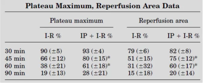
There is a significant correlation between PM and (RA), and the duration of ischemia (Table 1). Data obtained in ischemic groups differ significantly from each other (P 0.05). The groups
with IP had significantly (P < 0.05) higher flow rates than groups without preconditioning.
The graphs pertaining to the groups that underwent IP and those without IP are similar to each other; however, the graphs’ slopes related to IP were steeper. There is no significant
difference between the IP group and the group without IP at the 30- and 90-min ischemia sessions.
Histological Examination I-R groups
According to the applied classification, the appearance of the pathological alterations in lobe I (ischemic damaged lobe) was not directly related to the duration of ischemia. After 30 min of ischemia, only slight periodic stagnation-reperfusion was observed. After 45 min of hepatic ischemia, vacuolization appeared. Only 60 min of ischemia could induce significant venous dilation, vacuolization. Significant swelling and necrosis were observed after 90 min of ischemia (19 points).
Samples taken from lobe II (reperfusion damaged lobe), after reperfusion of 30 min ischemic liver, showed similar alterations as above; however, after 45 min of ischemia, increased venous dilation and periportal lymphocyte infiltration could also be observed. After 60 min of ischemia, these alterations were more pronounced. Finally, after 90 min
vacuolization, and venous dilation were most apparent and severe tissue hemorrhage could be detected (21 points).
Samples taken from lobe V of the (surviving animals on the seventh day) in the 30 min group displayed “restitutio ad integrum”. Furthermore, 45 min and 60 min groups showed evidence of vacuolization, necrosis, and periportal lymphocyte infiltration without fibrosis and bile duct proliferation. No animals survived 90 min of ischemia. (15 points)
IP + I-R groups
IP-treated groups did not show histological differences from the control I-R groups.
Histological samples after ischemia or reperfusion in the 30, 45, and 90 min IP + I-R groups did not show significant difference to the identical I-R groups. Number of necrotic cells were less.
Table 1: Significant difference between flowmetry data (PM and RA) with or without preconditioning. (*p<0,05)
Nevertheless in the 60 min IP+I-R groups the I-R injury was milder (ischemic lobe:14 pts, reperfusion lobe: 17 pts) compared with I-R groups (ischemic lobe: 16 pts, reperfusion lobe: 20 pts). In these IP groups no necrosis was observed.
Samples taken on the seventh day in the 30, 45, and 60 min IP + I-R groups showed less periportal lymphocyte infiltration and tissue destruction. The only surviving animal in the 90 min IP + I-R group showed considerable necrosis and lymphocyte migration.
TNF-α levels
TNF-α levels were significantly (P < 0.05) higher in the 30 min I-R group than in the control group. With increasing duration of ischemia, the TNF-α levels in the blood samples increased. Venous blood samples drawn at the 30th min of reperfusion contained significantly (P < 0.05) less TNF-α in the 30 min and 45 min IP + I-R groups than in the I-R groups. The difference was even more significant (P < 0.01) in the 60 min group. After 90 min IP + I-R, however, preconditioning did not cause a significant decline in the TNF-α level.
Serum Bilirubin, ALT, and LDH results
In the 30 min I-R group, there was no significant (P <0.05) increase in serum direct bilirubin, ALT, or LDH compared to the control sample, neither on the first nor on the seventh postoperative day. Similarly, the IP did not cause any significant (P < 0.05) decrease in these parameters.
After 45 and 60 min I-R, all examined parameters were significantly (P < 0.05) elevated on the first postoperative day compared to the control group. These parameters were significantly (P < 0.05) lower in the IP + I-R groups than in the I-R groups on the first
post-operative day. On the seventh post-operative day, none of these parameters were significantly (P < 0.05) elevated compared to those in the control group.
In the 90 min I-R group, all parameters were significantly (P < 0.05) higher during the first post-operative day than in the control group, yet there was no significant difference between the IP + I-R and the IR groups. For the single surviving animal in the 90-min IP + I- R group, the blood sample still demonstrated high serum direct bilirubin, ALT, and LDH levels.
Survival on the 7th day
Survival I-R vs. IP+I-R: 30 min: 100% vs. 100%; 45min: 90% vs. 100%; 60min: 60%
vs. 70%; 90min: 0% vs. 10%.
Redox parameters (2nd model, 60 min ischemia) Scavenger activity
The amount of ODFR scavenger activity was significantly lower in the IP+I-R, than in the I-R group (serum and liver sample).
Reducing power
IP reduced the reducing power (serum antioxidants level) significantly. The levels in the liver samples were not significantly lower in the IP groups.
Free SH-groups
The amount of free SH groups were lower in the IP group than in the I-R group. The results were not significant.
II. Human results
Patient Characteristic
Both subgroups (IPC and IP) were comparable in the cirrhotic and non-cirrhotic main groups regarding age and gender. Of the 160 randomized patients, 79 were men and 81 were
women with a mean age of 57.1 ± 1.6 years. The preoperative risk and liver function tests were similar in both subgroups (IPC and IP), but naturally not in the two main groups (cirrhosis–no cirrhosis). According to these, the groups were homogenous to compare.
Intraoperative Parameters
There were no significant difference in the length of the operation, duration of liver ischemia, indication for resection and number of resected liver segments between the subgroups A–B and C–D. The ratio of resected volume/total volume, residual liver
volume/total liver volume, the type and number of procedures were comparable between both subgroups (IPC and IP). The time of continuous inflow occlusion (IP group) exceeded 30 min in the non-cirrhotic groups (group A vs. B: 34 ± 3.2 vs. 33 ± 2.1 min), in patients with liver cirrhosis ischemia time was approximately 20 min (group C: 22 ± 2.2 vs. group D: 21± 1.8 min). The mean number of red blood cell (RBC) units in transfused patients during the first postoperative 48 hr was significantly lower in the IP groups (patients with normal liver—IPC:
2.8 ± 0.3 vs. IP: 1.6 ± 0.4; patients with cirrhotic liver—IPC: 1.9± 0.7 vs. 0.9 ± 0.1).
Postoperative Liver Injury
The degree of ischemia and reperfusion injury of the liver was assessed by postoperative peak serum AST, ALT, and bilirubin levels. Patients treated with ischemic preconditioning had a significantly lower peak AST (median peak 150 ± 14 and 169 ± 10 U/L) and ALT (median peak 136 ± 9 and 145± 12 U/L) on the 1st postoperative day when compared with the IPC groups (median peak AST 284± 12 and 362± 16 U/L, median peak ALT 262 ± 11 and 355± 16 U/L, P < 0.05). Bilirubin levels of subgroup A did not differ significantly from subgroup B (median peak 36 ± 8 vs. 30 ± 7 mmol/L). In all non-cirrhotic patients, AST, ALT, and bilirubin levels returned to normal values within 7 days (Figs. 1 and 2). In cirrhotic patients receiving IP prior to liver resection significantly lower (P < 0.01) peak AST, ALT and in contrast to noncirrhotic patients also significant lower peak bilirubin values (median peak bilirubin 59 ± 9 vs. 25 ± 6 mmol/L, P < 0.05) were measured when compared to the IPC group. In contrast to the non-cirrhotic patients, in patients with cirrhosis not receiving IP, none of the measured parameters returned to normal values after 7 days.
Redox parameters
Significantly less free radicals (median peak 66 ± 16 x 105 vs.101 ± 12 x 105 RLU) and more antioxidants (SH median peak 1.14 ± 0.05 vs. 0.44 ± 0.06 mmol/L, and RP median peak 0.18 ± 0.03 vs. 0.04 ± 0.002 mmol/L ASeqv.) were measured in IP group (B) after reperfusion, than in IPC group (A) in non-cirrhotic patients (P < 0.05), but these parameters returned to the preoperative values after 7 days (Figs. 3 and 4). In cirrhotic patients, the level of serum ODFRs were significantly higher (median peak 131 ± 16 x 105 vs. 80 ± 13 x 105 RLU, P < 0.05), the level of serum antioxidants were significantly lower (SH median peak 0.3
± 0.05 vs. 0.65 ± 0.04 mmol/L and RP median peak 0.02 ± 0.001 vs. 0.16 ± 0.02 mmol/L ASeqv., P < 0.05) in the IPC group (C) after reperfusion, than in IP (D) group. Without IP the level of serum antioxidants remained low, even on the 7th postoperative day.
Clinical Outcome
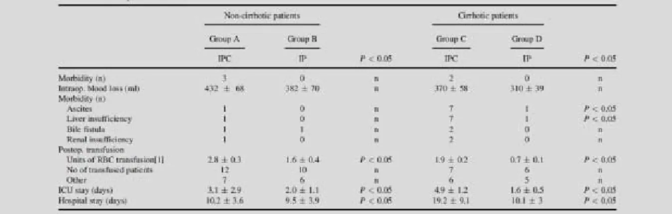
Postoperative morbidity and mortality rates, such as intraoperative blood loss, ICU, and hospital stay data are presented in Table 2.
There was no difference between normal liver IP and IPC groups regarding to postoperative outcome morbidity. In contrast, cirrhotic patients with IP had significantly lower morbidity rates (liver insufficiency, ascites formation), than patients with IPC.
ICU and hospital stay were also significantly shorter in the cirrhotic IP group, than in the IPC group. Although we observed a trend, that postoperative bleeding and the number of
transfused patients were less in the IP groups, these results were not significant.
Number of transfused RBC’s was significantly lower in the IP group in cirrhotic and non- cirrhotic patients.
CONCLUSIONS
1. I-R injury plays an important role after liver resections with afferent occlusion.
2. In the promotion and in the sustained I-R injury of the liver TNF-α has a key role.
3. ODFRs are responsible for hepatocyte and sinus endothel injury. The outcome of this process is mainly depending on the antioxidant pools and on the liver condition.
4. Laser Doppler Flowmetry is a suitable method to measure liver microcirculation under special circumstances, such as temperature, RBC concentration and tissue thickness.
5. After mathematical corrections of the flow graphs (LDF measurements were calculated after mathematical transformation of the ischemic flow; the latter was used as the baseline (0%)) were comparable.
6. Plateau Maximum (PM), Reperfusion Area (RA), and Reperfusion Maximum Time (RMT) made the evaulation of the flow graphs possible and suitable for statistical analysis. „KRT” ratio (KRT = RTx/RT0 x 100; as: %) made the interpretation of RT easier.
7. Thirty minutes of liver ischemia is tolerable, the 45 and the 60 min of ischemia cause severe microcirculatory damage (PM, RT). The 90 min ischemia caused early and irreversible microcirculation injury, which can not be restored after the observed 30 minutes of reperfusion.
Table 2: Morbidity, mortality
8. Ischemic preconditioning in 1 cycle (5min ischemia-10min reperfusion) before 45 and 60 minutes ischemia resulted in significantly better microcirculatory parameters. These benefits could not be observed after 30 and 90 minutes ischemia of the rat liver.
9. IP resulted in better (but not significantly) histological alteration than without IP.
10. According to the results of TNF-α levels the early inflammatory responseand Kupffer cell activation was lower in the 45 and 60 minutes ischemia group, if IP was performed. Liver lesion was similary milder in the IP groups as the transaminase levels showed.
11. The direct hepatocyte and sinus endothelial cell injury is caused by ODFRs. IP resulted in lower ODFRs and higher antioxidant levels after reperfusion.
12. IP in one cycle is a suitable method to decrease I-R injury of the rat liver in this model after 45 and 60 minutes ischemia.
13. As the results of the animal model showed, it is worth to transform the beneficial effect of IP to human liver resections.
14. According to the result of the human study IP before afferent exclusion of the livercaused milder liver lesion, than with IPC on non cirrhotic and cirrhotic patients too. Liver lesion was well interpreted by transaminase levels.
15. Serum bilirubin levels did not differ significantly from IPC group of non cirrhotic patients ont he 1st postoperative day..
16. AST, ALT and serum bilirubin levels normalized after 7 days in normal liver patients, independently from receiving preconditioning or not.
17. Cirrhotic patients with IP had also significantly lower peak AST and ALT levels, but serum bilirubin levels were also significantly lower, than in the IPC groups.
18. ODFRs (the effector of hepatocyte and sinus endothel cell injury after I-R) levels were lower, if ischemic preconditioning was applied.
19. These redox parameters normalized after 7 days in the non cirrhotic patients..
20. Significantly decreased antioxidant levels were observed after reperfusion in the IPC groups.
21. Redox parameters did not normalized after 7 days in the cirrhotic groups.
22. Postoperative liver failure rate (total bilirubin > 70umol/l, prothrombin< 40%, and/or asterixis, and/or alteration of consciousness) and ascites production of the IP groups did not differ significantly from the IPC groups with normal liver parenchyma.
23. Cirrhotic patients with IP had significantly less postoperative liver falure and ascites productuion than cirrhotic IPC patients..
24. The number of transfused patients did not differ in the IPC and IP groups. In contrast to this the amount of transfusion (IU) was significantly lower in the IP groups than in the IPC groups independently from the liver parencyma (non cirrhotic, or cirrhotic).
25. Because of the lower postoperative complication rates ICU and hospital stay were also significantly lower in the IP group, than in the IPC grou in cirrhotic and non cirrhotic patients too.
26. According to the above mentioned conclusions IP is a suitable method to decrease I-R injury of the liver independently from thecondition of the liver parenchyma. Using this method postoperative complication rates in cirrhotic patients can be lower.
PUBLICATIONS
I. Publications which underlie and are related to this Ph.D. work:
1. Hahn O, Blazovics A, Vali L, Kupcsulik PK. (2011) The effect of ischemic
preconditioning on redox status during liver resections-randomized controlled trial J Surg Oncol 104: 647–653.
IF: 2.100
2. Szijártó A, Hahn O, Lotz G, Schaff Zs, Madarász E, Kupcsulik P. (2006) Effect of ischemic preconditioning on rat liver microcirculation monitored with laser Doppler flowmetry
J Surg Res 131: 150-157.
IF: 2.038
II. Publications which do not underlie and are not related to this Ph.D. work:
1. Hahn O, Szijártó A, Lotz G, Schaff Zs, Vigváry Z, Váli L, Kupcsulik P. (2007) The effect of ischemic preconditioning prior to intraoperative radiotherapy on ischemic and on reperfused rat liver J Surg Res 142: 32-44. (2007)
IF: 1.836
2. Szijarto A, Batmunkh E, Hahn O, Mihály Z, Kreiss A, Kiss A, Lotz G, Schaff Zs, Váli L, Blazovics A, Gero D, Szabó C, Kupcsulik P (2007) Effect of PJ-34-PARP inhibitor on rat liver microcirculation and antioxidant status. J Surg Res 142: 72-80.
IF: 1.836
3. Szijártó A, Hahn O, Batmunkh E, Stangl R, Kiss A, Lotz G, Schaff Z, Váli L, Blázovics A, Gero D, Szabó C, Kupcsulik P, Harsányi L. (2007) Short-term alanyl-glutamine dipeptide pretreatment in liver ischemia-reperfusion model: Effects on microcirculation and antioxidant status in rats. Clin Nutr 26: 640-648.
IF: 2.878
4. Váli L, Hahn O, Kupcsulik P, Drahos Á, Sárvári E, Szentmihályi K, Pallai ZS, Kurucz T, Sípos P, Blázovics A (2008) Oxidative stress with altered element content and decreased ATP level of erythrocytes in hepatocellular carcinoma and colorectal liver metastases Eur J Gastroen Hepat 20: pp. 393-398.
IF: 2.080
5. Lukovich P, Hahn O, Tarjanyi M. (2011) Single-port cholecystectomy through the lateral ring of the left inguinal hernia. Surg Innov 18:(3). NP1-NP3.
IF: 2.126
6. Hahn O, Dudás I, Pajor P, Györke T, Korom Cs, Zsirka-Klein A, Kupcsulik P, Harsányi L (2013) ALPPS (Associated Liver Partition and Portal vein ligation for Staged hepatectomy) – gyorsabb, nagyobb májhypertrophia Magy Seb 66:(1) 21-26.
7. Hahn O, Kupcsulik P, Pajor P, Zsirka-Klein A, Dudás I, Györke T, Komáromi Krisztiánné Török É, Harsányi L (2013) Adaptációs mechanizmusok a gasztroenterológiában.
Programozott májregeneráció MBA 66:(1) 28-34.
8. Harsányi L, Hahn O. (2012) Epesebészet Magy Seb 65:(3) 150-153
Impact factors: 14,894