JACCREVIEW TOPIC OF THE WEEK
Multitarget Strategies to Reduce
Myocardial Ischemia/Reperfusion Injury
JACC Review Topic of the Week
Sean M. Davidson, PHD,a,*Péter Ferdinandy, PHD,b,c,*Ioanna Andreadou, PHD,dHans Erik Bøtker, MD, PHD,e Gerd Heusch, MD, PHD,fBorja Ibáñez, MD, PHD,g,h,iMichel Ovize, MD, PHD,jRainer Schulz, MD, PHD,k Derek M. Yellon, PHD, DSC,aDerek J. Hausenloy, MD, PHD,a,l,m,n,o,yDavid Garcia-Dorado, MD, PHD,i,p,q,y on behalf of the European Union CARDIOPROTECTION COST Action (CA16225)
ABSTRACT
Many treatments have been identified that confer robust cardioprotection in experimental animal models of acute ischemia and reperfusion injury. However, translation of these cardioprotective therapies into the clinical setting of acute myocardial infarction (AMI) for patient benefit has been disappointing. One important reason might be that AMI is multifactorial, causing cardiomyocyte death via multiple mechanisms, as well as affecting other cell types, including platelets,fibroblasts, endothelial and smooth muscle cells, and immune cells. Many cardioprotective strategies act through common end-effectors and may be suboptimal in patients with comorbidities. In this regard, emerging data suggest that optimal cardioprotection may require the combination of additive or synergistic multitarget therapies. This review will present an overview of the state of cardioprotection today and provide a roadmap for how we might progress towards successful clinical use of cardioprotective therapies following AMI, focusing on the rational combination of judi- ciously selected, multitarget therapies. This paper emerged as part of the discussions of the European Union (EU)- CARDIOPROTECTION Cooperation in Science and Technology (COST) Action, CA16225. (J Am Coll Cardiol 2019;73:89–99)
© 2019 The Authors. Published by Elsevier on behalf of the American College of Cardiology Foundation. This is an open access article under the CC BY-NC-ND license (http://creativecommons.org/licenses/by-nc-nd/4.0/).
ISSN 0735-1097 https://doi.org/10.1016/j.jacc.2018.09.086
FromaThe Hatter Cardiovascular Institute, University College London, London, United Kingdom;bDepartment of Pharmacology and Pharmacotherapy, Semmelweis University, Budapest, Hungary;cPharmahungary Group, Szeged, Hungary;dLaboratory of Pharmacology, Faculty of Pharmacy, National and Kapodistrian University of Athens, Athens, Greece;eDepartment of Cardiology, Aarhus University Hospital Skejby, Aarhus N, Denmark;fInstitute for Pathophysiology, West German Heart and Vascular Center, University of Essen Medical School, Essen, Germany;gCentro Nacional de Investigaciones Cardiovasculares Carlos III, Madrid, Spain;hCIBER de Enfermedades CardioVasculares, Madrid, Spain;iIIS-Fundación Jiménez Díaz University Hospital, Madrid, Spain;jINSERM U1060, CarMeN Laboratory, Université de Lyon and Explorations Fonctionnelles Cardiovasculaires, Hôpital Louis Pradel, Hospices Civils de Lyon, Lyon, France;kInstitute of Physiology, Justus-Liebig University Giessen, Giessen, Germany;
lCardiovascular & Metabolic Disorders Program, Duke-National University of Singapore Medical School, Singapore;mNational Heart Research Institute Singapore, National Heart Centre, Singapore;nYong Loo Lin School of Medicine, National University Singapore, Singapore;oTecnologico de Monterrey, Escuela de Ingenieria y Ciencias, Centro de Biotecnologia-FEMSA, Nuevo Leon, México;pDepartment of Cardiology, Vascular Biology and Metabolism Area, Vall d’Hebron University Hospital and Research Institute (VHIR), Barcelona, Spain; and theqUniversitat Autónoma de Barcelona, Barcelona, Spain. *Drs. Davidson and Ferdinandy are jointfirst authors.yDrs. Hausenloy and Garcia-Dorado are joint senior authors. This paper is based upon work FROM COST ACTION EU-CARDIOPROTECTION CA16225 supported by COST (European Cooperation in Science and Technology). Dr. Davidson was supported by the National Institute for Health Research University College London Hospitals Biomedical Research Centre. Dr.
Ferdinandy has been supported by the the National Research, Development and Innovation Office of Hungary (NVKP_16-1-2016- 0017; OTKA KH 125570; and OTKA 115378) and by the Higher Education Institutional Excellence Programme of the Ministry of Human Capacities in Hungary, within the framework of the Therapeutic Development thematic programme of the Semmelweis University. Dr. Bøtker was supported by The Danish Council for Strategic Research (11-108354), Novo Nordisk Foundation (Con- ditioning Based Intervention Strategies–ConBis), and Trygfonden. Dr. Heusch was supported by the German Research Foundation (SFB 1116, B 08). Dr. Ovize has been supported by the OPeRa (ANR-10-IBHU-0004 OPeRa) and the RHU MARVELOUS (ANR-16- RHUS-0009) programs. Dr. Schulz was funded by the German Research Foundation SFB/CRC 1213/B05. Dr. Hausenloy was supported Listen to this manuscript’s
audio summary by Editor-in-Chief Dr. Valentin Fuster on JACC.org.
CARDIOPROTECTION:
WHERE WE ARE TODAY
Despite success in animal studies, translation of cardioprotection to clinical practice has proven difficult (1,2). Several pharmacolog- ical treatments have failed, and although ischemic conditioning strategies are prom- ising, effects are weak and, in some cases, inconsistent (3). Differences between pre- clinical models of transient myocardial ischemia and the clinical scenario in patients, including age, comorbidities, and cotreat- ments (4,5), may help to explain the diffi- culties in translation in some cases. In others, insufficient preclinical data or incorrect study design may be responsible (1–3). However, the notion is emerging from experimental studies that an important reason for the weak, inconsistent results obtained in patients may be the presence of multiple, partially redundant mechanisms of cell death during ischemia–reperfusion whose relative importance may depend on the con- ditions. According to the hypothesis we raise herein, targeting 1 mechanism at a time may be insufficient to produce a strong and robust effect in clinical situations where many un- controlled variables usually coexist.
DIFFERENT TYPES OF
CARDIOPROTECTIVE STRATEGIES
Over the past 3 decades, many cardioprotective stra- tegies against myocardial ischemia–reperfusion injury (IRI) have been proposed (6). These can be broadly divided into several categories based on the protective modality, time of application, cellular target, and intracellular target (Central Illustration).
The cardioprotective modalities that have been the most studied are based on either the controlled application of episodes of brief ischemia and reper- fusion (ischemic conditioning), the administration of chemical substances (pharmacological), or the
application of physical measures, such as hypother- mia or electrical nerve stimulation.
Strategies based on ischemic conditioning include local pre-conditioning (IPC) and post-conditioning (IPost), and remote ischemic conditioning (RIC). The mechanisms of ischemic conditioning are incom- pletely understood but are probably multiple. IPC delays recovery of pHi, prevents uncoupling of nitric oxide synthases (NOS) and subsequent generation of reactive oxygen and nitrogen species, and increases protein kinase G (PKG), reperfusion injury salvage kinase (RISK) and survivor activating factor enhancement (SAFE) signaling in reperfused car- diomyocytes (7). RIC appears to share with IPC an effect on nitrotyrosylation and preservation of PKG (8), but also acts on mitochondrial function and ac- tivates the RISK and SAFE pathways(9,10).
Cardioprotective strategies can also be classified according to the time they are applied, that is, before, during, or after ischemia. Here, we limit our discus- sion to treatments applied after the onset of ischemia, because when patients with ST-segment elevation myocardial infarction (STEMI) present, their heart is already ischemic. However, studies suggest that some cardioprotective agents or interventions (e.g., car- iporide [11], hypothermia [12], metoprolol [13], glucose/insulin/potassium [GIK] [14], RIC [15]) may reduce myocardial infarction (MI) size when admin- istered during the acute ischemic phase. In fact, RIC and metoprolol may protect the heart from ongoing ischemic injury(16,17), providing an opportunity to deliver the cardioprotective agent or intervention to the acute myocardial infarction (AMI) patient in the ambulance on the way to the cardiac catheterization laboratory. However, for STEMI patients undergoing primary percutaneous coronary interventions (PPCI), administering the cardioprotective therapy before reperfusion by PPCI can be challenging, because it never should delay the onset of reperfusion.
Treatments that protect from reperfusion injury should generally be applied as early as possible dur- ing reperfusion because most cell death occurs during the first minutes of reflow. An example of this is IPost (18). In the case of drugs, it is generally A B B R E V I A T I O N S
A N D A C R O N Y M S
AMI= acute myocardial infarction
GIK= glucose/insulin/
potassium
IPC= ischemic pre- conditioning
IPost= ischemic post- conditioning
IRI= ischemia–reperfusion injury
MI= myocardial infarction MPTP= mitochondrial permeability transition pore
MVO= microvascular obstruction
NAC=N-acetylcysteine NOS= nitric oxide synthase PKG= protein kinase G PPCI= primary percutaneous coronary interventions
RIC= remote ischemic conditioning
RISK= reperfusion injury salvage kinase
SAFE= survivor activating factor enhancement
STEMI= ST-segment elevation myocardial infarction
by the British Heart Foundation (FS/10/039/28270), the National Institute for Health Research University College London Hospitals Biomedical Research Centre, Duke-National University Singapore Medical School, Singapore Ministry of Health’s National Medical Research Council under its Clinician Scientist-Senior Investigator scheme (NMRC/CSA-SI/0011/2017) and Collabo- rative Centre Grant scheme (NMRC/CGAug16C006), and the Singapore Ministry of Education Academic Research Fund Tier 2 (MOE2016-T2-2-021). Dr. Garcia-Dorado was supported by the Instituto de Salud Carlos III, CIBERCV-Instituto de Salud Carlos III, Spain (grant CB16/11/00479 to DGD, cofunded with European Regional Development Fund-FEDER contribution), and grants PIE/2013-00047 and PI 17/1397. Dr. Ferdinandy is the founder and CEO of Pharmahungary Group, a group of R&D companies. Dr. Bøtker is a shareholder in CellAegis Inc. Dr. Yellon has served on advisory boards for Novo Nordisk and MSD.
All other authors have reported that they have no relationships relevant to the contents of this paper to disclose.
Manuscript received September 6, 2018; accepted September 20, 2018.
preferable to administer them as early as possible during ischemia to ensure adequate myocardial con- centration at the onset of reperfusion. Prior studies have shown that delaying the administration of some cardioprotective therapies (e.g., sanglifehrin-A [19], IPost[20]) until after reperfusion had already taken place failed to reduce MI size. On the other hand, there are limited experimental data suggesting that some cardioprotective agents or interventions (e.g., an inhibitor of phosphoinositide 3-kinase g/d (PI3Kg/d)[21], antiapoptotic agents [22], or delayed RIC[23]) may reduce MI size even when administered after the onset of reperfusion (20 min to 3 h into reperfusion), providing an opportunity to deliver the cardioprotective agent after PPCI when the STEMI
patient is on the ward. However, this approach is based on the premise that MI size increases with reperfusion time, which remains controversial(4).
An additional consideration in the case of STEMI patients is that if drugs protecting against ischemic injury are given after coronary occlusion, they may fail to reach severely ischemic myocardium with little or no collateralflow(24,25). However, this situation may be rapidly changing due to the increasingly widespread use of potent antiplatelet drugs in these patients uponfirst medical contact(26), resulting in a growing proportion of infarcts being partially reper- fused before PPCI. Physical therapies, such as hypo- thermia, may also reach ischemic myocardium when applied before reperfusion.
CENTRAL ILLUSTRATION Multitarget Cardioprotective Strategies to Reduce Myocardial Infarction
Davidson, S.M. et al. J Am Coll Cardiol. 2019;73(1):89–99.
Cardioprotective strategies fall into 4 broad categories, which may be combined in different manners to achieve multitarget cardioprotection. PKG¼protein kinase G;
RISK¼reperfusion injury salvage kinase; SAFE¼survivor activating factor enhancement.
Cardioprotective strategies may be further catego- rized by their end targets (Central Illustration). The first group includes molecular targets involved in mainly necrotic cell death, such as ion exchangers and channels, proteases, reactive oxygen species, contractile elements, or constituents of the mito- chondrial permeability transition pore (MPTP). These strategies have generally been based on the use of pre-existing pharmacological tools and have rarely progressed to clinical trials. One exception is cyclo- sporine A, which targets the MPTP. However, cyclo- sporine A produced inconsistent preclinical results and failed in clinical trials. Other forms of cell death may occur during acute myocardial IRI, including apoptosis, autophagy, necroptosis, and pyroptosis, all of which may contribute in varying degrees tofinal MI size following acute IRI and provide new targets for cardioprotection. A second group of targets includes activation of endogenous cardioprotective signaling
pathways, including the NO/cGMP/PKG cascade, RISK and SAFE pathways, mitochondrial morphology, and cardiomyocyte metabolism. Inflammation contrib- utes to post-MI injury and forms an additional target for its reduction (21,27). Translation of these indi- vidual targets to patients has met with variable suc- cess(3,6), but they could form part of a multitarget strategy.
Finally, cardioprotective strategies may be aimed at either protecting cardiomyocytes or non- cardiomyocyte cells, such as platelets or leukocytes (6). Although cardiomyocytes are the working cells in the heart and the most susceptible to IRI, the myocardium also contains a large number of other cell types that are important players in myocardial IRI, including endothelial cells, fibroblasts, smooth muscle cells, and neuronal cells. Some factors released by the endothelium and fibroblasts (i.e., the “secre- tome”) such as microRNA (miRNA) and exosomes,
TABLE 1 Experimental Studies Illustrating the Potential for Additive Cardioprotection With Multiple Cardioprotective Agents or Interventions Having Distinct Targets Within the Cardiomyocyte
First Author, Year (Ref. #)
Experimental AMI Model
Cardioprotective Agents or
Interventions Cardioprotective Effect Signaling Pathways
Schwiebert et al., 2010(58)
In vivo rat Xenon (20%) at reperfusion Hypothermia (34C) at reperfusion
Additive effects on reducing MI size
Not investigated
Alburquerque-Béjar et al., 2015(8)
In vivo pig RIPerC
GIK or exenatide at reperfusion
Additive effects of RIPerC with either insulin or exenatide
RIPerC—less oxidative stress and reduced eNOS uncoupling GIK and exenatide—shift to
glycolysis Sun et al., 2016(59) In vivo mice NaHS (H2S donor) at reperfusion
SNAP (NO donor) at reperfusion
Additive effects on reducing MI size
NaHS—S-sulfhydration SNAP—S-nitrosylation
Experimental studies illustrating the potential for additive cardioprotective effects with 2 or more agents or interventions given acutely in combination during ischemia or shortly after reperfusion. Only those agents given in the short term in combination during ischemia or shortly after reperfusion, and demonstrating a reduction in infarct size that is additive are shown.
AMI¼acute myocardial infarction; eNOS¼endothelial nitric oxide synthase; GIK¼glucose/insulin/potassium; MI¼myocardial infarction; NaHS¼Sodium hydrosulfide;
NO¼nitric oxide; RIPerC¼remote limb ischemic per-conditioning during cardiac ischemia; SNAP¼S-nitroso-N-acetylpenicillamine.
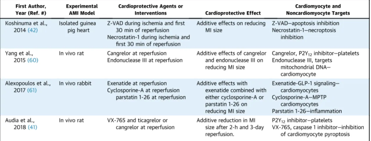
TABLE 2 Experimental Studies Illustrating the Potential for Additive Cardioprotection With Multiple Cardioprotective Agents or Interventions Combining Cardiomyocyte Targets With Noncardiomyocyte Targets*
First Author, Year (Ref. #)
Experimental AMI Model
Cardioprotective Agents or
Interventions Cardioprotective Effect
Cardiomyocyte and Noncardiomyocyte Targets Koshinuma et al.,
2014(42)
Isolated guinea pig heart
Z-VAD during ischemia andfirst 30 min of reperfusion Necrostatin-1 during ischemia and
first 30 min of reperfusion
Additive effects on reducing MI size
Z-VAD—apoptosis inhibition Necrostatin-1—necroptosis
inhibition
Yang et al., 2015(60)
In vivo rat Cangrelor at reperfusion Endonuclease III at reperfusion
Additive effects of cangrelor and endonuclease III on reducing MI size
Cangrelor, P2Y12inhibitor—platelets Endonuclease III, targets
mitochondrial DNA— cardiomyocyte Alexopoulos et al.,
2017(61)
In vivo rabbit Exenatide at reperfusion Cyclosporine-A at reperfusion
parstatin 1-26 at reperfusion
Additive effects with exenatide combined with either cyclosporine-A or parstatin 1-26 on reducing MI size
Exenatide-GLP-1 signaling—
cardiomyocytes Cyclosporine-A—MPTP
cardiomyocytes Parstatin 1-26—inflammation Audia et al.,
2018(41)
In vivo rat VX-765 and ticagrelor or cangrelor at reperfusion
Additive reduction in MI size after 2-h and 3-day reperfusion.
P2Y12inhibitor—platelets
VX-765, caspase 1 inhibitor—inhibition of cardiomyocyte pyroptosis
*Other criteria are as perTable 1.
MPTP¼mitochondrial permeability transition pore; VAD¼val-ala-asp; Z-VAD¼Z-val-ala-asp. Other abbreviations as inTable 1.
may contribute to cardioprotective signaling(28,29).
IRI may cause the death of noncardiomyocytes by various pathways including apoptosis(30). Further- more, IRI may disrupt the endothelial barrier favoring myocardial edema(31,32), and may activate endothe- lial cells causing them to interact with circulating blood cells that may plug the microvessels, release molecules that affect cardiomyocyte function and tolerance to IRI, and infiltrate the myocardium.
Platelets are some of thefirst hematopoietic cells to respond to IRI. Although activated platelets release factors that may exert cardioprotective effects during ischemia(33)strong evidence indicates that they may exacerbate reperfusion injury by mechanisms not dependent on vessel obstruction (34,35). They also form coaggregates with white blood cells (mainly neutrophils), and these plugs are distally embolized upon reperfusion contributing to microvascular obstruction (MVO)(27). MVO can also be caused by embolization from the recanalized coronary plaque and extrinsic compression secondary to edema for- mation upon reperfusion(36). Furthermore, areas of no-reflow and intramyocardial hemorrhage may develop due to extreme myocardial devastation(37).
However they arise, MVO and no-reflow have the po- tential to cause further cardiomyocyte necrosis(38) and are clearly associated with adverse prognosis in patients with AMI(32).
MULTITARGET STRATEGIES FOR CARDIOPROTECTION
We define “multitargeted cardioprotective therapy” as additive or synergistic cardioprotective effects of multiple cardioprotective agents or interventions directed to distinct targets. There are also some specific examples where a single agent is known to have effects on multiple distinct targets and can therefore also be considered as a multitargeted strategy. The combination of agents or interventions to achieve multitarget cardioprotection may be
broadly classified into 5 categories, although these are not mutually exclusive (Central Illustration, Tables 1 to 5). Each of these categories are discussed in the following sections using examples taken from animal experiments. Their applicability to patients is dis- cussed in the following text.
MULTIPLE CARDIOPROTECTIVE AGENTS OR INTERVENTIONS WITH DISTINCT TARGETS WITHIN THE CARDIOMYOCYTE. Conceptually, the simplest approach to multitarget cardioprotection is to combine 2 or more agents or interventions, each of which has a distinct target within the cardiomyocyte.
In this approach, it is important that each car- dioprotective agent or intervention is at maximal
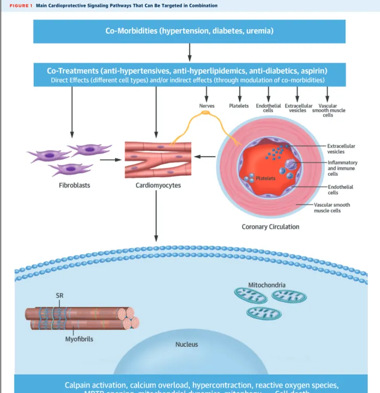
“dose” (i.e., not subthreshold), and that the combi- nation of agents or interventions confers additive benefit in terms of infarct size reduction. The intra- cellular targets can include prosurvival signaling pathways (e.g., the RISK, SAFE, and NO-cGMP-PKG cascades), cell death pathways (e.g., necrosis, apoptosis, autophagy, necroptosis, and pyroptosis), and cellular organelles (e.g., mitochondria, sarco- plasmic reticulum) (Figure 1). As such, maximal cardioprotection may require activation of comple- mentary prosurvival pathways and/or inhibition of deleterious cell death pathways, as recently proposed in the“multitarget hypothesis”(39).
There are several published examples of multi- target cardioprotective strategies directed to distinct signaling pathways within the cardiomyocyte (Table 1). For example, in a pig AMI model, the com- bination of limb RIC with either GIK or exenatide (a glucagon-like peptide-1 mimetic) at the time of reperfusion reduced infarct size to a greater extent than either intervention alone (8). Importantly, the interventions were shown to have distinct intracel- lular targets, with RIC decreasing oxidative stress (myocardial nitrotyrosine levels) and endothelial NOS (eNOS) uncoupling, and GIK and exenatide shifting cardiac metabolism toward increased glycolysis(8).
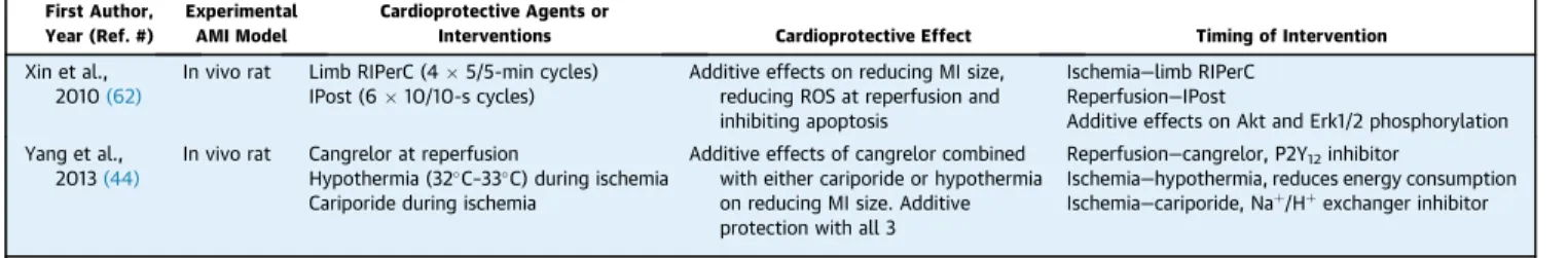
TABLE 3 Experimental Studies Illustrating the Potential for Additive Cardioprotection With Multiple Cardioprotective Agents or Interventions Targeting Distinct Time-Points During Ischemia and Reperfusion*
First Author, Year (Ref. #)
Experimental AMI Model
Cardioprotective Agents or
Interventions Cardioprotective Effect Timing of Intervention
Xin et al., 2010(62)
In vivo rat Limb RIPerC (45/5-min cycles) IPost (610/10-s cycles)
Additive effects on reducing MI size, reducing ROS at reperfusion and inhibiting apoptosis
Ischemia—limb RIPerC Reperfusion—IPost
Additive effects on Akt and Erk1/2 phosphorylation Yang et al.,
2013(44)
In vivo rat Cangrelor at reperfusion
Hypothermia (32C–33C) during ischemia Cariporide during ischemia
Additive effects of cangrelor combined with either cariporide or hypothermia on reducing MI size. Additive protection with all 3
Reperfusion—cangrelor, P2Y12inhibitor
Ischemia—hypothermia, reduces energy consumption Ischemia—cariporide, Naþ/Hþexchanger inhibitor
*Other criteria are as perTable 1.
IPost¼post-conditioning; ROS¼reactive oxygen species; other abbreviations as inTable 1.
MULTIPLE CARDIOPROTECTIVE AGENTS OR INTERVENTIONS WITH NONCARDIOMYOCYTE TARGETS.
Combining cardiomyocyte-targeted therapies with therapies that target noncardiomyocyte components in the heart (e.g., those improving tissue perfusion) may provide a more effective strategy for car- dioprotection (Table 2). One example is provided by P2Y12 inhibitors (such as ticagrelor and cangrelor), which are known to reduce infarct size(40). Because all patients with AMI receive a P2Y12receptor antag- onist, a cardioprotective agent, to be effective, must provide additive protection on this therapeutic background. In this regard, VX-765, an inhibitor of caspase 1–mediated pyroptosis, has been demon- strated in rats to provide such an additive benefit on a therapeutic background of the P2Y12inhibitors tica- grelor or cangrelor(41). In another example, targeting necroptosis with necrostatin-1 and apoptosis (pre- sumably in nonmyocytes) with Z-VAD during ischemia and reperfusion conferred additive reduc- tion in infarct size in isolated perfused guinea pig hearts(42).
There would appear to be a solid rationale for combining one agent targeting the microcirculation (MVO) with another targeting cardiomyocytes. Un- fortunately, there has been only marginal success to date in trying to relieve MVO and improve microcir- culatoryflow after MI, even experimentally. Some of the more promising candidates include the vasoactive compounds adenosine and NO (32). Recombinant human angiopoietin-like protein 4 (ANGPTL4) indeed reduced infarct size and prevented no-reflow and intramyocardial hemorrhage in mice(43).
MULTIPLE CARDIOPROTECTIVE AGENTS OR INTERVENTIONS TARGETING DISTINCT TIME-POINTS DURING ISCHEMIA AND REPERFUSION.Given the pos- sibility of intervening at 3 different time points in the setting of AMI (i.e., during ischemia, at reperfusion, and late into reperfusion), there is an opportunity to combine 2 or more cardioprotective therapies to target these 3 different phases in order to achieve additive cardioprotection (Table 3). An excellent example of this multitarget cardioprotective approach was provided by Yang et al. (44), who
TABLE 5 Experimental Studies Illustrating the Potential for Additive Cardioprotection With Multiple Cardioprotective Agents or Interventions Having Multiple Targets*
First Author, Year (Ref. #)
Experimental AMI Model
Cardioprotective Agents or
Interventions Cardioprotective Effect Signaling Pathways
Rastaldo et al., 2012(48)
Isolated rat heart
Hybrid molecule containing NO donor and antioxidant
Additive effects on reducing MI size
Mitochondrial KATPchannel
Lougiakis et al., 2016(47)
In vivo rabbit Hybrid molecule containing H2S-donor and adenosine analogue at reperfusion
Additive effects on reducing MI size
cGMP/PKG/phospholamban pathway
García-Ruiz et al., 2016(17)and Garcia-Prieto et al., 2017(27)
In vivo pig In vivo mouse
Intravenous metoprolol targeting simultaneously cardiomyocytes (reducing energy demand), and neutrophils (inhibiting migration and neutrophil–
platelet coaggregates)
Reduces infarct size when given at different times of ischemia duration, but effect is stronger when given earlier
Cardiomyocyte oxygen consumption reduction and neutrophil conformational re-arrangements
*Other criteria are as perTable 1.
cGMP¼cyclic guanosine monophosphate; PKG¼protein kinase G; other abbreviations as inTable 1.
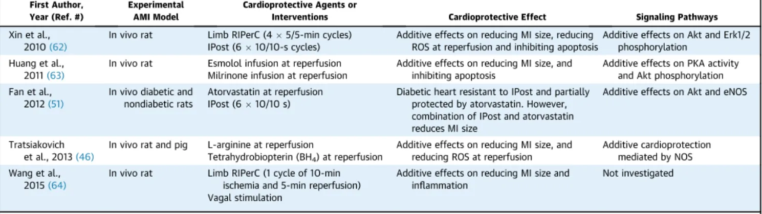
TABLE 4 Experimental Studies Illustrating the Potential for Additive Cardioprotection With Multiple Cardioprotective Agents Or Interventions Targeting the Same Signaling Pathway but With Potentiating Effects*
First Author, Year (Ref. #)
Experimental AMI Model
Cardioprotective Agents or
Interventions Cardioprotective Effect Signaling Pathways
Xin et al., 2010(62)
In vivo rat Limb RIPerC (45/5-min cycles) IPost (610/10-s cycles)
Additive effects on reducing MI size, reducing ROS at reperfusion and inhibiting apoptosis
Additive effects on Akt and Erk1/2 phosphorylation
Huang et al., 2011(63)
In vivo rat Esmolol infusion at reperfusion Milrinone infusion at reperfusion
Additive effects on reducing MI size, and inhibiting apoptosis
Additive effects on PKA activity and Akt phosphorylation Fan et al.,
2012(51)
In vivo diabetic and nondiabetic rats
Atorvastatin at reperfusion IPost (610/10 s)
Diabetic heart resistant to IPost and partially protected by atorvastatin. However, combination of IPost and atorvastatin reduces MI size
Additive effects on Akt and eNOS
Tratsiakovich et al., 2013(46)
In vivo rat and pig L-arginine at reperfusion
Tetrahydrobiopterin (BH4) at reperfusion
Additive effects on reducing MI size, and reducing ROS at reperfusion
Additive cardioprotection mediated by NOS Wang et al.,
2015(64)
In vivo rat Limb RIPerC (1 cycle of 10-min ischemia and 5-min reperfusion) Vagal stimulation
Additive effects on reducing MI size and inflammation
Not investigated
*Other criteria are as perTable 1.
NOS¼nitric oxide synthase; PKA¼protein kinase A; other abbreviations as inTables 1 and 3.
showed that triple therapy combining mild hypo- thermia and cariporide (a sodium-hydrogen exchanger inhibitor) during ischemia, with cangrelor added at reperfusion conferred additive protection.
MULTIPLE CARDIOPROTECTIVE AGENTS OR INTERVENTIONS TARGETING THE SAME SIGNALING PATHWAY BUT WITH ADDITIVE EFFECTS. In some situ- ations, 2 agents may act on the same signaling
FIGURE 1 Main Cardioprotective Signaling Pathways That Can Be Targeted in Combination
Different comorbidities and cotreatments can influence protection via effects on different cellular and subcellular targets, as indicated byarrows.
MPTP¼mitochondrial permeability transition pore; SR¼sarcoplasmic reticulum.
pathway, one potentiating the other’s cardioprotective effects (Table 4). For example, coadministration of the NOS substrate L-arginine and cofactor tetrahy- drobiopterin (BH4) just before reperfusion significantly reduced MI size in both rats and pigs, despite neither being protective on their own(45,46).
A SINGLE CARDIOPROTECTIVE AGENT OR INTERVENTION WITH MULTIPLE TARGETS.There are many examples of a single cardioprotective agent or single intervention having multiple targets (Table 5), and one would intuitively expect that these therapies would be more effective than a single-target agent or intervention.
For example, intravenous metoprolol administered before reperfusion reduces both infarct size and MVO in mice(27), pigs (17), and humans (13). Classically, metoprolol has been considered to reduce ischemic injury by reducing energy demands from car- diomyocytes, because it is more effective when given early during ischemia(17). However, metoprolol has recently been shown to act via theb1 adrenergic re- ceptors on neutrophils to decrease neutrophil–platelet coaggregate formation during reperfusion(17), which can explain the strong effect of metoprolol on MVO.
The dual-target benefits of metoprolol appear to be specific to this drug and not a class effect.
The endogenous cardioprotective strategies of IPC, IPost, and RIC are known to protect the heart through
a number of different signaling pathways and might therefore be assumed to confer a stronger car- dioprotective effect than a single-target agent.
A single miRNA or small interfering RNA may protect the heart against acute IRI through its effects on a variety of different target mRNAs. Hybrid mol- ecules may have 2 or more structural domains acting as 2 distinct pharmacophores to provide additive cardioprotection. For example, a hybrid compound that combines the adenine nucleus with a moiety that slowly releases hydrogen sulfide (H2S)–induced ad- ditive cardioprotection(47). A hybrid molecule con- sisting of a lipophilic NO donor and a lipophilic antioxidant compound protected the rat heart against acute IRI if given as a hybrid molecule, but not as a mixture(48).
THE IMPACT OF COMORBIDITIES AND COTREATMENTS
Since the first observations in animal studies in the late nineties, it has been well established that many of the signaling pathways involved in the protection by ischemic conditioning interventions are affected by several cardiovascular risk factors and comorbid- ities such as sex, age, hypertension, and metabolic diseases such as hyperlipidemia and diabetes(5). For
TABLE 6 Main Clinical Studies (Completed and Ongoing) Investigating Multitargeted Agents or Interventions Against AMI IRI in STEMI Patients Undergoing PPCI
First Author, Year (Trial) (Ref. #)
No. of Patients (Control/Intervention)
Multitargeted Treatment
Intervention Approach Primary Endpoint Outcome
Completed studies Eitel et al., 2015 (LIPSIA-COND) (54)
Control/IPost/RIPerCþIPost 232/232/232
Combined limb RIPerCþIPost
RIPerC: In hospital upper limb 3 cycles (5/5 min, 200 mm Hg), IPost: (1-min balloon inflation/1-min
deflation) started as soon as possible after reopening of the culprit coronary artery
Myocardial salvage index (edema and late gadolinium enhancement by CMR)
23% increase in salvage index No limb RIPerC alone
group
Pasupathy et al., 2017 (NACIAM) (56)
IV GTN/IV GTNþNAC 38/37
Combined NACþGTN
IV GTN:
IV NAC:
MI size (late gadolinium enhancement by CMR)
5.5% reduction in infarct size All patients received
GTN Actively recruiting
studies Ovize et al.,
(CARIOCA) (NCT03155022)
Estimated enrolment 355/355
Combined limb RIPerC and IPost
RIC: In-hospital, upper limb,
4 cycles (5/5 min, 200 mm Hg) initiated as soon as possible before PCI IPost: 4 cycles (1 min balloon inflation/
1 min deflation) started as soon as possible after reopening of the culprit coronary artery
Combined incidence of all- cause mortality; worsening of heart failure during initial hospitalization or rehospitalization for heart failure at 6 months after PPCI
Recruiting
Garcia-Dorado et al., COMBAT-MI (NCT02404376)
22 factorial design (RIC, exenatide, both, or neither) 107/107/107/107
Combined limb RIPerCþ exenatide
RIC: in-hospital, upper limb, 4 cycles (5/5 min, 200 mm Hg) Intravenous infusion of exenatide
initiated before reperfusion
Myocardial infarct size (late gadolinium enhancement by CMR)
Recruiting
CARIOCA¼Combined Application of Remote and Intra-Coronary Ischemic Conditioning in Acute Myocardial Infarction; CMR¼cardiac magnetic resonance; COMBAT-MI¼COMBinAtion Therapy in Myocardial Infarction; GTN¼nitroglycerin; IRI¼ischemia–reperfusion injury; IV¼intravenous; LIPSIA-COND¼Effect of Conditioning on Myocardial Damage in STEMI; NAC¼N-acetylcysteine;
NACIAM¼N-acetylcysteine in Acute Myocardial Infarction; PCI¼percutaneous coronary intervention; PPCI¼primary percutaneous coronary intervention; RIC¼remote ischemic conditioning;
STEMI¼ST-segment elevation myocardial infarction; other abbreviations as inTables 1 and 3.
example, conditioning stimuli are less effective in diabetic animals because they are less able to activate PI3K/Akt (49). A stronger stimulus or combination strategy against additional targets may be required to fully activate the protective pathways. For example, cardioprotection can be restored by administering an inhibitor of phosphatase and tensin homolog (PTEN)—a major negative regulator of PI3K/Akt—to maximize PI3K/Akt activation(50). Medications used to treat a comorbidity may either interfere or enhance cardioprotective signaling(6). Atorvastatin at reper- fusion combined with IPost was able to overcome the resistance of the diabetic murine heart to car- dioprotection by augmenting the activation of the Akt-eNOS pathway(51).
Most studies showing the interaction of certain comorbidities with cardioprotection have been per- formed in single comorbidity models with no specific treatment of the comorbidity. Although most animal experiments on IRI and protection from it were per- formed in young and otherwise healthy (therefore untreated) animals, patients recruited into clinical cardioprotection trials are usually of advanced age and have numerous comorbidities and related come- dications as well as short-term treatments related to AMI. An important example of comedications con- founding cardioprotection are anesthetics including propofol that can affect cardioprotection(6). There- fore, more studies in adequate animal models more closely mimicking the clinical situation with multiple comorbidities and related comedications would be ideal for finding drug targets (5). In this regard, it should be noted that existing clinical therapies post- MI already consist of many combination strategies.
The impact of comorbidities and cotreatments on cardioprotection has long been suspected; however, subgroup analyses performed in largescale clinical studies including patients with multiple comorbid- ities and comedications have not confirmed the con- founding effect of a particular single comorbidity or comedication (e.g., in the CIRCUS [Cyclosporine and Prognosis in Acute Myocardial Infarction (MI) Pa- tients] trial on cyclosporine A[52]).
CLINICAL STUDIES OF MULTITARGET THERAPY.The main target patient population for cardioprotection is those with STEMI undergoing immediate revascular- ization by PPCI. Current clinical studies of multitarget therapies are limited to combinations of different ischemic conditioning strategies, a combination of pharmacological treatments, or a combination of pharmacological and conditioning strategies, (Table 6), whereas physical measures such as hypo- thermia or nerve stimulation have not been studied in combination with other cardioprotective strategies.
As an example of a study investigating 2 in- terventions targeted primarily to cardiomyocytes, exenatide (53) and RIC (15), which have each demonstrated cardioprotective efficacy individually in STEMI patients undergoing PPCI, are being inves- tigated in combination in the COMBAT-MI (COMBi- nAtion Therapy in Myocardial Infarction) trial (NCT02404376).
In an investigation of 2 therapies administered at different time points, Eitel et al. (54) studied the combination of in-hospital RIC before reperfusion and intracoronary IPost after reopening the culprit coronary artery in 696 STEMI patients. Whereas IPost alone failed to improve myocardial salvage index assessed by cardiac magnetic resonance, combined RIC and IPost increased the salvage index. Because there was no group treated with RIC alone, the study could not confirm an additive effect. Another clinical study failed to observe an additive cardioprotective effect with limb RIC and IPost (55). The CARIOCA (Combined Application of Remote and Intra-Coronary Ischemic Conditioning in Acute Myocardial Infarc- tion) trial (NCT03155022), investigating the efficacy of combined in-hospital RIC before reperfusion and IPost on clinical outcome is ongoing.
In a study intended to test the potentiating effect of 2 different cardioprotective agents, the NACIAM (N-acetylcysteine in Acute Myocardial Infarction) trial (56)examined the effects of N-acetylcysteine (NAC) on infarct size in 75 patients with STEMI undergoing PPCI. NAC is an antioxidant and potentiates the ef- fects of nitroglycerine. With background nitroglycerin infusion administered to all, patients receiving NAC had an absolute 5.5% reduction in cardiac magnetic resonance imaging–assessed infarct size relative to placebo. However, the study design of the trial did not provide conclusive information about mechanisms involved, because all patients received nitroglycerin(56).
As an example of a single agent targeting multiple pathways, metoprolol has been studied in STEMI patients; however, results have been mixed(13,57).
FUTURE RECOMMENDATIONS
Extensive evidence accumulated over the past 30 years has shown that a multitude of cardioprotective therapies are effective at reducing infarct size in an- imal models of IRI(3,4,6,32). However, routinely used animal models of IRI do not adequately recapitulate the complex phenomenon of IRI in patients. Here, we hypothesize that to be effective in these models, and to effectively translate cardioprotection to patients, a multitarget cardioprotective therapy is necessary.
Combinations of interventions with solid preclinical
information on mechanism of action, efficacy, and safety, and that are easily applicable are good candi- dates to be moved to clinical trials. In designing such a trial, a factorial design may be used to prove addi- tive benefit of a combination, but this approach in- creases patient numbers needed. A better approach may be to first prove additive benefit in animal models and then test the combination in patients against control. Another important consideration is that STEMI patients receive comedications such as P2Y12inhibitors. Other factors such as the effect on arrhythmias and long-term cardiac remodeling should also be considered.
In light of the examples discussed in the previous sections, some promising examples of approaches to multitargeted cardioprotection include:
A combination of RIC with a drug with a different mechanism of action—this is being tested in the COMBAT-MI trial.
A combination of a drug that activates endoge- nous cardioprotective pathways (RISK, SAFE,
cGMP/PKG) with a drug that inhibits cell death pathways.
A drug targeting vascular injury/inflammation with a drug targeting cardiomyocyte death.
We hypothesize that the ideal multitargeted therapy might be one that can target MVO (e.g., intravenous cangrelor or ANGPL4), target car- diomyocytes (e.g., remote ischemic per-conditioning) and target inflammation (e.g., metoprolol). The timing of administration of these modalities could potentially be separated over time.
ADDRESS FOR CORRESPONDENCE: Dr. Sean M.
Davidson, The Hatter Cardiovascular Institute, Uni- versity College London, 67 Chenies Mews, London WC1E 6HX, United Kingdom. E-mail:s.davidson@ucl.
ac.uk. Twitter: @UCL. OR Dr. David Garcia-Dorado, Servicio de Cardiología, Hospital Universitari Vall d’Hebron, Passeig Vall d’Hebron, 119-129, 08035, Barcelona, Spain. E-mail: dgdorado@vhebron.net.
Twitter:@VHIR.
R E F E R E N C E S
1.Heusch G. Critical issues for the translation of cardioprotection. Circ Res 2017;120:1477–86.
2.Heusch G. Cardioprotection research must leave its comfort zone. Eur Heart J 2018;39:3393–5.
3.Hausenloy DJ, Botker HE, Engstrom T, et al.
Targeting reperfusion injury in patients with ST- segment elevation myocardial infarction: trials and tribulations. Eur Heart J 2017;38:935–41.
4.Heusch G, Gersh BJ. The pathophysiology of acute myocardial infarction and strategies of pro- tection beyond reperfusion: a continual challenge.
Eur Heart J 2017;38:774–84.
5.Ferdinandy P, Hausenloy DJ, Heusch G, Baxter GF, Schulz R. Interaction of risk factors, comorbidities, and comedications with ischemia/
reperfusion injury and cardioprotection by pre- conditioning, postconditioning, and remote con- ditioning. Pharmacol Rev 2014;66:1142–74.
6.Hausenloy DJ, Garcia-Dorado D, Botker HE, et al. Novel targets and future strategies for acute cardioprotection: position paper of the European Society of Cardiology Working Group on Cellular Biology of the Heart. Cardiovasc Res 2017;113:
564–85.
7.Inserte J, Hernando V, Vilardosa U, Abad E, Poncelas-Nozal M, Garcia-Dorado D. Activation of cGMP/protein kinase G pathway in post- conditioned myocardium depends on reduced oxidative stress and preserved endothelial nitric oxide synthase coupling. J Am Heart Assoc 2013;2:
e005975.
8.Alburquerque-Bejar JJ, Barba I, Inserte J, et al.
Combination therapy with remote ischaemic con- ditioning and insulin or exenatide enhances infarct size limitation in pigs. Cardiovasc Res 2015;107:
246–54.
9.Kleinbongard P, Skyschally A, Heusch G. Car- dioprotection by remote ischemic conditioning and its signal transduction. Pflugers Arch 2017;
469:159–81.
10.Heusch G. Molecular basis of cardioprotection:
signal transduction in ischemic pre-, post-, and remote conditioning. Circ Res 2015;116:674–99.
11.Ruiz-Meana M, Garcia-Dorado D, Pina P, Inserte J, Agullo L, Soler-Soler J. Cariporide pre- serves mitochondrial proton gradient and delays ATP depletion in cardiomyocytes during ischemic conditions. Am J Physiol Heart Circ Physiol 2003;
285:H999–1006.
12.Miki T, Liu GS, Cohen MV, Downey JM. Mild hy- pothermia reduces infarct size in the beating rabbit heart: a practical intervention for acute myocardial infarction? Basic Res Cardiol 1998;93:372–83.
13.Ibanez B, Macaya C, Sanchez-Brunete V, et al.
Effect of early metoprolol on infarct size in ST- segment-elevation myocardial infarction patients undergoing primary percutaneous coronary inter- vention: the Effect of Metoprolol in Car- dioprotection During an Acute Myocardial Infarction (METOCARD-CNIC) trial. Circulation 2013;128:1495–503.
14.Selker HP, Beshansky JR, Sheehan PR, et al. Out- of-hospital administration of intravenous glucose- insulin-potassium in patients with suspected acute coronary syndromes: the IMMEDIATE randomized controlled trial. JAMA 2012;307:1925–33.
15.Botker HE, Kharbanda R, Schmidt MR, et al.
Remote ischaemic conditioning before hospital admission, as a complement to angioplasty, and effect on myocardial salvage in patients with acute myocardial infarction: a randomised trial. Lancet 2010;375:727–34.
16.Kleinbongard P, Amanakis G, Skyschally A, Heusch G. Reflection of cardioprotection by remote ischemic perconditioning in attenuated ST- segment elevation during ongoing coronary oc- clusion in pigs: evidence for cardioprotection from ischemic injury. Circ Res 2018;122:1102–8.
17.Garcia-Ruiz JM, Fernandez-Jimenez R, Garcia- Alvarez A, et al. Impact of the timing of meto- prolol administration during STEMI on infarct size and ventricular function. J Am Coll Cardiol 2016;
67:2093–104.
18.Ovize M, Baxter GF, Di Lisa F, et al. Post- conditioning and protection from reperfusion injury: where do we stand? Position paper from the Working Group of Cellular Biology of the Heart of the European Society of Cardiology. Cardiovasc Res 2010;87:406–23.
19.Hausenloy DJ, Duchen MR, Yellon DM. Inhibit- ing mitochondrial permeability transition pore opening at reperfusion protects against ischaemia- reperfusion injury. Cardiovasc Res 2003;60:617–25.
20.Kin H, Zhao ZQ, Sun HY, et al. Postcondition- ing attenuates myocardial ischemia-reperfusion injury by inhibiting events in the early minutes of reperfusion. Cardiovasc Res 2004;62:74–85.
21.Doukas J, Wrasidlo W, Noronha G, et al.
Phosphoinositide 3-kinase gamma/delta inhibition limits infarct size after myocardial ischemia/
reperfusion injury. Proc Natl Acad Sci U S A 2006;
103:19866–71.
22.Souktani R, Pons S, Guegan C, et al. Car- dioprotection against myocardial infarction with PTD-BIR3/RING, a XIAP mimicking protein. J Mol Cell Cardiol 2009;46:713–8.
23.Basalay M, Barsukevich V, Mastitskaya S, et al. Remote ischaemic pre- and delayed
postconditioning - similar degree of cardioprotection but distinct mechanisms. Exp Physiol 2012;97:
908–17.
24.Hausenloy DJ, Yellon DM. Myocardial ischemia-reperfusion injury: a neglected thera- peutic target. J Clin Invest 2013;123:92–100.
25.Figueras J, Otaegui I, Marti G, et al. Area at risk and collateral circulation in afirst acute myocar- dial infarction with occluded culprit artery. STEMI vs non-STEMI patients. Int J Cardiol 2018;259:
14–9.
26.Ibanez B, James S, Agewall S, et al. 2017 ESC guidelines for the management of acute myocar- dial infarction in patients presenting with ST- segment elevation: the Task Force for the man- agement of acute myocardial infarction in patients presenting with ST-segment elevation of the Eu- ropean Society of Cardiology (ESC). Eur Heart J 2018;39:119–77.
27.Garcia-Prieto J, Villena-Gutierrez R, Gomez M, et al. Neutrophil stunning by metoprolol reduces infarct size. Nat Commun 2017;8:14780.
28.Abrial M, Da Silva CC, Pillot B, et al. Cardiac fibroblasts protect cardiomyocytes against lethal ischemia-reperfusion injury. J Mol Cell Cardiol 2014;68:56–65.
29.Giricz Z, Varga ZV, Baranyai T, et al. Car- dioprotection by remote ischemic preconditioning of the rat heart is mediated by extracellular vesi- cles. J Mol Cell Cardiol 2014;68:75–8.
30.Jose Corbalan J, Vatner DE, Vatner SF.
Myocardial apoptosis in heart disease: does the emperor have clothes? Basic Res Cardiol 2016;
111:31.
31.Garcia-Dorado D, Andres-Villarreal M, Ruiz- Meana M, Inserte J, Barba I. Myocardial edema: a translational view. J Mol Cell Cardiol 2012;52:
931–9.
32.Heusch G. The coronary circulation as a target of cardioprotection. Circ Res 2016;118:1643–58.
33.Russo I, Penna C, Musso T, et al. Platelets, diabetes and myocardial ischemia/reperfusion injury. Cardiovasc Diabetol 2017;16:71.
34.Barrabes JA, Inserte J, Mirabet M, et al.
Antagonism of P2Y12 or GPIIb/IIIa receptors re- duces platelet-mediated myocardial injury after ischaemia and reperfusion in isolated rat hearts.
Thromb Haemost 2010;104:128–35.
35.Vilahur G, Gutierrez M, Casani L, et al. Pro- tective effects of ticagrelor on myocardial injury after infarction. Circulation 2016;134:1708–19.
36.Fernandez-Jimenez R, Barreiro-Perez M, Mar- tin-Garcia A, et al. Dynamic edematous response of the human heart to myocardial infarction: im- plications for assessing myocardial area at risk and salvage. Circulation 2017;136:1288–300.
37.Garcia-Dorado D, Theroux P, Solares J, et al.
Determinants of hemorrhagic infarcts. Histologic observations from experiments involving coronary occlusion, coronary reperfusion, and reocclusion.
Am J Pathol 1990;137:301–11.
38.Heusch G, Kleinbongard P, Bose D, et al.
Coronary microembolization: from bedside to bench and back to bedside. Circulation 2009;120:
1822–36.
39.Rossello X, Yellon DM. The RISK pathway and beyond. Basic Res Cardiol 2017;113:2.
40.Cohen MV, Downey JM. The impact of irre- producibility and competing protection from P2Y12 antagonists on the discovery of car- dioprotective interventions. Basic Res Cardiol 2017;112:64.
41.Audia JP, Yang X-M, Crockett ES, et al. Cas- pase-1 inhibition by VX-765 administered at reperfusion in P2Y12 receptor antagonist-treated rats provides long-term reduction in myocardial infarct size and preservation of ventricular func- tion. Basic Res Cardiol 2018;113.
42.Koshinuma S, Miyamae M, Kaneda K, Kotani J, Figueredo VM. Combination of necroptosis and apoptosis inhibition enhances cardioprotection against myocardial ischemia-reperfusion injury.
J Anesth 2014;28:235–41.
43.Galaup A, Gomez E, Souktani R, et al. Protec- tion against myocardial infarction and no-reflow through preservation of vascular integrity by angiopoietin-like 4. Circulation 2012;125:140–9.
44.Yang XM, Cui L, Alhammouri A, Downey JM, Cohen MV. Triple therapy greatly increases myocardial salvage during ischemia/reperfusion in the in situ rat heart. Cardiovasc Drugs Ther 2013;
27:403–12.
45.Lux A, Pokreisz P, Swinnen M, et al. Concom- itant phosphodiesterase 5 inhibition enhances myocardial protection by inhaled nitric oxide in ischemia-reperfusion injury. J Pharmacol Exp Ther 2016;356:284–92.
46.Tratsiakovich Y, Gonon AT, Kiss A, et al.
Myocardial protection by co-administration of L- arginine and tetrahydrobiopterin during ischemia and reperfusion. Int J Cardiol 2013;169:83–8.
47.Lougiakis N, Papapetropoulos A, Gikas E, et al.
Synthesis and pharmacological evaluation of novel adenine-hydrogen sulfide slow release hybrids designed as multitarget cardioprotective agents.
J Med Chem 2016;59:1776–90.
48.Rastaldo R, Cappello S, Di Stilo A, Folino A, Losano G, Pagliaro P. A lipophilic nitric oxide donor and a lipophilic antioxidant compound protect rat heart against ischemia-reperfusion injury if given as hybrid molecule but not as a mixture. J Cardiovasc Pharmacol 2012;59:241–8.
49.Tsang A, Hausenloy DJ, Mocanu MM, Carr RD, Yellon DM. Preconditioning the diabetic heart: the importance of Akt phosphorylation. Diabetes 2005;54:2360–4.
50.Xue R, Lei S, Xia ZY, et al. Selective inhibition of PTEN preserves ischaemic post-conditioning cardioprotection in STZ-induced Type 1 diabetic rats: role of the PI3K/Akt and JAK2/STAT3 path- ways. Clin Sci (Lond) 2016;130:377–92.
51.Fan Y, Yang S, Zhang X, Cao Y, Huang Y.
Comparison of cardioprotective efficacy resulting from a combination of atorvastatin and ischaemic post-conditioning in diabetic and non-diabetic rats. Clin Exp Pharmacol Physiol 2012;39:938–43.
52.Cung TT, Morel O, Cayla G, et al. Cyclosporine before PCI in patients with acute myocardial infarction. N Engl J Med 2015;373:1021–31.
53.Lonborg J, Vejlstrup N, Kelbaek H, et al. Exe- natide reduces reperfusion injury in patients with
ST-segment elevation myocardial infarction. Eur Heart J 2012;33:1491–9.
54.Eitel I, Stiermaier T, Rommel KP, et al. Car- dioprotection by combined intrahospital remote ischaemic perconditioning and postconditioning in ST-elevation myocardial infarction: the random- ized LIPSIA CONDITIONING trial. Eur Heart J 2015;
36:3049–57.
55.Prunier F, Angoulvant D, Saint Etienne C, et al.
The RIPOST-MI study, assessing remote ischemic perconditioning alone or in combination with local ischemic postconditioning in ST-segment elevation myocardial infarction. Basic Res Cardiol 2014;109:400.
56.Pasupathy S, Tavella R, Grover S, et al. Early use of N-acetylcysteine with nitrate therapy in patients undergoing primary percutaneous coro- nary intervention for ST-segment-elevation myocardial infarction reduces myocardial infarct size (the NACIAM Trial [N-acetylcysteine in Acute Myocardial Infarction]). Circulation 2017;136:
894–903.
57.Roolvink V, Ibanez B, Ottervanger JP, et al.
Early intravenous beta-blockers in patients with ST-segment elevation myocardial infarction before primary percutaneous coronary interven- tion. J Am Coll Cardiol 2016;67:2705–15.
58.Schwiebert C, Huhn R, Heinen A, et al.
Postconditioning by xenon and hypothermia in the rat heart in vivo. Eur J Anaesthesiol 2010;27:
734–9.
59.Sun J, Aponte AM, Menazza S, Gucek M, Steenbergen C, Murphy E. Additive car- dioprotection by pharmacological postcondition- ing with hydrogen sulfide and nitric oxide donors in mouse heart: S-sulfhydration vs. S-nitro- sylation. Cardiovasc Res 2016;110:96–106.
60.Yang XM, Cui L, White J, et al. Mitochondrially targeted Endonuclease III has a powerful anti- infarct effect in an in vivo rat model of myocar- dial ischemia/reperfusion. Basic Res Cardiol 2015;
110:3.
61.Alexopoulos P, Panoutsopoulou K, Vogiatzis G, Koletsis E, Dougenis D, Tsopanoglou NE. Com- bined treatment with exenatide and cyclosporine A or parstatin 1-26 results in enhanced reduction of infarct size in a rabbit model. J Cardiovasc Pharmacol 2017;70:34–41.
62.Xin P, Zhu W, Ma S, et al. Combined local ischemic postconditioning and remote percondi- tioning recapitulate cardioprotective effects of local ischemic preconditioning. Am J Physiol Heart Circ Physiol 2010;298:H1819–31.
63.Huang MH, Wu Y, Nguyen V, et al. Heart protection by combination therapy with esmolol and milrinone at late-ischemia and early reperfusion. Cardiovasc Drugs Ther 2011;25:
223–32.
64.Wang Q, Liu GP, Xue FS, et al. Combined vagal stimulation and limb remote ischemic percondi- tioning enhances cardioprotection via an anti- inflammatory pathway. Inflammation 2015;38:
1748–60.
KEY WORDS cardioprotection, ischemia, myocardial infarction, reperfusion