Endothelial Dysfunction of Bypass Graft:
Direct Comparison of In Vitro and In Vivo Models of Ischemia-Reperfusion Injury
Gábor Veres1*, Péter Hegedűs1,2, EnikőBarnucz1, Raphael Zöller1, Stephanie Klein1, Harald Schmidt1, Tamás Radovits2, Sevil Korkmaz1, Matthias Karck1, Gábor Szabó1 1Department of Cardiac Surgery, University of Heidelberg, Heidelberg, Germany,2Heart Center, Semmelweis University, Budapest, Hungary
*gaborveres@yahoo.com
Abstract
Background
Although, ischemia/reperfusion induced vascular dysfunction has been widely described, no comparative study of in vivo- and in vitro-models exist. In this study, we provide a direct comparison between models (A) ischemic storage and in-vitro reoxygenation (B) ischemic storage and in vitro reperfusion (C) ischemic storage and in-vivo reperfusion.
Methods and Results
Aortic arches from rats were stored for 2 hours in saline. Arches were then (A) in vitro reoxy- genated (B) in vitro incubated in hypochlorite for 30 minutes (C) in vivo reperfused after het- erotransplantation (2, 24 hours and 7 days reperfusion). Endothelium-dependent and independent vasorelaxations were assessed in organ bath. DNA strand breaks were as- sessed by TUNEL-method, mRNA expressions (caspase-3, bax, bcl-2, eNOS) by quantita- tive real-time PCR, proteins by Western blot analysis and the expression of CD-31 by immunochemistry. Endothelium-dependent maximal relaxation was drastically reduced in the in-vivo models compared to ischemic storage and in-vitro reperfusion group, and no difference showed between ischemic storage and control group. CD31-staining showed sig- nificantly lower endothelium surface ratio in-vivo, which correlated with TUNEL-positive ratio. Increased mRNA and protein levels of pro- and anti-apoptotic gens indicated a significantly higher damage in the in-vivo models.
Conclusion
Even short-period of ischemia induces severe endothelial damage (in-vivo reperfusion model). In-vitro models of ischemia-reperfusion injury can be limitedly suited for reliable in- vestigations. Time course of endothelial stunning is also described.
OPEN ACCESS
Citation:Veres G, Hegedűs P, Barnucz E, Zöller R, Klein S, Schmidt H, et al. (2015) Endothelial Dysfunction of Bypass Graft: Direct Comparison ofIn VitroandIn VivoModels of Ischemia-Reperfusion Injury. PLoS ONE 10(4): e0124025. doi:10.1371/
journal.pone.0124025
Academic Editor:Xin-Liang Ma, Thomas Jefferson University, UNITED STATES
Received:October 23, 2014 Accepted:March 9, 2015 Published:April 15, 2015
Copyright:© 2015 Veres et al. This is an open access article distributed under the terms of the Creative Commons Attribution License, which permits unrestricted use, distribution, and reproduction in any medium, provided the original author and source are credited.
Data Availability Statement:All relevant data are within the paper.
Funding:The authors have no support or funding to report.
Competing Interests:The authors have declared that no competing interests exist.
Introduction
Vascular grafts are important therapeutic option for bypass surgery. The long-term benefit of bypass surgery depends largely on the long-term patency of bypass grafts, which are determined by several factors: the progress of heart/vascular disease, the run-off and the biological proper- ties of the implanted graft, injuries during surgical manipulation and the degree of ischemia/
reperfusion (IR) injury.
Regarding the importance of an intact endothelial layer for graft patency, numerous experi- mental animal models have been developed in an attempt to understand the developing patho- physiological processes of IR injury on the bypass graft. A simple, clinically relevant in vitro experimental model and/or an in vivo model for vascular IR injury would obviously offer great prospects in the field of vascular pharmacological research. While the complex nature of IR inju- ry cannot possibly be replicated fully in in vitro circumstances, these animal models must provide a reproducible paradigm allowing the investigation and evaluation of endothelial dysfunction.
Throughout the years, various in vitro animal models have been utilized to mimic endothe- lial dysfunction: (A) In vitro model of ischemia (hypoxia-reoxygenation) [1–3] and (B) in vitro models of ischemia-reperfusion [4–7] are two dominant models of vascular injury. However, it was proved that endothelial injury occurring in vessels during in vitro hypoxia and reoxygena- tion is too moderate to induce functional alterations of the endothelium, probably due to lack of activated leucocytes [8]. Several recent in vitro studies used successfully the hypochlorite to mimic the detrimental effect of reperfusion injury [4–7]. However, these in vitro models could not demonstrate a severely reduced vasorelaxation function after IR injury, which is not fully correlated the results after bypass surgery published by several animal and human studies. To observe the time course of vascular injury and the in vivo effect of IR injury, we developed a new rat model of arterial revascularization.
This study aims to compare models of in vivo induced reperfusion injury of transplanted rat aortic arches with the in vitro injury models where oxidative damage is caused by reoxygena- tion and/or incubation in hypochlorite. Additionally, time course of endothelial dysfunction and recovery was also studied using a newly developed in vivo IR model in rat.
Methods
Animals
Young male Lewis rats (250 to 330 g; Charles River, Sulzfeld, Germany) (N = 5–7/group) were used for our experiments. All procedures concerning animals were conformed to the Guide for the Care and Use of Laboratory Animals prepared by the Institute of Laboratory Animal Re- sources and published by the National Institutes of Health (NIH Publication No. 86–23, re- vised 1996). The investigations were reviewed and approved by the Ethical Committee for Animal Experimentation of Semmelweis University (Permit Number: 22.1/1934/3/2011).
Experimental groups
Control group aortic arches were cut to rings and freshly mounted for organ bath. Arches in all other groups went through 2 hours of 4°C storage in saline solution. In the ischemia group arches were then cut to rings and mounted for organ bath. Aortic rings in the 3. group follow- ing 2 hours ischemia were mounted and were incubated in 200μM HOCl for 30 min before organ bath measurements (the applied dose and timing were widely used in previous investiga- tions with hypochlorite-induced reperfusion injury [7,9,10]). Aortic arches for in vivo reper- fusion following the 2 hours ischemia were transplanted and reperfused for 2, 24 hours and 7
days. They were then explanted again and mounted in organ bath. The list of groups and treat- ments is shown onTable 1.
Aortic arch preparations for in vitro experiments
Rats were euthanized by an overdose of sodium-pentobarbital before exsanguination. Aortic arches were isolated and according to the experimental groups (control and in vitro groups) transferred to cold (4°C) Krebs–Henseleit solution (118 mmol/L NaCl, 4.7 mmol/L KCl, 1.2 mmol/L KH2PO4, 1.2 mmol/L MgSO4, 1.77 mmol/L CaCl2, 25 mmol/L NaHCO3, and 11.4 mmol/L glucose; pH = 7.4), physiological saline solution respectively, cleaned of superficial and loose connective tissue. Special attention was paid during the preparation to avoid damag- ing the endothelium. Aortic arches of the control group went through no cold ischemic storage, were cut to 4 mm rings and mounted after preparation in organ bath, and received no hypo- chlorite exposition. Aortic arches, excluded control group were cut to 4 mm pieces and stored in 4°C for 2 hours in 5ml cold saline containing tubes. Previously the tubes were equilibrated for 15 minutes with nitrous oxide to extrude oxygen from the solutions. After 2 hours of cold ischemic storage, rings were mounted to organ bath.
Aortic arch transplantation for in vivo experiments
Aortic transplantations were performed in isogeneic Lewis to Lewis rat strain, therefore no organ rejection can be expected. The rats were anaesthetized with a mixture of ketamine (100 mgkg-1) and xylazine (3 mgkg-1) intraperitoneally, tracheotomized and intubated to facilitate breathing. The animals were placed on controlled heating pads, and core temperature measured via a rectal probe was maintained at 37°C. Intraoperatively, the depth of anaesthesia was as- sessed using the toe pinch method. As analgesia, 0.05.mg/kg buprenorphin was injected subcu- taneously twice daily in the first three postoperative days. Aortic arches were isolated and transferred to cold (4°C) physiological saline solution. Explanted aortic arches were stored at 4°C for 2 hours in 5ml cold saline containing tubes. Previously the tubes were equilibrated for 15 minutes with nitrous oxide to extrude oxygen from the solutions. After the 2 hours ischemia, systemic anticoagulation was performed in the recipient rat and the aortic arch was heterotopi- cally transplanted by two end-to-end anastomoses into abdominal aorta of the isogenic recipi- ent. Two or 24 hours or 7 days after transplantation, animals were sacrificed with an overdose of sodium pentobarbital (150 mg/kg, intraperitoneally) and the implanted graft is harvested.
Graft segments were cut transversely into 4-mm width rings. Isolated aortic rings were mounted on hooks in individual organ baths (Radnoti Glass Technology, Monrovia, CA, USA).
In vitro
organ bath experiments
Isolated rings were mounted on stainless steel hooks in individual baths containing 25 ml of Krebs–Henseleit solution at 37°C and aerated with 95% O2and 5% CO2. Isometric force was
Table 1. Groups and treatments.
Groups Ischemia Reperfusion
(1) Control - -
(2) Ischemia 2h storage in saline -
(3) Ischemia + In vitro R 2h storage in saline 30min hypochlorite incubation (4) Ischemia + In vivo R (2h) 2h storage in saline 2h in vivo reperfusion (5) Ischemia + In vivo R (24h) 2h storage in saline 24h in vivo reperfusion (6) Ischemia + In vivo R (7d) 2h storage in saline 7d in vivo reperfusion
doi:10.1371/journal.pone.0124025.t001
recorded through force transducers (159901A, Radnoti Glass Technology, Monrovia, CA, USA), with IOX Software System (EMKA Technologies, Paris, France). Resting tension was ad- justed to 20mN (found optimal in preliminary experiments [11,12]) and the rings were al- lowed to equilibrate for at least 60 min. At 30 min intervals the medium was exchanged for fresh buffer. After equilibration period maximal contraction forces to potassium chloride (KCl, 80 mM) were determined and aortic rings were washed until the resting tension was
again obtained.
To stimulate free radical burst 200μM hypochlorite was added for 30 minutes to the baths of in vitro group of IR for modeling reperfusion, then washed out. Aortic preparations were pre- constricted with phenylephrine (10−6M) until a stable plateau was reached and relaxation re- sponses were examined by adding cumulative concentrations of endothelium-dependent dilator acetylcholine (10−9–10−4M). For testing relaxing response of smooth muscle cells, a direct ni- tric oxide donor, sodium nitroprusside (10−10–10−5M) was used. Half-maximal response (EC50) values were obtained from individual concentration–response curves by fitting experi- mental data to a sigmoidal equation using Origin 7.0 (Microcal Software, Northampton, USA).
Contractile responses to phenylephrine are expressed as percent of the maximal contraction in- duced by KCl. The sensitivity to vasorelaxants was assessed by pD2=−log EC50(M), vasore- laxation (and its maximum (Rmax)) is expressed as percent of the contraction induced by phenylephrine (10−6M).
Histopathologial processing
Aortic segments from each experimental group were fixed in buffered paraformaldehyde solu- tion (4%) and embedded in paraffin. Then, 4-μm-thick sections were placed on adhesive slides.
CD31 immunhistochemical staining
To detect the loss of endothelial cells in the lumen of the aortic arches, CD31 staining was per- formed. The endothel-covered area was measured by Cell^A imaging software (Olympus, Hamburg, Germany). During confocal analysis, the vessel was imaged. Endothelium covered areas of the vessels were demarcated on the screen by manual tracking and the re-endothelisa- tion expressed as a percentage of the lumen area. The evaluation was conducted by an investi- gator blinded to the experimental groups.
Terminal deoxynucleotidyl transferase-mediated dUTP nick-end labeling (TUNEL) reaction
To detect DNA strand breaks TUNEL assay was performed. Following the protocol of the com- mercial kit provided by the manufacturer (Roche Diagnostics, Mannheim, Germany) the sec- tions were incubated with 50μl of Terminal deoxynucleotidyl Transferase (TdT) enzyme and TUNEL Reaction mixture for 1 h at 37°C in the dark. The sections were then washed with PBS (1×) for 3×5 min. The slides were mounted using 40, 6-diamidino-2-phenylindole (DAPI)- Fluoromount-G (SouthernBiotech, Birmingham, USA), covered with cover glass and analyzed under a fluorescence microscope. The number of TUNEL-positive cells was expressed as the ratio of DAPI-TUNEL double-labeled nuclei to the total number of nuclei stained with DAPI.
Cells were counted in four fields characterizing each specimen, and an average value was calcu- lated for each experimental group. The evaluation was conducted by an investigator blinded to the experimental groups.
Quantitative Real-Time Polymerase Chain Reaction (rtPCR)
Total RNA was isolated from the chosen aortic rings with RNeasy Fibrous Tissue Mini Kit (Qiagen, Hilden, Germany) after homogenisation. RNA concentration and purity were deter- mined at 260, 280, and 230 nm wave-length with. Reverse transcription was performed with the QuantiTect Reverse Transcription Kit (Qiagen, Hilden, Germany) using 400μg RNA in a volume of 20μL. Quantitative real-time PCR was performed on the LightCycler480 system with the LightCycler480 Probes Master and Universal ProbeLibrary probes (Roche, Mann- heim, Germany). Efficiency of the PCR reaction was confirmed with standard curve analyis.
Every sample was quantified in duplicate, normalized to glyceraldehyde-3-phosphate dehydro- genase (GAPDH) expression. Primers were obtained from TIB Molbiol (Berlin, Germany), their sequences and UPL probes used are represented onTable 2. Evaluation was performed with LightCycler 480 SW1.5 software (Roche, Mannheim, Germany).
Western blotting
Proteins were extracted from tissue homogenates and Western blotting was perfomed for the quantification of caspase-3, Bax, eNOS and Bcl-2 fragment. Glyceraldehid-3-phopsphate dehy- drogenase was determined as housekeeping protein (Santa Cruz Biotechnology, Heidelberg, Germany). Target protein densities were normalized to housekeeping densities of the same sample.
Chemical Reagents
Sodium-hypochlorite solution was produced by Grüssing, Filsum, Germany. For anesthetic sodium-phenobarbital (MerialGmbH, Hallbergmoos, Germany) was used. Phenylephrin, acetyl- choline and sodium nitroprusside were obtained from Sigma-Aldrich (Taufkirchen, Germany).
Statistical Analysis
Data were tested for normal distribution with Shapiro-Wilk and expressed as the means±SEM.
Statistical significance was determined by ANOVA followed by Bonferroni multiple compari- son post hoc test. P<0.05 was considered statistically significant.
Results
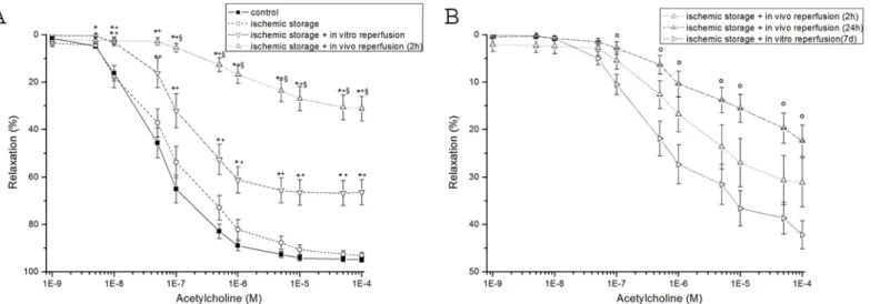
Vasomotor function
In aortic rings precontracted with PE, ACh induced a concentration-dependent relaxation (Fig 1A). Ischemic storage and in vitro reoxygenation had not effect on endothelium-dependent vasorelaxation, however a marked, significant decrease was shown in the ischemic storage and in vitro reperfusion group when compared to the control (Fig 1A). After short ischemia and 2 hours of warm reperfusion, a significant impairment of endothelial function of arterial rings
Table 2. The sequences for the forward (F) and reverse (R) primers (from 5’to 3’) and Universal Probe Library (UPL) probes.eNOS: endothelial nitric oxide synthase; GAPDH: glyceraldehyde-3-phosphate dehydrogenase.
Assay Sequence UPL probes
Bax F: 5’-TAGCAAACTGGTGCTCAAGG, R: 5’-GCCACCCTGGTCTTGGAT 69
Bcl-2 F: 5’-GTACCTGAACCGGCATCTG, R: 5’-GGGGCCATATAGTTCCACAA 75
Caspase-3 F: 5’-AAACCTCCGTGGATTCAAAA, R: 5’-AGCCCATTTCAGGGTAATCC 56
eNOS F: 5’-TGACCCTCACCGATACAACA, R: 5’-CGGGTGTCTAGATCCATGC 5
GAPDH F: 5’-CTACCCACGGCAAGTTCAAT, R: 5’-ATTTGATGTTAGCGGGATCG 111
doi:10.1371/journal.pone.0124025.t002
was demonstrated as compared to the control and in vitro groups. The endothelial dysfunction was indicated by the reduced maximal relaxation of aortic rings to ACh and the rightward shift of the concentration–response curve as compared with control (Fig 1). After 24 hours of warm reperfusion, the endothelial function worsened compared to the 2 hours reperfusion group without reaching the level of statistical significance (Fig 1B). Partially recovered endothelial function was observed after 7 days of warm reperfusion (Fig 1B). There was no significant differ- ence in Rmax for endothelium-independent vasorelaxation of the aortic rings to SNP between the experimental groups (Table 3). Contractile responses of aortic rings to PE (10-6M) are
Fig 1. Vasomotor function of rat aortic rings.(A) acetylcholine-induced endothelium-dependent vasorelaxation are shown in control, ischemic storage, ischemic storage and in vitro reperfusion, ischemic storage and in vivo 2h reperfusion groups. (B) acetylcholine-induced endothelium-dependent vasorelaxation are shown in control and ischemic storage and in vitro reperfusion groups. Values represent mean±SEM;*P<.05 versus control; +P<.05 versus ischemic storage; §P<.05 versus ischemic storage and in vitro reperfusion; ° p<0.05 vs. ischemic storage and in vivo 7d reperfusion.
doi:10.1371/journal.pone.0124025.g001
Table 3. Values of maximal relaxation (Rmax,%) and pD2to acetylcholine (ACh), to sodium nitroprusside (SNP) and contraction forces induced by phenylephrine (10-6M) in aortic rings in control, ischemic storage, ischemic storage and in vitro reperfusion, ischemic storage and in vivo 2h reper- fusion, ischemic storage and in vivo 24h reperfusion, ischemic storage and in vivo 7d reperfusion group.
Control Ischemic storage
Ischemic storage + in vitro reperfusion
Ischemic storage + in vivo 2h reperfusion
Ischemic storage + in vivo 24h reperfusion
Ischemic storage + in vivo 7d reperfusion Rmaxto ACh (%) 94,9±1,1 93,1±1,4 66,4±5,3a,b,e 31,2±5,2a,b,c 22,4±3,3a,b,c 42,2±3,0a,b,c,e pD2to ACh 7,3±0,1 7,2±0,1 6,9±0,1 5,9±0,2a,b,c 5,2±0,3a,b,c,d,f 5,9±0,4a,b Rmaxto SNP (%) 100,5
±0,2
102,5±1,1 99,5±1,6 100,9±1,0 98,7±1,3 102,0±2,2
pD2to SNP 8,2±0,1 7,9±0,1 7,5±0,1 7,9±0,2 7,7±0,1 7,6±0,1
Phenylephrine (%
of KCl)
86,4±4,3 95,4±4,4 115,6±4,9a 118,3±3,6a,b 117±6,5a 176,3±8,5a,b,c,d,e
Values represent mean±SEM
aP<.05 versus control
bP<.05 versus ischemic storage
cP<.05 versus ischemic storage + in vitro reperfusion
dP<.05 versus ischemic storage + in vivo 2h reperfusion
eP<.05 versus ischemic storage + in vivo 24h reperfusion
fP<.05 versus ischemic storage + in vivo 7d reperfusion.
doi:10.1371/journal.pone.0124025.t003
shown inTable 3. Significantly increased PE-induced maximum contraction was shown in the in vitro and in vivo groupscompared to the control group (Table 3).
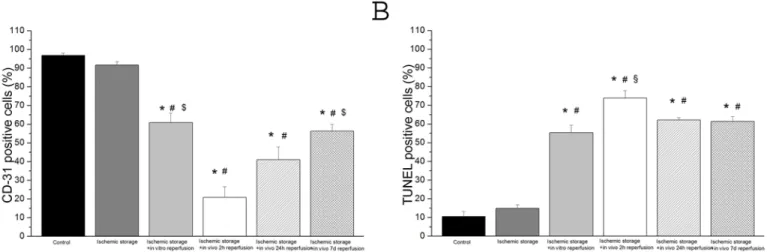
Expression of CD-31
The inner walls of all aortic segments (control+other groups) were covered with endothelium CD-31 presence was detected. The percentage of stained endothelium differed between the control and the other groups. In all groups, there were regions of endothelium, which showed no reaction. In the control group the endothelial surface showing positive reaction for the CD- 31 antigen was estimated as 96±1%, whereas vessels exposed to ischemic storage it was 91±4%, to ischemic storage and hypochlorite-induced reperfusion: 61±11% (Fig 2A). After 2h in vivo reperfusion CD-31 reaction was 21±6% and after 24h warm reperfusion: 41±7% (Fig 2A).
However, after 7 days in vivo reperfusion, the CD-31 reaction was significantly stronger (58
±5%) as compared to the 2h reperfusion group (Fig 2A).
TUNEL-positive nuclei
An increased density of TUNEL-positive nuclei indicates severe DNA fragmentation in the aortic wall (intima and media) of rings in all groups. In vitro/in vivo-induced reperfusion sig- nificantly increased DNA strand breaks (Fig 2B) as compared to the control.
In vivo
induced reperfusion injury caused pronounced changes of the mRNA expressions and protein levels
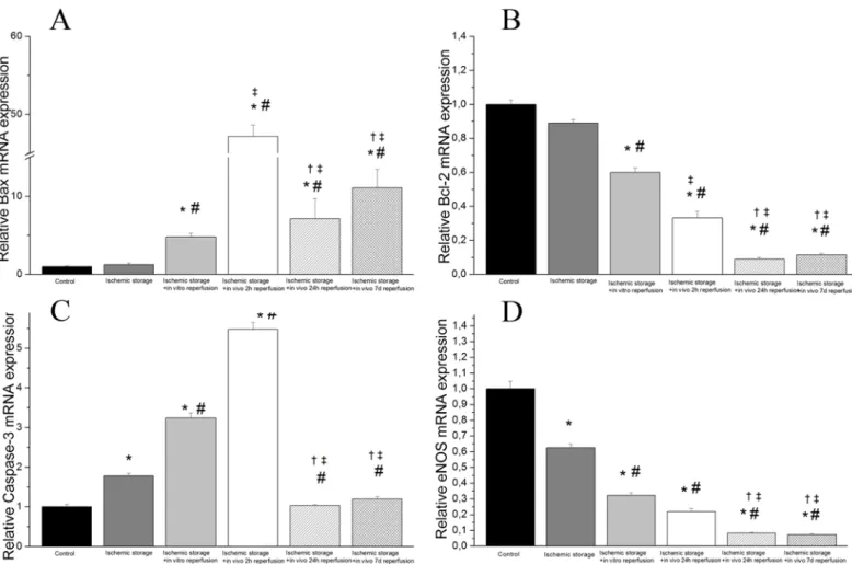
We observed a statistically relevant up-regulation in mRNA expression of two apoptotic genes caspase-3 and bax in the ischemic storage and in vitro reperfusion group and in the ischemic storage and in vivo reperfusion groups (after 2, 24 hours and 7 days reperfusion) as compare to the control (Fig 3). Furthermore, there was statistically significance up-regulation of these gene expressions in the ischemic storage and in vivo reperfusion groups when compared to the is- chemic storage and in vitro group (Fig 3). We also found a significant down-regulated mRNA expression of bcl-2 and eNOS in the in vitro R and in vivo R groups (after 2, 24 hours and 7 days reperfusion) compared to control (Fig 3).
Fig 2. DNA strand breaks and CD-31 positive cells in aortic rings.(A) endothelium-covered area (in percentage of the lumen area) (B) scoring of TUNEL staining (in percentage of total cell number) (Magnification x200, bar = 50μm) Values represent mean±SEM;*P<.05 versus control; #P<.05 versus ischemic storage; §P<.05 versus ischemic storage and in vitro reperfusion; $P<.05 versus ischemic storage and in vivo 2h reperfusion.
doi:10.1371/journal.pone.0124025.g002
Densitometric analysis of caspase-3 cleavage and Bax bands showed a significant increase in the ischemic storage and in vivo reperfusion groups (Fig 4) (after 2, 24 hours and 7 days reper- fusion) as compare to the control, ischemic storage and ischemic storage and in vitro reperfu- sion groups (Fig 4). Furthermore, we detected significantly reduced bcl-2 and eNOS protein levels in in the in vitro R and in vivo R groups (after 2, 24 hours and 7 days reperfusion) com- pared to control. The eNOS protein levels were also significantly reduced in the 2 and 24h in vivo reperfusion groups compared to the ischemic storage group (Fig 4). These results correlate with PCR findings.
Discussion
Our study is the first to directly compare bothin vitroandin vivoinduced R injury, showing dramatically impaired endothelial function after in vivo R injury when compared to the in vitro R injury. By testing the in vitro induced R injury, we are able to detect a marked endothe- lial injury, however we found no endothelial impairment in the ischemic storage model.
IR injury is a complex phenomenon with serious clinical consequences and has been in focus of intensive investigations. Changes in the health of the vascular endothelium may not only have implications concerning the short-term clinical course of patients after CABG, but
Fig 3. Gene expressions of (A) Bax, (B) Bcl-2, (C) caspase-3 and (D) endothelial nitric oxide synthase (eNOS) in aortic rings.Controls were given the arbitrary value of 1. Values represent median±quartiles;*P<.05 versus control; #P<.05 versus ischemic storage;‡P<.05 versus ischemic storage and in vitro reperfusion;†P<.05 versus ischemic storage and in vivo 2h reperfusion.
doi:10.1371/journal.pone.0124025.g003
may affect the late patency of coronary artery bypass grafts, the progression of atherosclerosis in the native coronary circulation and the long-term success of cardiac transplants [13,14].
The recognition that endothelial dysfunction is an early and rate-determining factor in the pathogenesis of IR injury has led to a major effort to characterize fully the vascular alterations associated with IR injury and to define the mechanisms that underlie this pathological process.
In spite of the intensive investigations of this field, the exact molecular mechanisms of the pathogenesis of endothelial dysfunction of bypass graft after IR injury still remain unclear.
Effects of ischemic storage and reoxygenation on endothelium
To date, there are only sporadic functional studies investigating in vitro vascular ischemia and reoxygenation injury [1–3,8,15], and the data are still remained contradictory. Isolated human endothelial cell culture investigations showed, that hypoxia and reoxygenation resulted in morphological signs of cell damage [16,17]. A few previous investigations could show endo- thelial dysfunction after in vitro hypoxia and reoxygenation [2,3,15], but it contradict the re- sults from other studies showing no changes in the endothelium-dependent vasorelaxation induced by ACh [1,8]. It was also shown that prolonged (>1–2 days) cold storage is able to in- duce endothelial dysfunction [4,18,19]. In the present study we demonstrated a weak, but not
Fig 4. Protein expressions of (A) cleaved bax, (B) bcl-2 (C), caspase-3 and (D) eNOS in aortic rings.Values represent median±SEM;*P<.05 versus control; #P<.05 versus ischemic storage;‡P<.05 versus ischemic storage and in vitro reperfusion.
doi:10.1371/journal.pone.0124025.g004
significant reduction of the endothelium-dependent vasorelaxation in the vessels after 2h ische- mic storage when compared to the control. Furthermore, we did not observe any difference in histopathological results (CD-31 and TUNEL) and mRNA expressions (Figs2and3). Interest- ingly, just the eNOS mRNA expression and protein level were significantly lower in ischemic storage group when compared to the control. In agree with Radovits et al. [8], we can speculate that the endothelial damage occurred by ischemia and reoxygenation is too moderate after a clinically relevant storage time (2h), probably due to the lack of activated leukocytes and there- fore is not suitable for reliable investigation of endothelial dysfunction.
Effects of ischemic storage and in vitro induced reperfusion injury on endothelium
Ischemia and then the rapid reintroduction of molecular oxygen per se result in a burst of re- active oxygen species (ROS) during the first minutes of reperfusion [20–22]. Activated leuco- cytes also generate a number of ROS, including O2-, H2O2and through the myeloperoxidase system, highly reactive hypochlorite. The luminal surface of blood vessels and vascular wall are the most exposed tissues to the deleterious effects of these reactive agents, which lead to loss of homeostatic functions of the vascular endothelium and thus capillary plugging, im- paired vascular permeability, adhesion and infiltration of leucocytes [23]. Due to its role in pathophysiological conditions, such as inflammation, atherosclerosis and reperfusion injury, hypochlorite is commonly used in in vitro models of vascular I/R injury to stimulate the ef- fect of activated leukocytes [4–7].
We showed in the present in vitro model of reperfusion injury an impaired endothelium- dependent acethylcholine-induced relaxation of aortic rings exposed to hypochlorite. The endothelium-independent vasorelaxation induced by SNP was unaffected by hypochlorite, indicating normal dilatative capacity of the vascular smooth muscle, however an enhanced PE-induced vasoconstriction was measured in this group. These functional data are consis- tent with the results of other studies in rabbit and rat models [6,24,25]. We also reported that exposure of rat aortic rings to hypochlorite resulted in formation of DNA stand breaks in the vessel wall, as evidenced by TUNEL assay. Consistently with these data, previous works on endothelial cells reported similar results [26]. Furthermore, we measured a reduc- tion of living endothelial cells of the aortic graft, as demonstrated in the CD-31 immunohis- tochemical assay (Fig 2). In addition, we reported here enhanced signs of apoptosis (up- regulation of Bax and Caspase-3 levels, down-regulation of bcl-2 by PCR and Western blot experiments).
Previous in vitro models of reperfusion injury could also not observe a dramatically reduced vasorelaxation function after IR injury [4,6,7]. Human endothelial cell cultured experiments, however, showed that the addition of neutrophils upon reoxygenation enhances endothelial cell injury [17,27]. The observation suggests that neutrophils become activated by some factor(s) re- leased by endothelial cells and that the activated neutrophils exacerbate endothelial cell injury.
However, experimental and clinical studies clearly showed that endothelial cells are more sensi- tive to IR injury than cardiomyocytes and pronounced endothelial responsiveness has also been described [28–30]. Furthermore, a human heart transplantation study clearly demonstrated that in contrast to cardiomyocytes the cell oedema of endothelium is still more pronounced after mid-term reperfusion than at the onset of ischemia [28].
We supposed that in vivo development of reperfusion injury could be the major cause of im- paired short and long-term patency rate and vasospasm of the arterial graft after CABG.
Effects of ischemic storage and in vivo induced reperfusion injury on endothelium
To observe the effect of in vivo reperfusion injury, a novel method of ischemic storage and in vivo reperfusion injury was developed. To the best of our knowledge, we demonstrated for the first time a severe endothelial dysfunction even also after 2 hour in vivo reperfusion. The peak of the endothelial damage occurred after 24 hours of reperfusion as demonstrated by dramati- cally damaged endothelium-dependent vasorelaxation, apoptosis rate of TUNEL-staining and increased mRNA expression of Bax and caspase-3 and the decreased levels of Bcl-2 and eNOS gene expressions. These data also correlate to the clinical findings of early complications in by- pass grafts, as Lockerman and colleagues found transient ST segment elevation occuring in the first 12–24 hours after CABG [31]. Furthermore, the severity of the endothelial dysfunction (endothelium-dependent vasorelaxation and mRNA expression, protein levels, results from TUNEL and CD-31 staining) occurred by in vivo reperfusion in our model was more detri- mental than in our in vitro model of IR injury.
Endothelial integrity and structure (as measured by lower expression of CD-31 on the endo- thelial surface in the aortic segments and enhanced DNA stand breaks) were also severely dam- aged in the in vivo-induced IR injury group. CD-31 is highly specific for endothelial cells and has a constant expression. After 2 hours/1 day reperfusion, a severely damaged endothelial layer of the arterial graft was also reported (Figs1Band4D). Surprisingly, we also showed that the func- tion and structure of the arterial graft partially recovered after 1 week of reperfusion (Fig 1B). A previous study also reported that the endothelium is capable of regaining its functional integrity within a few days by proliferation and migration after mechanical injury [32]. Nevertheless, it was also noted, that endothelial integrity after mechanical injury could not be fully restored, since there are marked functional and structural differences between native and regenerated en- dothelium [33]. Regenerated endothelium exhibits impaired endothelium-dependent relaxation and signs of apoptosis. The results of our experiments are in good agreement with previous re- ports on the regeneration of the endothelium, as in our investigation the function of the endothe- lium demonstrates partial recovery after 1 week of reperfusion, however it was not fully restored (impaired vasodilatative capacity, damaged endothelial layer) and there were also signs of apo- ptosis (up-regulation of Bax and Caspase-3 levels, marked TUNEL-staining). The endothelial stunning in our model was also demonstrated through the dynamic changes of several genes over time, which supported the results of CD-31 staining and changed of vasomotor function over time. The fact, that relaxation to the endothelium-independent vasodilator SNP was pre- served, after long-term storage and reperfusion in our model, indicated that the impaired vascu- lar response may not be attributed to a deficit in function of vascular smooth muscle.
Much of the data derived from studies of IR-induced vascular dysfunction are consistent with the role for NO as participant in the changed endothelium-dependent responses [23].
Due to a low NO level, the endothelium-dependent vasorelaxation is compromised. We showed that the eNOS mRNA expression and protein level (Figs3Dand4D) are drastically re- duced in the ischemic storage and in vivo reperfusion groups, thus it may be a theoretical ex- planation of the reduced vasorelaxation of the grafts.
Conclusion
In conclusion, the present study investigated the IR injury and impairment of vascular respon- siveness induced by in vitro or in vivo IR injury. Vascular models of in vitro reoxygenation and incubation with high dose hypochlorite may be adequate for modelling oxidative stress and so partially the reperfusion injury, but in vivo reperfusion models involve the full scale of damag- ing effects by restored arterial flow after ischemia. Thus, in vivo methods providing a better
understanding of IR mechanism and contribute to develop new preservation solutions. In the present study, mimicking surgical revascularization through aortic transplantation, we clearly demonstrated that the function and the structure of the implanted arterial graft were both se- verely damaged after short-term storage and warm reperfusion. The impaired endothelium-de- pendent vasorelaxation and the structural changes in the endothelium may play important roles in the pathogenesis of early graft thrombosis and vasospasm after CABG. However, the functional and structural changes in time could partially recover. Further studies are needed to evaluate the preservation effect of preservation solutions (heparinised blood, Custodiol, UW, Tiprotec). In addition, a deep mechanistic understanding on molecular level of the observed al- terations needs to be investigated in other clinically relevant experimental model of
bypass grafting.
Author Contributions
Conceived and designed the experiments: GV PH EB RZ S. Klein HS TR S. Korkmaz MK GS.
Performed the experiments: GV PH EB RZ S. Klein HS TR S. Korkmaz GS. Analyzed the data:
GV PH EB RZ S. Klein HS TR S. Korkmaz MK GS. Contributed reagents/materials/analysis tools: GV PH EB RZ S. Klein TR S. Korkmaz MK GS. Wrote the paper: GV PH TR S. Korkmaz MK GS.
References
1. Yang BC, Nichols WW, Lawson DL, Mehta JL. Agonist-induced tension determines vascular reactivity during anoxia and reoxygenation. Life Sci 1992; 50: 1805–12. PMID:1598068
2. Yokoyama S, Korthuis RJ, Benoit JN. Hypoxia-reoxygenation impairs endothelium-dependent relaxa- tion in isolated rat aorta. The American journal of physiology. 1996; 270: 1126–31.
3. Besse S, Tanguy S, Boucher F, Bulteau AL, Riou B, de Leiris J, et al. Aortic vasoreactivity during pro- longed hypoxia and hypoxia-reoxygenation in senescent rats. Mech Ageing Dev 2002; 123: 275–285.
PMID:11744040
4. Radovits T, Lin LN, Zotkina J, Koch A, Rauen U, Kohler G, et al. Endothelial dysfunction after long-term cold storage in HTK organ preservation solutions: effects of iron chelators and N-alpha-acetyl-L-histi- dine. J Heart Lung Transplant 2008; 27: 208–216. doi:10.1016/j.healun.2007.11.002PMID:18267229 5. Sand C, Peters SL, Pfaffendorf M, van Zwieten PA. Effects of hypochlorite and hydrogen peroxide on
cardiac autonomic receptors and vascular endothelial function. Clin Exp Pharmacol Physiol 2003; 30:
249–253. PMID:12680842
6. Veres G, Hegedus P, Barnucz E, Zoller R, Radovits T, Korkmaz S, et al. Addition of vardenafil into stor- age solution protects the endothelium in a hypoxia-reoxygenation model. Eur J Vasc Endovasc Surg 2013; 46: 242–248. doi:10.1016/j.ejvs.2013.05.006PMID:23751217
7. Barnucz E, Veres G, Hegedus P, Klein S, Zoller R, Radovits T, et al. Prolyl-hydroxylase inhibition pre- serves endothelial cell function in a rat model of vascular ischemia reperfusion injury. J Pharmacol Exp Ther 2013; 345: 25–31. doi:10.1124/jpet.112.200790PMID:23388095
8. Radovits T, Zotkina J, Lin LN, Karck M, Szabo G. Endothelial dysfunction after hypoxia-reoxygenation:
do in vitro models work? Vascul Pharmacol 2009; 51: 37–43. doi:10.1016/j.vph.2009.01.009PMID:
19275967
9. Radovits T, Zotkina J, Lin LN, Bomicke T, Arif R, Gero D, et al. Poly(ADP-Ribose) polymerase inhibition improves endothelial dysfunction induced by hypochlorite. Exp Biol Med 2007; 232: 1204–12. PMID:
17895528
10. Radovits T, Arif R, Bomicke T, Korkmaz S, Barnucz E, Karck M, et al. Vascular dysfunction induced by hypochlorite is improved by the selective phosphodiesterase-5-inhibitor vardenafil. Eur J Pharmacol 2013; 710: 110–9. doi:10.1016/j.ejphar.2013.04.012PMID:23623933
11. Korkmaz S, Radovits T, Barnucz E, Neugebauer P, Arif R, Hirschberg K, et al. Dose-dependent effects of a selective phosphodiesterase-5-inhibitor on endothelial dysfunction induced by peroxynitrite in rat aorta. Eur J Pharmacol 2009; 615: 155–62. doi:10.1016/j.ejphar.2009.05.020PMID:19482016 12. Radovits T, Bomicke T, Kokeny G, Arif R, Loganathan S, Kecsan K, et al. The phosphodiesterase-5 in-
hibitor vardenafil improves cardiovascular dysfunction in experimental diabetes mellitus. Br J Pharma- col 2009; 156: 909–919. doi:10.1111/j.1476-5381.2008.00098.xPMID:19298393
13. Luscher TF. Vascular biology of coronary bypass grafts. Curr Opin Cardiol 1991; 6: 868–876. PMID:
10149593
14. Pearson PJ, Evora PR, Schaff HV. Bioassay of EDRF from internal mammary arteries: implications for early and late bypass graft patency. Ann Thorac Surg 1992; 54: 1078–1084. PMID:1449290 15. Carrasco-Martin C, Alonso-Orgaz S, De la Pinta JC, Marques M, Macaya C, Barrientos A, et al. Endo-
thelial hypoxic preconditioning in rat hypoxic isolated aortic segments. Exp Physiol 2005; 90: 557–569.
PMID:15769880
16. Hummitzsch L, Zitta K, Bein B, Steinfath M, Albrecht M. Culture media from hypoxia conditioned endo- thelial cells protect human intestinal cells from hypoxia/reoxygenation injury. Exp Cell Res 2014; 322:
62–70. doi:10.1016/j.yexcr.2013.12.022PMID:24394542
17. Inauen W, Granger DN, Meininger CJ, Schelling ME, Granger HJ, Kvietys PR. Anoxia-reoxygenation- induced, neutrophil-mediated endothelial cell injury: role of elastase. Am J Physiol 1990; 259: 925–31.
18. Ebner A, Poitz DM, Augstein A, Strasser RH, Deussen A. Functional, morphologic, and molecular char- acterization of cold storage injury. J Vasc Surg 2012; 56: 189–198. doi:10.1016/j.jvs.2011.12.012 PMID:22398374
19. Garbe S, Zatschler B, Muller B, Dieterich P, Ebner A, Rauen U, et al. Preservation of human artery func- tion following prolonged cold storage with a new solution. J Vasc Surg 2011; 53: 1063–1070. doi:10.
1016/j.jvs.2010.10.093PMID:21227623
20. Maathuis MH, Leuvenink HG, Ploeg RJ. Perspectives in organ preservation. Transplantation 2007; 83:
1289–1298. PMID:17519776
21. Wilbring M, Tugtekin SM, Zatschler B, Ebner A, Reichenspurner H, Matschke K, et al. Even short-time storage in physiological saline solution impairs endothelial vascular function of saphenous vein grafts.
Eur J Cardiothorac Surg 2011; 40: 811–815. doi:10.1016/j.ejcts.2011.01.024PMID:21376620 22. Chu Y, Wu YC, Chou YC, Chueh HY, Liu HP, Chu JJ, et al. Endothelium-dependent relaxation of ca-
nine pulmonary artery after prolonged lung graft preservation in University of Wisconsin solution: role of L-arginine supplementation. J Heart Lung Transplant 2004; 23: 592–598. PMID:15135376
23. Sellke FW, Boyle EM Jr., Verrier ED. Endothelial cell injury in cardiovascular surgery: the pathophysiol- ogy of vasomotor dysfunction. Ann Thorac Surg 1996; 62: 1222–1228. PMID:8823128
24. Radovits T, Lin LN, Zotkina J, Gero D, Szabo C, Karck M, et al. Poly(ADP-ribose) polymerase inhibition improves endothelial dysfunction induced by reactive oxidant hydrogen peroxide in vitro. Eur J Pharma- col 2007; 564: 158–166. PMID:17397824
25. Thomas SR, Schulz E, Keaney JF Jr. Hydrogen peroxide restrains endothelium-derived nitric oxide bio- activity—role for iron-dependent oxidative stress. Free Radic Biol Med 2006; 41: 681–688. PMID:
16864001
26. Kang YH, Chung SJ, Kang IJ, Park JH, Bunger R. Intramitochondrial pyruvate attenuates hydrogen peroxide-induced apoptosis in bovine pulmonary artery endothelium. Mol Cell Biochem 2001; 216:
37–46. PMID:11216862
27. Inauen W, Granger DN, Meininger CJ, Schelling ME, Granger HJ, Kvietys PR. An in vitro model of is- chemia/reperfusion-induced microvascular injury. Am J Physiol 1990; 259: 134–9.
28. Koch A, Bingold TM, Oberlander J, Sack FU, Otto HF, Hagl S, et al. Capillary endothelia and cardio- myocytes differ in vulnerability to ischemia/reperfusion during clinical heart transplantation. Eur J Cardi- othorac Surg 2001; 20: 996–1001. PMID:11675187
29. Boyle EM Jr., Pohlman TH, Cornejo CJ, Verrier ED. Endothelial cell injury in cardiovascular surgery: is- chemia-reperfusion. Ann Thorac Surg 1996; 62: 1868–1875. PMID:8957415
30. Pearson PJ, Schaff HV, Vanhoutte PM. Acute impairment of endothelium-dependent relaxations to ag- gregating platelets following reperfusion injury in canine coronary arteries. Circ Res 1990; 67: 385–393.
PMID:2115821
31. Lockerman ZS, Rose DM, Cunningham JN Jr., Lichstein E. Postoperative ST-segment elevation in cor- onary artery bypass surgery. Chest 1986; 89: 647–651. PMID:3486097
32. Tada T, Tateyama H, Fujiyoshi Y, Eimoto T. Arterial Smooth-Muscle Cell Response Induced by Loss of Endothelium Continues Even after Apparent Completion of Endothelial Repair. Ann Ny Acad Sci. 1990;
598: 507–9.
33. Fournet-Bourguignon MP, Castedo-Delrieu M, Bidouard JP, Leonce S, Saboureau D, Delescluse I, et al. Phenotypic and functional changes in regenerated porcine coronary endothelial cells: increased uptake of modified LDL and reduced production of NO. Circ Res 2000; 86: 854–861. PMID:10785507