We do not accept corrections in the form of edited manuscripts.
In order to ensure the timely publication of your article, please submit the corrections within 48 hours.
If you have any questions, please contactphysiology.production.office@frontiersin.org.
Author Queries Form
Query No. Details required Author’s Response
Q1 The citation and surnames of all of the authors have been highlighted.
Please check all of the names carefully and indicate if any are incorrect. Please note that this may affect the indexing of your article in repositories such as PubMed.
Q2 Confirm that the email address in your correspondence section is accurate.
Q3 Please ask the following authors toregisterwith Frontiers (athttps://
www.frontiersin.org/Registration/Register.aspx) if they would like their names on the article abstract page and PDF to be linked to a Frontiers profile. Please ensure to provide us with the profile link(s) when submitting the proof corrections. Non-registered authors will have the default profile image displayed.
Ágnes Szántai János Pálóczi Judit Pipis Paola Poggi
Q4 If you decide to use previously published,copyrighted figuresin your article, please keep in mind that it is your responsibility, as the author, to obtain the appropriate permissions and licenses and to follow any citation instructions requested by third-party rights holders. If obtaining the reproduction rights involves the payment of a fee, these charges are to be paid by the authors.
Q5 Ensure that all the figures, tables and captions are correct.
Q6 Verify that all the equations and special characters are displayed correctly.
Q7 Ensure to add all grant numbers and funding information, as after publication this is no longer possible.
Q8 Please ensure that any supplementary material is correctly published at this link: https://www.frontiersin.org/articles/10.3389/fphys.2019.
01564/full#supplementary-material(you may need to copy-paste the link directly in your browser).
Please provide new files if you have any corrections.
Note that ALL supplementary files will be deposited to FigShare and receive a DOI. Notify us of any previously deposited material.
Citations and author names
were checked and they are correct.
I confirm, the email is correct.
Non-registered authors can be displayed with default profile picture.
Copyrighted figures were not used.
Figures, tables and captions are correct.
Special characters are displayed correctly, equations were not used.
Grant numbers and funding informations are correct.
Supplementary material is correctly
published.
Q12 Please confirm if the Ethics statement included here is fine.
Q13 Please explain the part lables “A,B” in Supplementary Figure S1 caption.
Q12: Please replace ethical statement:
The animal study was reviewed and approved by the local ethics committee at the University of Szeged animal ethics committee (I-74-52/2012 MAB) and at Semmelweis University, Budapest, Hungary, and by the National Scientific Ethical Committee on Animal
Experimentation and permitted by the government (Food Chain Safety and Animal Health Directorate of the Government Office for Pest County (PE/EA/1784-7/2017)
Q13: Please correct the first sentence of the figure legend: "Simulated
ischemia/reperfusion (SI/R) injury caused significant cell death of cardiac myocytes, both in neonatal (NRCM) (A) and adult (ARCM) (B) cardiomyocytes.
Please see below.
Please see below.
1 2 3 4 5 6 7 8 9 10 11 12 13 14 15 16 17 18 19 20 21 22 23 24 25 26 27 28 29 30 31 32 33 34 35 36 37 38 39 40 41 42 43 44 45 46 47 48 49 50 51 52 53 54 55 56 57
58 59 60 61 62 63 64 65 66 67 68 69 70 71 72 73 74 75 76 77 78 79 80 81 82 83 84 85 86 87 88 89 90 91 92 93 94 95 96 97 98 99 100 101 102 103 104 105 106 107 108 109 110 111 112 113 114 ORIGINAL RESEARCH published: xx December 2019 doi: 10.3389/fphys.2019.01564
Edited by:
Vincenza Cifarelli, Washington University in St. Louis, United States Reviewed by:
Claudia Penna, University of Turin, Italy Didier Morin, Centre National de la Recherche Scientifique (CNRS), France
Q2 *Correspondence:
Anikó Görbe gorbe.aniko@
med.semmelweis-univ.hu
Specialty section:
This article was submitted to Lipid and Fatty Acid Research, a section of the journal Frontiers in Physiology
Received:17 June 2019 Accepted:12 December 2019 Published:xx December 2019
Citation:
Makkos A, Szántai Á, Pálóczi J, Pipis J, Kiss B, Poggi P, Ferdinandy P, Chatgilialoglu A and Görbe A (2019) A Comorbidity Model of Myocardial Ischemia/Reperfusion Injury and Hypercholesterolemia in Rat Cardiac Myocyte Cultures.
Front. Physiol. 10:1564.
doi: 10.3389/fphys.2019.01564
A Comorbidity Model of Myocardial Ischemia/Reperfusion Injury and
Hypercholesterolemia in Rat Cardiac Myocyte Cultures
Q1 Q3 András Makkos1, Ágnes Szántai2, János Pálóczi2, Judit Pipis3, Bernadett Kiss1,2,
Paola Poggi4, Péter Ferdinandy1,2,3, Alexandros Chatgilialoglu4and Anikó Görbe1,2,3*
1Cardiometabolic Research Group, Department of Pharmacology and Pharmacotherapy, Semmelweis University, Budapest, Hungary
Q9 ,2Department of Biochemistry, University of Szeged, Szeged, Hungary,3Pharmahungary Group, Szeged, Hungary,
4Remembrane S.r.L, Imola, Italy
Introduction: The use of comorbidity models is crucial in cardioprotective drug development. Hypercholesterolemia causes endothelial and myocardial dysfunction, as well as aggravates ischemia/reperfusion (I/R)-induced myocardial injury. Endogenous cardioprotective mechanisms against I/R are impaired in hyperlipidemic and hyperglycemic in vivo animal models. Therefore, our aim was to develop a medium throughput comorbidity cell-based test system of myocardial I/R injury, hypercholesterolemia and hyperglycemia that mimics comorbidity conditions.
Methods: Cardiac myocytes isolated from neonatal or adult rat hearts were cultured in control or in three different hypercholesterolemic media with increasing cholesterol content (hiChol) or hiChol + hyperglycemic medium, respectively. Each group was then subjected to simulated ischemia/reperfusion (SI/R) or corresponding normoxic condition, respectively. Cholesterol uptake was tested by Filipin staining in neonatal cardiac myocytes. Cell viability, total cell count and oxidative stress, i.e., total reactive oxygen species (ROS) and superoxide level were measured by fluorescent assays.
Results:Neonatal cardiac myocytes took up cholesterol from the different hiChol media at a concentration-dependent manner. In normoxia, viability of hiChol neonatal cardiac myocytes was not significantly changed, however, superoxide levels were increased as compared to vehicle. After SI/R, the viability of hiChol neonatal cardiac myocytes was decreased and total ROS level was increased as compared to vehicle. HiChol combined with hyperglycemia further aggravated cell death and oxidative stress in normoxic as well as in SI/R conditions. Viability of hiChol adult cardiac myocytes was significantly decreased and superoxide level was increased in normoxia and these changes were further aggravated by SI/R. HiChol combined with hyperglycemia further aggravated cell death, however level of oxidative stress increased only in normoxic condition.
115 116 117 118 119 120 121 122 123 124 125 126 127 128 129 130 131 132 133 134 135 136 137 138 139 140 141 142 143 144 145 146 147 148 149 150 151 152 153 154 155 156 157 158 159 160 161 162 163 164 165 166 167 168 169 170 171
172 173 174 175 176 177 178 179 180 181 182 183 184 185 186 187 188 189 190 191 192 193 194 195 196 197 198 199 200 201 202 203 204 205 206 207 208 209 210 211 212 213 214 215 216 217 218 219 220 221 222 223 224 225 226 227 228 Q6
Q7
Conclusion: HiChol rat cardiac myocytes showed reduction of cell viability and increased oxidative stress, which were further aggravated by SI/R and with additional hyperglycemia. This is the first demonstration that the combination of the current hypercholesterolemic medium and SI/R in cardiac myocytes mimics the cardiac pathology of the comorbid heart with I/R and hypercholesterolemia.
Keywords: cardiac myocytes, ischemia/reperfusion injury (I/R injury), hypercholesterolemia (HC), cell culture, hypercholesterolemia and hyperglycemia
INTRODUCTION
Ischemic heart disease is still the leading cause of death worldwide; therefore, there is an unmet clinical need for the development of efficient cardioprotective therapies. In the last few decades, a wide variety of cardioprotective interventions and pharmacological treatments were found effective in experimental animal models and in cell cultures. However, their clinical translation has been largely disappointing (Hausenloy et al., 2017). One of the major problem is that thein vitropreclinical testing of drug candidates apply cell lines and in vivo,ex vivo testing apply young, healthy animals, thus neglecting the presence of cardiovascular risk factors and comorbidities.
Ischemic heart disease is typically associated with metabolic diseases such as diabetes, obesity, hyperlipidemia and hypercholesterolemia, which predispose the subject to atherosclerosis and the development of coronary artery diseases (CADs) (Benjamin et al., 2017). Hypercholesterolemia is widely accepted as a principal risk factor for CAD (Ferdinandy et al., 2014) and can increase the myocardial damage due to ischemia/reperfusion injury and interfere with responses to cardioprotective interventions (Andreadou et al., 2017). Most of the preclinical studies have shown that hyperlipidemia (but not atherosclerosis) leads to a significant aggravation of myocardial ischemia/reperfusion injury and to an attenuation of the cardioprotective effect of preconditioning (Ferdinandy et al., 2007, 2014;Andreadou et al., 2017). One of the first articles reporting the loss of rapid pacing-induced preconditioning in hypercholesterolemic rabbits was released in Szilvassy et al.
(1995). The loss of the infarct size-limiting effect of ischemic preconditioning (Gorbe et al., 2011; Babbar et al., 2013) and late ischemic preconditioning (Yadav et al., 2012) have been shown in different models of diet-induced hyperlipidemia in rats. Detrimental effect of hypercholesterolemia could be due to either increased production and/or decreased removal of highly reactive oxygen and/or nitrogen species (ROS and RNS), such as superoxide, hydrogen peroxide, hydroxyl radicals, and peroxynitrite (Csonka et al., 2016). Diabetes mellitus is a major independent risk factor for acute coronary syndrome (ACS) and causes increased mortality among diabetic individuals (Sethi et al., 2012). Numerous mechanisms have been proposed to contribute to the formation of diabetic cardiomyopathy and myocardial contractile function, including oxidative stress (Singh et al., 2018).
The investigation of mechanisms behind ischemia/reperfusion injury in the presence of hyperlipidemia and other metabolic comorbidities is crucial for testing potential cardioprotective
compounds and interventions. Ischemia/reperfusion injury can be modeled with induction of hypoxia/anoxia in a hypoxic chamber, which can be further combined with the application of hypoxic medium. The aforementioned model is widely used in primary cardiac myocyte cultures and cell lines as well (Lecour et al., 2014;Lindsey et al., 2018). We reported previously that simulated ischemia/reperfusion injury causes significant cell death in neonatal rat cardiac myocytes, which can be reversed with an NO-donor treatment (Gorbe et al., 2010). Simulation of hyperlipidemia and hypercholesterolemia in vitro is less standardized in the literature. There are only few studies, where lipoprotein or oxidized lipoprotein supplementation was used in cardiac myocyte cultures to inducein vitrohyperlipidemia (Cal et al., 2012a,b).
Currently, there is a lack of in vitro cell based platforms able to mimic such pathological conditions and to become the gold standard in the development of new effective drug candidates. Therefore, the aim of the present study was to set an in vitro medium throughput test system of primary isolated cardiac myocytes, which can be subjected to simulated ischemia/reperfusion and mimicsin vivo hypercholesterolemia and hyperglycemia. Severity of cell injury and level of oxidative stress could reflect the possible cardioprotective or cardiotoxic effects of tested compounds during preclinical phase of drug development.
MATERIALS AND METHODS
These experiments conform to the National Institutes of Health Guide for the Care and Use of Laboratory Animals (NIH Pub.
No. 85-23, Revised 1996) and were approved by the local ethics committee at the University of Szeged.
Study Design
In the present study, we used both primary isolated neonatal and adult rat cardiac myocyte adherent cultures. The following groups were investigated:
(1) normochol (normocholesterolemic control, cell culture medium supplemented with the vehicle of HiChol supplementations)
(2) HiChol 1 (cell culture medium supplemented with hypercholesterolemic medium 1)
(3) HiChol 2 (cell culture medium supplemented with hypercholesterolemic medium 2)
(4) HiChol 3 (cell culture medium supplemented with hypercholesterolemic medium 3).
229 230 231 232 233 234 235 236 237 238 239 240 241 242 243 244 245 246 247 248 249 250 251 252 253 254 255 256 257 258 259 260 261 262 263 264 265 266 267 268 269 270 271 272 273 274 275 276 277 278 279 280 281 282 283 284 285
286 287 288 289 290 291 292 293 294 295 296 297 298 299 300 301 302 303 304 305 306 307 308 309 310 311 312 313 314 315 316 317 318 319 320 321 322 323 324 325 326 327 328 329 330 331 332 333 334 335 336 337 338 339 340 341 342
Makkos et al. Ischemic Hypercholesterolemic Comorbidity in Cardiomyocytes
Each group was tested under the following conditions:
(a) Standard culturing under normoxic condition (b) Simulated ischemia/reperfusion injury (SI/R)
(c) Simulated ischemia/reperfusion injury + treatment with NO donor drug or its vehicle (a well-known cardioprotective compound) under SI/R
(d) Additional hyperglycemia (high concentration of glucose combined with HiChol supplementation, refers to metabolic disease condition) under normoxic condition (e) Additional hyperglycemia + simulated ischemia/
reperfusion injury.
Isolation of Neonatal Cardiac Myocytes
Neonatal cardiac myocytes (NRCM) were isolated from new- born (1–3 day old) Wistar rats as described previously (Csont et al., 2010; Bencsik et al., 2014). Briefly, rats were disinfected with 70% ethanol and then euthanized by cervical dislocation.
The hearts were rapidly removed and placed in ice cold PBS.
Ventricles were separated and minced with fine forceps. Tissue fragments were digested in 0.25% trypsin for 25 min in a conical tube at 37◦C. After digestion, the cell suspension was centrifuged (250×gfor 15 min at 4◦C). Pellet was resuspended in culture medium [Dulbecco’s modified Eagle’s medium (DMEM), supplemented with 10% fetal bovine serum (FBS),L-Glutamine, and Antibiotic/Antimycotic]. This cell suspension was preplated in 6-well plates at 37◦C for 90 min to enrich the culture with cardiac myocytes. The non-adherent myocytes were collected and cells were counted and then plated at a density of 105cells/well in a 24-well plate.
Isolation of Adult Cardiac Myocytes
Male adult Wistar rats (150 g) were used. Surgery was performed under sodium pentobarbital anesthesia and each animal was heparinised (500 IU/kg) through femoral vein. For cardiac myocyte (ARCM) isolation, hearts were cannulated and perfused retrograde with butanedione monoxide supplemented Krebs–
Henseleit solution to wash out the clots and blood. After a 2–4 min solution was changed to collagenase II (8000 U/mL) containing Krebs solution and perfused for 30–45 min. The ventricles were removed and chopped in small pieces and digestion continued for 10 min more. The cell suspension was filtrated and pelleted under gravity, repeated 2–3 times. Under these steps, the Ca2+ concentration was increased gradually up to 1 mM. The ratio of the rod shape viable cells was controlled visually under the isolation at each step of the phasic increase of Ca2+. We considered isolated adult cardiomyocytes viable when spontaneously contracting and showing rod shape. After cell counting, the cells were plated in laminin-coated wells of a 24-well plate (7500 cell/well) (Markou et al., 2011). To start SI/R experiment minimum 50% viable cells were required by cell counting.
Tailored Refeed
RSupplements
In order to mimic the elevated concentration of cholesterol typical of hypercholesterolemic conditions on cultured primary cardiac myocytes, we identified three increasing cholesterol
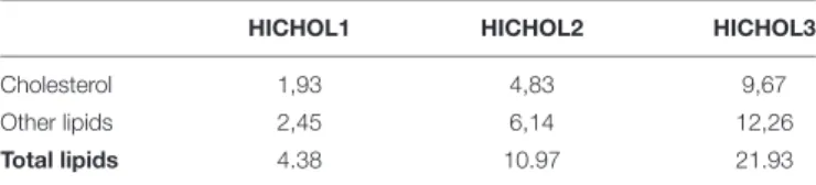
concentrations suitable for obtaining the desired responses by the cells. However, an in vivo hypercholesterolemic condition is usually overlapped by a general hyperlipidemia/dyslipidemia, characterized by a wider array of dysregulated lipids and influenced by multiple factors belonging to genetics, lifestyle and diet. For this reason, we decided to integrate the cholesterol-based supplements with selected lipids, able to generate a more heterogeneous and authentic hypercholesterolemic/hyperlipidemic phenotype in in vitro primary cardiac myocytes. The three tailored RefeedR supplements (hiChol1, hiChol2, hiChol3) used in this study were therefore developed by integrating the desired levels of cholesterol with selected adjuvant lipids, in order to strengthen the hypercholesterolemic biological effects and create a more accurate in vitro model. RefeedR supplements (Remembrane Srl, Imola, Italy) are a completely defined combination of non-animal derived lipids (NuCheckPrep, Inc., Elysian, MN, United States; Sigma Aldrich, St. Louis, MO, United States;
Applichem an ITW, Inc., Chicago, IL, United States) solubilized in 1 mL of ethanol (Sigma Aldrich). 1.5 mL of RefeedR was diluted in 500 mL of complete cell growth medium, the resulting ethanol concentration being less than 1% (vol/vol) in the final medium. The specific tailored RefeedR composition is shown inTable 1. Similar Refeed compositions for different purposes have been previously developed, as described (Poggi et al., 2015;
Chatgilialoglu et al., 2017;Cavallini et al., 2018).
Medium Supplementation and Treatment of Cardiac Myocytes
Neonatal cardiac myocytes were kept at 37◦C in a standard CO2 incubator (humidified atmosphere of 5% CO2) and supplied with growth medium (10% FBS containing DMEM) for 24 h and with proliferation medium (1% FBS) for another 48 h. The adult cardiac myocytes were cultured with same conditions with serum supplemented media for 3 h (5% FBS containing M199) and with growth media (serum free M199) for 48 h (Experimental protocol:Figure 1). Cholesterol supplements (hiChol1, hiChol2, or hiChol3) or vehicle (0.3% ethanol) were added to each series (3µL into 1 mL culture media) (Figure 1). NO- donorS-nitroso- N-acetyl penicillamine (10-6 M) was applied during simulated ischemia and reperfusion. High glucose medium contained 4.5 g/L glucose.
Determination of Cholesterol Content of the Cells by Filipin Staining
To measure the cholesterol content of the cultured cells Filipin staining was used that enables semi-quantification of free
TABLE 1 |Composition of RefeedRused forin vitrosupplementation (hypercholesterolemic medium/hiChol) of cardiac myocytes.
HICHOL1 HICHOL2 HICHOL3
Cholesterol 1,93 4,83 9,67
Other lipids 2,45 6,14 12,26
Total lipids 4.38 10.97 21.93
Data are the amount (mg) per 500 mL of complete medium.
343 344 345 346 347 348 349 350 351 352 353 354 355 356 357 358 359 360 361 362 363 364 365 366 367 368 369 370 371 372 373 374 375 376 377 378 379 380 381 382 383 384 385 386 387 388 389 390 391 392 393 394 395 396 397 398 399
400 401 402 403 404 405 406 407 408 409 410 411 412 413 414 415 416 417 418 419 420 421 422 423 424 425 426 427 428 429 430 431 432 433 434 435 436 437 438 439 440 441 442 443 444 445 446 447 448 449 450 451 452 453 454 455 456 FIGURE 1 | (A)Neonatal
Q4 cardiomyocytes
Q5
were cultured in normo glycemic or hyperglycemic medium supplemented with vehicle or hypercholesterolemic supplementation (hiChol). Cholesterol staining was to show the effect of hiChol supplementation. Cell viability and oxidative stress, i.e., total reactive oxygen species (ROS) and superoxide level was measured by fluorescent assays after 72 h cultivation. Each group was subjected to normoxia or simulated ischemia/reperfusion injury (SI/R), respectively. Viability and oxidative stress was measured after normoxia or SI/R.(B)In adult rat cardiomyocytes treated with vehicle or hiChol supplements cell viability and oxidative stress was measured under normoxia or after SI/R injury.
cholesterol in biological membranes (Maxfield and Wustner, 2012;Wilhelm et al., 2019). NRCMs were incubated in 300µL warm D-PBS based Filipin working solution (100 ug/ml) (Sigma, F4767) for 30 min at 37◦C. Then we fixed them with 2% paraformaldehyde (10 min at room temperature). After the fixation, cells were permeabilized (digitonin at 500 uM),
and then propidium iodide (PI) dye (50 µM, dissolved in D-PBS) was added and incubated for 5 min to assess the cell number. Filipin data were quantified by using a fluorescent microscope (Olympus Fluoview 1000, excitation wavelength:
340 nm; emission wavelength: 410 nm), whereas 20–23 random areas of cell cultures (four different cultures per group) were
457 458 459 460 461 462 463 464 465 466 467 468 469 470 471 472 473 474 475 476 477 478 479 480 481 482 483 484 485 486 487 488 489 490 491 492 493 494 495 496 497 498 499 500 501 502 503 504 505 506 507 508 509 510 511 512 513
514 515 516 517 518 519 520 521 522 523 524 525 526 527 528 529 530 531 532 533 534 535 536 537 538 539 540 541 542 543 544 545 546 547 548 549 550 551 552 553 554 555 556 557 558 559 560 561 562 563 564 565 566 567 568 569 570
Makkos et al. Ischemic Hypercholesterolemic Comorbidity in Cardiomyocytes
taken and the integrated density of fluorescence intensity was analyzed by the NIH software ImageJ.
Simulated Ischemia/Reperfusion (SI/R)
To simulate ischemia/reperfusion injury we used a combination of hypoxic atmosphere (mixture of 95% N2 and 5% CO2) in a three-gas incubator and a hypoxic solution (in mM: NaCl 119, KCl 5.4, MgSO4 1.3, NaH2PO4 1.2, HEPES 5, MgCl2 0.5, CaCl2 0.9, Na-lactate 20, BSA 0.1% pH 6.4, 310 mOsm/L). The culture medium was removed and replaced with the hypoxic solution (without supplementation). Parallel normoxic control was performed, where the culture medium was replaced with normoxic solution (in mM: NaCl 125, KCl 5.4, NaH2PO4 1.2, MgCl2 0.5, HEPES 20, MgSO4 1.3, CaCl2 1, glucose 15, taurine 5, creatine-monohydrate 2.5, and BSA 0.1%, pH 7.4, 310 mOsm/L) and the cells kept in the normoxic incubator (Csont et al., 2010; Gorbe et al., 2010; Bencsik et al., 2014;
Paloczi et al., 2016). Hypoxic and normoxic solutions were used without modification according to Li et al. (2004). The length of ischemia was 4 h for the neonatal (NRCM) and 30 min for the adult (ARCM) cells. After the ischemic period, the culture medium was replaced and the cells were reoxygenated for 2 h. Cholesterol supplementation was applied again during simulated reperfusion. See for protocol figure (Figure 1). The length of the simulated ischemia is based on our preliminary results and literature. The European Society of Cardiology Working Group Cellular Biology of the Heart has recommended that the combined ischemic and reperfusion times should be selected to result in 50% cell death (Lecour et al., 2014), then cardioprotection can be tested.
Viability Assays
To assess cell viability, calcein and propidium iodide stainings were performed. Cells were washed with warm D-PBS and calcein solution (1µM) was added and incubated for 30 min at room temperature in dark chamber. Then the calcein solution was replaced with fresh D-PBS and the fluorescence intensity of each well was detected by fluorescent plate reader (FluoStar Optima, BMG Labtech). Fluorescence intensity was measured in well scanning mode (scan matrix: 10 × 10; scan diameter:
10 mm; bottom optic; no of flashes/scan point: 3; temp: 37◦C;
excitation wavelength: 490 nm; emission wavelength: 520 nm) (Bencsik et al., 2014).
To express the viability in a ratio of the total cell number we used propidium iodide staining. Propidium iodide (50µM) and digitonin (500 µM) were added and incubated for 7 min.
Then the propidium iodide solution was replaced with warm D-PBS and fluorescence intensity of each well was detected;
excitation wavelength: 544 nm; emission wavelength: 620 nm (Bencsik et al., 2014).
Oxidative Stress Measurements
The presence of general reactive oxygen species (ROS) production was detected with 2,7-dichlorodihydroflourescein diacetate (DCFH-DA) (Sigma; D6883). This fluorogenic dye is widely used to measure general level of oxidative stress, as it measures hydroxyl, peroxyl and other ROS activity within
the cell according to manufacturers instruction. The presence of superoxide was detected with an oxidative fluorescent dye dihydroethidium (DHE) (Sigma; D7008). Cardiac myocytes were rinsed with Dulbecco’s Phosphate Buffered Saline (D-PBS), then incubated in 100µL of 10µM DHE or DCFH-DA at room temperature for 60 min in a dark chamber. Then the dye solution was replaced with warm D-PBS and fluorescence intensity of each well was detected; excitation wavelength: 530 nm; emission wavelength: 620 nm in case of DHE (Csont et al., 2007) and excitation/emission at 495 nm/529 nm in case of DCFH-DA, as described (Csont et al., 2007;Tao et al., 2007;Kalyanaraman et al., 2012;Ludke et al., 2017).
RESULTS
Cholesterol Uptake of Neonatal Rat Cardiac Myocytes
Normoxic neonatal cardiac myocytes were treated with cholesterol containing medium with increasing concentrations (hiChol1, hiChol2, hiChol3) of cholesterol to test the uptake by the cells. Filipin staining reflected the cholesterol content of the cardiac myocytes and propidium iodide counterstain reflected the total cell count (representative imagesFigure 2A).
Fluorescence signal analysis showed that cholesterol uptake from the hiChol supplements was efficient and cholesterol content increased in cardiac myocytes at concentration dependent manner (Figure 2B).
Effect of Hypercholesterolemic Supplementation and Simulated
Ischemia/Reperfusion Injury on Neonatal Cardiac Myocytes
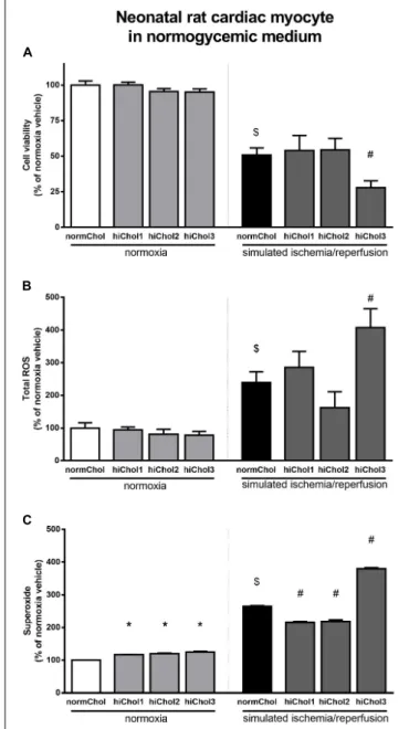
Under normoxic conditions, the cell viability of neonatal cardiac myocytes was not influenced by the hypercholesterolemic supplementation (Figure 3A). Under normoxic conditions there were no differences in total ROS levels between the groups too (Figure 3B). However, superoxide levels were significantly elevated in all groups (Figure 3C), reflecting some detrimental effect in presence of high level of cholesterol.
Simulated ischemia/reperfusion (SI/R) injury caused significant cell death of normocholesterolemic cardiac myocytes (Supplementary Figure S1A) compared to normoxic groups.
Cardiac myocyte viability was significantly decreased with the administration of hiChol3 (Figure 3A). SI/R injury alone increased both total ROS and superoxide levels in normocholesterolemic (normChol) groups, which were further increased in presence of hypecholesterolemic supplementation (hiChol3) (Figures 3B,C).
Effect of Metabolic Disease Condition and Simulated Ischemia/Reperfusion Injury in Neonatal Cardiac Myocytes
In normoxic condition, when hypercholesterolemic supplementation was applied in combination with high
571 572 573 574 575 576 577 578 579 580 581 582 583 584 585 586 587 588 589 590 591 592 593 594 595 596 597 598 599 600 601 602 603 604 605 606 607 608 609 610 611 612 613 614 615 616 617 618 619 620 621 622 623 624 625 626 627
628 629 630 631 632 633 634 635 636 637 638 639 640 641 642 643 644 645 646 647 648 649 650 651 652 653 654 655 656 657 658 659 660 661 662 663 664 665 666 667 668 669 670 671 672 673 674 675 676 677 678 679 680 681 682 683 684 FIGURE 2 | (A)Representative images of membrane cholesterol content in neonatal cardiac myocytes after cholesterol supplementation (hiChol1, hiChol2, hiChol3) measured with Filipin staining (for cholesterol content) and propidium iodide staining (for total cell count).(B)Membrane cholesterol levels of neonatal cardiac myocytes evaluated (Image J software) after cultivation with different supplements (hiChol1, hiChol2, hiChol3). Data are expressed as mean±SEM compared to normoxia vehicle control group (100%).∗p<0.05 vs. normoxia vehicle (one-way ANOVA, Tukey’spost hoc),n= 20–23.
glucose in medium, reduced cell viability was detected at higher concentration of cholesterol (hiChol2 and hiChol3) (Figure 4A). In these groups, total ROS and superoxide levels increased correspondingly (Figures 4B,C). Simulated ischemia-reperfusion further decreased cell viability in hiChol2 and hiChol3, while total ROS and superoxide levels increased (Figure 4).
Effects of Hypercholesterolemic Supplementation and Simulated Ischemia/Reperfusion Injury in Adult Cardiac Myocytes
We tested the sensitivity of cardiac myocytes isolated from adult rats to hypercholesterolemia. Cell viability was significantly reduced after hiChol2 supplementation of adult cardiac myocytes in normoxia (Figure 5A). The total ROS level was not influenced, but superoxide level was elevated by hiChol2 under normoxic condition (Figures 5B,C).
Simulated ischemia/reperfusion injury caused significant cell death of adult cardiac myocytes (Supplementary Figure S1B).
The reduction of cell viability by cholesterol supplementation was further increased when hypercholesterolemia was combined with simulated ischemia/reperfusion injury (Figure 5A). Both total ROS and superoxide showed markedly elevated levels
when hypercholesterolemic supplementation and simulated ischemia/reperfusion was combined (Figures 5B,C).
Effect of Metabolic Disease Condition and Simulated Ischemia/Reperfusion Injury in Adult Cardiac Myocytes
In normoxic condition, when hypercholesterolemic supplementation was applied in combination with high glucose in medium, reduced cell viability was detected at higher concentration of cholesterol (hiChol2 and hiChol3) (Figure 6A).
In these groups, total ROS and superoxide levels increased correspondingly in normoxic condition (Figures 6B,C).
Simulated ischemia-reperfusion caused similar rate of cell death in hiChol2 and hiChol3 as in normoxic cells, while interestingly total ROS did not changed, superoxide levels increased only in hiChol3 group (Figure 6).
Cardioprotection Against Simulated Ischemia/Reperfusion Injury in Hypercholesterolemic Neonatal and Adult Cardiac Myocytes
The NO-donor S-nitroso-N-acetyl penicillamine (SNAP) significantly decreased cell death induced by SI/R injury in neonatal normocholesterolemic cardiac myocytes (Figure 7A).
685 686 687 688 689 690 691 692 693 694 695 696 697 698 699 700 701 702 703 704 705 706 707 708 709 710 711 712 713 714 715 716 717 718 719 720 721 722 723 724 725 726 727 728 729 730 731 732 733 734 735 736 737 738 739 740 741
742 743 744 745 746 747 748 749 750 751 752 753 754 755 756 757 758 759 760 761 762 763 764 765 766 767 768 769 770 771 772 773 774 775 776 777 778 779 780 781 782 783 784 785 786 787 788 789 790 791 792 793 794 795 796 797 798
Makkos et al. Ischemic Hypercholesterolemic Comorbidity in Cardiomyocytes
FIGURE 3 |Neonatal rat cardiac myocyte viability was measured(A)with calcein AM staining after cultivation with/without hiChol1-3 supplements under normoxic conditions and combined with simulated ischemia/reperfusion injury (SI/R). Total ROS(B)and superoxide(C)level was measured in neonatal cardiac myocytes treated with cholesterol supplements (hiChol1-3) under normoxia or after SI/R. Data are expressed as mean±SEM, in comparison to normoxia vehicle control group (100%). $p<0.05 normoxia vehicle vs. SI/R vehicle (t-test);∗p<0.05 vs. normoxia vehicle (one-way ANOVA, LSD post hoc); #p<0.05 vs. SI/R vehicle (one-way ANOVA, LSDpost hoc);
n= 5–11 “N number denotes the number of wells originated from several technical repeats.”
The protective effect of SNAP was abolished in each hiChol supplemented groups (Figure 7B). SNAP significantly decreased rate of cell death induced by SI/R injury in adult normocholesterolemic cardiac myocytes (Figure 8A). Protective effect of SNAP was abolished in each hiChol supplemented groups (Figure 8B).
FIGURE 4 |Neonatal rat cardiac myocyte cells cultured in hyperglycemic medium with/without hiChol1-3 supplements. Viability(A)was measured with calcein AM staining in normoxia or after SI/R injury. Total ROS(B)and superoxide(C)level were also measured in normoxia or after SI/R. Data are expressed as mean±SEM, in comparison to normoxia vehicle control group (100%). $p<0.05 normoxia vehicle vs. SI/R vehicle (t-test);∗p<0.05 vs.
normoxia vehicle (one-way ANOVA, LSDpost hoc); #p<0.05 vs. SI/R vehicle (one-way ANOVA, LSDpost hoc);n= 6–12.
DISCUSSION
In the present study, we showan in vitro medium throughput cell-based test system of primary isolated cardiac myocytes subjected to simulated ischemia/reperfusion in combination with hypercholesterolemia using tailored hypercholesterolemic supplementation with or without hyperglycemia. HiChol- supplemented rat cardiac myocytes showed reduction of cell viability and increased oxidative stress, which were further
799 800 801 802 803 804 805 806 807 808 809 810 811 812 813 814 815 816 817 818 819 820 821 822 823 824 825 826 827 828 829 830 831 832 833 834 835 836 837 838 839 840 841 842 843 844 845 846 847 848 849 850 851 852 853 854 855
856 857 858 859 860 861 862 863 864 865 866 867 868 869 870 871 872 873 874 875 876 877 878 879 880 881 882 883 884 885 886 887 888 889 890 891 892 893 894 895 896 897 898 899 900 901 902 903 904 905 906 907 908 909 910 911 912 FIGURE 5 |Adult myocardial cell viability(A)was measured in normoxia or
after simulated ischemia-reperfusion (SI/R) injury with/without hiChol1-3 supplements. Total ROS(B)and superoxide(C)level was measured too. Total ROS(B)and superoxide(C)level were also measured in normoxia or after SI/R. Data are expressed as mean±SEM, in comparison to normoxia vehicle control group (100%). $p<0.05 normoxia vehicle vs. SI/R vehicle (t-test);
∗p<0.05 vs. normoxia vehicle (one-way ANOVA, LSDpost hoc); #p<0.05 vs. SI/R vehicle (one-way ANOVA, LSDpost hoc);n= 5–14 on(A,C);n= 3–5 on(B).
aggravated by SI/R and additional hyperglycemia. Moreover, HiChol supplementation blocked the cardiocytoprotective effect the positive control NO-donor SNAP. These results are in accordance to results observed in in vivo settings with myocardial infarction and metabolic disease. This is the first demonstration that the combination of the current hypercholesterolemic/metabolic disease medium and SI/R in
FIGURE 6 |Adult rat cardiac myocyte cells cultured in hyperglycemic medium with/without hiChol supplements. Viability(A)was measured with calcein AM staining in normoxia or after SI/R injury. Total ROS(B)and superoxide(C)level were also measured in normoxia or after SI/R. Data are expressed as mean±SEM, in comparison to normoxia vehicle control group (100%).
$p<0.05 normoxia vehicle vs. SI/R vehicle (t-test);∗p<0.05 vs. normoxia vehicle (one-way ANOVA, LSDpost hoc); #p<0.05 vs. SI/R vehicle (one-way ANOVA, LSDpost hoc);n= 6–12.
cardiac myocytes mimics the cardiac pathology of the comorbid heart with I/R and hypercholesterolemia/metabolic disease. This in vitro model can be suitable for testing potential drug candidates for cardioprotection.
Hypercholesterolemia is widely accepted as a principal risk factor for CAD (Ferdinandy et al., 2014). Hypercholesterolemia has direct negative effects on the myocardium itself, in addition to the development of atherosclerosis and CAD. In the
913 914 915 916 917 918 919 920 921 922 923 924 925 926 927 928 929 930 931 932 933 934 935 936 937 938 939 940 941 942 943 944 945 946 947 948 949 950 951 952 953 954 955 956 957 958 959 960 961 962 963 964 965 966 967 968 969
970 971 972 973 974 975 976 977 978 979 980 981 982 983 984 985 986 987 988 989 990 991 992 993 994 995 996 997 998 999 1000 1001 1002 1003 1004 1005 1006 1007 1008 1009 1010 1011 1012 1013 1014 1015 1016 1017 1018 1019 1020 1021 1022 1023 1024 1025 1026
Makkos et al. Ischemic Hypercholesterolemic Comorbidity in Cardiomyocytes
FIGURE 7 | (A)SI/R andS-niroso-N-penicillinamine (SNAP) effect on the cell viability of neonatal cardiomyocytes was detected with calcein-AM.(B)Effect of Hypercholesterolemia on protective effect of SNAP against SI/R was tested in each concentration (hiChol1, hiChol2, hiChol3). Data are expressed as mean±SEM, in comparison to normoxia vehicle control group (100%).
$p<0.05
Q10 normoxia vehicle vs. SI/R vehicle;#p<0.05 vs. SI/R vehicle (one-way ANOVA, LSDpost hoc);n= 13–15.
present study, we observed a concentration-dependent uptake of cholesterol by cardiac myocytes, which formed lipid droplets mainly visible in the cytoplasm. HiChol-supplemented normoxic neonatal rat cardiac myocytes did not show reduced cell viability, but adult rat cardiac myocytes did. Similarly, direct harmful effect of hypercholesterolemia on myocardium has been shown in several experimental animal models. After 10 weeks of cholesterol feeding, both systolic and diastolic impairments were detected without hypertrophy or elevated blood pressure in rabbits (Huang et al., 2004). Reduced myocardial strain was detected with speckle tracking echocardiography in rabbit after 2- and 3-month atherogenic feeding, without atherosclerosis (Liu et al., 2014). It was shown in a hypercholesterolemic rat model that sterol esters affect membrane composition, increase erythrocyte osmotic fragility and decrease antioxidant enzyme levels (Sengupta and Ghosh, 2014). In the present study, the presence of hypercholesterolemia induced an increased level of superoxide formation in both neonatal and adult rat cardiac myocytes in normoxic condition. This finding is in line
FIGURE 8 | (A)SI/R andS-niroso-N-penicillinamine (SNAP) effect on the cell viability of adult cardiomyocytes was detected with calcein-AM.(B)Effect of Hypercholesterolemia on protective effect of SNAP against SI/R was tested in each concentration (hiChol1, hiChol2, hiChol3). Data are expressed as mean±SEM, in comparison to normoxia vehicle control group (100%).
$p<0.05 normoxia vehicle vs. SI/R vehicle;#p<0.05 vs. SI/R vehicle (one-way ANOVA, LSDpost hoc);n= 5–12.
with in vivo data, where increased formation superoxide has been observed in hypercholesterolemic rat myocardium (Onody et al., 2003). Elevated oxidative stress associated with high left ventricular diastolic pressure were observed in in vivo and ex vivoisolated diet-induced hypercholesterolemic rat hearts as well (Varga et al., 2013). These results shows that the present in vitro hypercholesterolemic/metabolic disease cell culture model mimics the in vivo settings regarding the deteriorative effects on cardiac myocytes via increased oxidative stress.
As already widely reported in the literature (Lin et al., 2015), lipid dysregulation is often present as a cause or a consequence of many human diseases. Commercially availablein vitromodels do not take into account the influence of lipid dysregulation on most cell properties. Therefore, there is an urgent need for a new generation of in vitro models that would be able to mimic pathologies or predisposing conditions also through the consideration of the cell lipidome. Mammalian in vitro cells are able to synthesize internally the majority of lipids, lipid building blocks and related precursors they need. However, their
1027 1028 1029 1030 1031 1032 1033 1034 1035 1036 1037 1038 1039 1040 1041 1042 1043 1044 1045 1046 1047 1048 1049 1050 1051 1052 1053 1054 1055 1056 1057 1058 1059 1060 1061 1062 1063 1064 1065 1066 1067 1068 1069 1070 1071 1072 1073 1074 1075 1076 1077 1078 1079 1080 1081 1082 1083
1084 1085 1086 1087 1088 1089 1090 1091 1092 1093 1094 1095 1096 1097 1098 1099 1100 1101 1102 1103 1104 1105 1106 1107 1108 1109 1110 1111 1112 1113 1114 1115 1116 1117 1118 1119 1120 1121 1122 1123 1124 1125 1126 1127 1128 1129 1130 1131 1132 1133 1134 1135 1136 1137 1138 1139 1140
preference is to uptake lipids from the cell culture medium, if they are available. Consequently, in the presence of an adequate external source of lipids, most cellular enzymes are down regulated or switched-off. This is why the lipid composition of in vitrocells can be modulated by strictly controlling their external supply and a carefully planned feeding strategy grants the possibility to develop efficientin vitromodels mimicking real in vivoconditions (Poggi et al., 2015;Chatgilialoglu et al., 2017).
The scope of this work was to develop a hypercholesterolemic comorbidity model of primary cardiac myocytes. In our opinion, the supplementation of increasing concentrations of cholesterol only was a too simplistic way to operate; in fact, in vivohypercholesterolemic conditions are often interconnected with a broader hyperlipidemia/dyslipidemia, characterized by a wider array of dysregulated lipids and influenced by multiple factors belonging to genetics, lifestyle and diet (Castro Cabezas et al., 2018). Frequently, a hypercholesterolemic condition is generated or corroborated by a poor diet quality based on saturated fats and pro-inflammatory lipids (Marais, 2013; Arsenault et al., 2017). For this reason, we decided to integrate the cholesterol-based supplements with selected lipids, thus generating a more heterogeneous and authentic hypercholesterolemic/hyperlipidemic phenotype for our primary cardiac myocyte in vitro model. The three tailored RefeedR supplements were therefore developed by integrating the desired levels of cholesterol with selected adjuvant lipids, in order to strengthen the hypercholesterolemic biological effects and create a more accurate in vitro model. In our present neonatal rat cardiac myocyte model, hypercholesterolemic supplementation was taken up by cells in a concentration dependent manner and did not influence viability of neonatal cells. Filipin fluorescence intensity showed lipid droplets mainly located in cell cytoplasm.
In another study, cardiac myocyte labeled with Filipin shows highest level of cholesterol content in plasma membrane, but also detectable signals can be captured from Golgi apparatus and outer nuclear membrane (Severs, 1982).
There are other, less-controlled external types of lipid supplementation described in the literature in cell culture models, showing direct harmful effect of cholesterol. Cal et al.
(2012a,b) describe that the cholesterol uptake from VLDL or LDL lipoprotein levels can affect the regulation of LPR- 1 (lipoprotein receptor-related protein 1) receptor expression and the cholesterol accumulation in the ischemic myocardium.
Castellano et al. (2011) described the VLDL effect on Ca2+ handling and how the hypoxia can further exacerbate this effect. Oxidized forms of lipoproteins can be harmful also directly for the myocardium. Therefore, the present tailored hypercholesterolemic supplementation is suitable for controlled induction of hypercholesterolemiain vitro.
In the present study, simulated ischemia/reperfusion was combined with hypercholesterolemic medium. Simulated ischemia/reperfusion induced cell death aggravated harmful effect of hypercholesterolemia in neonatal as well as in adult cardiac myocytes. This finding is in line with majority of in vivo animal models of ischemia/reperfusion, in which hypercholesterolemia aggravated the ischemia/reperfusion injury of the myocardium (Andreadou et al., 2017). In the
present model, decreased viability of cardiac myocytes was associated with increased levels of total ROS and superoxide anion. One of the most important free radicals generated during hypercholesterolemia is superoxide anion (Landmesser et al., 2000;Napoli and Lerman, 2001). Increased level of ROS and its fundamental role in ischemia/reperfusion injury is an extensively studied phenomenon (Perrelli et al., 2011; Moris et al., 2017; Sinning et al., 2017; Cadenas, 2018; Hernandez- Resendiz et al., 2018). ROS mediated signaling pathway is defined as “redox signaling” (Moris et al., 2017) which was not directly investigated in the present study. ROS modulates several downstream signaling pathways, i.e., the activity of NFkB, which is a well-studied redox-sensitive transcription factor (Frantz et al., 2001). Hypercholesterolemia was the first cardiovascular risk factor to be associated with the loss of cardioprotection due to deterioration of several signaling mechanisms (Ferdinandy et al., 2007, 2014), including disruption of NO-cGMP-PKG pathway (Giricz et al., 2009), KATP signaling (Csonka et al., 2014), Connexin43 distribution (Gorbe et al., 2011), inhibition of opening of mitochondrial permeability transition pores (Yadav et al., 2010), among several other (Andreadou et al., 2017).
To further validate our system, we used a well-known cardioprotective NO-donor to test if its cardiocytoprotective effect is also blocked by hyperchoelsteolemia in our in vitro system. Here we have found that the NO-donor SNAP protected both neonatal and adult normocholesterolemic cardiac myocytes against SI/R injury, but not the hypercholesterolemic cardiac myocytes. These results further validated our present in vitro I/R and hypercholesterolemic model is suitable for testing cardioprotective in the presence of hypercholesterolemic comorbidity.
Ischemic heart disease associates with several risk factors and comorbidities, like aging and diabetes. Several studies investigated the effect of hyperglycemia on ischemic heart and cardioprotection in different experimental animal models of diabetes and in diabetic patients. Studies showed that the presence of diabetes might interfere with the cardioprotective mechanisms, attenuating the effectiveness of these therapeutic strategies (Ferdinandy et al., 2014). Therefore, here we investigated the presence of hyperglycemia in addition to hypercholesterolemia in isolated primary cardiac myocytes. Here we have found that the combination of hypercholesterolemia and hyperglycemia mimicking metabolic disease worsened the survival of cardiac myocytes even in normoxic condition.
Reduction in cell viability and increase in the level of oxidative stress were further aggravated in ischemic neonatal cardiac myocytes. In case of adult cardiac myocytes, SI/R injury interestingly total ROS did not changed, and superoxide levels increased only in hiChol3 group. We have previously found that acute hyperglycemiain vivodid not influence infarct size in rat acute myocardial model, but abolished cardioprotective effect of remote ischemic preconditioning (Baranyai et al., 2015). In a diabetic mice model, the exacerbation of heart failure after MI has been observed via increasing NAD(P)H oxidase-derived superoxide. These results further prove the validity of our presentin vitroI/R and hypercholesterolemic/metabolic disease model is suitable for testing cardioprotective compounds in the