. . . .
. . . .
Inhibition of AP-1 signaling by JDP2 overexpression protects cardiomyocytes against hypertrophy
and apoptosis induction
Christian Hill
1†, Alona Wu¨rfel
1†, Jacqueline Heger
1, Bettina Meyering
1,
Klaus-Dieter Schlu¨ter
1, Martin Weber
1, Peter Ferdinandy
2, Ami Aronheim
3, Rainer Schulz
1, and Gerhild Euler
1*
1Physiologisches Institut, Justus-Liebig-Universita¨t Giessen, Aulweg 129, 35392 Gießen, Germany;2Department of Pharmacology and Pharmacotherapy, Semmelweis University, Budapest, Hungary; and3Rappaport Institute Haifa, Haifa, Israel
Received 4 January 2013; revised 26 March 2013; accepted 14 April 2013; online publish-ahead-of-print 23 April 2013 Time for primary review: 13 days
Aims Expression and activity of the transcription factor AP-1 are enhanced during cardiac remodelling and heart failure pro- gression. In order to test if AP-1 inhibition may limit processes contributing to cardiac remodelling, ventricular cardio- myocytes of mice with cardiac overexpression of the AP-1 inhibitor JDP2 were analysed under stimulation of hypertrophy, apoptosis, or contractile function.
Methods and results
Three models of JDP2 overexpressing mice were analysed: JDP2 was overexpressed either life-long, for 7 weeks, or 1 week. Then cardiomyocytes were isolated and stimulated withb-adrenoceptor agonist isoprenaline (ISO, 50 nM).
This enhanced cross-sectional area and the rate of protein synthesis in WT but not in JDP2 overexpressing cardiomyo- cytes. To induce apoptosis, cardiomyocytes were stimulated with 3 ng/mL TGFb1. Again, JDP2 overexpression pre- vented apoptosis induction compared with WT cells. Determination of contractile function under electrical stimulation at 2 Hz revealed enhancement of cell shortening, and contraction and relaxation velocities under increasing ISO concentrations (0.3 – 30 nM) in WT cells. This inotropic effect was abrogated in JDP2 overexpression cells. Respon- siveness to increased extracellular calcium concentrations was also impaired in JDP2 overexpressing cardiomyocytes.
Simultaneously, a reduction of SERCA expression was found in JDP2 mice.
Conclusion A central role of AP-1 in the induction of hypertrophy and apoptosis in cardiomyocytes is demonstrated. Besides these protective effects of AP-1 inhibition on factors of cardiac remodelling, AP-1-inhibition impairs contractile function.
Therefore, AP-1 acts as a double-edged sword that mediates mal-adaptive cardiac remodelling, but is required for main- taining a proper contractile function of cardiomyocytes.
- - - -
Keywords Transcription factor AP-1 † Hypertrophy † Apoptosis † Contractile function † Cardiac † Remodeling
1. Introduction
Cardiac hypertrophy and apoptosis are main predictors and causes for heart failure development, which finally results in contractile dysfunction of the heart. Interestingly, all these events are often accompanied by ele- vations of the transcription factor AP-1:in vivo, increased levels of AP-1 are found under pressure overload,1in the phase of LV remodelling after myocardial infarction,2or after isoprenaline infusion in rats.3
AP-1 is a dimer of jun and fos family members. Thus, dominant- negative expression of either of these subunits results in the impairment
of AP-1. Using this technique in isolated cardiomyocytes already has demonstrated an important role of AP-1 in cardiac hypertrophy and apoptosis. In neonatal cardiomyocytes, Omura et al. 4 expressed dominant-negative mutant c-jun, resulting in depressed AP-1 activity.
This abrogated hypertrophic responses to endothelin 1 as well as to thea-adrenoceptor agonist phenylephrine. Dominant-negative expres- sion of c-Fos, that also blocked AP-1 activity in neonatal cardiomyocytes, inhibited the induction of the pathological gene profile under stimulation with phenylephrine, i.e. blocking expression of beta-myosin heavy chain and atrial/brain natriuretic peptides (ANP/BNP), and prevented down
†C.H. and A.W. contributed equally to this work.
*Corresponding author. Tel:+49 641 9947246; fax:+49 641 9947239, Email: gerhild.euler@physiologie.med.uni-giessen.de
Published on behalf of the European Society of Cardiology. All rights reserved.&The Author 2013. For permissions please email: journals.permissions@oup.com.
doi:10.1093/cvr/cvt094
by guest on December 23, 2014Downloaded from
regulation of sarcoplasmic reticulum Ca2+-ATPase (SERCA).5 The latter finding indicates that AP-1 may, in addition to its role in hyper- trophic growth, also modulate contractile function of cardiomyocytes.
In adult cardiomyocytes of rat, the involvement of AP-1 in hyper- trophic growth could be confirmed by the use of decoy- oligonucleotides, that intracellularly scavenge AP-1, thereby inhibiting hypertrophic growth under phenylephrine stimulation.6In addition to its involvement in hypertrophic signalling, AP-1 contributes to apoptosis induction, since transfection of adult cardiomyocytes of rat with AP-1-decoy-oligonucleotides abolished apoptosis under stimulation with the growth factor TGFb1or with nitric oxide.7In addition to AP-1, activation of transcription factors of the SMAD family are needed in order to induce apoptosis in cardiomyocytes. Adenoviral SMAD4 overexpression in cardiomyocytes can even turn the a-adrenergic induced, pro-hypertrophic signal of AP-1 into an apoptotic stimulus.8
To dissect the role of AP-1in vivo, the working group of Aronheim and colleagues9 generated a transgenic mice model with cardiac specific overexpression of the AP-1 inhibitor JDP2 (c-jun dimerization protein 2). Both, JDP2 and AP-1 belong to the b-ZIP family of DNA-binding pro- teins. AP-1 is formed by the dimerization of jun- and fos-family members, resulting in a DNA-binding and transcription activating complex. In hypertrophic or apoptotic ventricular cardiomyocytes, c-Jun, Jun B, c-Fos, and Fos B were identified as subunits of AP-1 dimers.7Association of JDP2 with jun-family members prevents the formation of transcrip- tion promoting AP-1 dimers.10Additionally, JDP2 can bind directly to AP-1-specific promoter elements. This, again results in the abrogation of AP-1-dependent transcription.
Thus, cardiac-specific JDP2 overexpression results in specific inhib- ition of AP-1 signalling and may thus interfere with cardiac hypertrophy and apoptosis. First studies on these transgenic mice, that overexpressed JDP2 heart-specifically under control of a tetracycline-regulateda-MHC promoter, revealed the development of massive atrial dilatation, which was reversible upon abolishment of JDP2 expression by the tetracycline system (tet-off-system).9In histological sections of JDP2 overexpressing hearts, hypertrophic cardiomyocytes were detected in atria, but not in the ventricles.
Only under stimulation of ventricular cardiomyocytes with hyper- trophy or apoptosis inducing agents, AP-1 is up-regulated, and only under such conditions, JDP2 may interfere with the processes of ven- tricular remodelling. Therefore, the question arises, if ventricular cardi- omyocytes of transgenic JDP2 mice are resistant to the induction of hypertrophy and apoptosis? Furthermore, influence of JDP2 overex- pression on contractile function of cardiomyocytes was of interest, in order to get comprehensive data about effects of AP-1 inhibition on pro- cesses of ventricular remodelling. To answer these questions, hyper- trophic growth underb-adrenergic stimulation, apoptosis induction by TGFb1, and contractile responses tob-adrenoceptor stimulation or enhanced calcium concentrations were compared in WT and JDP2-overexpressing cardiomyocytes.
2. Methods
The investigation conforms to the Directive 2010/63/EU of the European Parliament. Use of animals was registered at the Justus-Liebig-University (registration-no.: 419-M).
2.1 JDP2 overexpressing mice
JDP2 mice, crossings with the C57BL6/FVB-line, had been generated in Haifa, Israel. They are double transgenic, carrying a heterocygote JDP2 gene with a
minimal promoter and a heterocygote transactivator gene under the control of the heart specifica-MHC promoter. The transactivator could be regu- lated by the antibiotic doxycycline (Dox) in a tet-off system: Feeding the animals with Dox blocked the interaction between the transactivator and the promoter of the JDP2 gene, thereby preventing JDP2 overexpression.
To investigate short- and long-term effects of AP-1 inhibition, duration of JDP2 expression was controlled by Dox-feeding of animals for different times (Figure1A). Therefore, three groups with constitutive, chronic, or acute JDP2 overexpression were generated by the following feeding proto- cols: In the first group, mice were kept without Dox-feeding their whole life through. This resulted in an embryonic and life-long, constitutive AP-1 inhib- ition. In the second group, breeding pairs and newborn mice were fed with Dox until the first week after birth. This guaranteed the absence of JDP2 overexpression during embryonic and juvenile development of mice, which was followed by a chronic AP-1 inhibition due to JDP2 overexpression for 7 weeks. In the third group, breeding pairs and new born mice were fed with Dox for 10 weeks followed by an acute AP-1 inhibition in the last week of life. As control, littermates of transgenic mice which did not overexpress JDP2 (wild-types) were used. Also WT mice received three different Dox-diet protocols. At the age of 9 weeks, all mice were used for isolation of hearts or ventricular cardiomyocytes.
2.2 Cell isolation and culture
Mice were anaesthetized by isofluran inhalation. After cervical dislocation hearts were extracted, and retrograde-perfused in a Langendorff apparatus with a collagenase-containing calcium-free buffer equilibrated at 378C, pH 7.4. After separation of cardiomyocytes from other cardiac cells by centrifu- gation, medium was re-adjusted to a physiological calcium concentration and suspended in basal culture medium. Cardiomyocytes were then plated on laminin-coated culture dishes. After 2 h, medium was changed and cells could be stimulated. The basal culture medium (CTT) was modified medium 199 including Earl´s salts, 2 mM L-carnitine, 5 mol/L taurine, 100 000 IU/L penicillin, 100 mg streptomycin, and 10mmol/L cytosine-beta-L-arabinofuranoside.
2.3 Electrophoretic mobility shift assay
Hearts were homogenized in swelling buffer (10 mM Tris – HCl, pH 7.9, 10 mM KCl, 1 mM MgCl2, and 1 mM DTT). After incubation for 1 h on ice, nuclei were pelleted by centrifugation at 900 rpm for 10 min. Pellets were homogenized in 10 mM Tris – HCl, pH 7.9, 300 mM saccharose, 1.5 mM MgCl2, 1 mM DTT, and 0.3% Triton X-100 and again centrifuged as described earlier. Pellets were suspended in storage buffer (10 mM HEPES, pH 7.5, 50 mM KCl, 300 mM NaCl, 1 mM EDTA, 1 mM DTT, 1 mM PMSF, and 20% glycerol) on ice for 30 min and centrifuged at 13 000 rpm at 48C for 5 min. The resulting supernatants were used for retardation assays: TPA re- sponse element (TRE) oligonucleotides, containing complementary sequences of the AP-1 binding domain (5´-ATCCGCTTGATGAGTCAGCC GGAA-3´) were hybridized, fluorescent labelled with Cy3-dCTP, and incu- bated with nuclear extracts in the presence of 0.5mg of poly(dIdC) at 308C for 30 min. The samples were run on 4% native polyacrylamide gels.
For the identification of binding proteins, subsequent to the incubation of nuclear extracts with the oligonucleotide, 0.5mg of antibodies specific for JDP2 or c-jun (Santa Cruz) were added to the reaction mixture and incu- bated for another 30 min at 308C. Gels were exposed on fluorescence imager (BioRad).
2.4 Real-time PCR
Total RNA from left ventricles was extracted with Trizol (Invitrogen) as described by the manufacturer. This was followed by DNAse treatment and reverse transcription with QuantiTect Reverse Transcription Kit from Quiagen. For each assayed gene, annealing temperature and the number of cycles resulting in a linear amplification range were tested. RT – PCR was per- formed in an automated thermal cycler and detected with the Biorad detec- tion system (Biorad) using SYBR Green fluorescence for quantification. The
by guest on December 23, 2014Downloaded from
calculations of the results were carried out according to the 22DDCtmethods as described.11Gene expression was related to B2M as housekeeping gene.
The following primers were used: b1-adrenoceptor from Quiagen,b2- adrenoceptor: 5´-TGGTACCGTGCCACCCACAA-3´, 5´-AAGACCATC ACCACCAGGGGCA-3´; B2M: 5´-GCTATCCAGAAAACCCCT CAA-3´, 5´-CATGTCTCGATCCCAGTAGACGGT-3´; phospholamban: 5´-GCAAT ACCTCACTCGCTCGGCTATC-3´, 5´-TGGAGATTCTGACGTGCTTGC TGAG-3´; NCX: 5´-CTACCAGGTCCTAAGTCAACAG-3´, 5´-TGCGTGC CTCTTCAAGATG; TGFb1: 5´-GTCCTTGCCCTCTACAACCA-3´, 5´-GT TGGACAACTGCTCCACCT-3´; SMAD2: 5´-GGAACCTGCATTCTGGT GTT-3´, 5´-ACGTTGGAGAGCAAGCCTAA-3´; SMAD3: 5´-TTCACTGA CCCCTCCAACTC-3´, 5´-CTCCGATGTAGTAGAGCCGC-3´; SERCA:
5´-TGACTGGTGATGGTGTGAATG-3´, 5´-GATGAGGTAGCGGATGAA CTG-3´
2.5 Immunoblot analysis
Proteins were extracted from frozen hearts of transgenic mice with chronic JDP2 overexpression and from corresponding hearts of WT mice. Hearts were homogenized in RIPA buffer (50 mmol/L Tris/HCl, pH 7.5, 150 mmol/L NaCl, 1% Nonidet P-40, 0.5% deoxycholat, 0.1% SDS, 1 mM PMSF, 1 mM EDTA, 1 mg/l pepstatin). Nucleic acids were digested with ben- zonase. Samples were denatured in Laemmli buffer at 908C for 5 min, loaded on 12.5% SDS-gels, and blotted on PVDF membranes. For detection of SERCA2A expression, specific antibodies were purchased from Santa Cruz. For loading controls, actin antibodies were used. Protein bands were detected by horseradish peroxidase-labelled secondary antibodies using ECL as detection system (Pierce). SERCA-specific signals were normal- ized against actin.
2.6 Hypertrophy assays
Isolated cardiomyocytes were incubated with theb-adrenoreceptor agonist isoprenaline (ISO, 50 nM) for 24 h. As parameters for hypertrophy,
cross-sectional area and the rate of protein synthesis were used. For meas- uring the cross-sectional area, myocyte size was determined on micrographs digitalized by a charge-coupled device camera as described elsewhere.12 Width/diameter of randomly taken cells was determined at the widest point of each myocyte using the software program Adobe Photoshop 5.5.
Cross-sectional area of cardiomyocytes was calculated by the following formula: cross-sectional area¼radius2×p.
To determine the rate of protein synthesis, incorporation of [L-14C] phenyl- alanine (0.1 mCi/L) over 24 h was analysed. Incorporation of radioactivity into acid-insoluble cell mass was determined as described previously.13
2.7 Apoptosis assay
To induce apoptosis, cardiomyocytes were incubated with 3 ng/mL of trans- forming growth factorb1(TGFb1) for 4.5 h. Then, cells were stained with annexin V-FITC (10mL/mL)/propidiumiodide (5mL/mL) for 15 min. Apop- totic cardiomyocytes stained with annexin V-FITC, resulting in a green fluor- escence when exited at 450 – 480 nm, and excluded propidiumiodide, a DNA dye unable to pass the plasma membrane. Necrotic cells have lost their physical integrity of their plasma membrane and appear double-stained with annexin V-FITC/propidium iodide, which fluoresces in the red when exited at 510 – 550 nm. For the quantification of apoptosis and necro- sis, 200 randomly distributed cardiomyocytes were counted in each experiment.
2.8 Cell contraction
Cell shortening was analysed as described previously.14Briefly, cardiomyo- cytes were incubated with different concentrations of isoprenaline (0.3 – 30 nM) or calcium (2.5 and 5 mM), and stimulated at 2 Hz for 1 min at room temperature. Analysis of cell contraction was performed using cell-edge detection system. Cells were stimulated via two AgCl electrodes with biphasic electrical stimuli composed of two equal but opposite rect- angular 50 V stimuli of 5 ms duration. Only rod-shaped cells that contracted Figure 1 (A) Regulation of AP-1 inhibition via Dox-feeding in transgenic JDP2 mice. Constitutive JDP2 overexpression resulting in embryonic and life- long AP-1 inhibition was achieved in the absence of Dox-diet. In the group with chronic AP-1 inhibition, Dox was fed during the embryonic development and over the first week after birth. During this time, JDP2 was not overexpressed. In the following 7 weeks, mice received Dox-diet no more, resulting in chronic AP-1 inhibition over 7 weeks. In the acute group, mice were treated with Dox during embryonic development and for 7 weeks after birth. Thus, acute JDP2 overexpression or AP-1 inhibition was present for 1 week only. (BandC) AP-1 and JDP2 binding activity in the hearts of wild-type and JDP2 overexpressing mice. (B) Nuclear extracts of hearts from WT and JDP2 mice with chronic JDP2 overexpression were prepared and tested in EMSAs for binding activity to the TRE-oligo. Enhanced binding activity is found in extracts from JDP2 overexpressing mice. (C) Subunit composition of the complex that binds to the TRE-oligo was analysed. Therefore, c-jun and JDP2 antibodies were added to the binding reaction. Specific antibody-protein interaction reduced the binding activity to the TRE-oligo.
by guest on December 23, 2014Downloaded from
regularly during the whole time of measurement were used. Every 15 s, cell shortening, contraction, and relaxation velocity were measured using a line camera. The mean of four measurements per cell was used as average value of each individual cardiomyocyte. Cell-shortening data were normalized to the individual diastolic cell length (dL/L %).
2.9 Statistics
Data are given as means+standard deviation fromndifferent culture pre- parations. Statistical comparisons were performed by ANOVA (One-Way Analysis of Variance) and Tukey-test or Student’s t-test. A P-value of ,0.05 was considered statistically significant.
3. Results
3.1 Constitutive JDP2 overexpression represses hypertrophic growth and apoptosis
First, we analysed the effects of life-long, constitutive JDP2 overexpres- sion on hypertrophy and apoptosis. Therefore, animals were fed with a regular diet without Dox supplementation, so that already during em- bryogenesis and juvenile development, as well as in adult mice, JDP2 was overexpressed. Thus, AP-1 was inhibited in all these stages (Figure1A). Ventricular cardiomyocytes of 9-week-old adult wild-type and transgenic JDP2 mice were isolated. As can be seen inFigure2A andB, continuous JDP2 overexpression had no influence on hyper- trophic growth, because in the absence of any stimulation, the cardio- myocyte size of WT and JDP2 mice are similar. As was shown by Sabri et al.,15cardiomyocytes of mice develop hypertrophic growth upon b-adrenergic stimulation. Therefore, we used the b-adrenoceptor agonist isoprenaline (ISO) for growth stimulation. Prior to ISO stimula- tion, mRNA expression of b-adrenoceptors in left ventricles was analysed to be sure that JDP2 overexpression does not influence b-adrenoceptor expression. As determined by RT – PCR, JDP2 and WT mice expressed the same b1- and b2-adrenoceptor levels (0.97-and 1.04-fold in JDP2 vs. WT left ventricles,n¼5, n.s.). Then, car- diomyocytes were stimulated with ISO (50 nM) for 24 h, and the cross- sectional area was determined as a parameter for hypertrophic growth.
In WT cardiomyocytes, ISO increased the cross-sectional area compared with unstimulated control cells to 116.7+15.7% (n¼7;
P,0.05 vs. control). In contrast, JDP2 overexpressing cardiomyocytes were protected against enlargement of cross-sectional area under ISO (103+6.8%n¼6; n.s. vs. control) (Figure2). To confirm these findings, the rate of protein synthesis, as another parameter for hypertrophic growth, was determined. In WT cardiomyocytes, incorporation of
14C-phenylalanine over 24 h was enhanced by ISO (50 nM) to 117.9+10.5%, (n¼15;P,0.05 vs. control) (Figure2B). Cardiomyo- cytes of transgenic JDP2 mice did not show an increase in
14C-phenylalanine incorporation upon ISO stimulation (99.9+18.8%;
n¼11;P,0.05 vs. control).
In order to analyse the effects of JDP2 overexpression on apoptosis induction, transforming growth factor beta (TGFb1) was chosen as an apoptotic stimulus, because TGFb1 is found up-regulated during cardiac remodelling and is known to induce apoptosis in rat cardiomyo- cytes via AP-1/SMAD signalling.7To be sure that TGFb/SMAD signalling is not influenced by JDP2 overexpressionper se, mRNA expression of TGFb1, SMAD2, and SMAD3 in the left ventricles of WT and JDP2 mice were compared. RT – PCR revealed no overt changes in the ex- pression of TGFb1-mRNA (0.8-fold vs. WT ventricles,n¼5, n.s.),
SMAD2-mRNA (1.7-fold vs. WT ventricles, n¼7, n.s.), and SMAD3-mRNA (1.5-fold vs. WT ventricles,n¼7, n.s.).
Then ventricular cardiomyocytes of WT and JDP2 overexpressing mice were stimulated with 3 ng/mL TGFb1 for 4.5 h, and apoptosis was detected by staining with annexin and propidiumiodid. In WT cardiomyo- cytes, TGFb1enhanced apoptosis from 0.8+0.6% in control to 1.6+ 1.1% (n¼25;P,0.05 vs. control) (Figure3). In cardiomyocytes overex- pressing the AP-1 inhibitor JDP2, TGFb1did not induce apoptosis (0.5+ 0.5% apoptotic cells in unstimulated controls vs. 0.6+0.6% in stimulated JDP2 cells;n¼7; n. s. vs. unstimulated controls).
Thus, constitutive, life-long inhibition of AP-1 by JDP2 prevents induc- tion of hypertrophic growth and apoptosis in ventricular cardiomyocytes.
3.2 Chronic or acute JDP2 overexpression represses hypertrophic growth and
apoptosis
Constitutive overexpression of any transgene may interfere with the developmental processes which may provoke many changes in organ systems and cell signalling. Although we could not find obvious Figure 2 Constitutive JDP2 overexpression protects cardio- myoytes against hypertrophic growth. Cardiomyocytes of WT and constitutively JDP2 overexpressing mice were stimulated with 50 nM ISO for 24 h. (A) Representative pictures of WT (a, b) and constitutively JDP2 overexpressing cardiomyocytes (c, d). Only in WT cardiomyo- cytes an increase in cell size was visible after stimulation with ISO. (B) For quantitative analysis of hypertrophic growth, the cross-sectional area of cardiomyocytes (WT:n¼7, JDP2:n¼6) and the rate of protein synthesis (WT:n¼15, JDP2:n¼7) were determined. Data are means+SD ofnindependent preparations. *Differences from un- stimulated controls withP,0.05.#Differences between stimulated WT and JDP2 overexpressing cardiomyocytes withP,0.05.
by guest on December 23, 2014Downloaded from
differences in cell size or apoptotic parameters of mice constitutively overexpressing JDP2 compared with WT mice, we wanted to exclude any developmental side effects of JDP2 overexpression. Therefore, breeding pairs and newborn mice were fed with Dox until the first week after birth. This guaranteed the absence of JDP2 overexpression during embryonic and juvenile development of mice, followed by a chronic phase of JDP2 overexpression.
For confirmation of efficient JDP2 overexpression, binding activity in nuclear extracts of WT- and JDP2-overexpressing hearts to the TRE oligo was analysed in electrophoretic mobility shift assays (EMSAs). As depicted inFigure1B, JDP2 overexpression enhanced binding to the TRE oligo. Densitometric analysis of these band shifts revealed a 127+10% increased binding activity in JDP2 mice (n¼3,P,0.05 vs.
WT). Since JDP2 can form heterodimers with c-jun in order to bind to the TRE sequence, we analysed subunit composition of the binding complex by the use of specific antibodies. While the band shift from nuclear extracts of WT mice was reduced by c-jun antibodies only, the shift from nuclear extracts of JDP2 mice was reduced by c-jun and JDP2 antibodies (Figure1C). This indicates that enhanced binding activity in JDP2 mice is due to JDP2 overexpression. Binding of c-jun/JDP2 het- erodimers or JDP2 alone to AP-1 binding sites can then block AP-1-mediated gene transcription.
In addition to the constitutive and chronic JDP2 overexpression, it was of interest, if short-term overexpression may be sufficient for protective effects on cardiomyocytes. Therefore, a third group of animals received continuous Dox feeding. One week prior to the isolation of cardiomyo- cytes, Dox feeding was stopped resulting in an acute AP-1 inhibition in these animals (Figure1A).
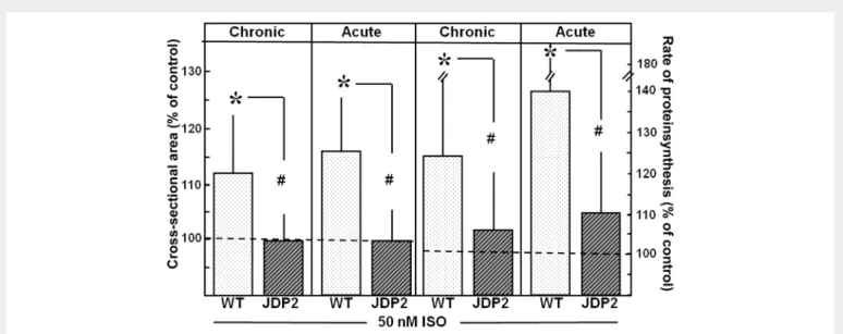
Cardiomyocytes of mice with chronic and acute JDP2 overexpression were stimulated with 50 nM ISO for the induction of hypertrophic
growth. As depicted in Figure 4, this stimulation did not enhance the cross-sectional area or the rate of protein synthesis. Thus, cardio- myocytes of chronic or acute overexpressing JDP2 mice were protected against hypertrophic growth stimulation. Furthermore, chronic or acute JDP2 overexpression protected cardiomyocytes against apoptosis induction, since TGFb1(3 ng/mL) did not increase the number of apop- totic cells for these mice (Figure5).
3.3 JDP2 overexpression impairs contractile function of cardiomyocytes
Induction of cardiac hypertrophy and apoptosis finally results in heart failure that is associated with contractile dysfunction. Therefore it was of interest, if AP-1 inhibition by JDP2 overexpression may influence con- tractile function of cardiomyocytes, and the contractile response to b-adrenergic stimulation with ISO was then tested.
Cardiomyocytes of WT mice responded to increasing ISO concentra- tions with enhanced cell shortening, reaching a maximum cell shortening of 14.6+3.1%dL/L at 30 nM ISO (P,0.05 vs. unstimulated control with 7.5+3.1%dL/L). This observation goes along with an enhance- ment of contraction and relaxation velocity (Figure6). In cardiomyocytes of mice with chronic JDP2 overexpression, contraction parameters at low ISO levels (0.3 – 10 ng ISO) were already decreased (Figure 6).
This impaired contractile responsiveness to ISO stimulation was even more pronounced at higher ISO concentrations. Even 30 nM ISO did not increase cell shortening in cardiomycytes of JDP2 mice (7.4+2.0 with 30 nM ISO vs. 6.3+3.1 unstimulated, n.s.,n¼54 cells). This indi- cates that AP-1-inhibition abrogates positive contractile responses to b-adrenergic stimulation of cardiomyocytes. To analyse, if contractile function is also reduced in the presence of other stimuli, cardiomyocyte contraction under increased calcium concentration was investigated.
Also here, a strong enhancement of cell shortening was found in WT cells (10.2+2.5%dL/L under 2.5 mM calcium vs. 13.6+2.7 under 5 mM calcium,P,0.05,n¼27). In JDP2 overexpressing cardiomyo- cytes, cell shortening was already reduced at 2.5 mM calcium (6.7+1.8%dL/L,P,0.05 vs. WT,n¼27), and remained significantly reduced at higher calcium concentrations (8.8+2.4%dL/L,P,0.05 vs. WT,n¼27) (Figure6B).
Such reduced contractile responses can be due to altered calcium handling in the cell. The main determinant of calcium handling in cardio- myocytes is the calcium pump SERCA, which is regulated by phospho- lamban. Therefore, we compared their mRNA expression levels in left ventricles of WT and chronically JDP2 overexpressing mice. While PLB mRNA-expression and expression of the sodium – calcium exchan- ger NCX was unaffected, SERCA2A-expression was reduced 0.4-fold (P,0.05 vs. WT,n¼5) in the left ventricles of JDP2 overexpressing mice. In western blots, reduction of SERCA2A was confirmed on the protein level: SERCA2A was found reduced to 59.5+13.4% in the hearts of JDP2 mice (P,0.05 vs. WT,n¼4).
4. Discussion
The main findings of our study are that AP-1 inhibition protects ventricu- lar cardiomyocytes against the induction of hypertrophy and apoptosis.
But simultaneously, AP-1 inhibition depresses contractile responses to b-adrenergic stimulation or increased calcium concentrations. Thus, AP-1 is identified as a mediator of mal-adaptive responses in cardiac re- modelling. But at the same time it is required to maintain proper con- tractile function of cardiomyocytes. Therefore, care should be taken Figure 3 Constitutive JDP2 overexpression protects cardiomyo-
cytes against TGFb1-induced apoptosis. Cardiomyocytes of WT and JDP2 overexpressing mice were incubated with 3 ng/mL TGFb1for 4.5 h. Cells were stained by annexin/propidiumiodide, and apoptotic cells were counted. Data are means+SD ofnindependent prepara- tions (WT:n¼25, JDP2:n¼7). *Differences from unstimulated con- trols withP,0.05.
by guest on December 23, 2014Downloaded from
when thinking about the use of AP-1 inhibitors for the prevention of cardiac remodelling.
In this study, we used transgenic JDP2 mice in order to analyse the effects of AP-1-inihibition on hypertrophic growth, apoptosis induction, and contractile function of ventricular cardiomyocytes. In these mice, JDP2 expression is under the control of thea-MHC promoter, which
guarantees cardiac-specific overexpression. Efficient JDP2 overexpres- sion in the hearts of these mice has already been demonstrated by Kehatet al.9using western blots and immunofluorescence. We now demonstrate in EMSAs that the binding activity to AP-1 binding sites (TRE) is enhanced in the nuclear extracts of JDP2 mice. Since JDP2 cannot promote gene transcription, binding of JDP2 will block AP-1-mediated gene transcription. However, this seems to be a partial inhibition since JDP2 antibodies could not totally abolish the binding ac- tivity to the TRE-oligo.
Phenotypic characterization of these mice revealed atrial dilatation.
However, no ventricular phenotype was obvious, although involvement of AP-1 in hypertrophic growth and apoptosis of cardiomyocytes was demonstrated by different groups. We also did not detect any sign of hypertrophic growth, since cell size was similar in WT and JDP2 overex- pressing cardiomyocytes. Furthermore, TGFb1and SMAD2/3 expres- sion, factors that are involved in AP-1-mediated apoptosis induction, were not elevated in the left ventricles of JDP2 mice. In addition, the levels of apoptosis in WT and JDP2 overexpressing cardiomyocytes were similar.
Induction of AP-1 expression and activity is found under pathophysio- logical conditions in the heart. This indicates that AP-1 may be of import- ance only, if pathophysiological stimuli are present in the heart. Since analysis of transgenic JDP2 mice was performed in the absence of any pathogenic stimulation, the lack of ventricular phenotypes does not sur- prise. Therefore, in this study, cardiomyocytes of JDP2 overexpressing mice were stimulated by factors that are known to induce hypertrophy or apoptosis in cardiomyocytes via AP-1 signalling. Since JDP2 overex- pression could be blocked by feeding mice doxocycline, we had the opportunity to distinguish groups with (i) constitutive, life-long overex- pression, (ii) with chronic overexpression for 7 weeks, and (iii) one group with JDP2 overexpression for 1 week prior to the isolation of car- diomyocytes. As described by Kehatet al.,97 days are required for JDP2 re-expression following doxycycline treatment.
Figure 4 Protection against hypertrophic growth stimulation under chronic and acute JDP2 overexpression. Cardiomyocytes of WT and JDP2 over- expressing mice were stimulated with 50 nM ISO for 24 h. As parameters of hypertrophic growth, cross-sectional area and rate of protein synthesis were determined. Data are expressed as per cent increase relative to untreated controls (dashed line). Data are means+SD ofnindependent preparations (cross-sectional area: WT:n¼18, JDP2:n¼10; acute: WT and JDP2:n¼8; protein synthesis: chronic: WT:n¼19, JDP2:n¼11; acute: WT:n¼18, JDP2:n¼11). *Differences from unstimulated controls withP,0.05.#Differences between stimulated WT and JDP2 overexpressing cardiomyocytes withP,0.05.
Figure 5 Protection against apoptosis under chronic and acute JDP2 overexpression. Cardiomyocytes of WT and JDP2 overexpressing mice were incubated with 3 ng/mL TGFb1 for 4.5 h. Cells were stained by annexin/propidiumiodide, and apoptotic cells were counted. Data are means+SD of n independent preparations (chronic: WT:n¼10, JDP2:n¼13; acute: WT and JDP2:n¼11).
*Differences from unstimulated controls withP,0.05.
by guest on December 23, 2014Downloaded from
Stimulation of cardiomyocytes with theb-adrenoceptor agonist iso- prenaline induced hypertrophic growth only in WT, but not in JDP2 over- expressing cardiomyocytes. This demonstrates that AP-1 inhibition prevents hypertrophic growth. Interestingly, in freshly isolated cardio- myocytes of rats, AP-1 dependence of hypertrophy was shown only undera-adrenergic and not underb-adrenergic stimulation.6This may be explained by a different receptor distribution among the two species. The ratio between eithera- andb- or betweenb1- andb2- recep- tors of cardiomyocytes has been shown to be different in mice or rat.16 Furthermore, a generalized defect in a1-adrenoceptor signalling was
described in mice cardiomyocytes.15These findings explain why hyper- trophy is coupled toa-adrenoceptors in rats but not in mice. Independent of the hypertrophic stimulus, via a-adrenoceptors in rats or via b-adrenoceptors in mice, AP-1 is a mediator of the growth response in both situations. Thus, we can conclude that adrenergically induced hyper- trophic growth signalling in cardiomyocytes is primarily mediated via AP-1 signalling. Abrogation of AP-1 signalling can, therefore, prevent hyper- trophic growth, that is one major predictor for heart failure development.
A further factor, contributing to heart failure progression is apoptosis.
TGFb1, which has been shown to induce apoptosis in ventricular Figure 6 Chronic JDP2 overexpression impairs contractile responses. Cardiomyocyte contractions from WT and chronically JDP2 overexpressing mice were analysed at 2 Hz for 1 min at room temperature. (A) Cardiomyocytes were incubated with different concentrations of isoprenaline (0.3, 1, 3, 10, and 30 nM). Cell shortening, as well as contraction and relaxation velocities were determined. Cell shortening was normalized to the individual diastolic cell length (dL/L %). Data are means+SD of 54 cells from two independent culture preparations. *Differences from unstimulated controls withP,0.05.
#Differences between WT and JDP2 overexpressing cardiomyocytes withP,0.05. (B) Single-cell recordings of cell shortening at 2 Hz are depicted.
Cardiomyocytes were incubated with 30 nM isoprenaline (30 nM) or calcium (2.5 and 5 mM).
by guest on December 23, 2014Downloaded from
cardiomyocytes of rat, also induces apoptosis in cardiomyocytes of WT mice. This induction was abrogated in JDP2 overexpressing cardiomyo- cytes. This protection was independent from the time point of JDP2 overexpression. These findings reveal a central role of AP-1 in apoptotic processes in the heart.
Besides the effects on hypertrophy and apoptosis, a major determin- ant of pump function is the contractile function of cardiomyocytes them- selves. Indications that AP-1 may also control this parameter, come from studies in neonatal cardiomyocytes: expression of dominant negative c-Fos in these cells has been shown to influence SERCA expression, a calcium transporter that is controlling the main calcium reservoir for cardiomyocytes contraction.5Now, in our study, we show that con- tractile function of cardiomyocytes, determined as per cent cell shorten- ing, as well as contraction and relaxation velocities under electric stimulation at 2 Hz, are slightly decreased in JDP2 overexpressing cardi- omyocytes. This reduced contractile response due to AP-1 inhibition gets even more pronounced, when cells are stimulated with isopren- aline. While WT cardiomyocytes show an enhanced cell shortening and faster contraction and relaxation velocities with increasing amounts of isoprenaline, JDP2 overexpressing cells fail to respond to b-adrenergic stimulation. Since the expression ofb-adrenoceptors in WT and JDP2-overexpressing ventricles was the same, the reduced b-adrenergic responsiveness must be due to other reasons. And indeed, abrogation of contractile responses was also detected when ex- posing JDP2 cardiomyocytes to increased calcium concentrations.
Looking at genes related to contraction, a decrease in SERCA expression is evident in JDP2 overexpressing cardiomyocytes. This can cause a re- duction in Ca2+-reuptake in the sarcoplasmatic reticulum. The internal cellular Ca2+-reservoir is thereby decreased and the cell is unable to respond to inotropic stimulations.
In human end-stage heart failure, like in dilated or ischaemic cardiomy- opathies, a chronic AP-1 activation is found.2Our results now suggest that this AP-1 activation may contribute to hypertrophic and apoptotic processes in the human heart, thereby provoking adverse cardiac re- modelling. Thus, treatment of patients with AP-1 inhibitors might reduce these remodelling processes. However, due to the additional positive action of AP-1 on cardiomyocyte contractile function, AP-1 ac- tivity should not be blocked continuously.
In conclusion, using transgenic mice overexpressing the AP-1 inhibitor JDP2, a central role of AP-1 in the induction of hypertrophy and apop- tosis in cardiomyocytes is demonstrated. Besides these protective effects of AP-1 inhibition on factors promoting cardiac remodelling, AP-1-inhibition negatively influences cardiomyocytes by impairment of contractile function. Thus, when thinking about the development of AP-1 inhibitors for the prevention of cardiac remodelling care should be taken.
Acknowledgements
The authors thank Birgit Sto¨rr and Nadine Woitasky for excellent tech- nical assistance. This study is part of the thesis from C.H. and A.W.
Conflict of interest:none declared.
Funding
This study was supported by the Deutsche Forschungsgemeinschaft, EU 121/2-3, and the Thyssen Stiftung, Az. 10.11.2.142.
References
1. Nadruz W Jr, Kobarg CB, Kobarg J, Franchini KG. c-Jun is regulated by combination of enhanced expression and phosphorylation in acute-overloaded rat heart.Am J Physiol Heart Circ Physiol2004;286:H760 – H767.
2. Frantz S, Fraccarollo D, Wagner H, Behr TM, Jung P, Angermann CEet al. Sustained ac- tivation of nuclear factor kappa B and activator protein 1 in chronic heart failure.Cardi- ovasc Res2003;57:749 – 576.
3. Takemoto Y, Yoshiyama M, Takeuchi K, Omura T, Komatsu R, Izumi Yet al. Increased JNK, AP-1 and NF-kappa B DNA binding activities in isoproterenol-induced cardiac re- modeling.J Mol Cell Cardiol1999;31:2017 – 2030.
4. Omura T, Yoshiyama M, Yoshida K, Nakamura Y, Kim S, Iwao Het al. Dominant negative mutant of c-Jun inhibits cardiomyocyte hypertrophy induced by endothelin 1 and phenyl- ephrine.Hypertension2002;39:81 – 86.
5. Jeong MY, Kinugawa K, Vinson C, Long CS. AFos dissociates cardiac myocyte hyper- trophy and expression of the pathological gene program. Circulation 2005;111:
1645 – 1651.
6. Taimor G, Schlu¨ter KD, Best P, Helmig S, Piper HM. Transcription activator protein 1 mediates alpha- but not beta-adrenergic hypertrophic growth responses in adult cardiomyocytes.Am J Physiol Heart Circ Physiol2004;286:H2369 – H2375.
7. Schneiders D, Heger J, Best P, Piper HM, Taimor G. SMAD proteins are involved in apoptosis induction in ventricular cardiomyocytes.Cardiovasc Res2005;67:87 – 96.
8. Heger J, Peters SC, Piper HM, Euler G. SMAD-proteins as a molecular switch from hyper- trophy to apoptosis induction in adult ventricular cardiomyocytes.J Cell Physiol2009;
220:515 – 523.
9. Kehat I, Heinrich R, Ben-Izhak O, Miyazaki H, Gutkind JS, Aronheim A. Inhibition of basic leucine zipper transcription is a major mediator of atrial dilatation.Cardiovasc Res2006;
70:543 – 554.
10. Aronheim A, Zandi E, Hennemann H, Elledge SJ, Karin M. Isolation of an AP-1 repressor by a novel method for detecting protein-protein interactions.Mol Cell Biol1997;17:
3094 – 3102.
11. Pfaffl MW. A new mathematical model for relative quantification in real-time RT-PCR.
Nucleic Acids Res2001;29:e45.
12. Scha¨fer M, Ponicke K, Heinroth-Hoffmann I, Brodde OE, Piper HM, Schlu¨ter KD.
b-Adrenoceptor stimulation attenuates the hypertrophic effect ofa-adrenoceptor stimulation in adult rat ventricular cardiomyocytes.J Am Coll Cardiol2001;37:300 – 307.
13. Pinson A, Schlu¨ter KD, Zhou XJ, Schwartz P, Kessler-Icekson G, Piper HM.a- and b-adrenergic stimulation of proteinsynthesis in cultured adult ventricular cardiomyo- cytes.J Mol Cell Cardiol1993;25:477 – 490.
14. Langer M, Lu¨ttecke D, Schlu¨ter KD. Mechanism of the positive contractile effect of nitric oxide on rat ventricular cardiomyocytes with positive force/frequency relationship.
Pflugers Arch2003;447:289 – 297.
15. Sabri A, Pak E, Alcott SA, Wilson BA, Steinberg SF. Coupling function of endogenous alpha(1)- and beta-adrenergic receptors in mouse cardiomyocytes.Circ Res2000;86:
1047 – 1053.
16. Hilal-Dandan R, Kanter JR, Brunton LL. Characterization of G-protein signaling in ventricular myocytes from the adult mouse heart: differences from the rat.J Mol Cell Cardiol2000;32:1211 – 1221.
by guest on December 23, 2014Downloaded from