ORIGINAL ARTICLE
Cardioprotection by Farnesol: Role of the Mevalonate Pathway
GergőSzűcs&Zsolt Murlasits&Szilvia Török&
Gabriella F. Kocsis&János Pálóczi&Anikó Görbe&
Tamás Csont&Csaba Csonka&Péter Ferdinandy
Published online: 15 May 2013
#Springer Science+Business Media New York 2013
Abstract
Purpose Farnesol is a key metabolite of the mevalonate pathway and known as an antioxidant. We examined wheth- er farnesol treatment protects the ischemic heart.
Methods Male Wistar rats were treated orally with 0.2, 1, 5, and 50 mg/kg/day farnesol/vehicle for 12 days, respectively.
On day 13, the effect of farnesol treatment on cardiac ischemic tolerance and biochemical changes was tested. Therefore, hearts were isolated and subjected either to 30 min coronary occlusion followed by 120 min reperfusion to measure infarct size or to 10 min aerobic perfusion to measure cardiac mevalonate pathway end-products (protein prenylation, cho- lesterol, coenzyme Q9, coenzyme Q10, dolichol), and 3- nitrotyrosine (oxidative/nitrosative stress marker), respective- ly. The cytoprotective effect of farnesol was also tested in cardiomyocytes subjected to simulated ischemia/reperfusion.
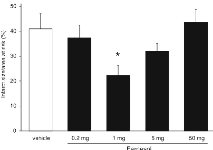
Results Farnesol pretreatment decreased infarct size in a U- shaped dose–response manner where 1 mg/kg/day dose reached a statistically significant reduction (22.3±3.9 % vs.
40.9±6.1 % of the area at risk, p<0.05). Farnesol showed a similar cytoprotection in cardiomyocytes. The cardioprotective dose of farnesol (1 mg/kg/day) significantly increased the marker of protein geranylgeranylation, but did not influence protein farnesylation, cardiac tissue cholesterol, coenzyme Q9, coenzyme Q10, and dolichol. While the cardioprotective dose of farnesol did not influence 3-nitrotyrosine, the highest dose of farnesol (50 mg/kg/day) tested did not show cardioprotection, however, it significantly decreased cardiac 3-nitrotyrosine.
Conclusions This is the first demonstration that oral farnesol treatment reduces infarct size. The cardioprotective effect of farnesol likely involves increased protein geranylgeranylation and seems to be independent of the antioxidant effect of farnesol.
Keywords Ischemia/reperfusion . Protein geranylgeranylation . Peroxynitrite . Farnesol . Mevalonate pathway
Introduction
Myocardial ischemia followed by reperfusion results in the development of tissue necrosis referred to as myocardial infarction. Myocardial infarction is one of the leading causes of morbidity and mortality globally. Therefore, the development of effective cardioprotective agents against ischemia/reperfusion injury is of great importance and re- mains a foremost experimental goal [1].
We have previously shown that farnesol restored the protective effect of preconditioning (an endogenous C. Csonka and P. Ferdinandy contributed equally to this work.
G. Szűcs
:
S. Török:
G. F. Kocsis:
J. Pálóczi:
A. Görbe:
T. Csont
:
C. Csonka (*)Cardiovascular Research Group, Department of Biochemistry, University of Szeged, 6720(9 Dóm tér, Szeged, Hungary e-mail: csonka.csaba@med.u-szeged.hu
C. Csonka
e-mail: csaba.csonka@pharmahungary.com Z. Murlasits
Department of Health and Sport Sciences, University of Memphis, 106 Fieldhouse, Memphis, TN 38152, USA
Z. Murlasits
:
T. Csont:
C. Csonka:
P. FerdinandyPharmahungary Group, 6722(6 Hajnóczy u., Szeged, Hungary P. Ferdinandy
Department of Pharmacology and Pharmacotherapy, Semmelweis University, 1089(4 Nagyvárad tér, Budapest, Hungary
adaptive mechanism against ischemia/reperfusion injury) in cholesterol fed rats [1, 2]; however, it is not known if farnesol itself is able to protect the heart against ischemia/reperfusion injury. Farnesol (3,7,11-trimethyl- 2,6,10-dodecatriene-1-ol), a 15-carbon sesquiterpenoid mol- ecule naturally occurring mainly in aromatic fruits [3] is a powerful antioxidant [3–5] and is able to modulate the mevalonate pathway [6]. Mammalian cells are capable of converting farnesol to farnesyl-pyrophosphate [6] which is a key branching point in the mevalonate pathway [7] (Fig.1) and can be further metabolized to the mevalonate pathway end-products i.e. cholesterol, coenzyme Q, and dolichol [7].
Moreover, farnesyl-pyrophosphate is a precursor for the prenylation (farnesylation and/or geranylgeranylation) of several intracellular proteins. Protein prenylation is a com- mon post-translational modification of several intracellular proteins [7,8] (Fig.1) and prerequisite for their physiolog- ical function. Examples of prenylated proteins include theγ subunit of heterotrimeric G-proteins, nuclear lamins, and some members of the Ras superfamily of small GTPases e.g.: Ras, Rho, Rac, Rab subfamily [9–11]. Although sev- eral G-proteins are involved in the signal-transduction of ischemia/reperfusion injury and cardioprotection, the role of their prenylation has not been investigated in ischemia/
reperfusion injury.
Cholesterol, a well-known risk factor for coronary heart disease and atherosclerosis, is the bulk end-product of the mevalonate pathway [7]. Nevertheless, very little is known about the role of the mevalonate pathway intermediates in the mechanism of ischemia/reperfusion injury.
Farnesyl pyrophosphate is also a precursor for the synthesis of different forms of coenzyme Q such as coenzyme Q10 that is the most common coenzyme Q in humans and Q9 that is most common in rats [12] (Fig.1). Coenzyme Q plays a major role in the mitochondrial electron-transport chain and serves as an endogenous antioxidant [12]. Coenzyme Q is protective against myocardial ischemia/reperfusion-injury in animal
studies [13,14] and it is a registered drug for adjuvant therapy of heart failure worldwide.
Dolichol, another derivative of farnesyl pyrophosphate (Fig.1), is the most prevalent polyisoprenyl glycosyl carrier in eukaryotes involved in C- [15] and O-mannosylaton of proteins, the formation of glycosylphosphatidylinositol an- chors [16] and the N-glycosylation of proteins [17]. The role of dolichol in ischemia/reperfusion is not known.
Farnesol has been shown to exert an antioxidant effect in- vivo [3–5] and to restore reduced glutathione and glutathi- one reductase and glutathione peroxidase levels [3–5]. The above findings may support the potential cardioprotective effect of farnesol, since reactive oxygen and nitrogen spe- cies e.g. peroxynitrite formed nonenzymatically from NO and superoxide, plays a pivotal role in ischemia/reperfusion injury [18,19].
Therefore, it is plausible to hypothesize that farnesol via its antioxidant effect and possible increase of coenzyme Q, pro- tein prenylation and dolichol formation may protect myocar- dium against ischemia/reperfusion injury. Accordingly, the aim of the study was to examine the cardioprotective effect of farnesol and its cellular mechanism.
Materials and Methods
This investigation conformed to the National Institutes of Health Guide for the Care and Use of Laboratory Animals (NIH Pub. No. 85–23, Revised 1996) and was approved by the local ethics committee at the University of Szeged.
Experimental Design and Isolated Heart Perfusion
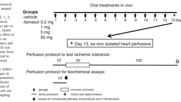
Here we assessed the effect of subchronic farnesol treatment on ischemic tolerance and biochemical changes of the heart.
Male Wistar rats (300–400 g,n=108 in the entire study) were kept under controlled temperature with 12/12 h light/dark cycles. They received a standard rodent chow and tap water ad libitum. Rats were randomly assigned to the following groups: oral administration of vehicle (2.5 % methylcellulose) or 0.2, 1, 5, and 50 mg/kg/day farnesol (SAFC Supply Solution, St Louis, MO), respectively for 12 days. The length of farnesol administration and doses were applied according to previous studies [3,20].
On day 13, the effect of farnesol on cardiac ischemic tolerance and biochemical changes were tested. Therefore, rats were anesthetized, heparin (500 U/kg iv) was adminis- tered, and hearts were isolated and perfused at 37 °C in Langendorff mode with oxygenated Krebs-Henseleit buffer with constant pressure as previously described [2, 20].
Hearts were subjected either to (i) 30 min coronary occlu- sion followed by 120 min reperfusion to measure infarct size
HMG-CoA Mevalonate
Cholesterol Isopentenyl-PP
Farnesyl-PP Geranyl-PP
Prenylated proteins (farnesylation, geranylgeranylation)
Dolichol Coenzyme Q
HMG-CoA reductase
Farnesol
Dietary cholesterol Fig. 1 The mevalonate pathway
in all groups (n= 12–14 except for the 0.2 mg/kg/day farnesol treated group where n= 8) or to (ii) 10 min of perfusion to wash out blood before tissue sampling for biochemical assays from selected groups as appropriate (seeResults). Cardiac levels of mevalonate pathway end- products (prenylated proteins, cholesterol, coenzyme Q9, Q10, and dolichol;n=13 in the vehicle and the 1 mg/kg/day farnesol treated group), and 3-nitrotyrosine (marker of oxidative/nitrosative stress, n=7 in the vehicle and the 1 and 50 mg/kg/day farnesol treated group) were measured from the tissue samples in separate experiments, respective- ly (Fig.2). Background morphological and hemodynamic parameters such as body weight, heart weight, and coronary flow before ischemia were measured from all animals served for infarct size measurement and assessment of bio- chemical parameters (Table1).
Infarct Size Determination
To assess ischemic tolerance of the heart, infarct size was measured as follows. At the end of the perfusion protocol, the coronary artery was reoccluded and 5 ml of 0.1 % Evans blue dye was injected into the aorta to delineate the area at risk zone. Stained hearts were frozen, sliced, and incubated at 37 °C in 1 % triphe- nyltetrazolium chloride to delineate infarcted tissue.
Slices were then fixed and quantified by planimetry u s i n g I n f a r c t s i z e 2 . 4 s o f t w a r e ( I n f a r c t s i z e™, Pharmahungary Group, Szeged, Hungary). Infarct size was expressed as a percentage of the area at risk. The area at risk was calculated as a percentage of the total ventricular area [20, 21].
Measurement of Myocardial Protein Geranylgeranylation and Farnesylation
The nucleophilic cleavage of the allylic thiol bond by 2- naphthol provides for quantitative determination of the cysteine-bound prenyl groups of prenylated proteins. The level of cysteine-bound farnesyl and geranylgeranyl groups were measured by HPLC method as previously described [22]. Briefly, 200 mg tissue samples were delipidated by extraction with ethanol followed by diethyl ether.
Delipidated samples were mixed with 2.5 ml reagent mix- ture (20 mg/mL potassium naphthoxide dissolved in diox- ane). Then dioxane was removed with a stream of nitrogen gas and 100μl dimethylformamide was added to the sam- ples and were heated at 100 °C for 8 h. The reaction products were extracted with 400μl n-hexane and the upper phase was injected to YL9100 HPLC system on a YMC- Pack ODS-A 250 mm×4.6 mm ID 5 μm using 95–100 % acetonitrile as mobile phase. Calibration curves were made using N-acetyl-S-farnesyl-L-cysteine and N-acetyl-S- geranylgeranyl-L-cysteine standards (Sigma, St. Louis, MO) underwent the same preparation procedure. Therefore protein farnesylation and geranylgeranylation were expressed as protein-bound farnesyl and geranylgeranyl group in ng/mg protein.
Measurement of Myocardial Cholesterol
Tissue cholesterol was measured (Cholesterol/Cholesterol Ester Quantification kit; BioVision, Mountain View, CA) from ventricular homogenates according to the manufac- turer’s instructions. Ten mg ventricular homogenates were
30’ 120’
10’
12 10 11 9
8
6 7
4 5
1 2 3
Day 13, ex-vivo isolated heart perfusions Oral treatments in vivo
13 day
*
*
Groups: -vehicle
-farnesol 0.2 mg 1 mg 5 mg 50 mg
coronary occlusion aerob perfusion infarct size determination
assays for mevalonate pathway end-products and 3-nitrotyrosine gavage
Perfusion protocol to test ischemic tolerance:
10’
Perfusion protocol for biochemical assays:
Fig. 2 Experimental protocol.
Male Wistar rats were treated with vehicle (2.5 % methylcellulose) or 0.2, 1, 5, and 50 mg/kg/day farnesol, respectively for 12 days per os by gavage. On day 13, hearts were isolated to test the effect of the different treatments on cardiac ischemic tolerance and biochemical changes. To test ischemic tolerance, hearts from all groups were subjected to 30 min coronary occlusion followed by 120 min reperfusion to measure infarct size. To measure changes in cardiac biochemical parameters from selected groups, hearts were subjected to 10 min of aerobic perfusion to wash out blood before tissue sampling
used for tissue cholesterol measurement. Myocardial cho- lesterol was expressed in ng/mg protein.
Measurement of Myocardial Coenzyme Q
The level of cardiac coenzyme Q9 and Q10 were measured by a HPLC method following lipid extraction with n-hexane as previously described [23]. Coenzyme Q9 and Q10 were detected at 275 nm using an YL 9160 PDA detector follow- ing separation with a YL9100 HPLC system on a C18 column (YMC Basic, 50 mm × 4.6 mm ID 3 μm). Four hundred mg ventricular homogenate was used for myocar- dial coenzyme Q measurement. Calibration curves were made using coenzyme Q9 and Q10 standards (Sigma, St.
Louis, MO). Myocardial coenzyme Q9 and Q10 were expressed in ng/mg protein.
Measurement of Myocardial Dolichol
The level of cardiac dolichol was measured by HPLC meth- od as previously described [24]. Briefly, minced cardiac tissues (400 mg) were mixed with 0.5 ml 0.25 % pyrogallol in methanol and 0.5 ml 60 % KOH. Hydrolysis was performed in a water bath at 100 °C for 30 min; the mixture was extracted three times with diethyl ether:petroleum ether (1:1). The pooled extracts were washed with methanol:diethyl ether (1:1) and were dried under nitrogen. The samples were resolved with isopropanol:acetonitrile (65:35). Dolichol was detected at 210 nm using an YL 9160 PDA detector following separation with an YL9100 HPLC system on C18 column (YMC-Pack ODS-AQ, 10 mm×4.0 mm ID 3μm and YMC- Pack ODS-AQ, 150 mm×4.6 mm ID 5 μm). Calibration
curve was made using C90-Dolichol standard (Larodan, Malmö, Sweden). Myocardial dolichol was expressed in ng/mg protein.
Measurement of Nitrotyrosine
Cardiac free 3-nitrotyrosine content, as markers for peroxynitrite-induced oxidative/nitrosative stress, was mea- sured after 10 min of perfusion. Cardiac free 3-nitrotyrosine level was measured by enzyme-linked immunosorbent assay (ELISA; Cayman Chemical, Ann Arbor, MI) from heart tissue samples according to the instructions. Briefly, super- natants of 50 mg ventricular tissue homogenate was incu- bated overnight with anti-nitrotyrosine rabbit IgG specific to free 3-nitrotyrosine and nitrotyrosine acetylcholinesterase tracer in precoated (mouse anti-rabbit IgG) microplates followed by development with Ellman’s reagent. Free 3- nitrotyrosine content was normalized to protein content of cardiac homogenate and expressed as ng/mg protein.
Measurement of Protein Concentration
Protein concentrations were measured by the BCA Protein Assay kit (Thermo, Rockford, IL) according to the instructions.
Primary Cardiomyocyte Culture Experiments
In order to investigate if farnesol has a direct cardiocytoprotective effect, we investigated whether farnesol protects cardiomyocytes subjected to simulated ischemia/reperfusion. Neonatal cardiomyocyte cultures were prepared as described previously Table 1 Body weight, animal weight, area at risk and cardiac func-
tional parameters in vehicle or 0.2, 1, 5, and 50 mg/kg/day farnesol- treated groups for 12 days followed by either 10 min of aerobic perfusion for tissue sampling or 30 min coronary occlusion followed by 120 min reperfusion to measure infarct size. Body weight, heart
weight, and coronary flow before ischemia were measured from all animals served for infarct size measurement and assessment of bio- chemical parameters to yield sample sizen=6–34 in different groups.
Values are mean±SEM (n)
Farnesol (mg/kg/day) Vehicle 0.2 1 5 50
Body weight (g) 350±5(34) 337±8(8) 353±5(32) 334±3(14) 361±6(20)
Heart weight (g) 1.28±0.02(34) 1.27±0.06(8) 1.30±0.03(32) 1.21±0.04(14) 1.27±0.04(20)
Area at risk (%) 54.9±3.2(10) 43.7±4.4(8) 46.2±4.6(11) 64.9±3.6(12) 50.1±2.6(13)
Coronary flow (mL/min)
Before isch 17.9±0.5(33) 17.2±1.6(8) 18.0±0.6(32) 16.6±0.7(14) 16.9±1.1(18)
During isch 10.1±1.1(11) 9.3±1.0(8) 9.8±0.5(12) 10.1±0.8(12) 9.0±0.7(13)
0–5 min of rep 88.0±6.2(11) 72.6±5.3(8) 82.8±6.6(12) 88.0±7.7(12) 75.1±4.0(13)
End of rep 12.6±1.3(11) 11.5±1.4(8) 14.4±1.1(12) 14.3±1.5(12) 11.6±0.8(13)
Heart rate (beats/min)
Before isch 284±18(14) 270±24(8) 284±17(12) 277±24(13) 303±9(13)
End of isch 250±27(7) 275±35(8) 288±28(11) 292±31(11) 308±19(7)
End of rep 288±9(6) 279±23(7) 239±28(8) 259±35(8) 294±17(8)
[25]. Cells were kept in normoxic incubator and sup- plied with proliferation medium for 1 day (DMEM + 10 % FBS). Then farnesol (0.0032–250 μM) was ad- ministered in differentiation medium (DMEM + 1 % FBS) for 2 days. After farnesol treatment, the culture medium was changed to hypoxic solution [26] and plates were placed into a hypoxic chamber, and cells were exposed to a constant flow of a mixture of 95 % N2 and 5 % CO2 at 37 °C for 4 h. Simulated ischemia was followed by 2 h simulated reperfusion using dif- ferentiation medium and normoxia (Fig. 3). To assess cell viability cardiomyocytes were incubated with 1 μM calcein acetoxymethyl ester (calcein-AM, Sigma, St Louis, MO) at room temperature for 30 min. Fluores- cence intensity was measured with a fluorescence plate reader (Fluostar Optima, BMG Labtech, Ortenberg, Germany) at 490-nm excitation and 520-nm emission filters. Simulated ischemia/reperfusion resulted in 21 % decrease in cell viability as compared to normoxia (4 h normoxia with normoxic solu- tion followed by simulated reperfusion, data not shown).
Statistics
All values are presented as mean±SEM. Differences among means were analyzed by Student’s unpairedt-test or one- way ANOVA followed by Fisher LSD post hoc test, respec- tively. All comparisons were made versus the vehicle- treated or vehicle-treated ischemia/reperfusion group. Sta- tistical significance was defined asp<0.05.
Results
To assess the cardioprotective effect of different oral doses of farnesol, infarct size was measured after 30 min regional ischemia and 120 min of reperfusion. Infarct size was sig- nificantly decreased by 1 mg/kg/day farnesol (22.3±3.9 % vs. 40.9 ±6.1 %, p< 0.05, Fig. 4). However, 0.2, 5, and 50 mg/kg/day farnesol treatment did not significantly de- crease infarct size (37.3±5.1 %, 32.0±3.1 %, 43.5±5.1 % vs. 40.9±6.1 %, respectively, Fig.4). Therefore, the infarct size reducing effect of farnesol showed a U-shaped dose–
response relationship. Farnesol did not affect body weight, heart weight, coronary flow and heart rate (Table1).
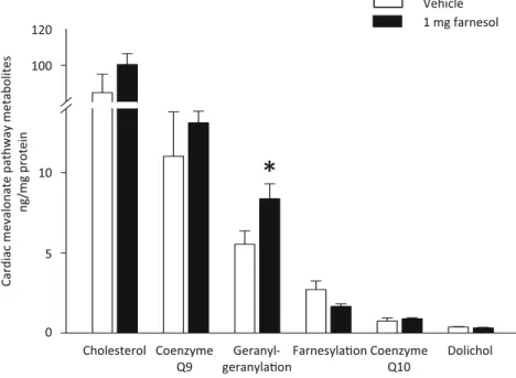
In separate sets of experiments, to assess the effect of cardioprotective (1 mg/kg/day) dose of farnesol on mevalonate pathway end-products, we measured cardiac protein prenylation, cholesterol, coenzyme Q, and dolichol.
We found that oral farnesol treatment significantly increased cardiac protein-bound geranylgeranyl level (8.4±0.9 vs. 5.5
±0.8 ng/mg protein,p<0.05) (Fig.5). However, farnesol did not influence cardiac protein-bound farnesyl level (1.6±0.2 vs. 2.7±0.5 ng/mg protein), cardiac cholesterol (100.4 ±4.5 vs. 88.8±7.8 ng/mg protein), coenzyme Q9 and Q10 (13.1±
0.7 vs. 11.0±2.8 ng/mg protein and 0.89±0.06 vs. 0.74±
0.19 ng/mg protein, respectively), and dolichol levels sig- nificantly (0.32±0.03 vs. 0.37±0.03 ng/mg protein) (Fig.5).
To assess the antioxidant effect of the cardioprotective dose of farnesol in the heart, in separate experiments, we measured cardiac 3-nitrotyrosine level as marker of cardiac peroxynitrite which is a major player in cardiac oxidative and nitrosative stress. We found that the cardioprotective
Viability Assay Simulated
Ischemia Normoxic
Differentiation
2 day 4 h 2 h
Simulated Reperfusion 1 day
Normoxic Proliferation
0- 250 µM farnesol Cardio-
myocyte isolation
Fig. 3 Experimental protocol of cardiomyocyte culture experiments.
To assess the protective effect of farnesol, neonatal rat cardiomyocytes were treated with 0.0032, 0.016, 0.08, 0.4, 2, 10, 50, and 250μM farnesol or its vehicle (0.1 % DMSO), respectively. To test ischemic
tolerance, cardiomyocytes were subjected 4 h simulated ischemia and 2 h simulated reperfusion. Cell viability was assessed by calcein viability assay
0 10 20 30 40 50
vehicle 0.2 mg 1 mg 5 mg 50 mg
Infarct size/area at risk (%)
*
Farnesol
Fig. 4 Effect of farnesol on myocardial infarct size. Infarct size is expressed as a percentage of the area at risk of isolated hearts that was subjected to 12 days vehicle or 0.2, 1, 5, and 50 mg/kg/day farnesol treatment followed by 30 min coronary occlusion followed by 120 min reperfusion to measure infarct size. Values are mean±SEM;n=8–13 in each group. *p<0.05 vs. vehicle
dose of farnesol (1 mg/kg/day) did not affect cardiac 3- nitrotyrosine level (Fig. 6). Therefore, to further assess if farnesol at a higher dose may show an antioxidant effect, in separate experiment, we measured 3-nitrotyrosine in the 50 mg/kg/day farnesol-treated group. This dose of farnesol (50 mg/kg/day) significantly decreased cardiac 3-nitrotyrosine level (1.2±0.2 vs. 2.4±0.5 ng/mg protein,p<0.05) (Fig. 5), however, it did not show any cardioprotective effect (see above, Fig.4).
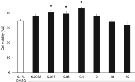
To assess if farnesol has a direct cardiocytoprotective effect, we measured cell viability of cardiomyocytes subjected to simulated ischemia/reperfusion. Farnesol (0.0032–50 μM) protected cardiomyocytes in a bell- shaped concentration-response manner similarly to that ob- served in the isolated heart experiments (0.016μM farnesol:
40.4±1.4 AU, 0.08 μM farnesol: 39.6±1.5 AU, 0.4 μM farnesol: 43.1 ± 1.0 AU vs. 34.8 ± 1.2 AU, respectively, Fig. 7). However, 250 μM farnesol showed a cytotoxic effect (data not shown).
Discussion
This is the first demonstration that oral farnesol treatment reduces infarct size in the rat heart following ischemia/
reperfusion. We also found that farnesol shows a direct cytoprotective effect in cardiomyocytes subjected to simu- lated ischemia/reperfusion. Furthermore, we found that the in-vivo cardioprotective dose of farnesol (1 mg/kg/day for 12 days) increased the geranylgeranylation of cardiac pro- teins, however, it did not affect other mevalonate pathway- derived end-products (cardiac cholesterol, coenzyme Q, dolichol) and cardiac peroxynitrite marker 3-nitrotyrosine level significantly. This shows that the cardioprotective ef- fect of farnesol is independent of its antioxidant effect but may involve changes in protein geranylgeranylation (Fig.8).
It is known that mammalian cells can utilize farnesol for protein prenylation [6] and for synthesis of other mevalonate
*
Farnesol 0
1 2 3
vehicle 1 mg 50 mg
Cardiac 3-nitrotyrosine (ng/mg protein)
Fig. 6 Cardiac 3-nitrotyrosine from rats oral treated with vehicle or 1 and 50 mg/kg/day farnesol for 12 days. Values are mean±SEM;n=7 in each group, *p<0.05 vs. vehicle
Fig. 5 Cardiac mevalonate pathway metabolites (cardiac cholesterol, coenzyme Q10, protein geranylgeranylation, protein farnesylation, co- enzyme Q9, and dolichol) from rats orally treated with vehicle or 0.2,
1, 5, and 50 mg/kg/day farnesol for 12 days. Values are mean±SEM;
n=6–8 in each group. *p<0.05 vs. vehicle
pathway derivatives (cholesterol, coenzyme Q, and dolichol) [6] (Fig. 1). Bentinger et al. have shown in rats that farnesol could be phosphorylated in vivo to form farnesyl-pyrophosphate, which could re-enter biosynthetic reactions [27]. Our present results support this finding since administration of exogenous farnesol increased total myo- cardial protein geranylgeranylation and reduced infarct size.
Prenylation (protein farnesylation and geranylgeranylation) is a lipid post-translational modification of proteins involving the irreversible covalent attachment of either farnesyl (15-car- bon) or geranylgeranyl (20-carbon) isoprenoid to conserved cysteine residues at or near the C-terminus of numerous cellular proteins [28]. Prenyl groups act as hydrophobic membrane anchors [9] and play a key role in the maturation of proteins [10], regulation of protein targeting, function of proteins, and controlling apoptosis [9, 29]. Large number of proteins is known to be prenylated, such as e.g. γ subunit of heterotrimeric G-proteins, nuclear lamins, and some members of the Ras superfamily of small GTPases e.g.: Ras, Rho, Rac, Rab subfamily [9–11]. Some of these proteins have been shown to be involved in cardioprotection. Brar et al. also showed that cardioprotective effect of urocortin-II against ischemia/reperfusion injury in rat heart was completely abolished by the Ras inhibitor manumycin A [30]. It was also shown that infarct size was increased in cardiac specific RhoA knockout mice hearts and was significantly
decreased in cardiac-specific RhoA transgenic mice [31]. It was also reported that activation of c-Jun N- terminal kinase through the Rac1/cdc42-TAK-1 pathway promotes survival of cardiac myocytes after hypoxia- reoxygenation [32]. Members of the Rab subfamily are geranylgeranylated [9] while the other small GTPases either farnesylated or geranylgeranylated [29]. In our present study, we measured total protein farnesylation and geranylgeranylation and found that farnesol treat- ment significantly increased total myocardial protein geranylgeranylation but not farnesylation. However, the reason for the discrepancy between farnesol-induced protein geranylgeranylation and farnesylation remained unknown and it was not investigated in our present study. Protein prenylation is catalyzed by prenyl transferase enzymes, which can be classified into two main functional classes: (i) the CAAX prenyl transferases including farnesyl transferase and geranylgeranyl transferase type 1, [29], and (ii) the Rab geranylgeranyl transferase (geranylgeranyl transferase type 2) [9]. CAAX prenyltransferase activities are highly selective for their isoprenoid diphosphate substrates: farnesyl transfer- ase for farnesyl-pyrophosphate and geranylgeranyl transferase type 1 for geranylgeranyl-pyrophosphate. However, farnesyl transferase can also bind geranylgeranyl-pyrophosphate with low affinity, yet the enzyme is unable to transfer the geranylgeranyl group to substrate proteins, indicating that Fig. 8 Proposed mechanism of
cardioprotection by farnesol
0 10 20 30 40
0.1%
DMSO
0.0032 0.016 0.08 0.4 2 10 50
Cell viability (AU)
* *
Farnesol (µM)
*
*
Fig. 7 Viability assay for neonatal rat cardiomyocytes treated with 0.0032, 0.016, 0.08, 0.4, 2, 10, and 50μM farnesol or its vehicle (0.1 % DMSO), respectively for 2 days. Values are mean±SEM;
n=16–24 well. *p<0.05 vs.
vehicle
geranylgeranyl-pyrophosphate is an inhibitor of farnesyl transferase [29]. This might explain our findings regard- ing the difference between farnesol-induced protein geranylgeranylation and farnesylation.
Farnesyl-pyrophosphate can be utilized for synthesis of cholesterol, coenzyme Q, and dolichol [7]. In our present study, we have observed that cardiac tissue cholesterol con- tent expressed in ng/mg protein was approximately 10-fold higher than the level of geranylgeranyl group of protein;
however, farnesol treatment did not modify cardiac choles- terol production.
In our present study, we have found that the cardiac tissue content of coenzyme Q9 was comparable to that of protein- bound geranylgeranyl group. Cardiac level of coenzyme Q10 content was approximately 10-fold less than coenzyme Q9 or protein-bound geranylgeranyl group. Our result is in concor- dance with the findings of Matejíková et al., who reported that the rat heart contains approximately 10-fold more coenzyme Q9 than coenzyme Q10 [33]. Coenzyme Q is a well-known antioxidant and cardioprotective molecule [13, 34]. In our present study, farnesol treatment failed to alter coenzyme Q levels, which suggest that farnesol-induced cardioprotection is independent of changes in coenzyme Q levels. In our experi- ment the dolichol content of cardiac tissue did not change, but it was 10-fold lower than protein-bound geranylgeranyl content.
The role of dolichol in ischemia/reperfusion is not known in the literature. However, it was shown that dolichol kinase deficiency causes congenital dilated cardiomyopathy in patients [35, 36].
Farnesol has been shown to exert antioxidant effects in vivo [3–5]. However, in our present study the cardioprotective dose of farnesol (1 mg/kg/day) failed to decrease peroxynitrite for- mation, a major player in oxidative/nitrosative stress. However, 50 mg/kg/day farnesol, which did not show any cardioprotective effect, significantly decreased 3-nitrotyrosine a marker of peroxynitrite, demonstrating an antioxidant effect of farnesol. These results show that the cardioprotective effect of farnesol is independent from its antioxidant effect.
We have found here that oral farnesol reduced infarct size in a U shaped dose-dependent manner showing the maxi- mum efficacy at 1 mg/kg/day dose. Moreover, here we also demonstrated that farnesol exerts a similar concentration- dependent direct cytoprotective effect in cardiomyocytes in vitro. These results show that the cardioprotective effect of farnesol is not dependent on any systemic effect; however, it might be based on a direct cardiocytoprotective action. The reason for the inefficiency of higher farnesol doses (5 and 50 mg/kg/day in in-vivo experiments and 2, 10, 50, 250μM in neonatal rat cardiomyocyte experiments) has not been revealed in this study. However, one may speculate that the potential pro-apoptotic effect of farnesol may interfere with its cardioprotective effect at higher doses. Indeed, Chagas et al. found in a partial hepatectomy model that
farnesol (250 mg/kg for 2 consecutive weeks) induced apoptosis in rat hepatocytes [37]. Joo et al. reported that 250 μM farnesol induced apoptosis in human lung carcinoma cell line [38]. Indeed, we have found here that 250 μM farnesol radically decreased the viability of cardiomyocytes.
The present study has clearly shown the cardioprotective effect of farnesol and revealed several aspects of its mecha- nism; however its cellular mechanism has not been fully explored. An obvious limitation of the present study is that total protein farnesylation and geranylgeranylation were measured and individual protein prenylation was not exam- ined. Further studies are necessary to identify the specific geranylgeranylated proteins, which may play a role in the cardioprotective effect of farnesol.
In conclusion, here we have demonstrated for the first time in the literature that oral farnesol treatment reduces infarct size possibly via a direct cardiocytoprotective action.
Furthermore, we have shown that the cardioprotective effect of farnesol likely involves increased protein geranylgeranylation and seems to be independent of the other end-products of mevalonate pathway and the antioxidant effect of farnesol.
Acknowledgments This work was supported by a grant from the National Innovation Office (5LET_STATIN_08, TAMOP-4.2.2-08/1/
2008-0013, TAMOP-4.2.1/B-09/1/KONV-2010-0005, TÁMOP-4.2.2/
B-10/1-2010-0012, and TAMOP-4.2.2/A-11/1/KONV-2012-0035) and a grant from the Hungarian Scientific Research Fund (OTKA PD 106001). A. Görbe and T. Csont hold a “János Bolyai Fellowship” from the Hungarian Academy of Sciences.
References
1. Ferdinandy P, Schulz R, Baxter GF. Interaction of cardiovascular risk factors with myocardial ischemia/reperfusion injury, preconditioning, and postconditioning. Pharmacol Rev.
2007;59:418–58.
2. Ferdinandy P, Csonka C, Csont T, Szilvassy Z, Dux L. Rapid pacing-induced preconditioning is recaptured by farnesol treatment in hearts of cholesterol-fed rats: role of polyprenyl derivatives and nitric oxide. Mol Cell Biochem. 1998;186:27–34.
3. Qamar W, Sultana S. Farnesol ameliorates massive inflammation, oxidative stress and lung injury induced by intratracheal instilla- tion of cigarette smoke extract in rats: an initial step in lung chemoprevention. Chem Biol Interact. 2008;176:79–87.
4. Jahangir T, Khan TH, Prasad L, Sultana S. Farnesol prevents Fe- NTA-mediated renal oxidative stress and early tumour promotion markers in rats. Hum Exp Toxicol. 2006;25:235–42.
5. Khan R, Sultana S. Farnesol attenuates 1,2-dimethylhydrazine induced oxidative stress, inflammation and apoptotic responses in the colon of Wistar rats. Chem Biol Interact. 2011;192:193–200.
6. Crick DC, Andres DA, Waechter CJ. Farnesol is utilized for protein isoprenylation and the biosynthesis of cholesterol in mam- malian cells. Biochem Biophys Res Commun. 1995;211:590–9.
7. Goldstein JL, Brown MS. Regulation of the mevalonate pathway.
Nature. 1990;343:425–30.
8. Resh MD. Trafficking and signaling by fatty-acylated and prenylated proteins. Nat Chem Biol. 2006;2:584–90.
9. Leung KF, Baron R, Seabra MC. Thematic review series: lipid posttranslational modifications. geranylgeranylation of Rab GTPases. J Lipid Res. 2006;47:467–75.
10. Reddy S, Comai L. Lamin A, farnesylation and aging. Exp Cell Res. 2012;318:1–7.
11. Takemoto M, Liao JK. Pleiotropic effects of 3-hydroxy-3- methylglutaryl coenzyme a reductase inhibitors. Arterioscler Thromb Vasc Biol. 2001;21:1712–9.
12. Aberg F, Appelkvist EL, Dallner G, Ernster L. Distribution and redox state of ubiquinones in rat and human tissues. Arch Biochem Biophys. 1992;295:230–4.
13. Greenberg S, Frishman WH. Co-enzyme Q10: a new drug for cardiovascular disease. J Clin Pharmacol. 1990;30:596–608.
14. Hano O, Thompson-Gorman SL, Zweier JL, Lakatta EG. Coen- zyme Q10 enhances cardiac functional and metabolic recovery and reduces Ca2+overload during postischemic reperfusion. Am J Physiol. 1994;266:2174–81.
15. Doucey MA, Hess D, Cacan R, Hofsteenge J. Protein C- mannosylation is enzyme-catalysed and uses dolichyl-phosphate- mannose as a precursor. Mol Biol Cell. 1998;9:291–300.
16. Takeda J, Kinoshita T. GPI-anchor biosynthesis. Trends Biochem Sci. 1995;20:367–71.
17. Kornfeld R, Kornfeld S. Assembly of asparagine-linked oligosac- charides. Annu Rev Biochem. 1985;54:631–64.
18. Ferdinandy P, Schulz R. Nitric oxide, superoxide, and peroxynitrite in myocardial ischaemia-reperfusion injury and preconditioning. Br J Pharmacol. 2003;138:532–43.
19. Yasmin W, Strynadka KD, Schulz R. Generation of peroxynitrite contributes to ischemia-reperfusion injury in isolated rat hearts.
Cardiovasc Res. 1997;33:422–32.
20. Kocsis GF, Pipis J, Fekete V, et al. Lovastatin interferes with the infarct size-limiting effect of ischemic preconditioning and postconditioning in rat hearts. Am J Physiol Heart Circ Physiol.
2008;294:2406–9.
21. Csonka C, Kupai K, Kocsis GF, et al. Measurement of myocardial infarct size in preclinical studies. J Pharmacol Toxicol Methods.
2010;61:163–70.
22. Epstein WW, Lever D, Leining LM, Bruenger E, Rilling HC.
Quantitation of prenylcysteines by a selective cleavage reaction.
Proc Natl Acad Sci U S A. 1991;88:9668–70.
23. Rousseau G, Varin F. Determination of ubiquinone-9 and 10 levels in rat tissues and blood by high-performance liquid chromatogra- phy with ultraviolet detection. J Chromatogr Sci. 1998;36:247–52.
24. Dini B, Dolfi C, Santucci V, et al. Effects of ageing and increased haemolysis on the levels of dolichol in rat spleen. Exp Gerontol.
2001;37:99–105.
25. Csont T, Gorbe A, Bereczki E, et al. Biglycan protects cardiomyocytes against hypoxia/reoxygenation injury: role of nitric oxide. J Mol Cell Cardiol. 2010;48:649–52.
26. Li X, Heinzel FR, Boengler K, Schulz R, Heusch G. Role of connexin 43 in ischemic preconditioning does not involve intercellular communication through gap junctions. J Mol Cell Cardiol. 2004;36:161–3.
27. Bentinger M, Grunler J, Peterson E, Swiezewska E, Dallner G.
Phosphorylation of farnesol in rat liver microsomes: properties of farnesol kinase and farnesyl phosphate kinase. Arch Biochem Biophys. 1998;353:191–8.
28. Ali BR, Nouvel I, Leung KF, Hume AN, Seabra MC. A novel statin-mediated“prenylation block-and-release”assay provides in- sight into the membrane targeting mechanisms of small GTPases.
Biochem Biophys Res Commun. 2010;397:34–41.
29. Lane KT, Beese LS. Thematic review series: lipid posttrans- lational modifications. Structural biology of protein farnesyltransferase and geranylgeranyltransferase type I. J Lip- id Res. 2006;47:681–99.
30. Brar BK, Jonassen AK, Egorina EM, et al. Urocortin-II and urocortin-III are cardioprotective against ischemia reperfusion in- jury: an essential endogenous cardioprotective role for corticotro- pin releasing factor receptor type 2 in the murine heart.
Endocrinology. 2004;145:24–35.
31. Xiang SY, Vanhoutte D, Del Re DP, et al. RhoA protects the mouse heart against ischemia/reperfusion injury. J Clin Invest.
2011;121:3269–76.
32. Dougherty CJ, Kubasiak LA, Frazier DP, et al. Mitochondrial signals initiate the activation of c-Jun N-terminal kinase (JNK) by hypoxia-reoxygenation. FASEB J. 2004;18:1060–70.
33. Matejikova J, Kucharska J, Pancza D, Ravingerova T. The effect of antioxidant treatment and NOS inhibition on the incidence of ischemia-induced arrhythmias in the diabetic rat heart. Physiol Res. 2008;57 Suppl 2:55–60.
34. Molyneux SL, Florkowski CM, Richards AM, Lever M, Young JM, George PM. Coenzyme Q10; an adjunctive therapy for con- gestive heart failure? N Z Med J. 2009;122:74–9.
35. Kapusta L, Zucker N, Frenckel G et al. From discrete dilated cardiomyopathy to successful cardiac transplantation in congenital disorders of glycosylation due to dolichol kinase deficiency (DK1- CDG). Heart Fail Rev. 2012;18:187-96
36. Lefeber DJ, de Brouwer AP, Morava E, et al. Autosomal reces- sive dilated cardiomyopathy due to DOLK mutations results from abnormal dystroglycan O-mannosylation. PLoS Genet.
2011;7:e1002427.
37. Chagas CE, Vieira A, Ong TP, Moreno FS. Farnesol inhibits cell proliferation and induces apoptosis after partial hepatectomy in rats. Acta Cir Bras. 2009;24:377–82.
38. Joo JH, Liao G, Collins JB, Grissom SF, Jetten AM. Farnesol- induced apoptosis in human lung carcinoma cells is coupled to the endoplasmic reticulum stress response. Cancer Res. 2007;67:
7929–36.