This is an Accepted Manuscript, which has been through the Royal Society of Chemistry peer review process and has been accepted for publication.
Accepted Manuscripts are published online shortly after
acceptance, before technical editing, formatting and proof reading.
Using this free service, authors can make their results available to the community, in citable form, before we publish the edited article. We will replace this Accepted Manuscript with the edited and formatted Advance Article as soon as it is available.
You can find more information about Accepted Manuscripts in the
author guidelines.Please note that technical editing may introduce minor changes to the text and/or graphics, which may alter content. The journal’s standard Terms & Conditions and the ethical guidelines, outlined in our author and reviewer resource centre, still apply. In no event shall the Royal Society of Chemistry be held responsible for any errors or omissions in this Accepted Manuscript or any consequences arising from the use of any information it contains.
Accepted Manuscript
NJC
New Journal of Chemistry A journal for new directions in chemistry www.rsc.org/njc
ISSN 1144-0546
PAPER Jason B. Benedict et al.
The role of atropisomers on the photo-reactivity and fatigue of diarylethene-based metal–organic frameworks
Volume 40 Number 1 January 2016 Pages 1–846
NJC
New Journal of Chemistry A journal for new directions in chemistry
This article can be cited before page numbers have been issued, to do this please use: J. Nasim, K. Witek, A. kincses, A. Y. Abdin, E. esawska, M. A. Mar, M. Gajdács, G. Spengler, W. Nitek, G. Latacz, E. Karczewska, K. Kononowicz, J. Handzlik and C. Jacob, New J. Chem., 2019, DOI: 10.1039/C9NJ00563C.
New Journal of Chemistry ARTICLE
a.Division of Bioorganic Chemistry, School of Pharmacy, Saarland University, Campus B2.1, D-66123 Saarbruecken, Saarland, Germany
b. Department of Technology and Biotechnology of Drugs, Faculty of Pharmacy, Jagiellonian University, Medical College, ul. Medyczna 9, 30-688 Cracow, Poland
c. Department of Pharmaceutical Microbiology,Faculty of Pharmacy, Jagiellonian University, Medical College, ul. Medyczna 9, 30-688 Cracow, Poland
d.Department of Medical Microbiology and Immunobiology, Faculty of Medicine, University of Szeged, Dóm tér 10, H-6720 Szeged, Hungary
e. Department of Chemistry, Institute of Biology, Pedagogical University, Podchorążych 2, 30-084 Kraków, Poland
f. Faculty of Chemistry, Jagiellonian University, ul. Gronostajowa 2, 30-387 Kraków, Poland
† Footnotes relating to the title and/or authors should appear here.
Electronic Supplementary Information (ESI) available: [details of any supplementary information available should be included here]. See DOI: 10.1039/x0xx00000x
Received 00th January 20xx, Accepted 00th January 20xx DOI: 10.1039/x0xx00000x www.rsc.org/
Pronounced activity of aromatic selenocyanates against multidrug resistant ESKAPE bacteria
Muhammad Jawad Nasima,b Karolina Witek b,c, Annamária Kincsesd, Ahmad Yaman Abdina, Ewa Żesławskae, Małgorzata Anna Marćb, Márió Gajdácsd, Gabriella Spenglerd, Wojciech NitekF, Gniewomir Lataczb, Elżbieta Karczewskac, Katarzyna Kieć-Kononowiczb, Jadwiga Handzlikb,* and Claus Jacoba,*
Selenocyanates represent an interesting class of organic selenium compounds. Due to their similarity with better known natural (iso-)thiocyanates, they promise high biological activity and may also be metabolized to other Reactive Selenium Species, such as selenols, diselenides and seleninic acids. Thirteen arylmethyl selenocyanates (1-13) have been synthesized and evaluated for potential antimicrobial, nematicidal and cytotoxic activity. The compounds exhibit pronounced antimicrobial activity against various strains of Gram-positive and Gram-negative bacteria and yeasts, including multidrug resistant strains. The results obtained so far demonstrate that these arylmethyl selenocyantes are also non-mutagenic and with limited cytotoxicity against human cells. Here, benzylselenocyanate (1) represents the most active anti-ESKAPE agent, with potent activity against multidrug resistant MRSA strains (HEMSA 5) - with a competitive MIC value of just 0.76 µg mL-1 (3.88 µM) whereas it exhibits low(er) cytotoxicity (IC50 = 31 µM) and no mutagenicity against mammalian cells. Because of this selective antimicrobial activity, aromatic selenocyanates may provide an interesting lead in the development of antimicrobial agents, particularly in the context of drug resistance.
.
Introduction
Since the discovery of the first modern antibiotics almost one hundred years ago, substances such as the penicillin have served as important and effective weapons in the prevention and treatment of a wide spectrum of infectious diseases. Nonetheless, over- and misuse of antibiotics, among various other reasons, have led to a surge of resistance in pathogenic bacteria, which has now become
one of the biggest threats and challenges facing humanity.1 The phenomenon of resistance to available antibiotics has attracted the attention of scientists for over two decades now, and has led to a considerable demand for the development of new types of antibiotics against such resistant strains of bacteria and fungi.
Fortunately, Nature itself is an affluent source of antibiotics. Natural substances acquired from medicinal and culinary plants, such as garlic and mustard, have been employed extensively as antibiotics throughout History.2-4 More recently, such secondary metabolites have become important leads in the development of new and effective medicines.5-7 These phytochemicals include alkaloids, flavonoids, terpenes, polyphenols and, particularly, organosulfur compounds (OSCs).8-11 OSCs are distributed widely in Nature, for instance as allicin in garlic, ergothioneine in mushrooms, and thiocyanates and isothiocyanates in cruciferous vegetables, to name just a few (Figure 1).12-14 Isothiocyanates and thiocyanates are reactive, electrophilic substances belonging to the class of natural Reactive Sulfur Species (RSS) and are cherished for their ability to randomly modify cysteine proteins and enzymes of the cellular thiolstat.15 Such a wider “oxidative onslaught” in microorganisms frequently results in pronounced toxicity, even in otherwise resistant organisms, and RSS are often seen as promising candidates in the battle against resistant bacteria and fungi.14-18
3
4 5 6 7 8 9 10 11 12 13 14 15 16 17 18 19 20 21 22 23 24 25 26 27 28 29 30 31 32 33 34 35 36 37 38 39 40 41 42 43 44 45 46 47 48 49 50 51 52 53 54 55 56 57 58 59 60
New Journal of Chemistry Accepted Manuscript
Published on 14 March 2019. Downloaded by Uppsala University on 3/17/2019 8:11:19 AM.
View Article Online DOI: 10.1039/C9NJ00563C
Compared to RSS, Reactive Selenium Species (RSeS) are considerably less common in Biology. Despite the overarching importance of this trace element in humans, we actually find only a handful of selenium secondary metabolites in higher organisms, such as the ergothioneine-analogue selenoneine in blue tuna, whereas most selenium in higher organisms is incorporated in the amino acids selenocysteine and selenomethioneine and forms part of a range of selenoproteins and selenoenzymes.19-21
The lack of a wider spectrum of selenium containing secondary metabolites is rather surprising and somewhat disappointing. A remarkable upsurge in activity and reactivity is often observed for the selenium analogues of organosulfur compounds, and “going for selenium” in form of selenium analogues of naturally occurring RSS is quite attractive from the perspective of drug design, especially at a time when certain selenium-containing organic compounds are receiving renewed attention. Selenazole compounds, such as ebselen (2-Phenyl-1,2-benzoselenazol-3-one), not only mimic the activity of the selenium enzyme glutathione peroxidase, they have already entered clinical trials in the context of mania and hypomania.22-25 Moreover, naturally occurring (iso-)thiocyanates, such as allyl isocyanate in mustard oil, and selenium analogues thereof, e.g. primarily (iso-)selenocyanates, represent a particularly promising class of compounds. Unfortunately, the most obvious analogues of such natural antibiotics are either not stable chemically or extraordinarily difficult to synthesise and to handle because of intense odour and inherent toxicity.26, 27 This is particularly the case for the respective, chemically simple isoselenocyanates, whilst replacement of sulfur with selenium in aromatic isothiocyanate has been reported to enhance the reactivity of compounds towards thiols in proteins.28 Additionally, isoselenocynates demonstrate a higher reactivity towards GSH,
target several cellular proteins, possess a greater ability to modulate the redox cycle in the cells and also induce elevated levels of apoptosis as compared to isothiocyanates.28 Since isoselenocyanates have been explored already for biological applications, for instance against several kinds of cancers (e.g. lung, colon, liver, prostate and breast) and infective diseases (e.g.
malaria, tuberculosis and leishmaniasis) we have shifted our focus to aromatic selenocyanates, which have received less attention and also promise greatly improved physico-chemical properties.29-32 These compounds are intriguing as they are not only active on their own, they are also often metabolized to a range of other RSeS, such as selenols, diselenides and seleninic acids.33-37 Once again, such organoselenium compounds are not “exotic” and - either directly or as metabolites - play significant roles in Biology.38 Although aromatic selenocyanates so far have mostly been studied when attached to some other “bioactive” scaffolds which may have influenced or even dominated their biological activity, they have also been reported to exhibit leishmanicidal activity. 35, 39, 40
Moreover, the dietary intake of benzyl selenocyanate has been reported to inhibit the incidence of small intestinal and colon adenocarcinoma.41 Since hardly any wider investigations of such - chemically quite stable - benzylselenocyanates are found in the more biological literature, we have turned our attention to this class of compounds first. Here we report the synthesis of aromatic selenocyanate compounds, including four novel compounds (2, 8, 9 and 13), a pronounced and even somewhat selective antimicrobial activity against pathogenic and resistant microorganisms and a set of relevant pharmacological parameters which indicate a low(er) risk to human cells.
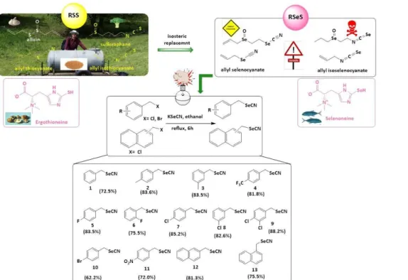
Figure 1. An “isosteric replacement” of sulfur for selenium is found in Nature where it leads from Reactive Sulfur Species (RSS), such as ergothioneine, to Reactive Selenium Species (RSeS), such as selenoneine. In pharmaceutical research, this strategy encompasses a range of (iso-)selenocyanates based on naturally occurring allyl(iso-)thiocyanates. Notably, the direct analogues of allyl(iso-)thiocyanates are rather unpleasant substances. The aromatic counterparts (1-13), shown here with their synthesis and respective percentage yields, are more promising candidates in the context of modern drug development.
2 3 4 5 6 7 8 9 10 11 12 13 14 15 16 17 18 19 20 21 22 23 24 25 26 27 28 29 30 31 32 33 34 35 36 37 38 39 40 41 42 43 44 45 46 47 48 49 50 51 52 53 54 55 56 57 58 59
New Journal of Chemistry Accepted Manuscript
Published on 14 March 2019. Downloaded by Uppsala University on 3/17/2019 8:11:19 AM.
View Article Online DOI: 10.1039/C9NJ00563C
Results and discussion
Chemical Synthesis. Thirteen arylmethyl selenocyanates (1-13, Figure 1) were designed and synthesized based on basic pharmacokinetic and pharmacodynamic considerations.42-44 The compounds were produced employing appropriate commercially available arylmethyl halides and potassium selenocyanate (KSeCN), according to the general procedure described by Wheeler and Merriam, and with minor modifications (Figure 1).45 All of the compounds - of which four are novel - were obtained in good yields (62–88 %) and structures of compounds 1-13 were confirmed by 1H and 13C-NMR. Molecular mass and purity were determined by LC- MS. (see Experimental Section and Electronic Supplementary Information (ESI)).
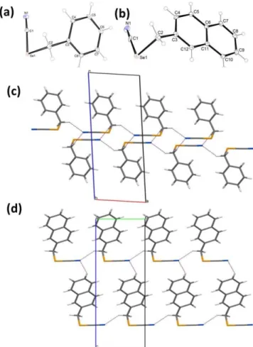
Crystallographic Studies. In order to provide a deeper insight into the structural properties, two selected compounds (1 and 12) were studied by X-ray crystallographic analysis (Figure 2 and Table S1 in ESI). In both crystal structures, the unit cells consisted of four molecules. The values of bond lengths formed by the selenium atom for C(sp)-Se were 1.850 Å and 1.837 Å for compound 1 and 12, respectively, whereas for C(sp3)-Se the bond length was 1.991 Å for both structures. Similar values were observed in other crystal structures with selenium atoms. The search of the CSD demonstrated that the geometry of the methyleneselenocyanate moiety in 1 and 12 is not exceptional among structures containing this fragment. The crystal structure of 1 has been determined earlier lacking hydrogen atoms.46
Figure 2. The molecular structure of (a) 1 and (b) 12, with the appropriate atomic numbering scheme. Displacement ellipsoids are
drawn at 30 % probability level. Partial packing views, indicating the intermolecular interactions in a layer of (c) 1 and (d) 12. The intermolecular interactions are depicted as dashed lines.
Antimicrobial Activity. Once available, compounds 1-13 were pre- screened for potential antimicrobial activity against selected Gram- positive and Gram-negative bacteria, fungi and multicellular nematodes. The antimicrobial activity was evaluated in terms of Minimum Inhibitory Concentrations (MICs) and values were compared to standard reference antimicrobial agents (Table 1).47-52 Overall, a significant and on occasion astonishingly selective antimicrobial activity against highly pathogenic organisms has been observed, frequently overcoming the kind of multidrug resistance traditional antibiotics are faced with. The impact on cultured human cells was considerably more modest, pointing towards a possible selectivity against some - particularly nasty - pathogens.
Indeed, most of compounds displayed significant activities against both, Gram-positive and Gram-negative members of a most obnoxious family of bacteria from the perspective of resistance, i.e.
ESKAPE pathogens, which comprise of Enterococcus faecium, Staphylococcus aureus, Klebsiella pneumoniae, Acinetobacter baumannii, Pseudomonas aeruginosa, and Enterobacter species.53 In the case of Gram-positive bacteria, the compounds were examined against the reference strain (ATCC 25923) and the multidrug resistant (MDR) clinical isolate of Staphylococcus aureus (S. aureus, HEMSA 5). It is noticeable that all compounds, except compound 11, displayed MIC values against the MDR strain which were considerably lower than the ones for the reference antibiotic oxacillin.47, 48 In the case of the most active compound, i.e. benzyl selenocyanate (1) an excellent antimicrobial activity was observed against HEMSA 5. Amazingly, the potency of this compound against the resistant strain (MIC 0.76 µg mL-1 or 3.88 µM) was almost one hundred fold higher than the reference drug oxacillin (MIC 128 µg mL-1 or 318.86 µM) (Table 1).
Other compounds (2, 3, 6, 8, 9, 12 and 13) also demonstrated anti- Staphylococcal activity with MIC values below 10 µg mL-1(62.5 µM).
Once more, these selenocyanates did not discriminate between reference and resistant MDR strains of S. aureus, displaying similar antibacterial potency against both, with an activity comparable or even slightly higher against the MDR strain (12). Hence, selenocyanates appear to overcome bacterial MDR, most likely by circumventing the components responsible for resistance. Still, the exact biochemical causes for this “resistance busting activity” are unclear, and, as mentioned earlier, may also involve reactive metabolic products of selenocyanates, such as selenols, diselenides and seleninic acids, which together with the original selenocyanate may interfere with microbial targets to overcome resistance.
Notably, most of these compounds were considerably less active against the harmless Staphylococcus strain S. carnosus, with an almost twenty to thirty-fold selectivity for HEMSA 5 over S.
carnosus observed for compounds 12 and 1, respectively. Except for compounds 3, 6 and 13 which inhibited the growth of S. carnosus at the concentrations of 13.19, 6.69 and 15.39 µg mL-1 (62.5µM, 31.25 µM and 62.5µM), respectively, no significant activity against this reference strain could be found.
3 4 5 6 7 8 9 10 11 12 13 14 15 16 17 18 19 20 21 22 23 24 25 26 27 28 29 30 31 32 33 34 35 36 37 38 39 40 41 42 43 44 45 46 47 48 49 50 51 52 53 54 55 56 57 58 59 60
New Journal of Chemistry Accepted Manuscript
Published on 14 March 2019. Downloaded by Uppsala University on 3/17/2019 8:11:19 AM.
View Article Online DOI: 10.1039/C9NJ00563C
The antibacterial activity of the selenocyanates was not limited to Gram-positive bacteria. Compound 1 also demonstrated excellent antimicrobial activity against pathogenic Acinetobacter baumannii (A. baumannii) and Pseudomonas aeruginosa (P. aeruginosa). Once again, compound 1, with a MIC of 1.53 µg mL-1( 7.8 µM) against A.
baumannii and 6.12 µg mL-1 (31.25 µM) against P. aeruginosa, was more potent against these Gram-negative pathogens when compared to the respective reference drug, in this case piperacillin with a MIC of 16 µg mL-1 (30.92 µM) against P. aeruginosa.49, 50 Compounds 2, 3, 6, 9, 12 and 13 also demonstrated a significant, albeit slightly lower activity against A. baumannii (MIC values 6.57- 15.44 µg mL-1, i.e. 31.25-62.5 µM). Although the compounds exhibit selectivity for both Gram-positive and Gram-negative bacteria, the selectivity of compounds against pathogenic strains of Gram- negative bacteria is particularly stimulating from a pharmaceutical perspective, as Gram-negative bacteria are generally more difficult to target, in part due to the specific structure and limited permeability of their cell wall.54, 55
When evaluated against pathogenic yeast Candida albicans (C.
albicans), compounds 1, 3, 5, 7 and 11 revealed an encouraging growth inhibitory activity at concentrations below 20 µg mL-1 (62.5 µM). Compounds 5, 7 and 11 prevailed with MIC values even below 10 µg mL-1 (31.25 µM). The fungistatic effect, however, is lower compared to clinically relevant antifungal drugs, such as fluconazole and itraconazole ((MIC values 0.09-4 µg mL-1 (0.29- 13.06 µM) and 0.03-2 µg mL-1 (0.04-2.83 µM), respectively) (Table 1).46, 47 When compared to a non-pathogenic yeast, the fungicidal activity of compound 7 exhibited a high level of selectivity towards C. albicans, where it was more than 60-fold more active (MIC of 7.4 µg mL-1 (31.25 µM)) compared to baker’s yeast Saccharomyces cerevisiae (S. cerevisiae)(MIC 460.8 µg mL-1 (2 mM)). Compounds 5 and 11 exhibited similar activities, although their selectivity declined noticeably when compared to compound 7. Nonetheless, these two compounds still maintained remarkably low MIC values against the pathogenic yeast (6.69 µg mL-1 (31.25 µM) and 7.53 µg mL-1 (31.25 µM), respectively) compared to non-pathogenic S.
cerevisiae (MIC 53.52 and 60.25 µg mL-1 (31.25-250 µM), respectively). Compound 1, which was extraordinarily active against
bacteria, also possessed some activity against unicellular fungi. It exhibited slightly higher fungicidal activity against pathogenic C.
albicans (MIC 12.24 µg mL-1 (62.5µM)) compared to S. cerevisiae (MIC 24.48 µg mL-1 (62.5-125 µM)). Such outcomes are promising in terms of utilizing these compounds as antifungal agents, for instance in the treatment of fungal infections, such as cutaneous, oropharyngeal and vulvovaginal candidiasis etc.56-58
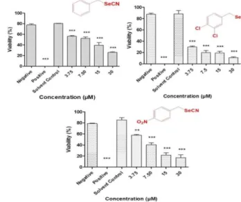
Nematicidal Activity. Although often ignored in developed countries, multicellular microorganisms, such as parasitic nematodes, still represent important targets in drug design which are particularly difficult to attack. In order to extend the scope of our preliminary studies, the series of aromatic selenocyanates (1- 13) was evaluated for nematicidal activity against the agricultural nematode S. feltiae, which often serves as a simple and reliably representative model of a multicellular organism (Figure 3).
Figure 3. Concentration-dependent activities of the most active selenocyanates (from left 1, 9 and 11) against S. feltiae. PBS buffer and ethanol (70 % v/v) and were employed as negative and positive controls, respectively. Values represent mean ± S.D. *** p < 0.001 and ** p < 0.01.
Compounds
Table 1. Antimicrobial activity of arylmethyl selenocyanates against selected bacteria from non-ESKAPE and ESKAPE families of bacteria and yeasts (1-13).
MICs (µg mL-1) S. carnosus S. aureus
ATCC25923
MRSA * HEMSA 5
A. baumannii 4184/2/5
P. aeruginosa
4600 C. albicans S. cerevisiae
1 24.48 ≤0.76 0.76 1.53 6.12 12.24 24.48
2 26.27 6.57 6.57 6.57–13.14 52.54 52.54 52.54
3 13.19 6.59 6.59 13.19 26.37 13.19 6.59
4 264.08 16.51 33.02 33.01 33.01 264.08 66.04
5 53.52 ≥26.76 >26.76 26.76 26.76 6.69 53.52
6 6.69 6.69 6.69 13.38 107.05–210.10 6.69 6.69
7 230.4 14.4–28.8 28.8 14.4–28.8 28.8 7.4 460.8
8 115.28 7.2–14.4 7.2–14.4 7.2–14.4 115.28 28.82 28.82
9 33.13 8.28 8.28 8.28–16.56 66.25 33.13 66.25
10 275.04 34.38 >34.38 34.38 34.38 275.04 68.76
11 30.125 482 482 >482 >482 7.53 60.25
12 123.09 15.44 7.72 15.44 246.17 61.55 123.09
13 15.39 7.69 7.69 15.39 246.16 30.78 61.56
Ref. 0.03 a 0.45 b 128 b <4 b ≤16 c 0.09–4 d 0.03–2 e
* MRSA; methicillin-resistant S. aureus; MIC values for reference antibiotics: a ampicillin, b oxacillin, c piperacillin d fluconazole and e itraconazole.48-53. Bold denotes significant values.
2 3 4 5 6 7 8 9 10 11 12 13 14 15 16 17 18 19 20 21 22 23 24 25 26 27 28 29 30 31 32 33 34 35 36 37 38 39 40 41 42 43 44 45 46 47 48 49 50 51 52 53 54 55 56 57 58 59
New Journal of Chemistry Accepted Manuscript
Published on 14 March 2019. Downloaded by Uppsala University on 3/17/2019 8:11:19 AM.
View Article Online DOI: 10.1039/C9NJ00563C
Figure 4. Different targets affected by a typical aromatic selenocyanate and relevant concentrations required to affect for these targets.
One may note the distinct differences in concentrations affecting resistant and non-resistant pathogens, normal and cancer cell lines (see text for details).
After a pre-screen to determine the concentration range relevant for this organism, compounds were evaluated at four different concentrations, i.e. at 3.75, 7.5, 15 and 30 µM. A remarkable, concentration-dependent decrease in the viability of the nematodes was observed for all compounds, which was pronounced even at the lowest concentrations employed.
Compounds 9 and 11 exhibited the most significant nematicidal activity and decreased the viability of nematodes to less than 40 % at the concentration of 3.75 and 15 µM. These compounds exhibited LD50 values of 0.28 µM and 4.90 µM against S. feltiae, respectively. Compound 1, which has shown considerable antibacterial and antifungal activity (see above), also exhibited significant nematicidal activity (LD50 = 6.85 µM) and decreased the viability of nematodes to less than 40 % at the concentration of 15 µM. Other selenocyanates were less active.
Although practical applications are more speculative at this time, these results indicate that the aromatic selenocyanates may also serve as excellent nematicidal agents, possibly against pathogenic nematodes affecting plants, animals and humans - clearly a matter of further investigation.
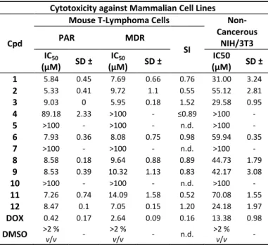
Cytotoxicity of Arylmethyl Selenocyanates against Mammalian Cells. In medicine, selenocyanates have been associated frequently with outright toxicity. In order to rule out any major cytotoxicity against mammalian cells and to investigate if there may be any selectivity against microorganisms, compounds 1-12 were investigated for their activity towards two cancer cell lines of mouse T-lymphoma, i.e., the sensitive (PAR) and the multidrug resistant cell line (MDR) transfected with the human MDR1 (ABCB1) gene which codes for the ABC transporter, and a normal NIH/3T3 mouse embryonic fibroblast cell line as control.
Compounds 1–12 exhibited some cytotoxicity against the non- cancerous NIH/3T3 mouse fibroblast cell line at concentrations ranging from 24 to above 100 µM, which was around two to five- fold lower when compared to the anticancer reference drug, doxorubicin and also significantly lower when pitched against the activity of these selenocyanates affecting various microorganisms (Table 2). Compounds 4, 5, 7 and 10 were the selenocyanates with the lowest cytotoxicity against both T-lymphoma cell lines, compared to doxorubicin. Intriguingly, compounds 1, 2, 3, 6, 8, 9,
11 and 12 were more cytotoxic - and also selective. They displayed significant cytotoxic activities against the parental and multidrug- resistant sublines of mouse T-lymphoma cells and were less cytotoxic against the non-cancerous NIH/3T3 cell line, pointing towards a slight, 3 to 10-fold selectivity against the cancer cells.
It is also notable, that the concentrations required for cytotoxicity in non-cancerous NIH/3T3 cells are generally two to three-fold lower compared to the concentrations required for antimicrobial activity (see below).
Table 2. Cytotoxicity of arylmethyl selenocyanates against non- cancerous and cancer cells.
Cytotoxicity against Mammalian Cell Lines
Cpd
Mouse T-Lymphoma Cells Non- Cancerous
NIH/3T3
PAR MDR
IC50 SI
(µM) SD ± IC50
(µM) SD ± IC50
(µM) SD ±
1 5.84 0.45 7.69 0.66 0.76 31.00 3.24
2 5.33 0.41 9.72 1.1 0.55 55.12 2.81
3 9.03 0 5.95 0.18 1.52 29.58 0.95
4 89.18 2.33 >100 - ≤0.89 >100 -
5 >100 - >100 - n.d. >100 -
6 7.93 0.36 8.08 0.75 0.98 59.94 0.35
7 >100 - >100 - n.d. >100 -
8 8.58 0.18 9.64 0.88 0.89 44.73 1.79
9 8.53 0.39 10.32 1.13 0.83 42.17 3.08
10 >100 - >100 - n.d. >100 -
11 7.26 0.74 14.09 1.58 0.52 70.08 1.55
12 8.47 0.1 7.05 0.15 1.20 24.18 1.97
DOX 0.42 0.17 2.64 0.09 0.16 13.38 0.98
DMSO >2 % v/v - >2 % v/v - n.d. >2 % v/v - PAR: parental T-lymphoma cells; MDR: multidrug resistant T- lymphoma cells overproducing efflux pump ABCB1; NIH/3T3: non- cancerous mouse embryonic fibroblast cells; DOX: doxorubicin;
DMSO: dimethyl sulfoxide; SD: standard deviation; SI: selectivity index; n.d.: not determined.
The substantial activity of compounds 1, 3 and 12 against MDR cells is worth noticing, despite the lack of any selectivity for particular cell lines. Aromatic selenocyanates 1-3, 6, 8, 9, 11 and 12 may not
3
4 5 6 7 8 9 10 11 12 13 14 15 16 17 18 19 20 21 22 23 24 25 26 27 28 29 30 31 32 33 34 35 36 37 38 39 40 41 42 43 44 45 46 47 48 49 50 51 52 53 54 55 56 57 58 59 60
New Journal of Chemistry Accepted Manuscript
Published on 14 March 2019. Downloaded by Uppsala University on 3/17/2019 8:11:19 AM.
View Article Online DOI: 10.1039/C9NJ00563C
only be of interest in the context of antimicrobial action - as anticipated initially - they may also represent a starting point in the search for anticancer agents with MDR-reversing properties. Such anti-cancer activity is a very complex issue and requires further investigations.
Pharmacological parameters associated with activity
Since the series of selenocyanates under investigation demonstrated pronounced antimicrobial and nematicidal activities, it was considered important to investigate their “drug-likeness”
profile. Four compounds (1, 2, 4 and 13) were selected for further investigations employing in vitro assays indicative of safety and absorption properties. The mutagenic potential and membrane permeability of compounds were evaluated employing a modified Ames fluctuation assay and a Parallel Artificial Membrane Permeability Assay (PAMPA) (see ESI for details).53, 59-62
Ames fluctuation assay. Since inorganic sodium selenite (Na2SeO3) is reputed for its toxic and mutagenic actions, the selenocyanates were evaluated for potential mutagenic potential. The Ames fluctuation assay was employed to calculate the mutagenic index (MI) and binomial B-values according to the method described in the literature.53 Compounds are generally considered mutagenic if the MI is above 2.0 and B is equal to or above 0.99.53, 59, 60 Neither the selenocyanates (1, 2, 4 and 13), nor the reference selenium compound ebselen displayed any mutagenic potential at concentrations of 1 µM and 10 µM (details in ESI), therefore ruling out any major mutagenic potential. Compound 1 exhibited an MI value of 1.15 (B= 0.74) and 1.20 (B= 0.81) at 1 µM and 10 µM concentrations, respectively.
In Vitro PAMPA Permeability. The PAMPA permeability screening test imitates the structural and biological conditions of the cell membrane and allows for a rapid and simple determination of the passive transport of a compound through biological membranes,
characterized by a permeability coefficient (Kp). Since some of the aromatic selenocyanates seem to enter cells readily and also appear to circumvent resistance based on efflux transporters, their transport properties were investigated employing a pre-coated PAMPA Plate System Gentest™ (Corning, Tewksbury, MA, USA), which provides good predictability and correlation of data for absorption in the human Caco-2 cell line. The concentrations of the compounds tested in the donor and acceptor compartments were estimated by capillary electrophoresis (CE) as described previously.60-62 The data on permeability obtained was compared to the one for selected reference drugs, i.e., highly permeable caffeine and lowly permeable norfloxacin (see ESI for details).
Notably, all compounds investigated, i.e. 1, 2, 4 and 13, exhibited good permeability with Kp values above the threshold for highly permeable compounds (i.e. > 1.5 × 10⁻⁶ cm s-1).62 In this context, the highest permeability was observed for compound 2 (Kp = 3.17 × 10⁻⁶ cm s-1) and compound 1 (Kp = 2.69 × 10⁻⁶ cm s-1) which are comparable to the permeability of the reference drug caffeine (kP
=3.61 × 10⁻⁶ cm s-1).
Whilst permeability may explain the ability of the selenocyanates investigated to enter - and to remain inside - target cells, the mode(s) of action underlying the biological activities associated with these compounds are still elusive. Here, one may speculate that selenocyanates may act as strong electrophiles, hence widely modifying cysteine thiols and possibly also amine groups in proteins and enzymes (Figure 5).63 Such redox modulating interactions with proteins and enzymes of the cellular thiolstat may also explain, in part, some of the selectivity observed for certain bacteria and cell lines. Other activities, possibly exerted by the various metabolic and breakdown products of the selenocyanates, may also be possible and clearly require further investigation.
Figure 5. Selenocyanates and their metabolites exhibit a wide spectrum of redox activity, which includes electrophilic attacks, oxidative modifications of thiol and selenol functions in proteins and enzymes, catalysis metal binding. The considerable impact on the cellular redox homeostasis in general, and on the cellular thiolstat, in particular, may explain the pronounced biological activities observed as part of this study.
2 3 4 5 6 7 8 9 10 11 12 13 14 15 16 17 18 19 20 21 22 23 24 25 26 27 28 29 30 31 32 33 34 35 36 37 38 39 40 41 42 43 44 45 46 47 48 49 50 51 52 53 54 55 56 57 58 59
New Journal of Chemistry Accepted Manuscript
Published on 14 March 2019. Downloaded by Uppsala University on 3/17/2019 8:11:19 AM.
View Article Online DOI: 10.1039/C9NJ00563C
Experimental
Chemical Synthesis. 1H NMR and 13C NMR spectra were recorded on a Varian Mercury-VX 300 MHz PFG instrument in DMSO-d6 at ambient temperature employing the solvent signal as an internal standard. The values of the chemical shifts are expressed in δ values and the coupling constants (J) in Hz.
Mass spectra were recorded on a UPLC–MS/MS system consisting of a Waters ACQUITY® UPLC® (Waters Corporation, Milford, MA, USA) coupled to a Waters TQD mass spectrometer (electrospray ionization mode ESI-tandem quadrupole). The purity of final products was confirmed by UPLC/MS to be higher than 95 %. Retention times (tR) are provided in min. Thin-layer chromatography (TLC) was performed on pre-coated Merck silica gel 60 F254 aluminium sheets.
General Procedure for the Synthesis of Selenocyanates.
Selenocyanates were synthesized following the general protocol described by Wheeler and Merriam with some modifications.45 Alkyl halides (10-20 mmol) were treated with KSeCN (12-25 mmol) in ethanol (10-20 mL). The reaction mixture was refluxed for 6 h and progress of the reaction was monitored periodically by TLC. After completion, the inorganic salt was separated by filtration and the filtrate was purified with charcoal, condensed and crystallized with ethanol, to yield crystals of arylmethylselenocyanates (1-13).
Compounds 1, 3-7 and 10-12 have been described in the literature before and analytical data for these compounds is in agreement with the values reported (see ESI).40, 64, 65
4-Methylbenzyl Selenocyanate (2). 4-Methylbenzyl chloride (1.4 g, 10 mmol), KSeCN (1.73 g, 12 mmol) and ethanol (10 mL) were employed. Compound 2 was obtained as light crystals.
Yield 83.6 % (1.76 g, 8.36 mmol); m.p.= 43-45 °C, TLC Rf (DCM, 100 %): 0.51. 1H NMR (DMSO-d6, ppm): δ 7.23 (d, J = 7.62 Hz, 2H, 2 C-H), 7.17 (d, J = 8.21 Hz, 2H, 2 C-H), 4.27 (s, 2H, CH2), 2.28 (t, J = 9.10 Hz, 3H, CH3). 13C NMR (DMSO-d6, ppm): δ 137.62,135.65, 129.61,129.22,105.39 (Se-CN), 33.08, 21.22.
LC–MS: purity 100 %, tR = 6.25, (ESI) m/z: calculated for C9H9NSe [M + H]+: 105.07, found: 105.02.
3-Chlorobenzyl Selenocyanate (8). 3-Chlorobenzyl chloride (3.22 g, 20 mmol), KSeCN (3.60 g. 25 mmol) and ethanol (20 mL) were employed. Compound 8 was obtained as light crystals. Yield 82.55 % (3.81 g, 16.51 mmol); m.p.= 40-42 °C, TLC Rf (DCM, 100 %): 0.75. 1H NMR (DMSO-d6, ppm): δ 7.42(m, 1H, CH), 7.37 (m, 2H, CH), 7.33 (m, 1H, CH), 4.28 (t, J = 9.15 Hz, 2H, CH2). 13C NMR (DMSO-d6, ppm): δ 141.45, 133.38, 130.95, 129.04, 128.17, 128.00, 105.23 (Se-CN), 31.96. LC–MS: purity 98.95 %, tR = 6.24, (ESI) m/z: calculated for C8H6ClNSe [M + H]+: 125.02, found: 125.02.
3,4-Dichlorobenzyl Selenocyanate (9). 3,4-Dichlorobenzyl chloride (3.91 g, 20 mmol), KSeCN (3.6 g. 25 mmol) and ethanol (20 mL) were employed. Compound 9 was obtained as yellow crystals. Yield 88.2 % (4.673 g, 17.64 mmol); m.p.= 76- 79 °C, TLC Rf (DCM, 100 %): 0.60. 1H NMR (DMSO-d6, ppm): δ 7.62 (m, 2H, CH), 7.37 (dd, J1 = 2.12 Hz, J2 = 2.09 Hz, 1H, CH), 4.28 (t, J = 9.15 Hz, 2H, CH2). 13C NMR (DMSO-d6, ppm): δ
140.24, 131.29 (4 C), 129.67, 105.19 (Se-CN), 31.23. LC–MS:
purity 99.49 %, tR = 6.86, (ESI) m/z: calculated for C8H5Cl2NSe [M + H]+: 158.98, found: 158.97.
1-(Selenocyanatomethyl)naphthalene (13). 1-Chloromethyl naphthalene (3.533 g, 20 mmol), KSeCN (3.6 g. 25 mmol) and ethanol (20 mL) were employed. Compound 13 was obtained as yellow crystals. Yield 75.5 % (3.71 g, 15.1 mmol); m.p.= 92- 94 °C, TLC Rf (DCM, 100 %): 0.64. 1H NMR (DMSO-d6, ppm): δ 8.27 (d, J = 8.21 Hz, 1H, CH), 7.96 (dd, J1 = 7.03 Hz, J2 = 7.03 Hz, 2H, CH), 7.49 (m, 4H, CH), 4.78 (t, J = 9.38 Hz, 2H, CH2). 13C NMR (DMSO-d6, ppm): δ 134.02, 130.87, 129.21, 128.61, 126.77, 125.73, 124.54, 105.33 (Se-CN), 31.09. LC–MS: purity 94.69 %, tR = 6.59, (ESI) m/z: calculated for C12H9NSe [M + H]+: 141.07, found: 141.03.
X-Ray Crystallography. Single crystals suitable for X-ray analysis were obtained in ethanol for 1 and in butan-2-ol for 12 by slow evaporation of the solvent at room temperature.
Intensity data of 1 was collected on the Bruker-Nonius Kappa CCD four circle diffractometer, whereas data for 12 was collected on an Oxford Diffraction SuperNova Diffractometer equipped with a Mo (0.71069 Å) Kα radiation source. The positions of non-hydrogen atoms were determined by the direct method employing the SIR-2014 programme.66 Hydrogen atoms bonded to carbons atoms were included at idealized positions and were refined utilising a riding model.
The aryl hydrogen atoms were constrained with C-H 0.93 Å, the methylene groups with C-H 0.97 Å and Uiso(H) = 1.2 Ueq.
The final refinements were performed by the SHELXL programme. ORTEP and MERCURY programmes were employed for molecular graphics.67-69
Compound 1: C8H7NSe, Mr = 196.11, crystal size 0.08 × 0.16 × 0.46 mm3, monoclinic, space group P21/c, a = 5.9880(1) Å, b = 7.4440(2) Å, c = 17.4880(5) Å, β = 96.277(2)°, V = 774.85 Å3, Z
= 4, T = 100(2) K, 6844 reflections collected, 1786 unique reflections [RINT = 0.0326], R1 = 0.0226, wR2 = 0.0521 [I >
2σ(I)], R1 = 0.0226, wR2 = 0.0536 [all data].
Compound 12: C12H9NSe, Mr = 246.16, crystal size 0.25 × 0.48
× 0.60 mm3, monoclinic, space group Ia, a = 8.2486(2) Å, b = 5.9838(1) Å, c = 20.4158(7) Å, β = 93.097(3)°, V = 1006.23 Å3, Z
= 4, T = 130(2) K, 4415 reflections collected, 2048 unique reflections [RINT = 0.0300], R1 = 0.0368, wR2 = 0.0921 [I >
2σ(I)], R1 = 0.0397, wR2 = 0.0947 [all data].
CCDC 1819893-1819894 contains the Supplementary Crystallographic Data for this manuscript. This data can be obtained free of charge from The Cambridge Crystallographic Data Centre via www.ccdc.cam.ac.uk/data_request/cif.
Antimicrobial Activity. The minimal inhibitory concentrations (MICs) were determined by the standard microdilution method in cation-adjusted Mueller-Hinton II Broth (MHB, Becton- Dickinson, Germany) according to the recommendations of the Clinical and Laboratory Standard Institute (CLSI).70 The compounds (1-13) were evaluated for their antimicrobial activity against a broad spectrum of microorganisms, including Gram-positive bacteria (S. carnosus and S. aureus), Gram- negative bacteria (A. baumannii and P. aeruginosa) and yeasts (C. albicans and S. cerevisiae). The values of MIC were recorded after 20 h and 24 h incubation of compounds for
3
4 5 6 7 8 9 10 11 12 13 14 15 16 17 18 19 20 21 22 23 24 25 26 27 28 29 30 31 32 33 34 35 36 37 38 39 40 41 42 43 44 45 46 47 48 49 50 51 52 53 54 55 56 57 58 59 60
New Journal of Chemistry Accepted Manuscript
Published on 14 March 2019. Downloaded by Uppsala University on 3/17/2019 8:11:19 AM.
View Article Online DOI: 10.1039/C9NJ00563C
bacteria and yeasts, respectively. Experiments were performed in triplicate and on three different occasions (i.e., a total of nine repeats for each individual measurement).
Nematicidal Activity. S. feltiae was obtained from Sautter and Stepper GmbH (Ammerbuch, Germany). The assay was performed according to an established literature protocol.71, 72 Results are provided as means ± SD. GraphPad Prism 5 was employed to perform the statistical analysis. Statistical significances were calculated by employing one-way ANOVA, with p < 0.05 considered to be statistically significant.
Cytotoxicity Assays. L5178 mouse T-cell lymphoma cells (PAR) (ECACC Cat. No. 87111908, obtained from FDA, Silver Spring, MD, USA) were transfected with pHa MDR1/A retrovirus, as described previously by Cornwell et al..73 The NIH/3T3 mouse embryonic fibroblast cell line (ATCC CRL-1658) was purchased from LGC Promochem, Teddington, UK. The cell line was cultured in Dulbecco’s Modified Eagle’s Medium (DMEM, containing 4.5 g L-1 glucose) supplemented with 10 % heat- inactivated fetal bovine serum. The cell line was incubated at 37 °C in a 5 % CO2, 95 % air atmosphere. Cytotoxicity assays were performed following the procedure described in the literature.74, 75
Mutagenicity assay. The Salmonella typhimurium TA100 strain with base pair substitution (hisG46 mutation, whose target is GGG) was purchased from Xenometrix, Allschwil, Switzerland, and employed in the Ames 384-well microtiter assay.76 Prior to the experiment, the Salmonella typhimurium TA100 strain was cultivated overnight (NB-2 liquid medium in the presence of 25 µg mL-1 ampicillin). Then, all of the compounds were assayed according to the microtitre liquid Ames fluctuation protocol described in the literature.76 NQNO was utilized as a positive control in the mutagenicity assays. This reagent causes point mutations in the genome as it induces G:C→A:T transitions in the Salmonella typhimurium TA-100 strain.76
In Vitro PAMPA Permeability Assay. Compounds 1, 2, 4, and 13 and reference substances were dissolved in PBS buffer (pH
= 7.4) from 10 mM DMSO stocks, according to a protocol described previously.60 The concentrations of compounds and references drugs - in this case caffeine, and norfloxacin - were estimated in the donor and acceptor compartments employing capillary electrophoresis (CE), and calibration curves were determined accordingly. Finally, the permeability coefficients (Kp, (cm s-1)) of the compounds tested were calculated employing the formula provided by the PAMPA Plate System manufacturer.60, 62
Conclusions
The comprehensive studies presented in the previous sections have provided new insights into the chemistry and biological activity of small aromatic selenocyanates, which may be valuable in the search for new antimicrobial agents. It is particularly noteworthy that several of the compounds investigated, in particular benzyl selenocyanate (1), exhibit considerable activity against Gram-positive and Gram-negative bacteria at concentrations below 1 μg mL-1, i.e. at
concentrations comparable to or even below the ones of traditional antibiotics, such as ampicillin, oxacillin and piperacillin. In the case of the most aggressive drug-resistant strains of S. aureus, the activity of some of these selenocyanates even seems to supersede the one of oxacillin, which is not active against these dangerous pathogens.
Similarly, an excellent activity has been noted against Gram- negative organisms, such as A. baumannii and P. aeruginosa, and against a pathogenic yeast, C. albicans. In the case of yeasts, a surprisingly high activity against infectious C. albicans and a surprisingly low activity against baker’s yeast has been observed for several compounds, which besides an astonishing antibacterial action also promises some interesting selectivity within the fungal kingdom.
Additional studies are now required to elucidate the underlying mode(s) of action and to enhance the activity and selectivity of these agents. Here, the notion of a random attack of such selenocyanates - and their respective metabolites - against a range of redox-sensitive cysteine proteins and enzymes composing the cellular thiolstat of the target organisms may serve as a first hypothesis (Figure 5). Such a more random attack is not uncommon within the realm of chalcogen redox chemistry and may also explain the ability of such electrophiles to overcome the traditional, more focussed mechanisms of drug resistance.
Here, it should be emphasized that such seemingly indiscriminate modifications of certain proteins and enzymes are not necessarily preventing selectivity, as the defence mechanisms against such oxidative onslaughts tend to differ dramatically between different target organisms, and also between targets and healthy human cells. Notably, our initial studies de facto hint at a low(er) (cyto-)toxicity against human cells, and relevant toxicity studies in higher organisms are clearly warranted now. As for other redox modulating drugs, such a low(er) activity may be due to the presence of a pronounced antioxidant defence in human cells, which is often lacking in smaller organisms. Still, this is speculative at this time and provides space for further studies, also addressing questions concerning any indiscriminatory activity and toxicity.
Furthermore, sulfur and selenium compounds are notorious for their unpleasant odour and one may indeed “sniff a rat”
here when considering the smell of several naturally occurring compounds, such as polysulfanes from garlic and allyl isothiocyanate in mustard oil.26, 77, 78
Since such a smell is due to high volatility of compounds, and aromatic selenocyanates are fairly stable solids, odour, and any apparent toxicity which may traditionally be associated with it, are not of any major concern. In fact, this aspect has been considered carefully as part of the selection of suitable compounds, as depicted in Figure 1.
In any case, selenocyanates and their closely related isoselenocyanates represent an interesting addition to the menu of selenium agents able to break through the resistance mechanisms of dangerous pathogens.47 Once developed and studied in more detail, they may spice up the search for the next generation of effective antibiotics with a specific culinary note of matured broccoli-mustard.
2 3 4 5 6 7 8 9 10 11 12 13 14 15 16 17 18 19 20 21 22 23 24 25 26 27 28 29 30 31 32 33 34 35 36 37 38 39 40 41 42 43 44 45 46 47 48 49 50 51 52 53 54 55 56 57 58 59
New Journal of Chemistry Accepted Manuscript
Published on 14 March 2019. Downloaded by Uppsala University on 3/17/2019 8:11:19 AM.
View Article Online DOI: 10.1039/C9NJ00563C
Conflicts of interest
There are no conflicts to declare
Author Contributions: M.J.N., K.W., G.S., J.H. and C.J. conceived and designed the experiments; M.J.N. synthesized the compounds;
E.Ż. and W.N. performed crystallographic studies; M.J.N. and K.W.
performed the experiments with microbes; A.K., M.G. and G.S.
performed the experiments with mammalian cells; M.A.M. and M.J.N. performed ADMET studies in vitro; G.L. and K.K.-K.
supervised ADMET studies in vitro; E.K. supervised microbiological studies; M.J.N., K.W., A.Y.A, C.J. and J.H. wrote the paper.
Acknowledgements
Authors acknowledge the financial support provided by the Jagiellonian University, Krakow, Poland (project K/ZDS/005593), University of Saarland, Saarbruecken, Germany and INTERREGVAGR program (BIOVAL, Grant No. 4- 09-21). We also acknowledge the support of Erasmus + mobility programme 2016–2017. Special thanks go to many other colleagues from the Academics International network (www.academiacs.eu) and “Pharmasophy” for their helpful discussions and advice. Gabriella Spengler was supported by the János Bolyai Research Scholarship of the Hungarian Academy of Sciences. Annamária Kincses and Márió Gajdács were supported by the UNKP-17-3 New National Excellence Program of the Ministry of Human Capacities.
Notes and references
1. J. Davies and D. Davies, Microbiol Mol Biol R, 2010, 74, 417-+.
2. M. G. Moloney, Trends Pharmacol Sci, 2016, 37, 689-701.
3. D. G. Brown, T. Lister and T. L. May-Dracka, Bioorg Med Chem Lett, 2014, 24, 413-418.
4. T. A. Wencewicz, Bioorgan Med Chem, 2016, 24, 6227-6252.
5. M. A. Adetumbi and B. H. S. Lau, Med Hypotheses, 1983, 12, 227-237.
6. F. Reyes-Jurado, A. Lopez-Malo and E. Palou, J Food Protect, 2016, 79, 309-315.
7. R. Subramani, M. Narayanasamy and K. D. Feussner, 3 Biotech, 2017, 7.
8. T. P. T. Cushnie, B. Cushnie and A. J. Lamb, Int J Antimicrob Ag, 2014, 44, 377-386.
9. T. P. Cushnie and A. J. Lamb, Int J Antimicrob Agents, 2005, 26, 343-356.
10. T. Daouda, K. Prevost, B. Gustave, D. A. Joseph, G. Nathalie, O. Raphael, D. Rubens, C. J. Claude, D. Mireille and T. Felix, J Essent Oil Bear Pl, 2014, 17, 607-616.
11. E. Coppo and A. Marchese, Curr Pharm Biotechnol, 2014, 15, 380-390.
12. J. Reiter, N. Levina, M. van der Linden, M. Gruhlke, C. Martin and A. J. Slusarenko, Molecules, 2017, 22.
13. M. D. Kalaras, J. P. Richie, A. Calcagnotto and R. B. Beelman, Food Chem, 2017, 233, 429-433.
14. M. O. Ko, M. B. Kim and S. B. Lim, J Microbiol Biotechn, 2016, 26, 2036-2042.
15. C. Jacob, Biochem Soc T, 2011, 39, 1247-1253.
16. V. Dufour, M. Stahl and C. Baysse, Microbiology+, 2015, 161, 229-243.
17. E. L. Thomas and T. M. Aune, Infect immun, 1978, 20, 456- 463.
18. J. D. Oram and B. Reiter, Biochem J, 1966, 100, 382-388.
19. Y. Yamashita, T. Yabu and M. Yamashita, World j biol chem, 2010, 1, 144-150.
20. D. H. Holben and A. M. Smith, J Am Diet Assoc, 1999, 99, 836-843.
21. J. Ey, E. Schomig and D. Taubert, J Agr Food Chem, 2007, 55, 6466-6474.
22. R. Collins, A. L. Johansson, T. Karlberg, N. Markova, S. van den Berg, K. Olesen, M. Hammarstrom, A. Flores, H. Schuler, L.
H. Schiavone, P. Brzezinski, E. S. J. Arner and M. Hogbom, Plos One, 2012, 7.
23. M. C. Yarema and S. C. Curry, Pediatrics, 2005, 116, E319- E321.
24. J. Kil, E. Lobarinas, C. Spankovich, S. K. Griffiths, P. J.
Antonelli, E. D. Lynch and C. G. Le Prell, Lancet, 2017, 390, 969- 979.
25. Ebselen as an add-on Treatment in Hypo/Mania, https://clinicaltrials.gov/ct2/show/record/NCT03013400, (accessed 02/02, 2018).
26. Y. S. Zhang, Mol Nutr Food Res, 2010, 54, 127-135.
27. X. Wu, Q. H. Zhou and K. Xu, Acta Pharmacol Sin, 2009, 30, 501-512.
28. M. A. Crampsie, M. K. Pandey, D. Desai, J. Spallholz, S. Amin and A. K. Sharma, Chem-Biol Interact, 2012, 200, 28-37.
29. E. E. Frieben, S. Amin and A. K. Sharma, J Med Chem, 2019, DOI: 10.1021/acs.jmedchem.8b01698.
30. T. Cierpial, J. Luczak, M. Kwiatkowska, P. Kielbasinski, L.
Mielczarek, K. Wiktorska, Z. Chilmonczyk, M. Milczarek and K.
Karwowska, Chemmedchem, 2016, 11, 2398-2409.
31. S. W. Emmert, D. Desai, S. Amin and J. P. Richie, Bioorg Med Chem Lett, 2010, 20, 2675-2679.
32. A. Sharma, A. K. Sharma, S. V. Madhunapantula, D. Desai, S.
J. Huh, P. Mosca, S. Amin and G. P. Robertson, Clin Cancer Res, 2009, 15, 1674-1685.
33. P. Du, U. M. Viswanathan, Z. J. Xu, H. Ebrahimnejad, B. Hanf, T. Burkholz, M. Schneider, I. Bernhardt, G. Kirsch and C. Jacob, J Hazard Mater, 2014, 269, 74-82.
34. P. Salama and C. Bernard, Tetrahedron Lett, 1995, 36, 5711- 5714.
35. Y. Baquedano, V. Alcolea, M. A. Toro, K. J. Gutierrez, P.
Nguewa, M. Font, E. Moreno, S. Espuelas, A. Jimenez-Ruiz, J.
A. Palop, D. Plano and C. Sanmartin, Antimicrob Agents Ch, 2016, 60, 3802-3812.
36. K. El-Bayoumy, P. Upadhyaya, V. Date, O. S. Sohn, E. S. Fiala and B. Reddy, Chem Res Toxicol, 1991, 4, 560-565.
37. K. El-Bayoumy, P. Upadhyaya, O. S. Sohn, J. G. Rosa and E. S.
Fiala, Carcinogenesis, 1998, 19, 1603-1607.
38. V. Gandin, P. Khalkar, J. Braude and A. P. Fernandes, Free Radical Bio Med, 2018, 127, 80-97.
3 4 5 6 7 8 9 10 11 12 13 14 15 16 17 18 19 20 21 22 23 24 25 26 27 28 29 30 31 32 33 34 35 36 37 38 39 40 41 42 43 44 45 46 47 48 49 50 51 52 53 54 55 56 57 58 59 60
New Journal of Chemistry Accepted Manuscript
Published on 14 March 2019. Downloaded by Uppsala University on 3/17/2019 8:11:19 AM.
View Article Online DOI: 10.1039/C9NJ00563C
39. S. Shaaban, A. Negm, M. A. Sobh and L. A. Wessjohann, Eur J Med Chem, 2015, 97, 190-201.
40. D. Plano, Y. Baquedano, D. Moreno-Mateos, M. Font, A.
Jimenez-Ruiz, J. A. Palop and C. Sanmartin, Eur J Med Chem, 2011, 46, 3315-3323.
41. J. R. Nayini, S. Sugie, K. Elbayoumy, C. V. Rao, J. Rigotty, O. S.
Sohn and B. S. Reddy, Nutr Cancer, 1991, 15, 129-139.
42. T. D. Bjornsson, Eur j drug metab pharmacokinet, 1997, 22, 1-14.
43. F. Lombardo, P. V. Desai, R. Arimoto, K. E. Desino, H. Fischer, C. E. Keefer, C. Petersson, S. Winiwarter and F. Broccatelli, J Med Chem, 2017, 60, 9097-9113.
44. A. Daina, O. Michielin and V. Zoete, Sci Rep-Uk, 2017, 7.
45. H. L. Wheeler and H. F. Merriam, J Am Chem Soc, 1901, 23, 283-299.
46. K. Maartmannmoe, K. A. Sanderud and J. Songstad, Acta Chem Scand A, 1984, 38, 187-200.
47. E. Castellucci Estevam, K. Witek, L. Faulstich, M. J. Nasim, G.
Latacz, E. Dominguez-Alvarez, K. Kiec-Kononowicz, M.
Demasi, J. Handzlik and C. Jacob, Molecules, 2015, 20, 13894- 13912.
48. R. S. C. Nunes, E. Del Aguila and V. M. F. Paschoalin, Biomed Res Int, 2015, DOI: Artn 48354810.1155/2015/483548.
49. J. Li, R. L. Nation, R. J. Owen, S. Wong, D. Spelman and C.
Franklin, Clin Infect Dis, 2007, 45, 594-598.
50. P. D. Tamma, A. E. Turnbull, A. M. Milstone, A. J. Hsu, K. C.
Carroll and S. E. Cosgrove, Clin Infect Dis, 2012, 55, 799-806.
51. P. Nenoff, U. Oswald and U. F. Haustein, Mycoses, 1999, 42, 629-639.
52. G. Kronvall and I. Karlsson, J Clin Microbiol, 2001, 39, 1422- 1428.
53. K. Witek, M. J. Nasim, M. Bischoff, R. Gaupp, P. Arsenyan, J.
Vasiljeva, M. A. Marc, A. Olejarz, G. Latacz, K. Kiec- Kononowicz, J. Handzlik and C. Jacob, Molecules, 2017, 22.
54. H. I. Zgurskaya, C. A. Lopez and S. Gnanakaran, Acs Infect Dis, 2015, 1, 512-522.
55. P. B. Savage, Ann Med, 2001, 33, 167-171.
56. E. Palese, M. Nudo, G. Zino, V. Devirgiliis, M. Carbotti, E.
Cinelli, D. M. Rodio, A. Bressan, C. Prezioso, C. Ambrosi, D.
Scribano, V. Pietropaolo, D. Fioriti and V. Panasiti, Int J Immunopath Ph, 2018, 32.
57. F. L. Mayer, D. Wilson and B. Hube, Virulence, 2013, 4, 119- 128.
58. B. Hebecker, J. R. Naglik, B. Hube and I. D. Jacobsen, Expert Rev Anti-Infe, 2014, 12, 867-879.
59. M. A. Marc, E. Dominguez-Alvarez, K. Sloczynska, P. Zmudzki, G. Chlon-Rzepa and E. Pekala, Appl Biochem Biotech, 2018, 184, 124-139.
60. G. Latacz, A. Lubelska, M. Jastrzebska-Wiesek, A. Partyka, A.
Sobilo, A. Olejarz, K. Kucwaj-Brysz, G. Satala, A. J. Bojarski, A.
Wesolowska, K. Kiec-Kononowicz and J. Handzlik, Chem Biol Drug Des, 2017, 90, 1295-1306.
61. H. Yu, Q. Wang, Y. Sun, M. Shen, H. Li and Y. Duan, Plos One, 2015, 10, e0116502.
62. X. X. Chen, A. Murawski, K. Patel, C. L. Crespi and P. V.
Balimane, Pharm Res, 2008, 25, 1511-1520.
63. E. R. Clark and M. A. S. Al-Turaihi, J Organomet Chem, 1977, 134, 181-187.
64. H. Suzuki, M. Usuki and T. Hanafusa, Synthesis, 1979, 1979, 705-707.
65. L. A. Jacob, B. Matos, C. Mostafa, J. Rodriguez and J. K.
Tillotson, Molecules, 2004, 9, 622-626.
66. M. C. Burla, R. Caliandro, B. Carrozzini, G. L. Cascarano, C.
Cuocci, C. Giacovazzo, M. Mallamo, A. Mazzone and G. Polidori, J Appl Crystallogr, 2015, 48, 306-309.
67. G. M. Sheldrick, Acta Crystallographica Section C-Struct Chem, 2015, 71, 3-8.
68. L. J. Farrugia, J Appl Crystallogr, 2012, 45, 849-854.
69. C. F. Macrae, P. R. Edgington, P. McCabe, E. Pidcock, G. P.
Shields, R. Taylor, M. Towler and J. van De Streek, J Appl Crystallogr, 2006, 39, 453-457.
70. CLSI, Methods for Dilution Antimicrobial Susceptibility Tests for Bacteria That Grow Aerobically; Approved Standard, Clinical and Laboratory Standards Institute, Wayne, PA , USA, 9 edn., 2012.
71. T. Schneider, A. Baldauf, L. A. Ba, V. Jamier, K. Khairan, M. B.
Sarakbi, N. Reum, M. Schneider, A. Roseler, K. Becker, T.
Burkholz, P. G. Winyard, M. Kelkel, M. Diederich and C. Jacob, J Biomed Nanotechnol, 2011, 7, 395-405.
72. D. Manikova, L. M. Letavayova, D. Vlasakova, P. Kosik, E. C.
Estevam, M. J. Nasim, M. Gruhlke, A. Slusarenko, T. Burkholz, C.
Jacob and M. Chovanec, Molecules, 2014, 19, 12258-12279.
73. M. M. Cornwell, I. Pastan and M. M. Gottesman, J Biol Chem, 1987, 262, 2166-2170.
74. D. Takacs, A. Csonka, A. Horvath, T. Windt, M. Gajdacs, Z.
Riedl, G. Hajos, L. Amaral, J. Molnar and G. Spengler, Anticancer Res, 2015, 35, 3245-3251.
75. G. Spengler, M. Evaristo, J. Handzlik, J. Serly, J. Molnar, M.
Viveiros, K. Kiec-Kononowicz and L. Amaral, Anticancer Res, 2010, 30, 4867-4871.
76. E. Zeiger, Methods Mol Biol, 2013, 529-523_521.
77. C. Jacob, Nat prod rep, 2006, 23, 851-863.
78. D. R. Allah, L. Schwind, I. Abu Asali, J. Nasim, C. Jacob, C. Gotz and M. Montenarh, Int J Oncol, 2015, 47, 991-1000.
2 3 4 5 6 7 8 9 10 11 12 13 14 15 16 17 18 19 20 21 22 23 24 25 26 27 28 29 30 31 32 33 34 35 36 37 38 39 40 41 42 43 44 45 46 47 48 49 50 51 52 53 54 55 56 57 58 59
New Journal of Chemistry Accepted Manuscript
Published on 14 March 2019. Downloaded by Uppsala University on 3/17/2019 8:11:19 AM.
View Article Online DOI: 10.1039/C9NJ00563C
New Journal of Chemistry ARTICLE
Received 00th January 20xx, Accepted 00th January 20xx DOI: 10.1039/x0xx00000x www.rsc.org/
Pronounced activity of aromatic selenocyanates against multidrug resistant ESKAPE bacteria
Muhammad Jawad Nasima,b Karolina Witek b,c, Annamária Kincsesd, Ahmad Yaman Abdina Ewa Żesławskae, Małgorzata Anna Marćb, Márió Gajdácsd, Gabriella Spenglerd, Wojciech NitekF, Gniewomir Lataczb, Elżbieta Karczewskac, Katarzyna Kieć-Kononowiczb, Jadwiga Handzlikb,* and Claus Jacoba,*
.
Selenocyanates demonstrate pronounced activity against bacteria of ESKAPE family, yeast and nematodes with limited cytotoxicity against human cells
2 3 4 5 6 7 8 9 10 11 12 13 14 15 16 17 18 19 20 21 22 23 24 25 26 27 28 29 30 31 32 33 34 35 36 37 38 39 40 41 42 43 44 45 46 47 48 49 50 51 52 53 54 55 56 57 58 59 60
New Journal of Chemistry Accepted Manuscript
Published on 14 March 2019. Downloaded by Uppsala University on 3/17/2019 8:11:19 AM.
View Article Online DOI: 10.1039/C9NJ00563C