Contents lists available atScienceDirect
Journal of Molecular and Cellular Cardiology
journal homepage:www.elsevier.com/locate/yjmcc
Late sodium current in human, canine and guinea pig ventricular myocardium
Balázs Horváth
a,b, Tamás Hézs ő
a, Norbert Szentandrássy
a,c, Kornél Kistamás
a,
Tamás Árpád ff y-Lovas
d, Richárd Varga
d, Péter Gazdag
d, Roland Veress
a, Csaba Dienes
a,
Dóra Baranyai
a, János Almássy
a, László Virág
d, Norbert Nagy
d, István Baczkó
d, János Magyar
a,e, Tamás Bányász
a, András Varró
d,f,⁎, Péter P. Nánási
a,g,⁎⁎aDepartment of Physiology, Faculty of Medicine, University of Debrecen, Debrecen, Hungary
bFaculty of Pharmacy, University of Debrecen, Debrecen, Hungary
cDepartment of Basic Medical Sciences, Faculty of Dentistry, University of Debrecen, Debrecen, Hungary
dDepartment of Pharmacology and Pharmacotherapy, Faculty of Medicine, University of Szeged, Szeged, Hungary
eDivision of Sport Physiology, Department of Physiology, Faculty of Medicine, University of Debrecen, Debrecen, Hungary
fDivision of Cardiovascular Pharmacology, Hungarian Academy of Sciences, Szeged, Hungary
gDepartment of Dental Physiology and Pharmacology, Faculty of Dentistry, University of Debrecen, Debrecen, Hungary
A R T I C L E I N F O
Keywords:
Late Na+current Ventricular repolarization Action potential voltage clamp Dog myocytes
Human myocytes
A B S T R A C T
Although late sodium current (INa-late) has long been known to contribute to plateau formation of mammalian cardiac action potentials, lately it was considered as possible target for antiarrhythmic drugs. However, many aspects of this current are still poorly understood. The present work was designed to study the true profile of INa- latein canine and guinea pig ventricular cells and compare them to INa-laterecorded in undiseased human hearts.
INa-latewas defined as a tetrodotoxin-sensitive current, recorded under action potential voltage clamp conditions using either canonic- or self-action potentials as command signals. Under action potential voltage clamp con- ditions the amplitude of canine and human INa-latemonotonically decreased during the plateau (decrescendo- profile), in contrast to guinea pig, where its amplitude increased during the plateau (crescendo profile). The decrescendo-profile of canine INa-latecould not be converted to a crescendo-morphology by application of ramp- like command voltages or command action potentials recorded from guinea pig cells. Conventional voltage clamp experiments revealed that the crescendo INa-lateprofile in guinea pig was due to the slower decay of INa-late in this species. When action potentials were recorded from multicellular ventricular preparations with sharp microelectrode, action potentials were shortened by tetrodotoxin, which effect was the largest in human, while smaller in canine, and the smallest in guinea pig preparations. It is concluded that important interspecies dif- ferences exist in the behavior of INa-late. At present canine myocytes seem to represent the best model of human ventricular cells regarding the properties of INa-late. These results should be taken into account when pharma- cological studies with INa-lateare interpreted and extrapolated to human. Accordingly, canine ventricular tissues or myocytes are suggested for pharmacological studies with INa-lateinhibitors or modifiers. Incorporation of present data to human action potential models may yield a better understanding of the role of INa-latein action potential morphology, arrhythmogenesis, and intracellular calcium dynamics.
1. Introduction
Although late Na+current (INa-late) is an important currentflowing during the action potential (AP) plateau in mammalian cardiomyocytes
with physiological and pathological significance recognized long ago [1–3], its pathophysiological role in LQT3 [4] and heart failure [5–8]
has been emphasized only in the last decades. INa-late- as an inward current - contributes to plateau formation and is responsible for largely
https://doi.org/10.1016/j.yjmcc.2019.12.015
Received 9 October 2019; Received in revised form 18 December 2019; Accepted 25 December 2019
Abbreviations:INa-late, late sodium current; AP, action potential; APD, action potential duration; APVC, action potential voltage clamp; TTX, tetrodotoxin; ATX-II, sea-anemone toxin
⁎Correspondence to: A. Varró, Department of Pharmacology and Pharmacotherapy, University of Szeged, H-6701 Szeged, Dóm tér 12, Hungary.
⁎⁎Correspondence to: P.P. Nánási, Department of Physiology, University of Debrecen, H-4012 Debrecen, Nagyerdei krt 98, Hungary.
E-mail addresses:varro.andras@med.u-szeged.hu(A. Varró),nanasi.peter@med.unideb.hu(P.P. Nánási).
Available online 17 January 2020
0022-2828/ © 2020 Elsevier Ltd. All rights reserved.
T
half of the transmembrane Na+entry [9–11]. As a consequence, the elevation of INa-lateresults in increased arrhythmia propensity (e.g.in heart failure) including prolongation of the action potential duration (APD), increased inhomogeneity of repolarization and occurrence of early as well as delayed afterdepolarizations [5,12–14]. Therefore, as a new concept, intensive efforts were made recently to develop selective inhibitors of INa-late[9,15,16].
Initially INa-latewas believed to be a consequence of the overlapping steady-state activation and inactivation functions of the Na+current (window Na+current) [17], now it is better explained by the slow inactivation kinetics of a small fraction of cardiac Na+channels (mode- II gating, bursting and late openings) [4,6]. In spite of its relative im- portance, many aspects of INa-lateare still poorly understood. In contrast to the detailed data obtained in rabbit [18], guinea pig [19] and porcine [20] myocytes, we have only a limited number of recordings of native human and canine INa-late, since these experiments were typically per- formed using conventional voltage clamp arrangements and many of them at room temperature [7,8,21–23]. Self action potential voltage clamp measurements, delivering the cell's own AP as a command signal, are not available in the literature either for canine or human ventricular cardiomyocytes. Since canine ventricular cells are believed to be a good model for human ventricular myocytes in general regarding their cel- lular electrophysiological properties [24–26], our goal was to monitor and compare the profiles of INa-latein ventricular cells obtained from canine, guinea pig and undiseased human hearts. The rationale of our work is given by the very limited availibilty of undiseased human ventricular tissues for experimental purposes, and our results show that canine myocytes - but not guinea pig cells - are reasonably suitable preparations for studying the properties of human INa-late.
2. Methods 2.1. Preparations
Adult mongrel dogs of either sex (35 animals) were anesthetized with intramuscular injections of 10 mg/kg ketamine hydrochloride (Calypsol, Richter Gedeon, Hungary) + 1 mg/kg xylazine hydro- chloride (Sedaxylan, Eurovet Animal Health BV, The Netherlands) ac- cording to a protocol approved by the local Animal Care Committee (license No: 9/2015/DEMÁB). All animal procedures conform to the guidelines from Directive 2010/63/EU of the European Parliament on the protection of animals used for scientific purposes. Bilateral pal- pebral reflex, jaw tone and response to bilateral painful stimuli (toe pinch) was assessed in every 2 min to monitor the depth of anesthesia.
The surgical procedure started after the palpebral reflex and the with- drawal response to painful stimuli had been absent on both sides, there had been no immediate respiratory response to painful stimuli and there had been a considerable drop in the jaw tone for at least 6 min (three consecutive assessments).
Male guinea pigs (22 animals) were heparinized and anesthetized with nembutal (100 mg/kg i.p.). After achieving deep anesthesia the hearts were rapidly removed and mounted on a Langendorffapparatus allowing for retrograde perfusion of the aorta.
Undiseased human hearts (n= 5) were obtained from organ donors who did not receive medication except furosemide, dobutamine or plasma expanders. The valves were utilized for pulmonary and aortic valve transplantation surgery, and the remaining unused ventricular tissues were used for experimental purposes. After explantation, the hearts were stored in cardioplegic solution at 5 °C prior to dissection.
The experimental protocols conform to the principles outlined in the Declaration of Helsinki and were approved by the University of Szeged and National Scientific and Research Ethical Review Boards (4991–0/
2010-1018EKU, 339/PI/010).
Left ventricular trabeculae were dissected from the hearts and were used for recording of AP using sharp microelectrodes.
2.2. Isolation of cardiomyocytes
Single canine and human myocytes were obtained by enzymatic dispersion using the segment perfusion technique, as described pre- viously [27]. Briefly, a wedge-shaped section of the ventricular wall supplied by the left anterior descending coronary artery was cannu- lated, dissected and perfused with a nominally Ca2+-free Joklik solu- tion (Minimum Essential Medium Eagle, Joklik Modification, Sigma- Aldrich Co. St. Louis, MO, USA) for 5 min. This was followed by 30 min long perfusion with Joklik solution supplemented with 1 mg/ml col- lagenase (Type II, Worthington Biochemical Co., Lakewood, NJ, USA;
representing final activity of 224 U/ml) and 0.2% bovine serum al- bumin (Fraction V., Sigma) containing 50μM Ca2+. After this, normal external Ca2+ concentration was gradually restored and cells were stored in Minimum Essential Medium Eagle until use.
Guinea pig ventricular cells were obtained using a standard retro- grade perfusion technique as previously described [19]. After mounting the aorta on a Langendorffdevice the heart was washed with oxyge- nized Tyrode solution for 5 min and further 3 min with Ca-free Tyrode solution to stop the heart. This superfusate was supplemented with 0.6 mg/ml collagenase (Type II, Worthington Biochemical Co., Lake- wood, NJ, USA) and 0.05 mg/ml protease (Type XIV, Sigma-Aldrich Co., St. Louis, MO, USA). After this procedure the left ventricle was minced into tissue chunks which were further incubated with enzyme solution for approximately 1 h. After harvesting the cells the normal external Ca2+concentration was restored.
2.3. Electrophysiology
Cells were placed in a 1 ml volume plexiglass chamber and con- tinuously superfused with a modified Tyrode solution supplied by a gravity driven system at a speed of 1–2 ml/min. The modified Tyrode solution contained (in mM): NaCl 121, KCl 4, CaCl2 1.3, MgCl2 1, HEPES 10, NaHCO3 25, glucose 10 at pH = 7.35. Osmolarity of the modified Tyrode solution was 300 ± 3 mOsm, measured with a vapor pressure osmometer (Vapro 5520, Wescor Inc., Logan, UT, USA).
During experiments the bath temperature was set to 37 °C by a tem- perature controller (Cell MicroControls, Norfolk, VA, USA). The cells were visualized by an inverted microscope (Eclipse TE2000-U or Diaphot 300, Nikon, Japan) placed in a Faraday cage on an anti-vi- bration table. Electrical signals were recorded with intracellular am- plifiers (MultiClamp 700A or 700B, Molecular Devices, Sunnyvale, CA, USA) and recorded with pClamp 10 software (Molecular Devices) after analogue-digital conversion (Digidata 1440A or 1332, Molecular Devices). Electrodes were fabricated from borosilicate glass having tip resistances of 2–3 MΩ after filling with pipette solution. Membrane currents were recorded using the whole-cell configuration of the patch- clamp technique. After establishing high (1–10 GΩ) resistance seal by gentle suction, the cell membrane beneath the tip of the electrode was disrupted by further suction or by applying 1.5 V electrical pulses for 1 ms. The series resistance was typically 4–8 MΩ. Experiments were discarded when the series resistance changed substantially during the measurement. The regular pipette solution contained (in mM): K-as- partate 120, KCl 30, MgATP 3, HEPES 10, Na2-phosphocreatine 3, EGTA 0.01, cAMP 0.002, KOH 10 at pH = 7.3. The osmolarity of the pipette solutions was 285 mOsm.
2.3.1. Action potential voltage clamp
Action potential voltage clamp (APVC) experiments were conducted according to the methods described previously [28–30]. Two types of APVC arrangements were applied. In the majority of experiments the cell's own AP was used as the command voltage (self APVC). In other experiments a previously recorded “canonic” midmyocardial action potential (possessing average parameters and configuration) was ap- plied to the voltage clamped cells as command signal (canonic APVC).
Current traces were recorded continuously under reference conditions
(I0), and after 5 min superfusion with the specific Na+channel inhibitor tetrodotoxin (ITTX). INa-latewas defined as a TTX-sensitive current, ob- tained by subtracting the post-TTX traces from the reference traces (INa- late= I0−ITTX). During the analysis of INa-latethe initial 15 ms after the AP upstroke (as indicated) was excluded from evaluation in order to omit the early Na+current peak. To account for trace-to-tracefluc- tuations and to reduce noise, 20 consecutive INa-latetraces were aver- aged, and the averaged curve was used for later analysis. INa-latewas usually normalized to cell capacitance, determined in each cell by ap- plying hyperpolarizations from +10 to−10 mV for 15 ms.
2.3.2. Conventional voltage clamp
Conventional voltage clamp experiments, using rectangular com- mand pulses, were performed with canine, human and guinea pig ventricular myocytes in order to study the density and inactivation characteristics of INa-late. In this case the external solution contained 1μM nisoldipine to block L-type Ca2+current and the rapid and slow delayed rectifier K+currents were blocked by application of 0.1μM dofetilide and 0.5μM HMR-1556, respectively. Test pulses were clamped to−20 mV from the holding potential of−120 mV before and after superfusion with 20μM TTX, and INa-latewas considered as a result of the pharmacological subtraction. These INa-laterecords (after exclu- sion of the initial 50 ms period) were fitted to a monoexponential function.
2.3.3. Recording of action potentials from multicellular preparations Left ventricular trabeculae, dissected from canine, human and guinea pig ventricles, were used for AP recording. Multicellular pre- parations were selected to prevent the limitations inherent to single myocyte studies, like absence of intercellular clefts, potential damage to channel proteinsetc., allowing better simulation ofin vivoconditions.
Transmembrane potentials were recorded using 3 M KCl filled sharp glass microelectrodes having tip resistance between 10 and 20 MΩ. These electrodes were connected to the input of a high impedance electrometer (MDE GmbH, Heidelberg, Germany). Preparations were paced through a pair of platinum electrodes using 1 ms wide rectan- gular current pulses with twice threshold amplitude. The pacing cycle length was initially set to 1 s for at least 60 min allowing the prepara- tions to equilibrate before starting the measurement. After taking re- cordings at this steady 1 s cycle length, the pacing cycle length was sequentially varied between 0.3 and 5 s. The 25th AP was measured at each cycle length, and the cycle length was then changed so that quasi- steady-state frequency-response relations could be obtained rapidly.
APs were digitized at 100 kHz using an ADA 3300 data acquisition board (Real Time Devices Inc., State Collage, PA, USA) and stored for later analysis. After taking control records at each cycle length the preparations were superfused with 2μM TTX for 20 min and the entire protocol was repeated in the presence of TTX. Efforts were made to maintain the same impalement throughout each experiment. If, how- ever, an impalement became dislodged, adjustment was attempted, and -1.0
-0.5 0.0
-0.5 0.0
-0.5 0.0
-0.5 0.0
-1.0 -1.0 -1.0
0 200 400
Time (ms)
0 200 400
Time (ms)
0 200 400
Time (ms)
0 200 400
Time (ms) I
Na-late(pAp/F ) I
Na-late(pAp/F )
I
Na-late(pAp/F ) I
Na-late(pAp/F )
0.1 1 10 100
TTX (µM)
EC50= 1.55±0.15 µM Hill co. = 0.98±0.12
r2= 0.996 (7)
(4) (9)
B
(6)C
EC50= 1.51±0.29 µM Hill co. = 0.88±0.19
r2= 0.98
0.1 1 10 100
20 40 60 80
TTX (µM)
(7)(4) (9)
(6)
30 50 70 90
I
Na-lateintegral (fC/pF)
0 10 0.00.1 0.2 0.3 0.4 0.5 0.6 0.7
3 µM TTX 10 µM TTX 20 µM TTX 1 µM TTX
-100 -50 0 50
V
m(mV)
-100-50 0 50
V
m(mV)
A
-100 -50 0 50
V
m(mV)
-100-50 0 50
V
m(mV)
I
Na-latedensity (pA/pF)
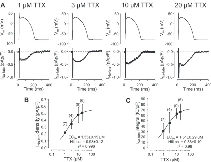
Fig. 1.Concentration-dependent effect of tetrodotoxin (TTX) in isolated canine ventricular myocytes under APVC conditions. For the sake of better comparability canonic APs were used as command signals (shown in the upper row). Furthermore, in order to exclude any contamination by L-type Ca2+current, these experiments were performed in the presence of 1μM nisoldipine. A: Representative records of INa-latedissected by increasing concentrations of TTX. Exposure to TTX lasted for 5 min in each case. B and C: Concentration-response curves obtained for INa-latemeasured 15 ms after the AP upstroke (B), and the net charge mediated by the current (C). The solid curve was obtained byfitting data to the Hill equation. Symbols and bars denote mean ± SEM values, numbers in parentheses indicate the number of cells studied, derived from 5 animals.
if the characteristics of the re-established impalement deviated by < 5% from the previous measurement, the experiment continued.
2.4. Statistics
Results are expressed as mean ± SEM values, the number of myo- cytes or multicellular preparations studied/derived from the number of animals is given in parenthesis. In the graphs individual data are de- noted by blue dots. Statistical significance of differences was evaluated using one-way ANOVA followed by Student'st-test. Differences were considered significant when p was < 0.05.
Chemicals used in the experiments were obtained from Sigma- Aldrich Co. (St. Louis, MO, USA).
3. Results
Primarily the optimal TTX concentration for dissection INa-late in canine ventricular myocytes had to be determined in order tofind a TTX concentration that is suitable to excise a sufficiently large portion of INa-late allowing visualization of the true profile of the current.
Representative records presented inFig. 1.A indicate that the effect of TTX tends to saturate at the concentration of 10μM. Based on the concentration-response curves obtained byfitting these data to the Hill equation (Fig. 1.B, C) 10μM TTX caused an approximately 87% in- hibition of the current. Therefore 10μM TTX was chosen to dissect INa- latein the forthcoming voltage clamp experiments bearing in mind that this approach is likely to somewhat underestimate the density and in- tegral of INa-late. Accordingly, the relative contribution of the high TTX-
affinity“neuronal”Na+channels were not monitored,i.e.the effects of lower (nanomolar) TTX concentrations were not studied. This may distort somewhat the EC50values obtained for TTX inFig. 1.B and C.
These measurements were performed in the presence of 1μM nisoldi- pine to suppress L-type Ca2+current, since large concentrations of TTX have been shown to inhibit Ca2+current as well [31,32].
As has been shown inFig. 1 (canonic APVC in nisoldipine) and Fig. 2.A (self APVC without nisoldipine), canine INa-late displayed a
“decrescendo” profile, i.e. its amplitude decreased monotonically during the time course of the AP, in contrast to guinea pig ventricular myocytes, where the amplitude of the current was increasing during the plateau and declined only on terminal repolarization (Fig. 2.D,“cres- cendo profile”). These differences are also reflected by the current- voltage relations (phase-plane trajectories) obtained under the APs in canine and guinea pig cells (Fig. 2.B and E). More quantitative ap- proach to demonstrate the profile of INa-lateis to measure the density of the TTX-sensitive current at 20%, 50% and 100% of APD, where the APD at 90% repolarization (APD90) was considered as 100% (Fig. 2.C and F). Accordingly, the density of INa-late at 20% APD was
−0.52 ± 0.09 pA/pF in dogversusthe−0.21 ± 0.05 pA/pF in guinea pig (p< .05), while at 100% APD INa-late had a greater density in guinea pig than dog (−0.78 ± 0.07 versus−0.03 ± 0.02 pA/pF, p < .05). At 50% APD the densities were not significantly different in the two species. Although the charge carried by INa-latewas higher in guinea pig than in dog (86.2 ± 10.8 fC/pF, n= 20/9 versus 67.1 ± 9.2 fC/pF,n= 7/4, p < .05) these current integrals were in a similar range.
As already mentioned above, INa-late current profiles in canine
DOG
GP
-100 -50 0 50
Vm(mV)
-2.0 -1.5 -1.0 -0.5 0.0
0 200 400
Time (ms)
-100 -50 0 50
-1.0 -0.5 0.0
Vm(mV)
B C
A
E F
D
-1.0 -0.5 0.0
Vm(mV)
-100 -50 0 50
-100 -50 0 50
Vm(mV)
-2.0 -1.5 -1.0 -0.5 0.0
0 200 400
Time (ms)
INa-late (pA/pF)INa-late(pA/pF)
(n = 7/4)
(n = 20/9) -2.0
-1.5 -1.0 -0.5 0.0
-2.0 -1.5 -1.0 -0.5 0.0
20%APD 50%APD 100%APD 20%APD 50%APD 100%APD
INa-late(pA/pF)INa-late(pA/pF) INa-late (pA/pF)INa-late(pA/pF)
Fig. 2.True profiles of INa-laterecorded in canine (A-C) and guinea pig (D-F) ventricular cells under self APVC conditions. Currents were dissected by 10μM TTX. A,D:
Representative records displaying INa-lateprofiles (command APs above). B,E: Phase-plane trajectories obtained from the same records. C,F: Densities of INa-late
measured at 20%, 50% and 100% of APD, where APD90was defined as 100%. Symbols and bars are mean ± SEM values, blue dots represent individual data, numbers in parentheses indicate the number of myocytes/number of animals studied.
cardiomyocytes were determined under APVC conditions in two slightly different experimental groups. In thefirst group canonic APVC was used and INa-latewas determined in the presence of 1μM nisoldipine (Fig. 1,n= 9/5). In the second group self APVC was used without ni- soldipine (Fig. 2.A-C, n= 7). Although the values were somewhat smaller in the presence of nisoldipine, no significant differences were found between the two groups at 20%, 50% or 100% APD (−0.48 ± 0.08 versus−0.52 ± 0.09 pA/pF, −0.30 ± 0.06 versus−0.34 ± 0.06 pA/pF and−0.06 ± 0.02 versus−0.03 ± 0.02 pA/pF, respectively). The integrals were also similar (63.1 ± 9.2 fC/pF, n = 9/5versus67.1 ± 9.2 fC/pF, n = 7/4, respectively, N.S.).
This comparison suggests that 10μM concentration of TTX does not interfere with the L-type Ca2+current.
Since AP configuration has been shown to influence current profiles under APVC conditions [28,29], attempts were made to convert the decrescendo type canine INa-lateinto a crescendo profile, characteristic of guinea pig myocytes. Therefore, duration-matched canonic guinea pig APs (Fig. 3.B,E) and voltage ramps resembling the ramp-like plateau of guinea pig cells (Fig. 3.C,F) were applied as command signals to canine cells. The ramp started from−80 mV, spent 10 ms at +40 mV and decayed to−20 mV during 200 ms. None of these interventions were capable to convert the canine decrescendo current profile to a crescendo guinea pig-like INa-lateprofile. The current integrals were also similar in canine cells independently of the canine or guinea pig origin of the command AP (63.1 ± 9.2 fC/pF,n= 9/5versus65.4 ± 10.7 fC/pF,n= 6/5, N.S.).
The sea-anemone toxin (ATX-II) has been shown to induce a current
in cardiac tissues resembling INa-lateby removing the fast inactivation machinery of Na+channels [33]. In guinea pig myocytes the profile of the TTX-sensitive current was similar in the absence and presence of 10 nM ATX-II (Fig. 4.A), although its amplitude was significantly in- creased by ATX-II (compare also Figs. 4.D and2.F),i.e. the current followed crescendo kinetics in both cases. In contrast, canine myocytes usually produced early afterdepolarizations when exposed to 10 nM ATX-II for 3 min (not shown), therefore the canine cells were treated with a lower, 1 nM concentration of ATX-II. The TTX-sensitive current in these canine cells displayed a plateau-like shape, i.e.the current densities were largely similar at 20%, 50% and 100% APD in the pre- sence of ATX-II (Fig. 4.B,E). This current profile, containing the sum of baseline INa-late plus the ATX-II-induced current component, was markedly different from that recorded in the absence of ATX-II (com- pare Figs. 4.E and 2.C). Therefore, in another series of experiments, where the ATX-II-induced current alone was visualized, the ATX-II-in- duced current showed moderate crescendo characteristics in canine myocytes (Fig. 4.C,F). The TTX-sensitive current integrals were sig- nificantly larger in the presence of ATX-II (1 nM in canine and 10 nM in guinea pig cells) comparing to untreated cells: 102 ± 13 fC/pF,n= 7/
4versus67 ± 9 fC/pF, n = 7/4 in canine; and 145 ± 19 fC/pF,n= 5/
3versus86 ± 11 fC/pF,n= 20/9 in guinea pig myocytes,p< .05 for both. The duration of action potentials (APD90) was significantly in- creased by 1 nM ATX-II in canine cells from 214 ± 13 ms to 257 ± 17 ms (lengthening of 43.2 ms, n = 7/4, p < .05), in contrast to guinea pig myocytes, where the increase induced by 10 nM ATX-II (from 192 ± 15 to 212 ± 20 ms, lengthening of 19.5 ms, n = 5/3),
0 200 400
-2.0 -1.5 -1.0 -0.5 0.0 -100 -50 0 50
Vm (mV)
Time (ms)
Dog AP to dog cell
Time (ms)
GP AP to dog cell
Vm (mV)
Ramp to dog cell
-100 -50 0 50
Vm (mV)
-2.0 -1.5 -1.0 -0.5 0.0
0 200 400
Time (ms)
0 200 400
-100 -50 0 50
-2.0 -1.5 -1.0 -0.5 0.0
C A B
) 3 / 5
= n ( )
5 / 6
= n ( )
5 / 9
= n (
20%APD 50%APD 100%APD
-1.0 -0.5 0.0
-1.0 -0.5 0.0
-1.0 -0.5 0.0
INa-late(pAp/F) INa-late(pA/pF) INa-late(pA/pF)
20%APD 50%APD 100%APD 20%APD 50%APD 100%APD
F D E
INa-late(pAp/F) INa-late(pA/pF) INa-late(pA/pF)
Fig. 3.Effect of the shape of command voltage on the profile of INa-latein canine myocytes.
A-C: Representative sets of records displaying command voltage protocols (upper row) and the recorded INa-lateprofiles dissected by 10μM TTX in the presence of 1μM nisoldipine (lower row). A canonic dog AP was delivered to a canine cell in panel A, in B a canonic guinea pig AP was applied to a canine cell, while in C a decaying voltage ramp was used as a command signal. D-F: INa-latedensities measured at 20%, 50% and 100% of APD (100% = APD90) under conditions shown in panels A-C. Symbols and bars are mean ± SEM values, blue dots represent individual data, numbers in parentheses indicate the number of myocytes/number of animals studied.
was not significant statistically. These results clearly demonstrate that canine myocytes are much more sensitive to ATX-II that the guinea pig cells, and more importantly, the profile of the ATX-II-induced current is different from the native INa-latein canine myocytes.
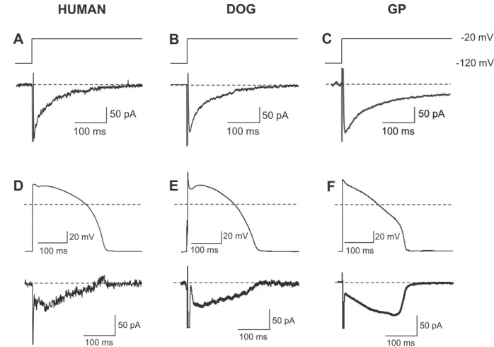
In the followings the profiles (under self APVC conditions) and the kinetic properties (using conventional voltage clamp techniques) of INa- latewere compared in human, canine and guinea pig ventricular cells (Fig. 5). The 20μM TTX-sensitive currents were similarly shaped in all types of myocytes when the membrane potential was switched from the holding potential of−120 mV to the test potential of−20 mV in the presence of 1μM nisoldipine (Fig. 5.A-C). When the decay of TTX- sensitive current wasfitted to a monoexponential function (excluding the initial 50 ms period) the inactivation time constants were 60 ± 3 ms (n= 13/7) in canine, 67 ± 5 ms (n= 5/3) in human, and 155 ± 16 ms (n = 5/3) in guinea pig cells. Thus the time course of inactivation of INa-latewas similar in human and canine myocytes, but was significantly (p< .05) slower in guinea pig. The corresponding current densities (obtained also by the monoexponentialfitting proce- dure) were−0.47 ± 0.05 pA/pF in human,−0.56 ± 0.04 pA/pF in canine and−0.54 ± 0.14 pA/pF in guinea pig myocytes, which values were not significantly different. Representative INa-lateprofiles recorded from a human, canine and guinea pig myocyte under self APVC con- ditions are presented inFig. 5.D-F. These current profiles were also very similar in canine and human cells (both sharing the decrescendo INa-late
profile), while markedly different from the crescendo type INa-latein guinea pig.
The rate-dependent effects of TTX on AP morphology in
multicellular ventricular preparations, obtained from the three species, is demonstrated inFig. 6. All preparations were superfused with 2μM TTX and the measurements were performed after equilibration. APD was shortened by TTX at each cycle length. The TTX-induced short- ening, recorded at the cycle length of 1 s, was the largest (67 ± 16 ms, n= 4/3) in human, intermediate (21 ± 4 ms,n= 5/5) in canine and the smallest (11 ± 3,n= 7/7) in guinea pig preparations. The resting membrane potential and the amplitude of AP were not altered by TTX.
The maximum velocity of depolarization (dV/dtmax) was moderately reduced by TTX from 310 ± 48 to 259 ± 24 V/s in human, from 231 ± 40 to 169 ± 33 V/s in canine, and from 226 ± 32 to 176 ± 21 V/s in guinea pig, however, these changes failed to reach the level of significance–except for the canine preparations.
4. Discussion and conclusions
This is thefirst study to demonstrate the profiles of human and canine ventricular INa-lateunder self APVC conditions. From this per- spective our results are in line with those of Murphy et al. [23] who have shown the profile of canine INa-lateusing canonic APs. Regarding undiseased human ventricular cells, this is the first report using the APVC technique. Our most importantfinding was to demonstrate that canine myocytes can be used as a reasonably good model to study human INa-late- in contrast to myocytes originating from other mammals including guinea pigs, rabbits and pigs, since all in these latter species the current displays a crescendo profile [18–20]. Important implication of this interspecies difference is that the relative contribution of INa-late
C
0 100 200 300 400 Time (ms) -100
-50 0 50
Vm(mV)
-2.0 -1.5 -1.0 -0.5 0.0
DOG
) 3 / 5
= n ( )
4 / 7
= n ( )
3 / 5
= n -2.0 (
-1.5 -1.0 -0.5 0.0
-2.0 -1.5 -1.0 -0.5 0.0
-2.0 -1.5 -1.0 -0.5 0.0
20%APD 50%APD 100%APD 20%APD 50%APD 100%APD 20%APD 50%APD 100%APD
F D E
DOG
0 100 200 300 400 -100
-50 0 50
-2.0 -1.5 -1.0 -0.5 0.0 Vm(mV)
Time (ms)
GP B
0 100 200 300 400 -100
-50 0 50
Time (ms) -2.0
-1.0 0
-1.5 -0.5 Vm(mV)
A
Current (pAp/F) Current (pA/pF) Current (pA/pF)
Current (pAp/F) Current (pA/pF) Current (pA/pF)
Fig. 4.Effect of ATX-II on the TTX-sensitive current profile in guinea pig and canine ventricular cells. In guinea pig myocytes (A, D) the TTX-sensitive current profile was similarly shaped (crescendo) in the absence (red records) or presence (black records) of 10 nM ATX-II, while in canine cells (B, E) the current profile was modified by 1 nM ATX-II. The control TTX-sensitive current records (red) are derived from different cells and presented exclusively for the sake of better comparison.
The profile of the ATX-II-induced current in canine cells is displayed in panels C and F.. Bottom panels display Na+current densities measured in the presence of ATX-II (D, E) and absence of ATX-II (F) at 20%, 50% and 100% of APD, where 100% = APD90. Symbols and bars are mean ± SEM values, blue dots represent individual data, numbers in parentheses indicate the number of myocytes/number of animals studied.
to action potential morphology (with the concomitant Na+and Ca2+
load resulting in increased arrhythmia propensity) increases with lengthening of APD in the “crescendo”group in contrast to the“de- crescendo” situation, and vice versa, it will be relatively smaller at shorter APDs. More explicitly, INa-lateis the largest in amplitude at the time of terminal repolarization in the“crescendo”type species, conse- quently, a given prolongation of APD (eg. due to application of a HERG channel inhibitor) is expected to cause a greater extra inward INa-late
current (together with larger Na+and Ca2+load), which in turn, may magnify the APD lengthening effect of the K+channel inhibitor. Since INa-lateis practically inactivated at this time in the“decrescendo”type preparations, changes in APD will barely modify the magnitude of Na+ entryviaINa-late. Using similar argumentation, the therapeutic effects of INa-late inhibitors (including the shortening of APD and reduction of cellular Na+and Ca2+content) is expected to be less pronounced in canine and human myocardium than in similar preparations from guinea pigs, rabbits and pigs. This should be taken into account when the results of pharmacological studies on INa-lateinhibitors or modifiers are interpreted or extrapolated to human, since some studies with such compounds have been performed in species displaying crescendo INa-late
profiles [34–36]. Based on the present results, canine ventricular tissues or myocytes are suggested for pharmacological studies with drugs in- teracting with INa-late.
We have also revealed why the guinea pig INa-lateis growing under the AP plateau, while canine and human INa-latedecreases during the time course of the AP. In canine and human myocytes the inactivation time constant of INa-latewas in the range of 60–67 ms at −20 mV, consequently the current became practically fully inactivated by the time of terminal repolarization. In contrast, the decay time constant
was much longer (156 ms) in guinea pig, thus the inactivation was slow enough to leave a significant portion of Na+cannels open by the time of terminal repolarization. Consequently, the increasing inward driving force acting on Na+ions could increase the amplitude of INa-lateduring the monotonic slow repolarization in guinea pig (and likely also in rabbit and pig). The well-known crescendo INa-lateprofile in guinea pig was originally explained with the non-equilibrium gating theory of Clancy et al. [37]. This model predicts the accumulation of INa-late
during the plateau as a consequence of the ramp-like configuration of phase-2 repolarization of the guinea pig AP. However, application of guinea pig APs as well as repolarizing ramps as command signals failed to convert the decrescendo INa-lateprofile of canine cells to crescendo, indicating that it is not the AP configuration, but rather the 2.5-fold slower inactivation kinetics, revealed by the conventional voltage clamp experiments, that accounts for the crescendo INa-lateprofile in guinea pig. Since a portion of INa-lateis attributed to the function of non- cardiac Na+channels in the canine heart [38], it is possible that the type or relative contribution of these TTX-sensitive channels or var- iances in their regulatory subunits are different in dogs (and humans) versusguinea pigs (and pigs or rabbits). However, elucidating the de- tails behind the different inactivation kinetics of INa-lateamong various species warrants further studies.
Here is to be mentioned that there is a wide variety of SCN5 mu- tations, responsible for LQT3 syndrome, and depending on the site and type of mutation many parameters of Na+channel gating, including the rate of inactivation and recovery as well as the voltage dependence of activation and inactivation, may be altered, however, augmentation of INa-lateis a critical and common feature in all cases [39]. Since only a very small fraction of the total Na+cahnnel population may contribute
DOG
50 pA 100 ms
E
20 mV 100 ms
100 ms 50 pA
HUMAN
50 pA 100 ms
A
D
20 mV 100 ms
50 pA 100 ms
-20 mV -120 mV
C
GP
F
50 pA 100 ms
50 pA 100 ms 20 mV 100 ms
50 pA 100 ms
B
Fig. 5.Comparison the properties of INa-laterecorded from myocytes digested from human (A,D), canine (B,E) and guinea pig (C,F) ventricular myocardium. In panels A-C the 20μM TTX-sensitive currents were activated with rectangular voltage pulses clamped to−20 mV from the holding potential of−120 mV in the presence of 1μM nisoldipine, 0.1μM dofetilide and 0.5μM HMR-1556, while in D-F TTX was used to dissect INa-lateunder self APVC conditions (without nisoldipine, dofetilide and HMR-1556).
GP DOG HUMAN Control TTX -100 -50 0
50 0 1 00 200 300 400 T ime (ms)
Membrane potential (mV) (ms) 90 APD
150
200
250
300
350 13 5
(n = 4/3) Cycle length (s)
Control TTX 2 µM
T ime (ms)
Membrane potential (mV)
Control TTX 0 1 00 200 300 400 -100 -50
0 50 (n = 7/7)
Control TTX 2 µM Cycle length (s)
90 APD (ms)
* * * * 012 34 5 100 150
200
250 ** **
Control TTX 2 µM (n = 5 /5) Cycle length (s)
90 APD (ms)
* 01 2345 150 200
250
300 * * * *
T ime (ms) * *
Membrane potential (mV)
Control TTX 0 100 200 300 400 -100 -50
0 50 B A * * * * *
* * * *
4 2 0
Fig.6.Effectsof2μMTTXonAPDinmulticellularhuman,canineandguineapigventricularpreparations.Theseexperimentswereperformedinarate-dependentmanner,wherethecyclelengthwasgraduallychanged between0.3and5s.RepresentativepairsofAPs,recordedatcyclelengthsof1saredepictedintheupperrows(A).B:Cyclelength-dependentshorteningeffectof2μMTTXonAPD.Symbolsandbarsrepresent mean±SEMvalues,asterisksindicatestatisticallysignificantdifferencesbetweencontrolandTTXdatadeterminedusingone-wayANOVAfollowedbyStudent'st-testforpaireddata.Differenceswereconsidered significantwhenpwas<0.05.Thenumbersinparenthesesdenotethenumberofpreparations/numberofanimalsstudied.to generation of INa-late(compare the few tens of pA amplitude of INa-late
with the several nA amplitude of the peak Na+current), the macro- scopic changes in INagating are less relevant from the pathophysiolo- gical point of view than the actual magnitude of INa-late.
Another important result of the present study was to show that the profile of the ATX-II-induced current recorded under APVC conditions in canine cells is markedly different from the shape of native canine INa- late, since the ATX-II-induced current displayed a crescendo profile in both species –similar to the native INa-lateof guinea pig, rabbit and porchine hearts, but different from the native INa-laterecorded from canine myocytes. This may be related to the well-known fact that ATX- II slows inactivation of Na+channels [33]. ATX-II is widely used for mimicking pharmacologically the consequences of an augmented INa- late, which is often seen under pathological conditions [5] and ATX-II is a widely used pharmacological tool to model this. In canine myocytes (i.e.in the cells appearing to be the best model for studying human INa- late), however, this approach may be misleading due to the differences observed between the native INa-lateand ATX-II-induced current pro- files. As a consequence, ATX-II modified Na+channels might also differ from the native channels in their in drug-sensitivity, making results obtained in the presence of ATX-II difficult to interpret.
APDs recorded from multicellular canine, human and guinea pig preparations were shortened by 2μM TTX, which effect was the largest in human, intermediate in canine and the smallest in guinea pig pre- parations. This sequence can not be explained by the known (and presently described) properties of INa-late, since the current densities and integrals were largely similar in the three species. It is more likely therefore that the magnitude of the TTX-induced shortening depends on the actual repolarization reserve of the myocardium [40,41], which is the smallest in human, intermediate in canine, and with the strong IKsis the largest in guinea pig ventricular myocardium [42,43]. This is a further argument in support of using canine myocytes for modeling human INa-late. This interspecies difference (i.e.dog vs.guinea pig) in repolarization reserve may explain also the higher ATX-II-sensitivity of dog comparing to guinea pig. In canine myocytes 1 nM ATX-II caused twice greater lengthening of AP than 10 nM ATX-II in guinea pig, while 10 nM ATX-II initiated early afterdepolarizations in canine cells.
In summary, we conclude that canine myocytes represent a rea- sonably suitable model of human ventricular cells regarding the prop- erties of INa-late. However, further detailed studies are required to de- scribe the gating kinetics of INa-late.
Funding
This work was funded by the National Research Development and Innovation Office (NKFIH-K115397 to P.P.N, NKFIH-PD120794 and NKFIH-FK128116 to B.H. NKFIH-PD125402 and NKFIH-FK129117 to N.N., and NKFIH-K119992 to A.V.). Further support was obtained from the GINOP-2.3.2.-15-2016-00040 and EFOP-3.6.2-16-2017-00006 pro- jects, which are co-financed by the European Union and the European Regional Development Fund. The work was also supported by the Hungarian Academy of Sciences (János Bolyai Research Scholarship to B.H. and N.N.), and by the ÚNKP-19-4 and ÚNKP-19-2 New National Excellence Program of the Ministry of Human Capacities (to B.H., D.B.).
Declaration of Competing Interest None declared.
References
[1] E. Coraboeuf, E. Deroubaix, A. Coulombe, Effect of tetrodotoxin on action potentials of the conducting system in the dog heart, Am. J. Phys. 236 (1979) H561–H567.
[2] E. Carmeliet, Slow inactivation of sodium current and voltage-dependent block by tetrodotoxin in rabbit cardiac Purkinjefibers, Biomed. Biochim. Acta 45 (1986) S163–S166.
[3] E. Carmeliet, Voltage-dependent block by tetrodotoxin of the sodium channel in
rabbit cardiac Purkinjefibers, Biophys. J. 51 (1987) 109–114.
[4] K.R. Chadda, K. Jeevaratnam, M. Lei, C.L.H. Huang, Sodium channel biophysics, late sodium current and genetic arrhythmic syndromes, Pflügers Arch. 469 (2017) 629–641.
[5] B. Horvath, D.M. Bers, The late sodium current in heart failure: pathophysiology and clinical relevance, ESC Heart Failure 1 (2014) 26–40.
[6] A.I. Undrovinas, V.A. Maltsev, J.W. Kyle, N. Silverman, H.N. Sabbah, Gating of the late Na+channel in normal and failing human myocardium, J. Mol. Cell. Cardiol.
34 (2002) 1477–1489.
[7] C.R. Valdivia, W.W. Chu, J. Pu, J.D. Foell, R.A. Haworth, WolffMR et al. increased late sodium current in myocytes from a canine heart failure model and from failing human heart, J. Mol. Cell. Cardiol. 38 (2005) 475–483.
[8] V.A. Maltsev, N. Silverman, H.N. Sabbah, A.I. Undrovinas, Chronic heart failure slows late sodium current in human and canine ventricular myocytes: implications for repolarization variability, Eur. J. Heart Fail. 9 (2007) 219–227.
[9] Y. Song, L. Belardinelli, Basal late sodium current is a significant contributor to the duration of action potential of guinea pig ventricular myocytes, Physiol. Rep.
(2017) e13295.
[10] V.A. Maltsev, A.I. Undrovinas, A multi-modal composition of the late Na+current in human ventricular cardiomyocytes, Cardiovasc. Res. 69 (2006) 116–127.
[11] D. Noble, P.J. Noble, Late sodium current in the pathophysiology of cardiovascular disease: consequences of sodium-calcium overload, Heart 92 (Suppl. 4) (2006) 1–5.
[12] A. Zaza, M. Rocchetti, The late Na+current - origin and pathophysiological re- levance, Cardiovasc. Drugs Ther. 27 (2013) 61–68.
[13] J.C. Shyrock, Y. Song, S. Rajamani, C. Antzelecitch, L. Belardinelli, The antiar- rhythmogenic consequences of increasing late INain the cardiomyocyte, Cardiovasc.
Res. 99 (2013) 600–611.
[14] S. Yu, G. Li, C.L.H. Huang, M. Lei, L. Wu, Late sodium current associated cardiac electrophysiological and mechanical dysfunction, Pflügers Arch. 470 (2018) 461–469.
[15] C. Antzelevitch, L. Belardinelli, The role of sodium channel current in modulating transmural dispersion of repolarization and arrhythmogenesis, J. Cardiovasc.
Electrophysiol. 17 (Suppl. 1) (2006) S79–S85.
[16] A.I. Undrovinas, L. Belardinelli, N.A. Undrovinas, H.N. Sabbah, Ranolazine im- proves abnormal repolarization and contraction in left ventricular myocytes of dogs with heart failure by inhibiting late sodium current, J. Cardiovasc. Electrophysiol.
17 (Suppl. 1) (2006) S169–S177.
[17] D. Attwell, I.S. Cohen, D.A. Eisner, M. Ohba, C. Ojeda, The steady state TTX-sen- sitive (“window”) sodium current in cardiac Purkinjefibres, Pflügers Arch. 379 (1979) 137–142.
[18] B. Hegyi, T. Bányász, L.T. Izu, L. Belardinelli, D.M. Bers, Y. Chen-Izu,β-Adrenergic regulation of late Na+current during cardiac action potential is mediated by both PKA and CaMKII, J. Mol. Cell. Cardiol. 123 (2018) 168–179.
[19] B. Horvath, T. Banyasz, Z. Jian, B. Hegyi, K. Kistamas, P.P. Nanasi, et al., Dynamics of the late Na+current during cardiac action potential and its contribution to afterdepolarizations, J. Mol. Cell. Cardiol. 64 (2013) 59–68.
[20] B. Hegyi, J. Bossuyt, L.G. Griffiths, R. Shimkunas, Z. Coulibaly, Z. Jian, et al., Complex electrophysiological remodeling in postinfarction ischemic heart failure, Proc. Natl. Acad. Sci. U. S. A. 115 (2018) E3036–E3044.
[21] V.A. Maltsev, H.N. Sabbah, R.S.D. Higgins, N. Silverman, M. Lesch, A.I. Undrovinas, Novel, ultraslow inactivating sodium current in human ventricular cardiomyocytes, Circulation 98 (1998) 2545–2552.
[22] A.C. Zygmunt, G.T. Eddelstone, G.P. Thomas, V.V. Nesterenko, C. Antzelevitch, Larger late sodium conductance in M cells contributes to electrical heterogeneity in canine ventricle, Am. J. Physiol. Heart Circ. Physiol. 281 (2001) H689–H697.
[23] L. Murphy, D. Renodin, C. Antzelevitch, J.M. Di Diego, J.M. Cordeiro, Extracellular proton depression of peak and late Na+current in the canine left ventricle, Am. J.
Physiol. Heart Circ. Physiol. 301 (2011) H936–H944.
[24] G. Szabo, N. Szentandrassy, T. Biro, B.I. Toth, G. Czifra, J. Magyar, et al., Asymmetrical distribution of ion channels in canine and human left-ventricular wall: epicardium versus midmyocardium, Pflugers Arch. 450 (2005) 307–316.
[25] N. Szentadrassy, T. Banyasz, T. Biro, G. Szabo, B.I. Toth, J. Magyar, et al., Apico- basal inhomogeneity in distribution of ion channels in canine and human ven- tricular myocardium, Cardiovasc. Res. 65 (2005) 851–860.
[26] N. Jost, K. Acsai, B. Horvath, T. Banyasz, I. Baczko, M. Bitay, et al., Contribution of IKrand IK1to ventricular repolarization in canine and human myocytes: is there any influence of action potential duration? Basic Res. Cardiol. 104 (2009) 33–41.
[27] B. Hegyi, B. Horváth, K. Váczi, M. Gönczi, K. Kistamás, F. Ruzsnavszky, et al., Ca2+- activated Cl−current is antiarrhythmic by reducing both spatial and temporal heterogeneity of cardiac repolarization, J. Mol. Cell. Cardiol. 109 (2017) 27–37.
[28] T. Banyasz, L. Fulop, J. Magyar, N. Szentandrassy, A. Varro, P.P. Nanasi, Endocardial versus epicardial differences in L-type calcium current in canine ven- tricular myocytes studied by action potential voltage clamp, Cardiovasc. Res. 58 (2003) 66–75.
[29] T. Bányász, J. Magyar, N. Szentandrássy, B. Horváth, P. Birinyi, J. Szentmiklósi, et al., Action potential clampfingerprints of K+currents in canine cardiomyocytes:
their role in ventricular repolarization, Acta Physiol. Scand. 190 (2007) 189–198.
[30] T. Banyasz, B. Horvath, Z. Jian, L.T. Izu, C.-I. Ye, Profile of L-type Ca2+current and Na+/Ca2+exchange current during cardiac action potential in ventricular myo- cytes, Heart Rhythm. 9 (2012) 134–142.
[31] B. Hegyi, B. Horváth, L. Bárándi, F. Papp, J. Magyar, T. Bányász, et al., Tetrodotoxin blocks L-type Ca2+channels in canine ventricular cardiomyocytes, Pflügers Arch.
464 (2012) 167–174.
[32] B. Hegyi, I. Komáromi, K. Kistamás, F. Ruzsnavszky, K. Váczi, B. Horváth, et al., Tetrodotoxin blockade on canine cardiac L-type Ca2+channels depends on pH and redox potential, Marine Drugs 11 (2013) 2140–2153.
[33] W.A. Catterall, Neurotoxins that act on voltage-sensitive sodium channels in ex- citable membranes, Ann. Rev. Pharmacol. Toxicol. 20 (1980) 15–43.
[34] R. Bonatti, A.F. Silva, J.A. Batatinha, L.F. Sobrado, A.D. Machado, B.B. Varone, et al., Selective late sodium current blockade with GS-458967 markedly reduces ischemia-induced atrial and ventricular repolarization alternans and ECG hetero- geneity, Heart Rhythm. 11 (2014) 1827–1835.
[35] L. Belardinelli, G. Liu, C. Smith-Maxwell, W.Q. Wang, N. El-Bizri, R. Hirakawa, et al., A novel, potent, and selective inhibitor of cardiac late sodium current sup- presses experimental arrhythmias, J. Pharmacol. Exp. Ther. 344 (2013) 23–32.
[36] S. Jia, J. Lian, D. Guo, X. Xue, C. Patel, L. Yang, et al., Modulation of the late sodium current by ATX-II and ranolazine affects the reverse use-dependence and proar- rhythmic liability of IKrblockade, Br. J. Pharmacol. 164 (2011) 308–316.
[37] C.E. Clancy, M. Tateyama, H. Liu, X.H. Wehrens, R.S. Kass, Non-equilibrium gating in cardiac sodium cahannels: an original mechanism of arrhythmia, Circulation 107 (2003) 2233–2237.
[38] M. Biet, H. Barajas-Martínez, A.T. Ton, J.F. Delabre, N. Morin, R. Dumaine, About half of the late sodium current in cardiac myocytes from dog ventricle is due to non- cardiac-type Na+channels, J. Mol. Cell. Cardiol. 53 (2012) 593–598.
[39] W. Song, W. Shou, Cardiac sodium channel Nav 1.5 mutations and cardiac ar- rhythmia, Pediatr. Cardiol. 33 (2012) 943–949.
[40] D.M. Roden, Long QT syndrome: reduced repolarization reserve and the genetic link, J. Intern. Med. 259 (2006) 59–69.
[41] A. Varró, I. Baczkó, Cardiac ventricular repolarization reserve: a principle for un- derstanding drug-related proarrhythmic risk, Br. J. Pharmacol. 164 (2011) 14–36.
[42] N. Jost, L. Virág, P. Comtois, B. Ördög, V. Szuts, Seprényi Gy, et al., Ionic me- chanisms limiting cardiac repolarization reserve in humans compared to dogs, J.
Physiol. 591 (2013) 4189–4206.
[43] S. Zicha, I. Moss, B. Allen, A. Varro, J. Papp, R. Dumaine, et al., Molecular basis of species-specific expression of repolarizing K+currents in the heart, Am. J. Physiol.
Heart Circ. Physiol. 285 (2003) H1641–H1649.