Contents lists available atScienceDirect
Journal of Molecular and Cellular Cardiology
journal homepage:www.elsevier.com/locate/yjmcc
Ablation of the calpain-targeted site in cardiac myosin binding protein-C is cardioprotective during ischemia-reperfusion injury
David Y. Bare fi eld
a,b,⁎,1, James W. McNamara
c,1, Thomas L. Lynch
a,d, Diederik W.D. Kuster
a,e, Suresh Govindan
a, Lauren Haar
d, Yang Wang
d, Erik N. Taylor
f, John N. Lorenz
g,
Michelle L. Nieman
g, Guangshuo Zhu
h, Pradeep K. Luther
i, Andras Varró
j, Dobromir Dobrev
k, Xun Ai
l, Paul M.L. Janssen
m, David A. Kass
h, Walter Keith Jones
d, Richard J. Gilbert
n,
Sakthivel Sadayappan
a,c,⁎⁎aDepartment of Cell and Molecular Physiology, Loyola University Chicago, Maywood, IL, USA
bCenter for Genetic Medicine, Northwestern University, Chicago, IL, USA
cHeart, Lung and Vascular Institute, Division of Cardiovascular Health and Disease, Department of Internal Medicine, University of Cincinnati, Cincinnati, OH, USA
dDepartment of Molecular Pharmacology and Therapeutics, Loyola University Chicago, Maywood, IL, USA
eDepartment of Physiology, Amsterdam Cardiovascular Sciences, VU University Medical Center, Amsterdam, the Netherlands
fDepartment of Physiology and Biophysics, Boston University, Boston, MA, USA
gDepartment of Pharmacology and Systems Physiology, University of Cincinnati College of Medicine, Cincinnati, OH, USA
hDivision of Cardiology, Department of Medicine, Johns Hopkins University School of Medicine, Baltimore, MD, USA
iMolecular Medicine Section, National Heart and Lung Institute, Imperial College London, London, UK
jDepartment of Pharmacology and Pharmacotherapy, Faculty of Medicine, University of Szeged, Szeged, Hungary
kInstitute of Pharmacology, West German Heart and Vascular Center, University Duisburg-Essen, Essen, Germany
lDepartment of Physiology and Biophysics, Rush University, Chicago, IL, USA
mDepartment of Physiology and Cell Biology, The Ohio State University College of Medicine, Columbus, OH, USA
nResearch Service, Providence VA Medical Center and Brown University, Providence, RI, USA
A R T I C L E I N F O
Keywords:
MYBPC3 cMyBP-C Calpain Cardioprotection Ischemia-reperfusion injury
A B S T R A C T
Cardiac myosin binding protein-C (cMyBP-C) phosphorylation is essential for normal heart function and protects the heart from ischemia-reperfusion (I/R) injury. It is known that protein kinase-A (PKA)-mediated phosphor- ylation of cMyBP-C prevents I/R-dependent proteolysis, whereas dephosphorylation of cMyBP-C at PKA sites correlates with its degradation. While sites on cMyBP-C associated with phosphorylation and proteolysis co- localize, the mechanisms that link cMyBP-C phosphorylation and proteolysis during cardioprotection are not well understood. Therefore, we aimed to determine if abrogation of cMyBP-C proteolysis in association with calpain, a calcium-activated protease, confers cardioprotection during I/R injury. Calpain is activated in both human ischemic heart samples and ischemic mouse myocardium where cMyBP-C is dephosphorylated and undergoes proteolysis. Moreover, cMyBP-C is a substrate for calpain proteolysis and cleaved by calpain at re- sidues 272-TSLAGAGRR-280, a domain termed as the calpain-target site (CTS). Cardiac-specific transgenic (Tg) mice in which the CTS motif was ablated were bred into a cMyBP-C null background. These Tg mice were conclusively shown to possess a normal basal structure and function by analysis of histology, electron micro- scopy, immunofluorescence microscopy, Q-space MRI of tissue architecture, echocardiography, and hemody- namics. However, the genetic ablation of the CTS motif conferred resistance to calpain-mediated proteolysis of cMyBP-C. Following I/R injury, the loss of the CTS reduced infarct size compared to non-transgenic controls.
Collectively, thesefindings demonstrate the physiological significance of calpain-targeted cMyBP-C proteolysis
https://doi.org/10.1016/j.yjmcc.2019.03.006
Received 16 January 2018; Received in revised form 7 March 2019; Accepted 8 March 2019
Nonstandard abbreviations and acronyms:CaMKII, Calcium-calmodulin-activated kinase II; CTS, Calpain-targeted site;ΔCTS, Ablation of calpain-targeted site;
cMyBP-C, Cardiac myosin binding protein-C; GQI, Generalized Q-space MRI; I/R, Ischemia-reperfusion; MHC, Myosin heavy chain;Mybpc3, Cardiac myosin binding protein-C gene;Myh7, Cardiacβ-myosin heavy chain gene; NTG, Non-transgenic mice;Nppa, Atrial natriuretic factor gene; PKA, Protein kinase A; ROI, Region of interest; RTD, Relative transmural depth; TTC, Triphenyl tetrazolium chloride; t/t, cMyBP-C null mice; WT, Wild-type mice
⁎Corresponding author at: Center for Genetic Medicine, Feinberg School of Medicine, Northwestern University, 303 E. Superior St., Chicago, IL 60304, USA.
⁎⁎Corresponding author at: Department of Internal Medicine, Heart, Lung and Vascular Institute, Division of Cardiovascular Health and Sciences, College of Medicine, University of Cincinnati, 231 Albert Sabin Way, Cincinnati, OH 45267-0575, USA.
E-mail addresses:david.barefield@northwestern.edu(D.Y. Barefield),sadayasl@ucmail.uc.edu(S. Sadayappan).
1Contributed equally.
Journal of Molecular and Cellular Cardiology 129 (2019) 236–246
Available online 09 March 2019
0022-2828/ © 2019 The Authors. Published by Elsevier Ltd. This is an open access article under the CC BY-NC-ND license (http://creativecommons.org/licenses/BY-NC-ND/4.0/).
T
and provide a rationale for studying inhibition of calpain-mediated proteolysis of cMyBP-C as a therapeutic target for cardioprotection.
1. Introduction
Ischemic heart disease is a leading global health concern that affects 5 million people annually [1–3]. Experimental models featuring cardiac ischemia-reperfusion (I/R) injury have been used to study a variety of detrimental effects, ranging from cell necrosis tofibrotic scarring [4–7], hypoxia [8,9], apoptosis [10–12], and protein degradation. Cardiac sarcomere proteins have been identified as specific substrates for post- ischemic proteolysis [13]. Following ischemia, multiple proteins and pathways associated with the cardiac sarcomere, including cardiac myosin binding protein-C (cMyBP-C), cardiac troponin I, and myosin light chain [13–15], are subject to post-translational modification and degradation. In particular, proteolysis of cMyBP-C during ischemia leads to impaired function in cells that survive I/R injury [15,16].
Cardiac MyBP-C is a thick and thin myofilament-interacting protein involved in regulating sarcomere structure and function in the heart [17]. Mutations that impair this protein's function have been linked to familial hypertrophic cardiomyopathy in > 60 million people world- wide [18–20], highlighting the clinical urgency of fully elucidating the function of cMyBP-C [17]. Previously, we showed that phosphorylation of cMyBP-C by protein kinase A (PKA) regulates myocardial function [21,22] and confers resistance to proteolysis and protection of cardiac tissue against ischemia-reperfusion (I/R) injury [22,23]. Cardiac MyBP- C is phosphorylated under physiological conditions by an array of sig- naling kinases, including PKA, protein kinase C, protein kinase D, ri- bosomal S6 kinase, glycogen synthase kinase 3β [24], and calcium- calmodulin-activated kinase II (CaMKII) [25]. Calcium activation of CaMKII can influence contractility by phosphorylation of cMyBP-C serine (S) 282 [25,26]. We have shown that phosphorylation of S282 is required for the subsequent phosphorylation of cMyBP-C and that these signaling events are required for many of the regulatory functions of cMyBP-C [21,27,28].
Reduced phosphorylation of cMyBP-C has been observed in mouse models of I/R injury [15], pathological hypertrophy [29], heart failure [30], cytoskeletal muscle LIM protein-knockout mice [21], myocardial stunning [15], as well as human cases of atrial fibrillation [31] and heart failure [32,33]. Such dephosphorylation is accompanied by pro- teolysis of cMyBP-C in association with thickfilament disruption, loss of contractile regulation, and contractile dysfunction [15,22]. Transgenic (Tg) mice expressing cMyBP-C with phosphorylatable serine residues mutated to phosphomimetic aspartic acids have reduced cMyBP-C proteolysis following I/R injury [22]. We previously reported that cMyBP-C proteolysis occurs between residues 271 and 272, located in the phosphorylatable M-domain, and results in the generation of a 40 kDa N′-terminal fragment (residues 1–271) that includes key reg- ulatory sites [34,35]. In addition to impaired regulatory function fol- lowing proteolysis, the cMyBP-C 40 kDa fragment has been shown to directly impair contractility in adult rat [14] and human [36] cardio- myocytes. Furthermore, the expression of this fragment leads to heart failurein vivo[34].
We established herein the role of calpain proteases in degrading cMyBP-C and generating the 40 kDa fragment in human and mouse ischemic myocardium. Moreover, we specifically characterized a mouse model that expresses a mutant cMyBP-C with the calpain target site (CTS) removed [37]. While this model showed no evidence of structural or functional impairment under normal conditions, we demonstrated that specific abrogation of calpain-mediated proteolysis of cMyBP-C in the ΔCTS model can offer a significant degree of cardioprotection during I/R injury in vivo. Under physiological conditions, these data suggest that preventing proteolysis of full-length cMyBP-C may result in
the preservation of myocardial tissue and provide cardioprotection during I/R injury.
2. Methods
2.1. Human heart samples
LV tissue samples were collected from nine non-failing human hearts that were not used for transplantation (5 males, 4 females, mean age 45 ± 3 years) and eight patients who had a myocardial infarction (7 males, 1 female, mean age 52 ± 3 years). The Institutional Review Boards at the University of Szeged, Hungary, Ohio State University, Columbus, OH, and Loyola University Chicago, Maywood, IL, USA, approved this study. Human hearts were obtained immediately (within seconds) post-explanation andflushed with a cardioplegic solution, as described previously [38,39]. The explanted hearts were quickly (within 10–30 min) transferred to the laboratory in a cold (~4 °C) cardioplegic solution containing (in mM) 110 NaCl, 16 KCL, 16 MgCl2, 10 NaHCO3, and 0.5 CaCl2. Upon arrival in the laboratory, samples wereflash-frozen and stored in−80 °C freezers. All hearts were pro- cured and treated with identical protocols, solutions, and timing, irre- spective of source. Human tissue experimentation conformed to the Declaration of Helsinki. Informed consent was acquired from cardiac transplant patients, while non-transplantable donor hearts were ac- quired in identical fashion in the operating room in collaboration with Lifeline of Ohio Organ Procurement.
2.2. Transgenic mouse models
All mouse models were maintained on the FvB/N background. The transgenic lines expressing either the cMyBP-CWT[21] or cMyBP-CΔCTS [37] transgenes were crossed to the cMyBP-C(t/t) mouse line (cMyBP- C(t/t)) [40–42] to study the effect of the transgenic protein without endogenous cMyBP-C (see discussion for further details). All procedures followed the protocol approved by the Institutional Animal Care and Use Committee of Loyola University Chicago and the University of Cincinnati and complied with the Guide for the Use and Care of La- boratory Animals published by the National Institutes of Health. All biochemical and functional assays were performed on 8- to 12-week-old mice of mixed sex with age- and sex-matched controls for each assay after preliminary experiments showed no gender differences.
2.3. Assessment of in vivo cardiac function
Echocardiography measurements were made on 8- to 10-week-old mice using a VisualSonics Vevo 2100 with an MS400 18–38 MHz transducer (FujiFilm, Toronto, Ontario) under 1% isoflurane anesthesia.
Cardiac function was measured from parasternal long axis M-mode recordings as previously described [40,43]. Pressure-volume catheter- ization was performed on anesthetized mice to determine cardiac he- modynamics using a pressure-volume catheter (SPR-839; Millar In- struments Inc., Houston, TX) on 8- to 12-week-old mice, as previously described [44].
2.4. Induction of I/R injury
For mouse ischemia/reperfusion studies shown, the mice were an- esthetized with a 90 mg/kg pentobarbital injection delivered I.P. and the chest was open through a left thoracotomy as previously described [45]. Cardiac I/R injury was induced by ligation of the left anterior
descending coronary artery for 60 min of ischemia, followed by 24 h of reperfusion in 8- to 10-week-old mice [22,45]. As a terminal procedure, the mice were anesthetized with a second 90 mg/kg pentobarbital in- jection delivered I.P., followed by removal of the heart. The heart was cannulated through the aorta and the heart was perfused with 1% tri- phenyl tetrazolium chloride (TTC), as previously described [45,46].
This was followed by the re-occlusion of the coronary artery (suture was left in place) and perfusion of the heart through the aorta of a 5%
Phthalo Blue Pigment solution (Heucotech); the occlusion was released and hearts were rinsed and cut into cross-sectional slices, weighed and photographed. The total area of the section, risk and non-risk regions as delineated by TTC and blue stains were quantified for each section using ImageJ software.
2.5. Cellular and molecular analyses
Histological analysis, electron microscopy, and immuno- fluorescence microscopy were performed, as described previously [40,43]. Analysis of cMyBP-C and the phosphorylation status of S273, S282, and S302 were performed by Western blot using custom-made antibodies that recognize each residue only when phosphorylated. All buffers used for isolating protein samples contained protease inhibitor (Roche, 4693159001) and phosphatase inhibitor cocktails (Sigma P5726, P0044). TaqMan primers were used to determine gene expres- sion with probes recognizing Myh7 and Nppa normalized to Gapdh (Applied Biosystems, Mm01319006g1; Mm01255748g1; 4352339E, respectively). Probes and templates were used with iTaq Probes Master Mix (BioRad 172-5131) and quantified using a BioRad CFX96 ther- mocycler. Mybpc3 expression was measured using SybrGreen inter- calating dye with forward 5′-ATATAGGCCGGGTCCACAA-3′ and re- verse 5′-GCAACAACCACAATGGTGTC-3′ primers (208 bp amplicon) normalized toActbamplified with forward 5′-GGCTGTATTCCCCTCCA TCG-3′and reverse 5′-CCAGTTGGTAACAATGCCATGT-3′(154 bp am- plicon) primers. All qPCR data were analyzed using the 2−ΔΔCqmethod.
2.6. Assessment of calpain activity
Calpain activity was assessed using the Calpain-Glo Protease Assay (Promega). In 96-well plates, 50μl of blanks, control samples con- taining purifiedμ-calpain (Calbiochem), or test samples were mixed with 50μl of Calpain-Glo™reagent with 2 mM CaCl2. Plates were agi- tated for 30 s at 300–500 RPM followed by incubation at room tem- perature for 5–30 min. The plates were read using a luminometer, de- tecting the luciferase signal generated following cleavage of the Calpain-Glo™reagent by calpain. To determine the effects of calpain activation on myofilament proteolysis, calpain was inhibited in vitro using 10 nM of the selective inhibitor MDL 28170 (EMD Biosciences, La Jolla, CA) in 200 mM imidazole, 20 mM L-cysteine, and 10 mM CaCl2at 37 °C. The inhibitor was dissolved in dimethyl sulfoxide (DMSO) and added to the perfusion buffer prior to heart perfusion at a concentration of 10μM.
2.7. Architectural analysis of the ventricular wall with generalized Q-space MRI
The theory and methods of Q-space MRI (GQI) for imaging myoarchitecture in human (in vivo) and rodent (ex vivo) models were previously described [47,48]. Employing GQI, multiple gradient pulses were applied with varying orientations and diffusion sensitivities to evaluate signal attenuation in 3D space, employing a 7 Tesla Bruker Biospec magnet equipped with a CryoProbe (Bruker Corp., Billerica, MA). MR scanning was accomplished with a multi-shot EPI pulse se- quence employing 512 gradient directions, B value of 750 s/mm2, 8 k- space segments, and a voxel size of 100 × 100 × 300μm. The resulting GQI image was assessed to discern the orientation and magnitude of the dominantfiber populations with the 3D directionality of thefiber po- pulations encoded to a red-green-blue scale. Intervoxelfiber tracts were constructed by employing streamline methods based on the alignment of the maximal diffusion vectors embodied in the diffusional probability
Fig. 1.Cardiac MyBP-C is dephosphorylated and degraded in ischemic human myocardium. (A) Representative sample of human infarcted heart tissue with annotated remote [R], border zone [B], and ischemic regions [I]. (B) A representative Coomassie blue-stained SDS-PAGE of myofilament protein preparations from the remote, border, and ischemic regions of an ischemic human heart reveals clear protein degradation in the ischemic region. (C) Calpain protease activity measured in the remote, border, and ischemic regions (n = 6). (D) Western blotting with remote, border, and ischemic protein samples shows full-length and degraded 40 kDa cMyBP-C (top panel) and cMyBP-C phosphorylated at residues S273, S282, and S302, with actin shown as a loading control (bottom). (E) The relative per- centage of full-length and degraded 40 kDa cMyBP-C in the remote, border, and ischemic regions (n = 9;
# comparison of full-length cMyBP-C among tissue regions,‡ comparison of 40 kDa cMyBP-C among tissue regions using one-way ANOVA with Holm- Sidak post-hoc test). (F) Quantification of phos- phorylated S273, S282, and S302 normalized against total full-length cMyBP-C (n = 8, top; 4, middle; 6, bottom). All data are mean ± SEM (* P < 0.05;
one-way ANOVA with Holm-Sidak post-hoc test).
(For interpretation of the references to colour in this figure legend, the reader is referred to the web ver- sion of this article.)
distribution function (pdf). Cardiacfiber orientation was analyzed for helix angle as a function of depth of the myocardium sampled, as previously described (33). In brief, regions of interest (ROI) were placed at various depths across the ventricular wall, and helix-angle at specific locations was quantified and plotted for each heart type. The relative transmural depth (RTD) was calculated for each ROI in a sample using the ratio of the number of ROI positions from the innermost tissue wall divided by the total number of ROIs required to traverse the cardiac wall. The RTD for each ROI in a sample was calculated and displayed as a percentage of the cardiac wall distance.
2.8. Statistics
Statistical analysis was performed using GraphPad Prism 6. Data in Figs. 1, 2, 4, 5, and S1 were analyzed by one-way ANOVA to determine group differences. Following ANOVA, group differences were evaluated by a Holm-Sidakpost-hoctest. Significance was defined at p < 0.05.
Data in Fig. 7 were also analyzed with a two-tailed Student's t-test.
Statistical comparisons of transmural helix angles were performed employing one-way ANOVA on a linear regression using least mean squares.
3. Results
3.1. Calpain proteases are activated and cMyBP-C is degraded in infarcted human hearts
To investigate the role of cMyBP-C proteolysis following ischemia, wefirst evaluated the active state of calpain and whether cMyBP-C is degraded in human heart samples following MI. Using samples of in- farcted human hearts, we isolated ischemic tissue, border zone tissue adjacent to the infarct area, and tissue from an area remote from the ischemic site (Fig. 1A). We also used non-ischemic human heart tissue from unused donor hearts as controls. Myofilament protein extractions from the ischemic region exhibited general protein degradation com- pared to remote and border tissues (Fig. 1B). Using a calpain activity assay, we observed significantly higher calpain activity in the border
zone and ischemic region compared to the remote area and control donor hearts (Fig. 1C). Western blotting revealed a reduction in full- length cMyBP-C and a concomitant increase in 40 kDa N′-terminal cMyBP-C proteolysis in the border and infarct tissues compared to the remote area (Fig. 1D and E). Dephosphorylation of residues S273, S282, and S302 is a required step that precedes cMyBP-C proteolysis and generation of the 40 kDa fragment, as previously reported [14,33].
Using antibodies that detect the phosphorylated epitope of these re- sidues, we observed decreased phosphorylation of cMyBP-C in human ischemic tissue compared to remote or border tissues (Fig. 1F).
3.2. I/R injury is associated with increased calpain activity and cMyBP-C proteolysis in mice
I/R injury or sham surgery was performed on non-transgenic (NTG) wild-type mice, and tissue from ischemic, border zone, and remote myocardium was identified with Phthalo Blue Pigment and TTC staining(Fig. 2A). Total protein lysates from these regions were as- sessed for full-length and degraded α-fodrin, a known substrate of calpain proteases [49]. An increase of the 145 kDaα-fodrin proteolysis product was identified in border zone and ischemic tissues compared to sham and remote samples (Fig. 2B). Calpain activity was significantly elevated in border and infarcted tissues compared to remote and sham tissue controls (Fig. 2C). Levels of total cMyBP-C, 40 kDa cMyBP-C, and phosphorylated cMyBP-C were assessed by Western blot (Fig. 2D). Full- length cMyBP-C was reduced in border and ischemic tissues compared to sham and remote samples (Fig. 2E). Levels of phosphorylated cMyBP- C residues normalized to total cMyBP-C revealed a significant reduction of phosphorylated S273 and S282 in ischemic tissue only (Fig. 2F-H).
3.3. Calpain proteases degrade cMyBP-C and generate a 40 kDa fragment In silicoanalysis [50] of the cMyBP-C amino acid sequence identified a candidate CTS with proteolysis expected between residue R271 and T272 (Fig. 3A). The CTS includes residues 272-TSLAGAGRR-280 and is consistent with the previously determined sequence of the 40 kDa N′- terminal fragment that contains cMyBP-C residues 1–271 [50]. To Fig. 2.Cardiac MyBP-C is dephosphorylated and degraded in a mouse ischemia/reperfusion model.
(A) TTC-staining of I/R-injured mouse hearts showed the remote regions stained blue [R], the border re- gion stained pink [B], and the infarct [I] region in white. The sham heart [S] was stained with TTC and blue dye and shows uniform blue staining. (B) The calpain proteolysis targetα-fodrin shows degrada- tion in protein samples from the border and ischemic regions. (C) Calpain activity was determined from sham, remote, border, and infarcted regions (n = 6, S; 6, R; 7, B; 7, I). (D) The levels of cMyBP-C from these regions were determined using Western blot with the anti-cMyBP-C2–14antibody, and the extent of cMyBP-C degradation was evaluated. The pre- sence of the 40 kDa cMyBP-C N′-terminal fragment was identified in the remote and infarct area.
Sarcomeric actin was used as a loading control (lower panel). Western blot analysis of cMyBP-C was performed to determine the level of cMyBP-C phos- phorylation using phospho-specific antibodies. (E) Quantification of total cMyBP-C and (F-H) phos- phorylated cMyBP-C at residues S273, S282, and S302 (n = 6, total protein; 4, phospho-protein). All data are mean ± SEM (* P < 0.05; one-way ANOVA with Holm-Sidakpost-hoctest). (For inter- pretation of the references to colour in thisfigure legend, the reader is referred to the web version of this article.)
establish cMyBP-C as a calpain substrate, we incubated 20μg of myo- filament protein from wild-type mouse hearts with increasing con- centrations ofμ-calpain at 37 °C for 1 h with 10 mM calcium. The results demonstrated a dose-dependent increase in the proteolysis of full- length cMyBP-C and generation of the 40 kDa fragment (Fig. 3B). To further demonstrate the specific calpain-mediated degradation of cMyBP-C, we incubated myofilament proteins with 1μg calpain in the presence and absence of 10 mM calcium and with 10 nM of the calpain inhibitor MDL 28170. In the presence of calcium, calpain could degrade cMyBP-C, whereas removal of calcium from the buffer or inclusion of MDL 28170 prevented the generation of the 40 kDa cMyBP-C proteo- lytic fragment (Fig. 3C). In order to compare calpain-mediated pro- teolysis of cMyBP-C and other known myofilament calpain substrates, 1 U ofμ-calpain was incubated with 20μg of myofilament protein for different durations with 10 mM calcium. Digested proteins were re- solved using 4–15% SDS-PAGE that revealed protein degradation after 12 h of calpain treatment (Fig. 3D). Western blot analysis using anti- cMyBP-C2–14antibodies showed a reduction in full-length cMyBP-C at 12 h of incubation with a corresponding rise in the 40 kDa N′-terminal fragment (Fig. 3E). Cardiac troponin T and troponin I are known targets of calpain proteolysis [13,51]. Western blotting for these troponins revealed similar proteolytic activity in both troponins and an increased amount of troponin proteolytic fragments with increasing calpain in- cubation time (Fig. 3F and G). These results confirm that calpain cleaves cMyBP-C into multiple fragments, principally including a 40 kDa fragment.
3.4. Transgenic mouse model with ablation of the CTS in cMyBP-C has normal levels and preserved phosphorylation of cMyBP-C
Previous investigations using transgenic mice with residues S273, S282, and S302 mutated to phosphomimetic aspartic acids were re- sistant to proteolysis of cMyBP-C and had significant improvement of cardiac function following ischemia [22]. Conversely, depho- sphorylation has been associated with increased cMyBP-C proteolysis following ischemic injury [35]. These data led us to investigate whether abrogation of calpain-mediated proteolysis of full-length cMyBP-C would be sufficient to confer the previously observed phosphorylation-
mediated cardioprotection. To test this question, we used a transgenic mouse with a deletion of the cMyBP-C CTS (residues 272-TSLAGA- GRR-280) [35], termed asΔCTS [37]. This transgenic cMyBP-CΔCTS construct includes an N′-terminal Myc tag and aMyh6 promoter for cardiac-specific expression (Fig. 4A). This construct was expressed in the cMyBP-C(t/t) mouse line that does not express endogenous cMyBP- C and normally has profound dilation and systolic dysfunction [41–43].
In order to control for transgenic overexpression of cMyBP-C, an ad- ditional transgenic mouse line was used that expresses full-length wild- type cMyBP-C with an N′-terminal Myc tag driven by the Myh6pro- moter [21]. This, too, was expressed in the cMyBP-C(t/t) background and is referred to as WT(t/t). A further description of the cMyBP-C(t/t) and transgenic mouse models of cMyBP-C is included in the discussion.
Deletion of the CTS residues 272-TSLAGAGRR-280 removes S273, although the amino acid sequence immediately preceding S282 remains largely unaltered (Fig. 4A). These phosphorylation sites are critical for regulating cardiac function [27]. To assess potential cardiac hyper- trophy following CTS deletion, we evaluated the heart weight-to-body weight ratio, which showed no differences among non-transgenic (NTG), WT(t/t), andΔCTS(t/t), but with a significant increase in the t/t group (Fig. 4B and C). Expression of the hypertrophic markersNppaand Myh7was significantly elevated in t/t samples compared to NTG, but with no elevation observed in WT(t/t) orΔCTS(t/t) (Fig. 4D and E).
Separation ofα- andβ-myosin heavy chain proteins using 6% glycerol SDS-PAGE and visualized with SYPRO Ruby staining showed an in- crease of the hypertrophic marker β-myosin heavy chain in the t/t group, again with no differences among NTG, WT(t/t), andΔCTS(t/t) (Fig. 4F).
Expression of the Mybpc3 transcript shows transgenic over- expression in both WT(t/t) andΔCTS(t/t) groups (Fig. 4G). Myofila- ment protein isolation from NTG, t/t, WT(t/t), andΔCTS(t/t) hearts resolved with SDS-PAGE and visualized with SYPRO Ruby showed normal stoichiometry of cMyBP-C and other myofilament proteins be- tween mouse lines, excepting the t/t group that does not express de- tectable levels of cMyBP-C (Fig. 4H). Western blotting of whole-heart protein lysates for cMyBP-C showed normal protein levels between the NTG, WT(t/t), andΔCTS(t/t) groups, confirming that cMyBP-C stoi- chiometry is not altered by transgenic overexpression, whereas no Fig. 3.Calpain proteases degrade cMyBP-C and generate the 40 kDa fragment. (A)In silicoanalysis of calpain proteolysis sites on cMyBP-C, with bar height representing the likelihood of cleavage, indicates R271 as a potential location of cleavage (dotted line), corresponding to the known sequence of the 40 kDa fragment. The calpain-target site is highlighted in black. (B) Incubation of myofilaments with increasing con- centration of calpain with 10 mM calcium for 1 h at 37 °C shows a dose-dependent increase in cMyBP-C proteolysis and the generation of the 40 kDa fragment with a SYPRO Ruby- stained SDS-PAGE loading control (bottom).(C) Proteolysis of cMyBP-C in myofilament fractions incubated with 1μg cal- pain for 1 h at 37 °C is prevented in the absence of calcium or in the presence of 10 nM of the calpain inhibitor MDL 28170.
(D) Myofilament protein fractions demonstrate proteolysis at time points following incubation of 20μg of total myofilament protein with 1 Uμ-calpain with 10 mM calcium.(E) Western blotting for cMyBP-C shows reduction in full-length cMyBP-C and an increase in 40 kDa cMyBP-C with longer calpain in- cubation. (F and G) The known calpain targets cTnT and cTnI show a reduction in full-length protein and an increase in degraded protein with increasing incubation time with cal- pain.
cMyBP-C was detected in the (t/t) group (Fig. 4I). To evaluate how the loss of CTS affects the phosphorylation of cMyBP-C, two-colourfluor- escent Western blots were performed to detect total and phosphorylated cMyBP-C and S273, S282, and S302 (Fig. 4J). Quantification of phos- phorylated cMyBP-C showed that the phosphorylated S273 epitope was lost in theΔCTS(t/t) group, but with no significant differences between NTG and WT(t/t) (Fig. 4K). Phosphorylation of S282 was not different between groups, whereas S302 phosphorylation showed a significant increase in the WT(t/t) and ΔCTS(t/t) groups compared to NTG (Fig. 4L, M). Taken together, these data demonstrate that the loss of cMyBP-C CTS does not result in overt cardiac pathology or alter normal cMyBP-C protein levels. Importantly, the deletion of CTS does not alter phosphorylation of the functionally critical S282 residue [27].
3.5. Loss of the calpain target site does not alter gross cardiac morphology Morphological analysis of theΔCTS(t/t) model was performed to assess the consequences of the change in the protein sequence.
Myocardial sections were stained with hematoxylin & eosin or Masson's trichrome to detectfibrosis (Fig. S1A). Staining revealed increasedfi- brosis in the t/t group, but no abnormalities among NTG, WT(t/t), and ΔCTS(t/t) hearts. Electron microscopy images of cardiac sarcomeres revealed normal M-line structure and proper orientation in NTG, WT(t/
t), andΔCTS(t/t) samples, while t/t sarcomeres showed disarray and improper M-line arrangement (Fig. S1B). Immunofluorescence micro- scopy performed on isolated cardiomyocytes showed typical cMyBP-C (green) C-zone doublet patterning between theα-actinin (red)-stained Z-disks (left panel) (Fig. S2A); no cMyBP-C was observed in the t/t samples. Anti-Myc antibodies were used to detect Myc-tagged WT and CTS transgenic protein constructs (green) (Fig. S2B). NTG and t/t groups showed no Myc staining, but WT(t/t) andΔCTS(t/t) showed Myc doublet staining (green) between theα-actinin (red)-stained Z- disks, which is consistent with proper cMyBP-C localization. The myoarchitecture of the left ventricular wall, as represented by the transmural helix angle transition (Fig. S3), was well preserved in the ΔCTS(t/t) mouse hearts (Fig. 5A and B) compared with WT(t/t) mouse hearts and previously published myoarchitectural studies [47,48].
3.6. ΔCTS(t/t) mice show normal cardiac function
M-mode echocardiography did not show changes in wall thickness or left ventricular diameter in theΔCTS(t/t) mice compared to NTG or WT(t/t) (Fig. 6A and B, Table S1). No significant change in % fractional shortening was measured among NTG, WT(t/t), andΔCTS(t/t) in con- trast to the significantly reduced systolic function and chamber dilation in the t/t group (Fig. 6B-D). Ejection fraction measured directly by left Fig. 4.Transgenic expression of cMyBP-C with ablation of the CTS. (A) Schematic representation of the transgenic cMyBP-C construct which includes anα-myosin heavy chain promoter, N′-terminal Myc tag sequence,Mybpc3cDNA, and an hGh polyadenylation site. TheΔCTS construct removes the region coding for CTS, amino acids 272-TSLAGAGRR-280. (B and C) HW:BW ratios showed cardiac hypertrophy in the t/t hearts, with no changes observed in WT(t/t) orΔCTS(t/t) compared to NTG (n = 8, 8, 7, 8). (D and E) Expression of the hypertrophic markersNppaandMyh7normalized toGapdhby qPCR showed a significant elevation in the t/t samples only (n = 3). (F) Separation of myosin heavy chain isoforms by SDS-PAGE to identify the hypertrophic markerβ-myosin heavy chain showed an increase in the t/t group only. (G) The expression level of totalMybpc3transcript normalized toActbby qPCR shows transgenic overexpression in the WT(t/t) andΔCTS(t/t) hearts. (H) Myofilament protein fractions from NTG, t/t, WT(t/t), andΔCTS(t/t) hearts resolved by SDS-PAGE and stained with SYPRO-Ruby showed no changes in cMyBP-C stoichiometry. (I) Western blotting of cMyBP-C from whole-heart lysate reveals normal cMyBP-C stoichiometry in the WT andΔCTS hearts compared to NTG with no detectable cMyBP-C in the t/t hearts. (J) Two-colourfluorescent Western blots of total and phospho-serine cMyBP-C. (K-M) Quantification of phosphorylated cMyBP- C at residues 273, 282, and 302 (n = 4). All data are mean ± SEM (* P < 0.05vs.NTG,‡P < 0.05vs.t/t, # P < 0.05vs.WT(t/t); one-way ANOVA with Holm- Sidakpost-hoctest).
ventricular pressure-volume catheterization revealed a similar pattern with a significant reduction in systolic function only in the t/t group (Fig. 6I). The left ventricular volumes at end systole and end diastole were significantly increased only in the t/t hearts, which is consistent with the dilated phenotype associated with this mouse model (Fig. 6H).
Preload recruitable stroke work was significantly decreased only in the t/t group, indicating impaired contractility (Fig. 6G). Diastolic function measured as the time to relaxation (τ-Weiss) was also significantly slower only in the t/t mice (Fig. 6J). These data, combined with normal protein levels, protein localization, and cardiac morphology, suggest that the ΔCTS(t/t) mouse is functionally indistinguishable from the NTG and WT(t/t) controls under normal conditions.
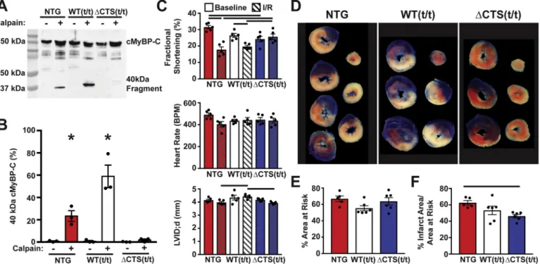
3.7. Ablation of the CTS of cMyBP-C prevents calpain proteolysis and is cardioprotective following I/R injury
To directly assess whether theΔCTS cMyBP-C is, in fact, resistant to calpain proteolysis, we incubated myofilament protein extracted from NTG, WT(t/t), andΔCTS(t/t) hearts withμ-calpain. NTG myofilament
samples treated withμ-calpain showed proteolysis of cMyBP-C with the appearance of the N′-terminal 40 kDa fragment, whereas calpain treatment ofΔCTS(t/t) samples did not produce any detectable cMyBP- C proteolysis (Fig. 7A and B). To determine if ablation of the CTS is protective following ischemic injury, NTG, WT(t/t), andΔCTS(t/t) mice were subjected to 60 min of ischemia followed by 24 h of reperfusion.
Echocardiography was performed pre- and post-ischemia to assess changes in cardiac function.ΔCTS(t/t) animals showed preserved sys- tolic function, with no significant reduction in % fractional shortening following ischemia (Fig. 7C, Table S2). Following ischemia, heart samples were stained with TTC and Phthalo Blue Pigment solution to identify the infarct area and the area at risk (Fig. 7D). Quantification of Fig. 5.Normal cardiac transmuralfiber helical progression in CTS(t/t) hearts
compared to NTG, WT(t/t), and t/t ascertained by generalized Q-space MRI (GQI) with tractography. (A) Quantification of CTS helical transmural fiber progression with individual readings (points), mean readings at each location (grey line) and the 95% confidence interval (black lines) shown as the function of relative transmural depth (RTD) in the myocardium from endocardium to epicardium (Endo:Epi) (n = 4 hearts). (B) Transmural helix anglefiber pro- gression shown as the linear regression of mean values for the NTG, t/t, WT(t/
t), and CTS(t/t) hearts shown as the function of the relative transmural depth in the myocardium. The (t/t) demonstrates significantly reduced helix angle progression compared to all other groups, which indicates a pathological ar- chitectural phenotype. (* P < 0.05 compared to NTG,‡P < 0.05 compared to t/t; one-way ANOVA on linear regression model with least squared means).
Fig. 6.Transgenic expression of ΔCTS in the t/t background shows normal cardiac structure and function. (A-C) Representative parasternal long axis M- mode echocardiography images showed dilation and reduced contractility only in the t/t hearts (n = 6, 6, 4, 7). (D) Representative pressure-volume loops. (E) Pressure-volume catheterization derived ejection fraction, (F) end diastolic volumes, (G) contractility shown by preload recruitable stroke work (PRSW), and (H) relaxation (Tau) were not significantly different in WT(t/t) andΔCTS(t/
t) compared to NTG, whereas deficits were observed in t/t hearts across all parameters (n = 6, 6, 6, 7). All data are mean ± SEM (* P < 0.05vs.NTG,‡ P < 0.05vs.t/t; one-way ANOVA with Holm-Sidakpost-hoctest).
the area at risk showed that the extent of ischemia was not significantly different between NTG, WT(t/t), andΔCTS(t/t) hearts, indicating that all groups received a similar surgical insult (Fig. 7E). However, the infarct area was significantly reduced in theΔCTS(t/t) hearts compared to NTG, whereas WT(t/t) infarct area was not significantly reduced compared to NTG (Fig. 7F). This result was recapitulated using a se- parate set of I/R surgeries on NTG andΔCTS(t/t) mice under the same conditions at a separate institution (Fig. S4).
4. Discussion
Myocardial dysfunction is a clinically important consequence of myocardial ischemia or infarction [1]. Impairments in calcium hand- ling, contractile protein phosphorylation, and sarcomere integrity often accompany contractile dysfunction, but the subcellular mechanisms responsible for these deficits remain incompletely defined [13,51].
Phosphorylation of cMyBP-C has a direct effect on the heart's con- tractile properties, as well as sarcomere organization, and contributes to cardioprotection during I/R injury [15,17,22]. Cardiac MyBP-C de- gradation during I/R injury may contribute to sarcomere disorganiza- tion and contractile dysfunction in the recovering myocardium [14,34].
4.1. Animal models of cMyBP-C
Many studies have used mouse models of cMyBP-C to establish the involvement of cMyBP-C in cardioprotection during ischemic injury. As in previous studies, we have used transgenic models of cMyBP-C ex- pressed on the cMyBP-C t/t background to evaluate the function of transgenic cMyBP-C constructs without confounding effects from
endogenous cMyBP-C. The cMyBP-C t/t model was engineered to have a truncating stop site inserted in exon 30 to model a human mutation with a similar truncation [42]. When the cMyBP-C t/t model was ori- ginally reported by McConnell et al. (1999), Western blot data showed a substantial amount of cMyBP-C in the t/t hearts [42]. However, in numerous subsequent reports by several research groups using this mouse line, no cMyBP-C protein has been detected by Western blot or immunofluorescence microscopy, using several high-quality antibodies [21,43,52]. While this discrepancy has never been explained, the pos- sibility remains that undetectable levels of mutant cMyBP-C could cause alterations in cardiac function. This possibility appears unlikely based on the evidence that models expressing various transgenic cMyBP-C constructs on the t/t background do not share the t/t model's pathology, despite rigorous cardiac phenotyping [21,22,40].
Transgenic cMyBP-C constructs have been used to elucidate the functions of phosphorylatable residues in the cMyBP-C M-domain [21,22,27,53], and in these studies, transgenic overexpression of WT cMyBP-C on the t/t background has never been shown to be sig- nificantly different from NTG controls at baseline or after I/R injury.
Differential phosphorylation of the three major serines, S273, S282, and S302, alter the ability of cMyBP-C to regulate cross-bridge cycling [17].
The deletion of CTS results in loss of the S273 site and alteration of residues near S282. Phosphorylation of S282 is critical for subsequent S302 phosphorylation and regulation of myofilament function [27];
therefore, evidence that S282 can still be phosphorylated after the loss of CTS is likely critical for the absence of any functional deficits in this model.
Fig. 7.Prevention of calpain proteolysis of cMyBP-C reduces infarct size following ischemia-reperfusion injury. (A) Western blot of cMyBP-C from NTG, WT(t/t), and ΔCTS(t/t) myofilaments incubated with and without calpain in 1.25 mM calcium shows the generation of the N′-terminal 40 kDa cMyBP-C proteolysis fragment. The fragment appears at a higher weight in the WT(t/t) sample due to the presence of a Myc tag. (B) Quantification of the percent of 40 kDa cMyBP-C fragment to total cMyBP-C (n = 3) (* P < 0.05; two-way ANOVA). (C) Echocardiography results from NTG, WT(t/t), andΔCTS(t/t) mice pre- and post-I/R show preserved systolic function inΔCTS(t/t) mice but not in NTG or WT(t/t) controls. Additionally, NTG and WT(t/t) hearts showed ventricular wall thinning following I/R, whereas this was not apparent inΔCTS(t/t) hearts (n = 5) (horizontal black bars represent p < 0.05 between indicated groups; two-way ANOVA). (D) Example tissue sections of NTG, WT(t/t), andΔCTS(t/t) hearts stained with TTC and Phthalo Blue pigment solution following I/R injury. After cutting into cross sections, tissue mass, area at risk, and infarct areas were measured. Blue myocardium represents the remote area, red stained myocardium represents the area-at-risk, and the white regions are infarcted tissue. (E) Quantification of these areas show the area at risk was similar between the three groups. (F) The % infarct area was significantly reduced inΔCTS (t/t) hearts. (n = 5, NTG; 6, WT(t/t); 6, ΔCTS(t/t)). Horizontal black bar represents p < 0.05 between indicated groups; one-way ANOVA). All data are mean ± SEM. (For interpretation of the references to colour in thisfigure legend, the reader is referred to the web version of this article.)
4.2. Calpain proteolysis of myofilament proteins
During cardiac ischemia, increased intracellular calcium activates calpain proteases [51,54,55], resulting in the degradation of myofila- ment proteins [56,57]. This proteolysis regulates physiological myofi- lament turnover [58] and hypertrophic remodeling during cardiac stress [59]. Inactive calpains localize to the myofilamentZ-disk under normal physiological conditions, emphasizing their importance in myofilament proteolysis [60]. Calpain proteolysis precedes the release of cardiac troponin fragments into the circulation following ischemic injury, and measurement of these fragments is the current gold standard for clinical diagnosis of myocardial ischemia [61,62]. Experimental methods have long used calpains to isolate specific myofilament com- ponents and have shown that these calcium-dependent, nonlysosomal cysteine proteases can degrade cMyBP-C [28]. Observations of cMyBP- C proteolysis in ischemic tissue have suggested calpain as the protease principally responsible for generating the 40 kDa cMyBP-C fragment [14,35]. We have now identified the exact location of calpain cleavage that contributes to cMyBP-C proteolysis and that it generates the pre- viously identified 40 kDa N′-terminal fragment. Future studies will evaluate the effects of calpain proteolysisin vivoin theΔCTS(t/t) during ischemia.
4.3. Regulation of cMyBP-C by proteolysis
Identification of this 40 kDa N′-terminal fragment of cMyBP-C in ischemic myocardium was performed by using cMyBP-C N′-terminal antibodies [34,35]. Viral expression of the 40 kDa cMyBP-C fragment in adult rat cardiomyocytes and addition of the recombinant 40 kDa fragment into permeabilized human cardiomyocytes have both resulted in impaired myofilament contractile function [14,36]. In addition, the proteolysis of cMyBP-C removes the N′-terminal domains from their functional location in the myofilament where these domains normally regulate actomyosin interactions [28]. Systemic release of cMyBP-C fragments causes an acute inflammatory response that may provide further insult to the heart during reperfusion [63]. The known cytotoxic effects of cMyBP-C proteolysis and the observed reduction in infarct size inΔCTS(t/t) mouse hearts following ischemic injury suggest that degradation of cMyBP-C may not only occur as a result of ischemic signaling events but can potentially exacerbate the progression of cell death within the ischemic myocardium [34].
The calpain proteolysis site is located within the phosphorylatable M-domain of cMyBP-C. Colocalization of these sites suggests a reg- ulatory interaction between the phosphorylation status of cMyBP-C and its degradation. A mouse line expressing a phospho-mimetic cMyBP-C transgene in which phosphorylatable sites S273, S282, and S302 were mutated to aspartic acid mitigates cMyBP-C degradation [22]. These mice exhibit cardioprotection during I/R injury similar to that observed from ischemic preconditioning and to an extent similar to that observed in the current study. This stands in contrast to cMyBP-C proteolysis that occurs with reduced cMyBP-C phosphorylation levels following hypoxia [35]. Accordingly, we have hypothesized that phosphorylation of cMyBP-C inhibits the ability of calpain to degrade cMyBP-C within the phosphorylatable M-domain. This could explain how cMyBP-C phos- phorylation prevents cMyBP-C proteolysis to confer cardioprotection following ischemia [21,22]; although whether cMyBP-C phosphoryla- tion prevents calpain proteolysis by altering the recognition site or sterically inhibiting the enzyme remain to be determined. While the myocardial tissue response to ischemia is multi-faceted, embodying spatially distributed patterns of apoptosis [10–12], hypoxia [8,9], and fibrogenesis [4–7], our currentfindings provide evidence that calpain- mediated cMyBP-C proteolysis is a pivotal event in the injury sequence occurringin vivo.
5. Conclusions
We assessed whether the abrogation of calpain-directed degradation of cMyBP-C is responsible for cardioprotection by ablating the calpain- target sequence in cMyBP-C and assessing the effect on infarct size.
Ablation of the CTS site in the phosphorylatable M-domain of cMyBP-C was associated with significantly reduced infarct size following I/R injury. Thesefindings support the beneficial effect of stabilizing cMyBP- C during ischemia to preserve myocardial function in reperfused tissue.
Previous efforts to explain cMyBP-C phosphorylation-mediated cardio- protection have focused on the importance of preserving cMyBP-C phosphorylation and the role of phosphorylation in altering myofila- ment contractility as the mechanism of cardioprotection. By demon- strating that cardioprotection occurs through the prevention of calpain- mediated cMyBP-C proteolysis, even in the absence of direct manip- ulation of cMyBP-C phosphorylation, we provide a rationale for a re- newed investigation into calpain and the CTS as therapeutic targets and the resultant regulation of calpain proteolysis during and after ischemic injury.
Acknowledgements
The cMyBP-CΔCTS mouse model was provided by Jeffrey Robbins, PhD, Cincinnati Children's Hospital.
Source of funding
The authors were supported by NIH National Heart, Lung, and Blood grants, including R01HL130356, R01HL105826, and K02HL114749 (to S.S.); R01DC011528 (to R.J.G.); and F32HL131304 (to D.Y.B), R01-HL131517 and R01-HL136389 (to D.D.), the American Heart Association Midwest Affiliate Research Programs, including Cardiovascular Genome-Phenome Study 15CVGPSD27020012 and Catalyst 17CCRG33671128 (to S.S.), Predoctoral Fellowship 11PRE7240022 (to D.Y.B.), Postdoctoral Fellowship 13POST14720024 (to S.G.), Postdoctoral Fellowship 13POST17220009 (to D.W.D.K.), Predoctoral Fellowship 15PRE22430028 (to T.L.L.IV.), Postdoctoral Fellowship 17POST33630095 (to J.W.M), and a British Heart Foundation Programme Grant RG/11/21/29335 (to P.K.L.).
Disclosures
Dr. Sadayappan provides consulting services to AstraZeneca, Merck, and Amgen unrelated to the content of this manuscript. No other dis- closures are reported.
Author contributions
S.S. conceived the research; D.Y.B., J.W.M., and S.S. designed ex- periments; D.Y.B., J.W.M., T.L.L.IV., D.W.D.K., S.G., L.H., Y.W., E.N.T., P.L., R.J.G., D.A.K., W.K.J., and S.S. performed research; A.V., D.D., X.A., and P.M.L.J. provided critical reagents; D.Y.B., D.W.D.K., L.H., J.W.M., and E.N.T. analyzed data; and D.Y.B., R.J.G., and S.S. wrote the manuscript.
Appendix A. Supplementary data
Supplementary data to this article can be found online athttps://
doi.org/10.1016/j.yjmcc.2019.03.006.
References
[1] J.A. Finegold, P. Asaria, D.P. Francis, Mortality from ischaemic heart disease by country, region, and age: statistics from World Health Organisation and United Nations, Int. J. Cardiol. 168 (2) (2013) 934–945.
[2] K. Thygesen, J.S. Alpert, A.S. Jaffe, M.L. Simoons, B.R. Chaitman, H.D. White,
E.S.C.A.A.H.A.W.H.F.T.F.f.U.D.o.M.I. Joint, C. Authors/Task Force Members, K. Thygesen, J.S. Alpert, H.D. White, S. Biomarker, A.S. Jaffe, H.A. Katus, F.S. Apple, B. Lindahl, D.A. Morrow, E.C.G. Subcommittee, B.R. Chaitman, P.M. Clemmensen, P. Johanson, H. Hod, S. Imaging, R. Underwood, J.J. Bax, J.J. Bonow, F. Pinto, R.J. Gibbons, S. Classification, K.A. Fox, D. Atar, L.K. Newby, M. Galvani, C.W. Hamm, S. Intervention, B.F. Uretsky, P.G. Steg, W. Wijns, J.P. Bassand, P. Menasche, J. Ravkilde, S. Registries Trials, E.M. Ohman, E.M. Antman, L.C. Wallentin, P.W. Armstrong, M.L. Simoons, S. Registries Trials, J.L. Januzzi, M.S. Nieminen, M. Gheorghiade, G. Filippatos, S. Registries Trials, R.V. Luepker, S.P. Fortmann, W.D. Rosamond, D. Levy, D. Wood, S.
Registries Trials, S.C. Smith, D. Hu, J.L. Lopez-Sendon, R.M. Robertson, D. Weaver, M. Tendera, A.A. Bove, A.N. Parkhomenko, E.J. Vasilieva, S. Mendis, E.S.C.C.f.P.
Guidelines, J.J. Bax, H. Baumgartner, C. Ceconi, V. Dean, C. Deaton, R. Fagard, C. Funck-Brentano, D. Hasdai, A. Hoes, P. Kirchhof, J. Knuuti, P. Kolh, T. McDonagh, C. Moulin, B.A. Popescu, Z. Reiner, U. Sechtem, P.A. Sirnes, M. Tendera, A. Torbicki, A. Vahanian, S. Windecker, R. Document, J. Morais, C. Aguiar, W. Almahmeed, D.O. Arnar, F. Barili, K.D. Bloch, A.F. Bolger, H.E. Botker, B. Bozkurt, R. Bugiardini, C. Cannon, J. de Lemos, F.R. Eberli, E. Escobar, M. Hlatky, S. James, K.B. Kern, D.J. Moliterno, C. Mueller, A.N. Neskovic, B.M. Pieske, S.P. Schulman, R.F. Storey, K.A. Taubert, P. Vranckx, D.R. Wagner, Third universal definition of myocardial infarction, J. Am. Coll.
Cardiol. 60 (16) (2012) 1581–1598.
[3] E.J. Benjamin, S.S. Virani, C.W. Callaway, A.M. Chamberlain, A.R. Chang, S. Cheng, S.E. Chiuve, M. Cushman, F.N. Delling, R. Deo, S.D. de Ferranti, J.F. Ferguson, M. Fornage, C. Gillespie, C.R. Isasi, M.C. Jimenez, L.C. Jordan, S.E. Judd, D. Lackland, J.H. Lichtman, L. Lisabeth, S. Liu, C.T. Longenecker, P.L. Lutsey, J.S. Mackey, D.B. Matchar, K. Matsushita, M.E. Mussolino, K. Nasir, M. O'Flaherty, L.P. Palaniappan, A. Pandey, D.K. Pandey, M.J. Reeves, M.D. Ritchey,
C.J. Rodriguez, G.A. Roth, W.D. Rosamond, U.K.A. Sampson, G.M. Satou, S.H. Shah, N.L. Spartano, D.L. Tirschwell, C.W. Tsao, J.H. Voeks, J.Z. Willey, J.T. Wilkins, J.H. Wu, H.M. Alger, S.S. Wong, P. Muntner, E. American Heart Association Council on, C. Prevention Statistics, S. Stroke Statistics, Heart disease and stroke statistics- 2018 update: a report from the American Heart Association, Circulation 137 (12) (2018) e67–e492.
[4] S.W. van den Borne, J. Diez, W.M. Blankesteijn, J. Verjans, L. Hofstra, J. Narula, Myocardial remodeling after infarction: the role of myofibroblasts, Nat. Rev.
Cardiol. 7 (1) (2010) 30–37.
[5] E.P. Daskalopoulos, B.J. Janssen, W.M. Blankesteijn, Myofibroblasts in the infarct area: concepts and challenges, Microsc. Microanal. 18 (1) (2012) 35–49.
[6] A.H. Li, P.P. Liu, F.J. Villarreal, R.A. Garcia, Dynamic changes in myocardial matrix and relevance to disease: translational perspectives, Circ. Res. 114 (5) (2014) 916–927.
[7] K.P. Quinn, K.E. Sullivan, Z. Liu, Z. Ballard, C. Siokatas, I. Georgakoudi, L.D. Black, Optical metrics of the extracellular matrix predict compositional and mechanical changes after myocardial infarction, Sci. Rep. 6 (2016) 35823.
[8] H.N. Sabbah, V.G. Sharov, S. Goldstein, Cell death, tissue hypoxia and the pro- gression of heart failure, Heart Fail. Rev. 5 (2) (2000) 131–138.
[9] D. Tekin, A.D. Dursun, L. Xi, Hypoxia inducible factor 1 (HIF-1) and cardiopro- tection, Acta Pharmacol. Sin. 31 (9) (2010) 1085–1094.
[10] A. Saraste, K. Pulkki, M. Kallajoki, K. Henriksen, M. Parvinen, L.M. Voipio-Pulkki, Apoptosis in human acute myocardial infarction, Circulation 95 (2) (1997) 320–323.
[11] G. Olivetti, F. Quaini, R. Sala, C. Lagrasta, D. Corradi, E. Bonacina, S.R. Gambert, E. Cigola, P. Anversa, Acute myocardial infarction in humans is associated with activation of programmed myocyte cell death in the surviving portion of the heart, J. Mol. Cell. Cardiol. 28 (9) (1996) 2005–2016.
[12] E. Teringova, P. Tousek, Apoptosis in ischemic heart disease, J. Transl. Med. 15 (1) (2017) 87.
[13] J.E. Van Eyk, F. Powers, W. Law, C. Larue, R.S. Hodges, R.J. Solaro, Breakdown and release of myofilament proteins during ischemia and ischemia/reperfusion in rat hearts: identification of degradation products and effects on the pCa-force relation, Circ. Res. 82 (2) (1998) 261–271.
[14] S. Govindan, J. Sarkey, X. Ji, N.R. Sundaresan, M.P. Gupta, P.P. de Tombe, S. Sadayappan, Pathogenic properties of the N-terminal region of cardiac myosin binding protein-C in vitro, J. Muscle Res. Cell Motil. 33 (1) (2012) 17–30.
[15] R.S. Decker, M.L. Decker, I. Kulikovskaya, S. Nakamura, D.C. Lee, K. Harris, F.J. Klocke, S. Winegrad, Myosin binding protein C phosphorylation, myofibril structure, and contractile function during low-flow ischemia, Circulation 111 (7) (2005) 906–912.
[16] T.L. Lynch, S. Sadayappan, Surviving the infarct: a profile of cardiac myosin binding protein-C pathogenicity, diagnostic utility, and proteomics in the ischemic myo- cardium, Proteomics Clin. Appl. 8 (7–8) (2014) 569–577.
[17] D. Barefield, S. Sadayappan, Phosphorylation and function of cardiac myosin binding protein-C in health and disease, J. Mol. Cell. Cardiol. 48 (5) (2010) 866–875.
[18] H. Watkins, D. Conner, L. Thierfelder, J.A. Jarcho, C. MacRae, W.J. McKenna, B.J. Maron, J.G. Seidman, C.E. Seidman, Mutations in the cardiac myosin binding protein-C gene on chromosome 11 cause familial hypertrophic cardiomyopathy, Nat. Genet. 11 (4) (1995) 434–437.
[19] G. Bonne, L. Carrier, J. Bercovici, C. Cruaud, P. Richard, B. Hainque, M. Gautel, S. Labeit, M. James, J. Beckmann, J. Weissenbach, H.P. Vosberg, M. Fiszman, M. Komajda, K. Schwartz, Cardiac myosin binding protein-C gene splice acceptor site mutation is associated with familial hypertrophic cardiomyopathy, Nat. Genet.
11 (4) (1995) 438–440.
[20] P.S. Dhandapany, S. Sadayappan, Y. Xue, G.T. Powell, D.S. Rani, P. Nallari, T.S. Rai, M. Khullar, P. Soares, A. Bahl, J.M. Tharkan, P. Vaideeswar, A. Rathinavel,
C. Narasimhan, D.R. Ayapati, Q. Ayub, S.Q. Mehdi, S. Oppenheimer, M.B. Richards, A.L. Price, N. Patterson, D. Reich, L. Singh, C. Tyler-Smith, K. Thangaraj, A common MYBPC3 (cardiac myosin binding protein C) variant associated with cardiomyo- pathies in South Asia, Nat. Genet. 41 (2) (2009) 187–191.
[21] S. Sadayappan, J. Gulick, H. Osinska, L.A. Martin, H.S. Hahn, G.W. Dorn 2nd, R. Klevitsky, C.E. Seidman, J.G. Seidman, J. Robbins, Cardiac myosin binding protein-C phosphorylation and cardiac function, Circ. Res. 97 (11) (2005) 1156–1163.
[22] S. Sadayappan, H. Osinska, R. Klevitsky, J.N. Lorenz, M. Sargent, J.D. Molkentin, C.E. Seidman, J.G. Seidman, J. Robbins, Cardiac myosin binding protein-C phos- phorylation is cardioprotective, Proc. Natl. Acad. Sci. U. S. A. 103 (45) (2006) 16918–16923.
[23] S. Sanada, H. Asanuma, O. Tsukamoto, T. Minamino, K. Node, S. Takashima, T. Fukushima, A. Ogai, Y. Shinozaki, M. Fujita, A. Hirata, H. Okuda, H. Shimokawa, H. Tomoike, M. Hori, M. Kitakaze, Protein kinase a as another mediator of ischemic preconditioning independent of protein kinase C, Circulation 110 (1) (2004) 51–57.
[24] D.W. Kuster, V. Sequeira, A. Najafi, N.M. Boontje, P.J. Wijnker, E.R. Witjas- Paalberends, S.B. Marston, C.G. Dos Remedios, L. Carrier, J.A. Demmers, C. Redwood, S. Sadayappan, J. van der Velden, GSK3beta phosphorylates newly identified site in the proline-alanine-rich region of cardiac myosin-binding protein C and alters cross-bridge cycling kinetics in human: short communication, Circ. Res.
112 (4) (2013) 633–639.
[25] M. Gautel, O. Zuffardi, A. Freiburg, S. Labeit, Phosphorylation switches specific for the cardiac isoform of myosin binding protein-C: a modulator of cardiac contrac- tion? EMBO J. 14 (9) (1995) 1952–1960.
[26] H.C. Hartzell, D.B. Glass, Phosphorylation of purified cardiac muscle C-protein by purified cAMP-dependent and endogenous Ca2+−calmodulin-dependent protein kinases, J. Biol. Chem. 259 (24) (1984) 15587–15596.
[27] S. Sadayappan, J. Gulick, H. Osinska, D. Barefield, F. Cuello, M. Avkiran, V.M. Lasko, J.N. Lorenz, M. Maillet, J.L. Martin, J.H. Brown, D.M. Bers, J.D. Molkentin, J. James, J. Robbins, A critical function for Ser-282 in cardiac myosin binding protein-C phosphorylation and cardiac function, Circ. Res. 109 (2) (2011) 141–150.
[28] M.J. Previs, S. Beck Previs, J. Gulick, J. Robbins, D.M. Warshaw, Molecular me- chanics of cardiac myosin-binding protein C in native thickfilaments, Science 337 (6099) (2012) 1215–1218.
[29] S.J. van Dijk, D. Dooijes, C. dos Remedios, M. Michels, J.M. Lamers, S. Winegrad, S. Schlossarek, L. Carrier, F.J. ten Cate, G.J. Stienen, J. van der Velden, Cardiac myosin-binding protein C mutations and hypertrophic cardiomyopathy: hap- loinsufficiency, deranged phosphorylation, and cardiomyocyte dysfunction, Circulation 119 (11) (2009) 1473–1483.
[30] A. El-Armouche, L. Pohlmann, S. Schlossarek, J. Starbatty, Y.H. Yeh, S. Nattel, D. Dobrev, T. Eschenhagen, L. Carrier, Decreased phosphorylation levels of cardiac myosin-binding protein-C in human and experimental heart failure, J. Mol. Cell.
Cardiol. 43 (2) (2007) 223–229.
[31] A. El-Armouche, P. Boknik, T. Eschenhagen, L. Carrier, M. Knaut, U. Ravens, D. Dobrev, Molecular determinants of altered Ca2+ handling in human chronic atrialfibrillation, Circulation 114 (7) (2006) 670–680.
[32] A.M. Jacques, O. Copeland, A.E. Messer, C.E. Gallon, K. King, W.J. McKenna, V.T. Tsang, S.B. Marston, Myosin binding protein C phosphorylation in normal, hypertrophic and failing human heart muscle, J. Mol. Cell. Cardiol. 45 (2) (2008) 209–216.
[33] O. Copeland, S. Sadayappan, A.E. Messer, G.J. Steinen, J. van der Velden, S.B. Marston, Analysis of cardiac myosin binding protein-C phosphorylation in human heart muscle, J. Mol. Cell. Cardiol. 49 (6) (2010) 1003–1011.
[34] M.A. Razzaque, M. Gupta, H. Osinska, J. Gulick, B.C. Blaxall, J. Robbins, An en- dogenously produced fragment of cardiac myosin-binding protein C is pathogenic and can lead to heart failure, Circ. Res. 113 (5) (2013) 553–561.
[35] S. Govindan, A. McElligott, S. Muthusamy, N. Nair, D. Barefield, J.L. Martin, E. Gongora, K.D. Greis, P.K. Luther, S. Winegrad, K.K. Henderson, S. Sadayappan, Cardiac myosin binding protein-C is a potential diagnostic biomarker for myo- cardial infarction, J. Mol. Cell. Cardiol. 52 (1) (2012) 154–164.
[36] N. Witayavanitkul, Y. Ait Mou, D.W. Kuster, R.J. Khairallah, J. Sarkey, S. Govindan, X. Chen, Y. Ge, S. Rajan, D.F. Wieczorek, T. Irving, M.V. Westfall, P.P. de Tombe, S. Sadayappan, Myocardial infarction-induced N-terminal fragment of cardiac myosin-binding protein C (cMyBP-C) impairs myofilament function in human myocardium, J. Biol. Chem. 289 (13) (2014) 8818–8827.
[37] Q. Yang, T.E. Hewett, R. Klevitsky, A. Sanbe, X. Wang, J. Robbins, PKA-dependent phosphorylation of cardiac myosin binding protein C in transgenic mice, Cardiovasc. Res. 51 (1) (2001) 80–88.
[38] N. Milani-Nejad, B.D. Canan, M.T. Elnakish, J.P. Davis, J.H. Chung, V.V. Fedorov, P.F. Binkley, R.S. Higgins, A. Kilic, P.J. Mohler, P.M. Janssen, The frank-Starling mechanism involves deceleration of cross-bridge kinetics and is preserved in failing human right ventricular myocardium, Am. J. Physiol. Heart Circ. Physiol. 309 (12) (2015) H2077–H2086.
[39] M.T. Elnakish, B.D. Canan, A. Kilic, P.J. Mohler, P.M. Janssen, Effects of zacopride, a moderate IK1 channel agonist, on triggered arrhythmia and contractility in human ventricular myocardium, Pharmacol. Res. 115 (2017) 309–318.
[40] D. Barefield, M. Kumar, J. Gorham, J.G. Seidman, C.E. Seidman, P.P. de Tombe, S. Sadayappan, Haploinsufficiency of MYBPC3 exacerbates the development of hypertrophic cardiomyopathy in heterozygous mice, J. Mol. Cell. Cardiol. 79 (2015) 234–243.
[41] B.K. McConnell, D. Fatkin, C. Semsarian, K.A. Jones, D. Georgakopoulos, C.T. Maguire, M.J. Healey, J.O. Mudd, I.P. Moskowitz, D.A. Conner, M. Giewat, H. Wakimoto, C.I. Berul, F.J. Schoen, D.A. Kass, C.E. Seidman, J.G. Seidman, Comparison of two murine models of familial hypertrophic cardiomyopathy, Circ.
Res. 88 (4) (2001) 383–389.
[42] B.K. McConnell, K.A. Jones, D. Fatkin, L.H. Arroyo, R.T. Lee, O. Aristizabal, D.H. Turnbull, D. Georgakopoulos, D. Kass, M. Bond, H. Niimura, F.J. Schoen, D. Conner, D.A. Fischman, C.E. Seidman, J.G. Seidman, Dilated cardiomyopathy in homozygous myosin-binding protein-C mutant mice, J. Clin. Invest. 104 (12) (1999) 1771.
[43] D. Barefield, M. Kumar, P.P. de Tombe, S. Sadayappan, Contractile dysfunction in a mouse model expressing a heterozygous MYBPC3 mutation associated with hy- pertrophic cardiomyopathy, Am. J. Physiol. Heart Circ. Physiol. 306 (6) (2014) H807–H815.
[44] T. Nagayama, E. Takimoto, S. Sadayappan, J.O. Mudd, J.G. Seidman, J. Robbins, D.A. Kass, Control of in vivo left ventricular contraction/relaxation kinetics by myosin binding protein C: protein kinase a phosphorylation dependent and in- dependent regulation, Circulation 116 (21) (2007) 2399–2408.
[45] X. Ren, Y. Wang, W.K. Jones, TNF-alpha is required for late ischemic pre- conditioning but not for remote preconditioning of trauma, J. Surg. Res. 121 (1) (2004) 120–129.
[46] M.C. Fishbein, S. Meerbaum, J. Rit, U. Lando, K. Kanmatsuse, J.C. Mercier, E. Corday, W. Ganz, Early phase acute myocardial infarct size quantification: va- lidation of the triphenyl tetrazolium chloride tissue enzyme staining technique, Am.
Heart J. 101 (5) (1981) 593–600.
[47] M.P. Hoffman, E.N. Taylor, G.E. Aninwene 2nd, S. Sadayappan, R.J. Gilbert, Assessing the multiscale architecture of muscular tissue with Q-space magnetic resonance imaging: review, Microsc. Res. Tech. 81 (2) (2018) 162–170.
[48] E.N. Taylor, M.P. Hoffman, D.Y. Barefield, G.E. Aninwene 2nd, A.D. Abrishamchi, T.L.t. Lynch, S. Govindan, H. Osinska, J. Robbins, S. Sadayappan, R.J. Gilbert, Alterations in multi-scale cardiac architecture in association with phosphorylation of myosin binding protein-C, J. Am. Heart Assoc. 5 (3) (2016) e002836.
[49] S. Fukuda, K. Harada, M. Kunimatsu, T. Sakabe, K. Yoshida, Postischemic re- perfusion induces alpha-fodrin proteolysis by m-calpain in the synaptosome and nucleus in rat brain, J. Neurochem. 70 (6) (1998) 2526–2532.
[50] D.A. DuVerle, Y. Ono, H. Sorimachi, H. Mamitsuka, Calpain cleavage prediction using multiple kernel learning, PLoS One 6 (5) (2011) e19035.
[51] C. Kositprapa, B. Zhang, S. Berger, J.M. Canty Jr., T.C. Lee, Calpain-mediated proteolytic cleavage of troponin I induced by hypoxia or metabolic inhibition in cultured neonatal cardiomyocytes, Mol. Cell. Biochem. 214 (1–2) (2000) 47–55.
[52] B.M. Palmer, B.K. McConnell, G.H. Li, C.E. Seidman, J.G. Seidman, T.C. Irving, N.R. Alpert, D.W. Maughan, Reduced cross-bridge dependent stiffness of skinned myocardium from mice lacking cardiac myosin binding protein-C, Mol. Cell.
Biochem. 263 (1) (2004) 73–80.
[53] S. Sadayappan, J. Gulick, R. Klevitsky, J.N. Lorenz, M. Sargent, J.D. Molkentin, J. Robbins, Cardiac myosin binding protein-C phosphorylation in a {beta}-myosin heavy chain background, Circulation 119 (9) (2009) 1253–1262.
[54] W.D. Gao, D. Atar, Y. Liu, N.G. Perez, A.M. Murphy, E. Marban, Role of troponin I proteolysis in the pathogenesis of stunned myocardium, Circ. Res. 80 (3) (1997) 393–399.
[55] J. Inserte, V. Hernando, D. Garcia-Dorado, Contribution of calpains to myocardial ischaemia/reperfusion injury, Cardiovasc. Res. 96 (1) (2012) 23–31.
[56] G. Neti, S.M. Novak, V.F. Thompson, D.E. Goll, Properties of easily releasable myofilaments: are they thefirst step in myofibrillar protein turnover? Am. J.
Physiol. Cell Physiol. 296 (6) (2009) C1383–C1390.
[57] A. Martin-Garrido, B.J. Biesiadecki, H.E. Salhi, Y. Shaifta, C.G. Dos Remedios, S. Ayaz-Guner, W. Cai, Y. Ge, M. Avkiran, J.C. Kentish, Monophosphorylation of cardiac troponin-I at Ser-23/24 is sufficient to regulate cardiac myofibrillar Ca(2+) sensitivity and calpain-induced proteolysis, J. Biol. Chem. 293 (22) (2018) 8588–8599.
[58] A.S. Galvez, A. Diwan, A.M. Odley, H.S. Hahn, H. Osinska, J.G. Melendez, J. Robbins, R.A. Lynch, Y. Marreez, G.W. Dorn 2nd, Cardiomyocyte degeneration with calpain deficiency reveals a critical role in protein homeostasis, Circ. Res. 100 (7) (2007) 1071–1078.
[59] C. Patterson, A.L. Portbury, J.C. Schisler, M.S. Willis, Tear me down: role of calpain in the development of cardiac ventricular hypertrophy, Circ. Res. 109 (4) (2011) 453–462.
[60] T. Kumamoto, W.C. Kleese, J.Y. Cong, D.E. Goll, P.R. Pierce, R.E. Allen, Localization of the Ca(2+)-dependent proteinases and their inhibitor in normal, fasted, and denervated rat skeletal muscle, Anat. Rec. 232 (1) (1992) 60–77.
[61] H.A. Katus, A. Remppis, S. Looser, K. Hallermeier, T. Scheffold, W. Kubler, Enzyme linked immuno assay of cardiac troponin T for the detection of acute myocardial infarction in patients, J. Mol. Cell. Cardiol. 21 (12) (1989) 1349–1353.
[62] T. Reichlin, W. Hochholzer, S. Bassetti, S. Steuer, C. Stelzig, S. Hartwiger, S. Biedert, N. Schaub, C. Buerge, M. Potocki, M. Noveanu, T. Breidthardt, R. Twerenbold, K. Winkler, R. Bingisser, C. Mueller, Early diagnosis of myocardial infarction with sensitive cardiac troponin assays, N. Engl. J. Med. 361 (9) (2009) 858–867.
[63] C. Lipps, J.H. Nguyen, L. Pyttel, T.L.t. Lynch, C. Liebetrau, G. Aleshcheva, S. Voss, O. Dorr, H.M. Nef, H. Mollmann, C.W. Hamm, S. Sadayappan, C. Troidl, N-terminal fragment of cardiac myosin binding protein-C triggers pro-inflammatory responses in vitro, J. Mol. Cell. Cardiol. 99 (2016) 47–56.
![Fig. 1. Cardiac MyBP-C is dephosphorylated and degraded in ischemic human myocardium. (A) Representative sample of human infarcted heart tissue with annotated remote [R], border zone [B], and ischemic regions [I]](https://thumb-eu.123doks.com/thumbv2/9dokorg/1090077.74390/3.892.55.563.634.1117/cardiac-dephosphorylated-degraded-ischemic-myocardium-representative-infarcted-annotated.webp)