Cardiac Resynchronization Therapy: Current Practice, Refining Implantation Methods, Effects on Ventricular Arrhythmias and New Indications
Ph.D. thesis
Valentina Kutyifa M.D.
Basic Medicine Doctoral School Semmelweis University
Supervisor: Béla Merkely M.D., Ph.D., D.Sc.
Official Reviewers: Lívia Jánoskúti M.D., Ph.D.
Gábor Duray M.D., Ph.D.
Head of the Final Examinatiom Committee: István Préda M.D., Ph.D., D.Sc.
Members of the Final Examinatiom Committee:
Gábor Veress M.D., Ph.D.
András Zsáry M.D., Ph.D.
Budapest 2012
Introduction
Heart Failure (HF) is a major public health problem worldwide with rising prevalence due to aging, improved medical treatment and successful prevention of cardiac events.
Cardiac resynchronization therapy (CRT) provides synchronization of the dyssynchronous left ventricular activation in heart failure patients with conduction abnormalities and severely reduced left ventricular function, resulting in an immediate decrease of left ventricular intra- and interventricular dyssynchrony, mitral regurgitation and an acute increase of LV dP/dt. During long-term follow-up, patients exhibit significant reduction of left ventricular end-diastolic (LVEDV) and left ventricular end- systolic volume (LVESV), and improvement in left ventricular ejection fraction (LVEF), this process is described as left ventricular reverse remodeling.
Cardiac resynchronization therapy or a combination of CRT with an Implantable Cardioverter Defibrillator (CRT-D) is proven to reduce heart failure symptoms, hospitalizations and mortality in patients with NYHA class III-IV drug-refractory heart failure, those with reduced left ventricular ejection fraction (LVEF≤35%) and QRS prolongation (QRS width≥120 ms).
Recent clinical trials, the Multicenter Automatic Defibrillator Implantation Trial – Cardiac Resynchronization Therapy (MADIT-CRT), the Resynchronization- Defibrillation in Ambulatory Heart Failure Trial (RAFT) and Resynchronization Reverses Remodeling in Systolic Left Ventricular Dysfunction (REVERSE) trials further broadened CRT indication to patients with mild HF and NYHA class I, II.
Aims
1. We evaluated the long-term echocardiographic and clinical outcome of patients implanted with CRT-P versus CRT-D in a single-center high-volume registry, and assessed the long-term survival of patients with an implanted CRT-P or CRT-D device.
2. We determined the prognostic significance of right to left ventricular interlead sensed electrical delay on the end point of all-cause mortality, in the CRT registry patients.
3. We evaluated the feasibility and safety of transseptal endocardial left ventricular lead implantation in a small patient cohort of the CRT registry. Furthermore, we determined if electroanatomic mapping guided left ventricular lead targeting would be associated with better clinical and echocardiographic improvement after CRT implantation.
4. We analyzed the association between LV lead position and the risk of VT/VF/Death or VT/VF in patients enrolled in the MADIT-CRT trial.
5. We investigated the association between left ventricular dyssynchrony, CRT-induced change in LV dyssynchrony and the risk of VT/VF/Death or VT/VF events in LBBB and non-LBBB patients in the MADIT-CRT trial.
6. We evaluated the effects of CRT upgrade in implantable cardioverter defibrillator patients with chronic RV apical pacing compared to pacemaker patients, and identified the predictors of long-term outcome in the CRT registry patients.
7. We evaluated the relationship between LVEF and the clinical outcome of mild heart failure patients enrolled in MADIT-CRT; the echocardiographic response to CRT-D in the trial; and the clinical benefit of CRT-D, with a specific focus on the subset of patients with more preserved LVEF enrolled in MADIT-CRT.
Methods
1. Patient population - CRT registry
From June 2000 to April 2011, 1122 consecutive patients underwent CRT implantation at the Semmelweis University Heart Center, Budapest, Hungary. During this time period, 494 of 1122 patients (44%) had measurements of right to left ventricular interlead sensed electrical delay at the implantation of the device. Four patients had undergone endocardial LV lead implantation between November 2007 and May 2010, guided by electroanatomical mapping. Patients met the indication criteria for CRT according to current guidelines.15 All patients had left bundle branch block (LBBB) or paced rhythm with LBBB-morphology. CRT was attempted or performed either via a transvenous or an epicardial approach. From December 2001 to September 2011, 198 consecutive patients had undergone CRT upgrade procedure at the Semmelweis University Heart Center, Budapest. Patients met the guideline criteria for CRT, including New York Heart Association (NYHA) class II, III or IV, QRS ≥ 120 ms, LVEF ≤ 35% and optimal medical treatment including beta-blocker, ACE-inhibitor or ARB therapy, diuretics and aldosterone antagonist, unless contraindicated or not tolerated by the patient.
Optimization of the medical therapy was performed according to current guidelines. All patients gave written informed consent before the procedure. Diagnostic coronary angiography and revascularization was performed in patients if indicated. Baseline clinical characteristics were recorded prior CRT implantation. Two-dimensional transthoracal echocardiography was performed before CRT implantation and during follow-up using commercially available systems (Toshiba Aplio, Toshiba Medical Systems Co, Ltd, Tokyo, Japan, and Philips iE33, Andover, Massachusetts, USA). Left ventricular end-diastolic and end-systolic diameters (LVEDD, LVESD) and left ventricular ejection fraction (LVEF) were measured according to standard methods. CRT device implantation was performed using transvenous, epicardial or transseptal approach.
Patients in sinus rhythm or those with paroxysmal atrial fibrillation were implanted a right atrial lead and right ventricular lead, while patients in permanent atrial fibrillation
had received right and left ventricular leads only. During the implantation procedure, after cannulating the coronary sinus, balloon catheter was used to perform coronary sinus venogram and to identify the target vein for CRT therapy, preferably the lateral or postero-lateral vein. Left ventricular pacing, sensing and impedance were measured.
Phrenic nerve stimulation was tested in supine body position using 10 V at 0.5 ms pacing of the LV lead at the end of CRT implantation. Commercially available LV leads and CRT devices were used. If the patient received a CRT device with ICD capabilities, defibrillation threshold testing was performed at implantation according to current standards to achieve a safety margin of at least 10 J. All patients were scheduled for outpatient visit one month after the implantation and every 6-month thereafter. Clinical status assessment and device follow-up was performed at each follow-up visit or at any meaningful clinical event. Two-dimensional echocardiography was performed 6 months after CRT upgrade and every 12-month thereafter or in case of heart failure progression.
Echocardiographic data available at last follow up (median 20 months, IQR: 10-38 months) were analyzed. The primary end point of this analysis was all-cause mortality.
Secondary end points included improvement in NYHA functional class, increase in left ventricular ejection fraction and decrease in left ventricular end-diastolic and end-systolic volumes. Mortality data were collected from medical records, phone follow-up, and from the mortality database of the Hungarian National Health Fund.
2. Electroanatomical mapping guided transseptal left ventricular lead implantation During the implantation procedure, the transseptal puncture point was marked on the CARTO map (Biosense Webster, Diamond Bar, CA) to facilitate relocating the puncture site from the subclavian access. A guide wire (0.035 inch*260 cm) was inserted into the left atrium (LA) and advanced into the left upper pulmonary vein. The dilator of the transseptal sheath was removed and an angioplasty balloon (6mm*20mm Maverick, Boston Scientific, Natick, MA, USA) was inserted into the LA. The transseptal sheath was withdrawn into the right atrium and the balloon was positioned across the septal puncture site. It was inflated 3 times with 12 atm for 5 seconds before its removal. The Quick Star deflectable catheter was inserted into the LA and advanced in the LV cavity via the right femoral vein. LV activation map was recorded. The Quick Star catheter was
advanced into the sheath and guided to the location of the transseptal puncture by CARTO location guidance. At this step, the Quick Star catheter was withdrawn into the sheath and the sheath was pushed against the left ventricular wall to ensure stable position. Active fixation bipolar pacing leads were implanted at the most delayed area of the left ventricle.
3. MADIT-CRT trial
From December 22, 2004, through April 23, 2008, a total of 1820 patients who had ischemic or non-ischemic cardiomyopathy, an ejection fraction (LVEF) less than 30%, prolonged intraventricular conduction with a QRS > 130 ms were randomized to receive CRT-D or ICD therapy in a 3:2 ratio in 110 hospital centers: 1271 patients at 88 centers in the United States, 22 patients at 2 centers in Canada, and 527 patients at 20 centers in Europe. Patients were excluded if they had an existing indication for CRT, if they received a pacemaker, had NYHA class III/IV less than 90 days before enrolment, underwent coronary artery bypass graft surgery or percutaneous coronary intervention, or had myocardial infarction within the past 90 days prior to enrolment.
3.1. Evaluation of LV lead locations
LV lead position was evaluated by biplane coronary venograms and anterior/posterior, lateral chest X-rays in patients enrolled in MADIT-CRT. At the time of CRT implantation, coronary venous angiograms were obtained in at least 2 orthogonal views (Right Anterior Oblique- RAO and Left Anterior Oblique- LAO) as well as fluoroscopic images in the same views after definitive LV lead placement. Anterior-posterior and lateral chest X-rays were performed after the procedure or prior to discharge. The stored images were copied onto a CD-ROM and sent to the core laboratory at the University of Rochester Medical Center for central reading. The study protocol recommended positioning the LV lead in the lateral or postero-lateral side-branch of the coronary sinus if possible. The final LV lead position was assessed in the longitudinal axis view (RAO 20°-40°) and the short axis view (LAO 20°-40°) together with the anterior/posterior and lateral chest-X ray. The LAO view, representing the short-axis view of the heart was used to classify the left ventricular wall into 3 equal parts; anterior, lateral and posterior. The
RAO view, representing the long axis of the heart, was used to distinct the lead position to be basal, mid-ventricular or apical. We were able to analyze LV lead location in 797 of 1089 (73%) patients who received CRT-D devices.
3.2. Evaluation of LV dyssynchrony
Echocardiography recordings were analyzed off-line at the Brigham and Women's Hospital, Boston, Massachusetts as an independent echocardiography core laboratory.
Left ventricular volumes were measured by Simpson’s disk method in the apical 4- and 2-chamber views and LVEF was calculated according to established American Society of Echocardiography protocols. The coefficients of variation for end-diastolic volume, end- systolic volume and LVEF were 5.2%, 6.2%, and 5.5%, respectively, as reported previously. LV mechanical dyssynchrony was measured using B-mode speckle tracking software (TomTec Imaging Systems, Unterschleissheim, Germany) as reported previously. The intra- and inter-observer variability for LV dyssynchrony was 13.8% and 15.4% for time-to-peak transverse strain, respectively as reported elsewhere. One- thousand and seventy-seven patients had digital echocardiograms of sufficient image quality to allow for 2D speckle tracking analysis, after excluding 607 patients with non- DICOM images and 136 patients with poor image quality. Paired echocardiograms from baseline and at 12 months eligible for 2D speckle tracking were available in 761 of 1077 patients.
3.3. Device programming and interrogation
Commercially available transvenous ICD and CRT-D devices (Boston Scientific) were used in the trial. Standard techniques were used to implant the devices. Device testing and programming were performed as reported in the study protocol. Devices were programmed to monitor + therapy, with protocol recommendation to a setting of ventricular tachycardia (VT) zone at 180 bpm, and ventricular fibrillation (VF) zone at 250 bpm. Sensitivity was programmed according to physician discretion. The protocol recommended to program VT zone first therapy to burst-type antitachycardia pacing (ATP), then shock therapy; second therapy should be shock at defibrillation threshold plus at least 10 J. The remaining therapies should be maximal energy shocks.
3.4. Patient follow-up
Patients had an outpatient visit 1-month after CRT-D or ICD implantation and every 3 months thereafter until the termination of the trial. All patients had clinical evaluation and ICD interrogation with retrieval of stored electrograms at each follow up visit.
Results
1. Evaluating the effects of CRT-D vs. CRT-P
Implantation of a CRT-D or CRT-P device resulted in immediate reduction of the QRS duration in both patient groups, with more pronounced decrease in the CRT-P group (CRT-D -26.6 ± 25.3 ms vs. CRT-P -36.0 ± 26.9 ms, p<0.001), and similar improvement of the NYHA functional class (CRT-D -0.86 ± 0.82 vs. CRT-P -0.74 ± 0.81, p = 0.168).
Significant improvement of LVEF was observed in patients with CRT-P vs. CRT-D (CRT-P 6.9 ± 10.3 vs. CRT-D 6.3 ± 19.3 %; p=0.49). Left ventricular end-diastolic and end-systolic diameters significantly decreased both in CRT-P and CRT-D patients (EDD mean decrease CRT-P -2.0 ± 9.8 mm vs. -0.4 mm ± 10.2 mm, p=0.08; ESD CRT-P -2.5 ± 10.2 vs. CRT-D -0.8 ± 11.7 mm, p=0.08).
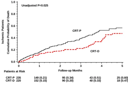
During the median follow-up of 28 months (IQR: 12-47), 378 (34%) patients died of any cause, 249 patients (36%) in the CRT-P arm and 129 patients (30%) in the CRT-D arm. There was no significant difference in the outcome between CRT-P and CRT-D patients (Kaplan-Meier 7-year cumulative event rate of CRT-P 63% vs. CRT-D 53%, p log-rank=0.531). Multivariate analysis after adjustment for ischemic etiology of cardiomyopathy, baseline LVEF and baseline urea showed consistent result to the univariate Kaplan-Meier model (CRT-D HR=0.78, 95 % CI: 0.57-1.06, p=0.11). In patients with ischemic cardiomyopathy, CRT-D treatment was associated with significant, 35% risk reduction in all-cause mortality as compared to patients with an implanted CRT-P (HR=0.65, 95 % CI: 0.44 - 0.95, p = 0.03, interaction p-value= 0.11) (Figure 1).
Figure 1. Cumulative probability of all-cause mortality in ischemic cardiomyopathy patients with implanted CRT-P or CRT-D.
Ischemic Patients Cumulative Probability of Death
0.0 0.2 0.4 0.6 0.8 1.0
Follow-up Months
0 1 2 3 4 5
Unadjusted P=0.025
CRT-P
CRT-D
Patients at Risk
CRT-P 235 149 (0.21) 95 (0.34) 43 (0.51) 25 (0.60)
CRT-D 220 152 (0.15) 90 (0.20) 40 (0.33) 18 (0.47)
2. Evaluating the prognostic significance of right to left ventricular interlead sensed electrical delay in CRT patients
During the median follow-up of 24 months (IQR: 12-42), 145 (29%) patients died of any cause, 80 patients (16%) with right to left ventricular interlead sensed electrical delay lower than 106.5 ms and 65 patients (13%) with right to left ventricular interlead sensed electrical delay greater than 106.5 ms.
CRT patients with right to left ventricular interlead sensed electrical delay lower than 106.5 ms had a 5-year cumulative mortality of 56% as compared to patients with right to left ventricular interlead sensed electrical delay greater than 106.5 ms who had a 41% 5-year cumulative mortality rate (p log-rank = 0.013) (Figure 2).
Figure 2. Cumulative probability of all-cause mortality in CRT patients stratified by right to left ventricular interlead sensed electrical delay.
CRT Patients Cumulative Probability of Death
0.0 0.2 0.4 0.6 0.8 1.0
Years after CRT implantation
0 1 2 3 4 5
Unadjusted P=0.013
IVD<=106.5
IVD>106.5
Patients at Risk
IVD<=106.5 247 178 (0.14) 115 (0.23) 64 (0.34) 34 (0.50) 17 (0.56) IVD>106.5 247 190 (0.09) 134 (0.17) 92 (0.23) 57 (0.31) 30 (0.41)
In the multivariate model after adjustment for age, female gender, left ventricular ejection fraction at baseline, ischemic etiology of cardiomyopathy, QRS duration at baseline, left ventricular end-systolic diameter at baseline, and permanent atrial fibrillation, right to left ventricular interlead sensed delay of greater than 106.5 ms was associated with significant, 48% risk reduction of all-cause mortality (95% CI: 0.31-0.88, p= 0.01).
3. Electroanatomical mapping guided endocardial transseptal LV lead implantation LV endocardial leads were successfully implanted in all patients. Electrical parameters during device implantation were as follows: LV signal amplitude 8.5±3.0 mV, LV pacing threshold 0.78±0.18 V, impulse width of 0.5 ms and LV lead impedance 520±177 Ohms.
No phrenic nerve stimulation occurred at 10 V; 0.5 ms with rapid LV pacing (100 bpm).
The procedure time was 92.5±22.2 minutes. Fluoroscopy time was 16.25±3.8 minutes.
There was no major hematoma or post-procedural bleeding observed. During the mean follow-up of 18 months, stable sensing and pacing parameters were found. We did not observe LV lead dysfunction, insulation failure or dyslocation. There were no signs of LV lead infection during the follow-up period.
Heart failure symptoms improved at least one NYHA class in all patients, LVEF improved significantly (28±5.2% to 41±6.6%, p=0.015). The grade of mitral regurgitation did not worsen during the follow-up period (p=0.28) (Table 4). We did not
observe any residual left-right shunts. No thromboembolic or hemorrhagic events occurred.
4. Left ventricular lead location and the risk of ventricular tachyarrhythmias
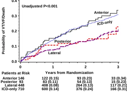
Lateral or posterior LV lead location was associated with significantly lower incidence of VT/VF or death (Figure 3) (p=0.002) and VT/VF as compared to anterior lead locations.
The findings were similar in both ischemic- and non-ischemic cardiomyopathy and in patients with LBBB, or non-LBBB ECG pattern. Patients with anterior LV lead locations had similar frequency of VT/VF/Death and VT/VF as ICD-only treated patients.
Figure 3. Kaplan–Meier Estimates of the Cumulative Probability of VT/VF/Death Episodes by device type and LV lead location
Probability of VT/VF/Death
0.0 0.1 0.2 0.3 0.4
Years from Randomization
0 1 2 3
Unadjusted P<0.001
Anterior
Posterior
Lateral
ICD-only
Patients at Risk
Anterior 146 122 (0.15) 93 (0.23) 33 (0.34)
Posterior 93 83 (0.11) 54 (0.13) 15 (0.23)
Lateral 448 408 (0.08) 264 (0.13) 117 (0.21)
ICD-only 710 600 (0.14) 376 (0.24) 166 (0.31)
In multivariate model, the lateral or posterior lead location was associated with significantly lower risk of VT/VF/Death (HR=0.58, p=0.004) as compared with anterior LV lead location, as well as when compared with ICD-only patients. Patients with anterior lead position showed similar risk of VT/VF/Death as patients with ICD-only (HR=1.04; 95% CI: 0.72 - 1.50; p=0.837).
5. Left ventricular dyssynchrony and the risk of ventricular tachyarrhythmias
Patients with non-LBBB (n=312) and LBBB (n=764) ECG pattern showed marked heterogeneity of LV dyssynchrony before device implantation irrespective of the QRS duration (r2=0.025, p<0.001). Non-LBBB or LBBB patients with increasing quartiles of baseline dyssynchrony did not show an increased risk of VT/VF or death and VT/VF. At 12-month follow-up, CRT-D patients with LBBB exhibited a greater decrease of LV dyssynchrony when compared to CRT-D patients with non-LBBB (n=125) (-56.5±81.1 ms vs. -27.1±85 ms, p=0.002). Bundle branch block pattern significantly modified the relationship between dyssynchrony and the outcome of VT/VF or death (p=0.03), while non-significant interaction was found between bundle branch pattern and dyssynchrony with regard of VT/VF (p=0.07).
In CRT-D patients with LBBB, the decrease in LV dyssynchrony was associated with significantly lower incidence of VT/VF/Death (p=0.014) as compared to patients with no change or worsening LV dyssynchrony. At one-year, CRT-D patients with LBBB and improving LV dyssynchrony showed significant, 63% risk reduction of VT/VF or death (p=0.02) after adjustment for relevant clinical covariates.
6. Effects of CRT upgrade in chronic right ventricular apical pacing patients
CRT upgrade was associated with an immediate significant reduction of the QRS duration in both patient groups (ICD -36.6 ± 25.3 ms vs. -43.3 ± 24.8 ms, p=0.082).
Baseline QRS duration was significantly longer in patients with previously implanted PM devices, therefore the absolute change was different however, the percent change was similar in both groups (ICD -21.1 ± 13.8 % vs. PM -22.8 ± 12.1%, p=0.276). After CRT upgrade, significant and similar improvement of NYHA functional class (ICD -0.67 ± 0.94 vs. PM -0.74 ± 0.77, p=0.745) was observed in patients with previously implanted PM or ICD. Improvement of the functional status was accompanied by significantly improved quality of life, assessed by the EQ-5D visual analog scale. After CRT upgrade, marked improvement of left ventricular ejection fraction was observed in PM (p<0.001) and ICD patients (p=0.003) as compared to baseline. However, PM patients derived significantly greater increase in LVEF as compared to ICD patients (p=0.02). Reduction in left ventricular end-diastolic and end-systolic diameter did not reach statistical
significance in both groups.
During the median follow-up of 21 months, 72 (36%) patients died of any cause (39 from PM, 33 from ICD group, p=0.98). There was no significant difference in mortality between PM and ICD patients (Kaplan-Meier 5-year cumulative event rate of PM 52% vs. 49%, p log-rank = 0.933). Multivariate model after adjustment for ischemic etiology of cardiomyopathy, baseline LVEF and baseline creatinine/urea ratio showed consistent results (ICD HR=1.18, 95 % CI: 0.60 - 2.29, p = 0.634).
7. Cardiac resynchronization therapy in patients with less severe ventricular dysfunction
At baseline, 914 patients (50.5%) had LVEF 26-30% (median=28.1%, Q1=27.1%, Q3=29.0%), and 199 patients (11%) comprised the LVEF group of ≤25%
(median=23.2%, Q1=21.8%, Q3=24.2%). The subgroup of LVEF>30% included 696 (38%) patients (in the range of 30.1-45.3%, median=31.8%, Q1=30.8%, Q3=33.1%), as evaluated by the echocardiography core laboratory. Patients with baseline LVEF≤25%
showed significantly higher cumulative incidence of HF or death episodes when compared to patients with LVEF 26-30% or LVEF>30%. Patients with LVEF≤25% had 55% higher risk of HF or death when compared to patients with LVEF 26-30% and 66%
higher risk when compared to patients with LVEF>30%. Patients with LVEF 26-30%
and LVEF>30% demonstrated similar risk of HF/Death.
Evaluating the treatment effects of CRT-D vs. ICD-only therapy, it showed borderline significance decreasing the cumulative incidence of HF/Death in patients with LVEF≤25% (p=0.062), while this effect was statistically significant among patients with LVEF 26-30% (p=0.003) and in those with LVEF>30%. Consistent with these findings, multivariate Cox-model showed CRT-D treatment associated with significant 43%
reduction in the risk of HF or death in patients with LVEF≤25% (p=0.03), 33% risk reduction in patients with LVEF 26-30% (p=0.007), and significant 44% risk reduction among those with LVEF>30% (p=0.003). The interaction p-value was not significant for all LVEF groups (all p-value for treatment-by-LVEF interactions >0.10). In patients with LBBB, even more striking risk reduction of HF/Death was observed. Non-LBBB patients did not show benefit of CRT-D irrespective of their baseline LVEF.
Conclusions
1. We demonstrated that heart failure patients implanted with CRT-D or CRT-P gain similar clinical and echocardiographic improvement after CRT implantation and in non- ischemic patients the mortality benefit is the same. However, patients with ischemic cardiomyopathy and an implanted CRT-D showed significant reduction in all-cause mortality as compared to ischemic patients implanted with a CRT-P device.
2. We showed that in CRT patients, right to left ventricular interlead sensed electrical delay of greater than 106.5 ms measured during the implantation procedure is associated with significantly lower all-cause mortality as compared to patients with right to left ventricular interlead sensed electrical delay less than 106.5 ms.
3. We reported successful electroanatomical mapping-guided LV endocardial lead implantation in four patients after unsuccessful permanent transvenous or epicardial LV lead placement. Electroanatomical mapping guidance to facilitate implanting LV endocardial leads was proven a useful method, which also might enhance identifying the optimal LV lead positions for CRT.
4. Our study on LV lead position and its impact on VT/VF/Death and VT/VF in MADIT- CRT showed that CRT-D with posterior or lateral left ventricular lead position is associated with reduced risk of ventricular arrhythmic events in comparison to anterior LV lead locations or ICD-only treated patients. However, CRT-D with anterior LV lead position does not increase arrhythmic events, clearly indicating that CRT carries no pro- arrhythmic effects.
5. This study demonstrated that CRT-induced improvement in LV dyssynchrony in patients with LBBB is associated with significant risk reduction of VT/VF/Death and VT/VF compared to patients with no improvement or worsening LV dyssynchrony.
6. It has been shown that CRT upgrade is feasible in patients with previously implanted ICD and patients get similar survival benefit as patients with previously implanted PM upgraded to CRT. However, the echocardiographic response was more pronounced in patients with a previously implanted PM compared to the ICD patient population.
7. It has been proven that the clinical benefit of CRT-D is present regardless of LVEF groups in patients enrolled in MADIT-CRT, including those with LVEF > 30% beyond the eligibility criteria. The echocardiographic response was directly correlated with increasing LVEF.
Publications
Publications closely related to the present thesis
V Kutyifa, B Merkely, S Szilagyi, E Zima, A Roka, A Kiraly, I Osztheimer, L Molnar, G Szeplaki, L Geller. Usefulness of electroanatomical mapping during transseptal endocardial left ventricular lead implantation. EUROPACE 14:(4) pp. 599-604. (2012)
V Kutyifa, A Kloppe, W Zareba, SD Solomon, S McNitt, A Barsheshet, S Polonsky, B Merkely, B Lemke, VK Nagy, AJ Moss, I Goldenberg. The Influence of Left Ventricular Ejection Fraction on the Effectiveness of Cardiac Resynchronization Therapy in MADIT- CRT. JACC, submitted
V Kutyifa, W Zareba, S McNitt, J Singh, WJ Hall, S Polonsky, I Goldenberg, DT Huang, B Merkely, PJ Wang, AJ Moss, HU Klein. Left Ventricular Lead Location and the Risk of Ventricular Arrhythmias in the MADIT-CRT trial. EUROPEAN HEART JOURNAL, submitted
V Kutyifa, P Bogyi, E Zima, VK Nagy, S Szilagyi, G Szeplaki, L Geller, B Merkely.
Effects of Upgrading to Cardiac Resynchronization Therapy in Pacemaker and Implantable Cardioverter Defibrillator Patients and Predictors of Long-Term Outcome.
EUROPACE, submitted
V Kutyifa, AC Pouleur, D Knappe, AA Ahmad, M Gibisnki, PJ Wang, S McNitt, B Merkely, I Goldenberg, SD Solomon, A J Moss, W Zareba. Dyssynchrony and the Risk of Ventricular Arrhythmias. JACC CARDIOVASCULAR IMAGING, submitted
V Kutyifa, OA Breithardt. How to assess the nonresponder to cardiac resynchronization therapy-a comprehensive stepwise approach. REVISTA ESPANOLA DE CARDIOLOGIA 65:(6) pp. 504-510. (2012), Review.
A Apor, V Kutyifa, B Merkely, S Szilagyi, P Andrassy, H Huttl, M Hubay, A Roka, L Geller. Successful cardiac resynchronization therapy after heart transplantation.
EUROPACE 10:(8) pp. 1024-1025. (2008)
Publications not related to the present thesis
B Merkely, A Róka, V Kutyifa, L Boersma, A Leenhardt, A Lubinski, A Oto, A Proclemer, J Brugada, PE Vardas, C Wolpert. Tracing the European course of cardiac resyncronization therapy from 2006 to 2008. EUROPACE 12:(5) pp. 692-701. (2010)
L Molnar, G Szűcs, E Zima, S Szilágyi, V Kutyifa, D Becker, L Geller, B Merkely.
Successful management and long term outcome of an accidental subclavian artery injury with a 9 french dilator during pacemaker implantation with collagen-based closure device. JOURNAL OF INTERVENTIONAL CARDIAC ELECTROPHYSIOLOGY 25:(3) pp. 217-218. (2009)
S Szilágyi, B Merkely, E Zima, V Kutyifa, G Szűcs, G Fülöp, L Molnár, Z Szabolcs, L Gellér. Minimal invasive coronary sinus lead reposition technique for the treatment of phrenic nerve stimulation. EUROPACE 10:(10) pp. 1157-1160. (2008)
S Szilágyi, B Merkely, A Róka, E Zima, G Fülöp, V Kutyifa, G Szűcs, D Becker, A Apor, L Geller. Stabilization of the coronary sinus electrode position with coronary stent implantation to prevent and treat dislocation. JOURNAL OF CARDIOVASCULAR ELECTROPHYSIOLOGY 18:(3) pp. 303-307. (2007)
A Nemes, T Forster, ML Geleijnse, V Kutyifa, K Neu, OI Soliman, FJ Ten Cate, M Csanády. The additional prognostic power of diabetes mellitus on coronary flow reserve in patients with suspected coronary artery disease. DIABETES RESEARCH AND CLINICAL PRACTICE 78:(1) pp. 126-131. (2007)